

γλώσσες
Σελίδες
Νομικός

Result
“I have used them in LI-COR IN CELL WESTERN method. They worked nicely. The method was according to the in-structions provided with the LI-COR kit with their buffers and antibodies.” - S. Morshed, Icahn School of Medicine at Mount Sinai and Bronx.
Gel electrophoresis information:N/A
Transfer information:N/A
Lane No. Antigen Loading amount Primary antibody Primary antibody dilution ratio
Secondary
antibody dilution ratio
Exposure time
Control-Ab Thyrocytes 50,000 cells/well STJ97398 Anti-
MAP LC3β 1:100 1:800 10Min
Cell Line: Rat Thyrocytes
Method of validation: LI-COR CELL Western
Primary Antibody: STJ97398 Anti-MAP LC3β anti-
body
Secondary Antibody LI-COR Antibody
Dilution ratio: 1:100
Protocol
Treatment of materials: Rat thyrocytes were treated with TSH receptor antibody for 24 hrs.
Cell fixing: 3.7% formaldehyde in 1xPBS, for 20 minutes at room temperature.
Washing: 1XPBS with 0.1% Triton X-100 for 5 minutes per wash. Repeat 4 times.
Blocking: LI-COR Blocking buffer, 1.5 hours at room temperature with moderate shaking.
Primary antibody probing: Overnight at 4°C.
Washing: 1XPBS with 0.1% Tween 20 for 5 minutes at room temperature.
Secondary antibody probing: LI-COR Antibody for 1 hour.
Washing: 1XPBS with 0.1% Tween 20 for 5 minutes at room temperature. Repeat 5 times.
Visualization: Analysed by LI-COR imaging 10 minutes.
Antibody Customer Review: STJ97398 Anti-MAP LC3β Antibody Antibody Specificity:
Antibody Rating:
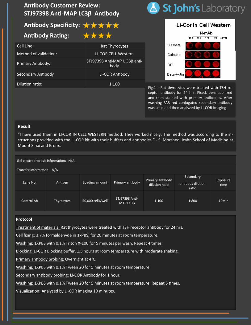
Fig.1 - Rat thyrocytes were treated with TSH re-ceptor antibody for 24 hrs. Fixed, permeabilized and then stained with primary antibodies. After washing FAR red conjugated secondary antibody was used and then analyzed by LI-COR imaging.
Top Related