


γλώσσες
Σελίδες
Νομικός

Crystal structures of human Ero1a reveal themechanisms of regulated and targeted oxidationof PDI
Kenji Inaba1,*, Shoji Masui1, Hiroka Iida1,Stefano Vavassori2, Roberto Sitia2,4
and Mamoru Suzuki3,4
1Division of Protein Chemistry, Post-Genome Science Center, MedicalInstitute of Bioregulation, Kyushu University, Maidashi, Higashi-ku,Fukuoka, Japan, 2Universita Vita-Salute San Raffaele Scientific Institute,Division of Genetics and Cell Biology, Milan, Italy and 3Institute forProtein Research, Osaka University, Osaka, Japan
In the endoplasmic reticulum (ER) of eukaryotic cells,
Ero1 flavoenzymes promote oxidative protein folding
through protein disulphide isomerase (PDI), generating
reactive oxygen species (hydrogen peroxide) as bypro-
ducts. Therefore, Ero1 activity must be strictly regulated
to avoid futile oxidation cycles in the ER. Although
regulatory mechanisms restraining Ero1a activity ensure
that not all PDIs are oxidized, its specificity towards PDI
could allow other resident oxidoreductases to remain
reduced and competent to carry out isomerization and
reduction of protein substrates. In this study, crystal
structures of human Ero1a were solved in its hyperactive
and inactive forms. Our findings reveal that human Ero1amodulates its oxidative activity by properly positioning
regulatory cysteines within an intrinsically flexible loop,
and by fine-tuning the electron shuttle ability of the loop
through disulphide rearrangements. Specific PDI targeting
is guaranteed by electrostatic and hydrophobic interac-
tions of Ero1a with the PDI b0-domain through its sub-
strate-binding pocket. These results reveal the molecular
basis of the regulation and specificity of protein disulphide
formation in human cells.
The EMBO Journal (2010) 29, 3330–3343. doi:10.1038/
emboj.2010.222; Published online 10 September 2010
Subject Categories: proteins; structural biology
Keywords: disulphide bond; ER quality control; human
Ero1a; redox homeostasis; X-ray crystal structure analysis
Introduction
Many secretory and membrane proteins form disulphide
bonds in the endoplasmic reticulum (ER), under the assis-
tance of numerous thiol-disulphide oxidoreductases (Sevier
and Kaiser, 2006; Appenzeller-Herzog and Ellgaard, 2008;
Hatahet and Ruddock, 2009). Elaborate redox networks
allow formation and isomerization of disulphide bonds
until the native state is attained. The major players in
oxidative reactions are protein disulphide isomerases (PDI)
and Ero1; both highly conserved from yeast to mammals
(Frand and Kaiser, 1998; Pollard et al, 1998; Cabibbo et al,
2000; Mezghrani et al, 2001). Ero1 generates disulphide
bonds de novo in conjunction with a flavin adenine dinucleo-
tide (FAD) cofactor and transfers them to PDI (Tu and
Weissman, 2004; Tavender and Bulleid, 2010). Protein dis-
ulphide isomerase, an ER-resident member of the thioredoxin
(Trx)-fold family, is composed of two redox-active Trx do-
mains with a CGHC motif (a- and a0-domains), two redox-
inactive Trx domains (b- and b0 -domains), an additional
a-helical domain (c-domain) and a linker loop (x-linker)
between the b0- and a’-domains. Crystal structures of yeast
PDI (Pdi1p) revealed that these domains are lined up in the
order a–b–b0–a0–c, and they can assume different spatial
arrangements, ‘twisted U-shape’ or ‘boat’, indicating confor-
mational flexibility of the enzyme (Tian et al, 2006, 2008).
Alternative conformations were also reported for human and
thermophilic fungus PDIs (Nguyen et al, 2008; Serve et al,
2010; Wang et al, 2010).
Since the discovery of an essential PDI–Ero1 oxidative
pathway in Saccharomyces cerevisiae, its operation mechan-
isms have been studied using genetic, biochemical and
structural approaches (Sevier and Kaiser, 2008). Recent
biochemical studies demonstrated that yeast Ero1 (Ero1p)
preferentially oxidizes the CXXC motif in the N-terminal Trx
domain (a-domain) of Pdi1p among the active sites of oxidor-
eductases present in the yeast ER, leading to a preferred
pathway for oxidizing the ER thiol pool (Vitu et al, 2010).
Furthermore, the crystal structure of Ero1p, in which its
N-terminal (Thr19–Thr55) and C-terminal (Asn425–Gln560)
regions were removed to obtain a high-quality protein crystal,
revealed the fold of its catalytic core region and the structure
of the FAD-containing reaction centre (Gross et al, 2004).
The generation of each disulphide bond by Ero1p can be
accompanied by the production of one molecule of hydrogen
peroxide (H2O2), a potential reactive oxygen species (ROS)
source (Gross et al, 2006). Although the cell can cope with
peroxides formed during routine oxidative protein folding
and sometimes uses them as a messenger in the cell-signal-
ling cascades (Veal et al, 2007) and possibly as a direct
protein disulphide introducer (Karala et al, 2009), ROS pro-
duction that exceeds the capacity of the cellular antioxidant
defense systems could induce harmful ER oxidative stress.
Overexpression of Ero1p was shown to cause a significant
increase in ROS (Haynes et al, 2004), whereas partial low-
ering of Ero1 activity by RNAi abrogated ROS accumulation
in ER-stressed animals and enhanced resistance to the lethal
effects of high levels of ER stress (Harding et al, 2003;
Marciniak et al, 2004). These observations suggested that
Ero1 activity must be tightly regulated in living cells. YeastReceived: 4 June 2010; accepted: 17 August 2010; published online:10 September 2010
*Corresponding author. Division of Protein Chemistry, Post-GenomeScience Center, Medical Institute of Bioregulation, Kyushu University,3-1-1 Maidashi, Higashi-ku, Fukuoka 812-8582, Japan.Tel.: þ 81 92 642 6433; Fax: þ 81 92 642 6433;E-mail: [email protected] authors contributed equally to this work
The EMBO Journal (2010) 29, 3330–3343 | & 2010 European Molecular Biology Organization | All Rights Reserved 0261-4189/10
www.embojournal.org
The EMBO Journal VOL 29 | NO 19 | 2010 &2010 European Molecular Biology Organization
EMBO
THE
EMBOJOURNAL
THE
EMBOJOURNAL
3330
Ero1p, indeed, was observed to exert an inbuilt feedback
regulation mechanism, in which two noncatalytic cysteine
pairs (Cys90–Cys349 and Cys150–Cys295) sense the ER redox
environment; their oxidation decreases Ero1p activity, thus
preventing potentially futile and cell-damaging cycles and ER
hyperoxidation (Sevier et al, 2007).
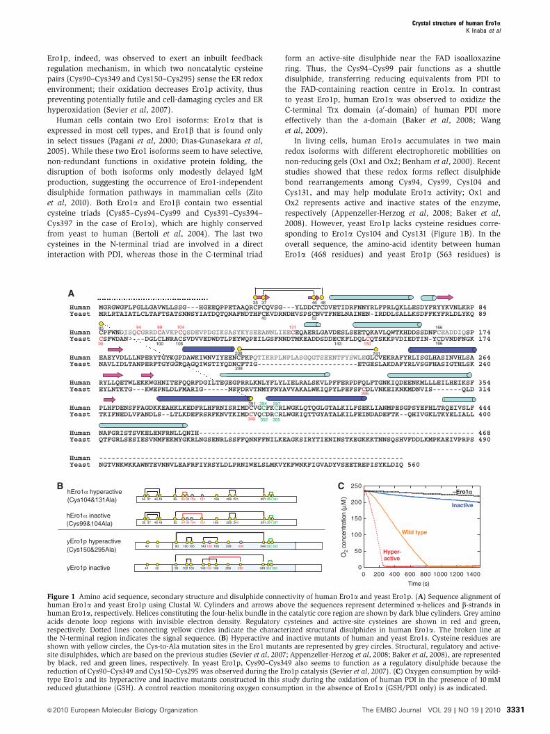
Human cells contain two Ero1 isoforms: Ero1a that is
expressed in most cell types, and Ero1b that is found only
in select tissues (Pagani et al, 2000; Dias-Gunasekara et al,
2005). While these two Ero1 isoforms seem to have selective,
non-redundant functions in oxidative protein folding, the
disruption of both isoforms only modestly delayed IgM
production, suggesting the occurrence of Ero1-independent
disulphide formation pathways in mammalian cells (Zito
et al, 2010). Both Ero1a and Ero1b contain two essential
cysteine triads (Cys85–Cys94–Cys99 and Cys391–Cys394–
Cys397 in the case of Ero1a), which are highly conserved
from yeast to human (Bertoli et al, 2004). The last two
cysteines in the N-terminal triad are involved in a direct
interaction with PDI, whereas those in the C-terminal triad
form an active-site disulphide near the FAD isoalloxazine
ring. Thus, the Cys94–Cys99 pair functions as a shuttle
disulphide, transferring reducing equivalents from PDI to
the FAD-containing reaction centre in Ero1a. In contrast
to yeast Ero1p, human Ero1a was observed to oxidize the
C-terminal Trx domain (a0-domain) of human PDI more
effectively than the a-domain (Baker et al, 2008; Wang
et al, 2009).
In living cells, human Ero1a accumulates in two main
redox isoforms with different electrophoretic mobilities on
non-reducing gels (Ox1 and Ox2; Benham et al, 2000). Recent
studies showed that these redox forms reflect disulphide
bond rearrangements among Cys94, Cys99, Cys104 and
Cys131, and may help modulate Ero1a activity; Ox1 and
Ox2 represents active and inactive states of the enzyme,
respectively (Appenzeller-Herzog et al, 2008; Baker et al,
2008). However, yeast Ero1p lacks cysteine residues corre-
sponding to Ero1a Cys104 and Cys131 (Figure 1B). In the
overall sequence, the amino-acid identity between human
Ero1a (468 residues) and yeast Ero1p (563 residues) is
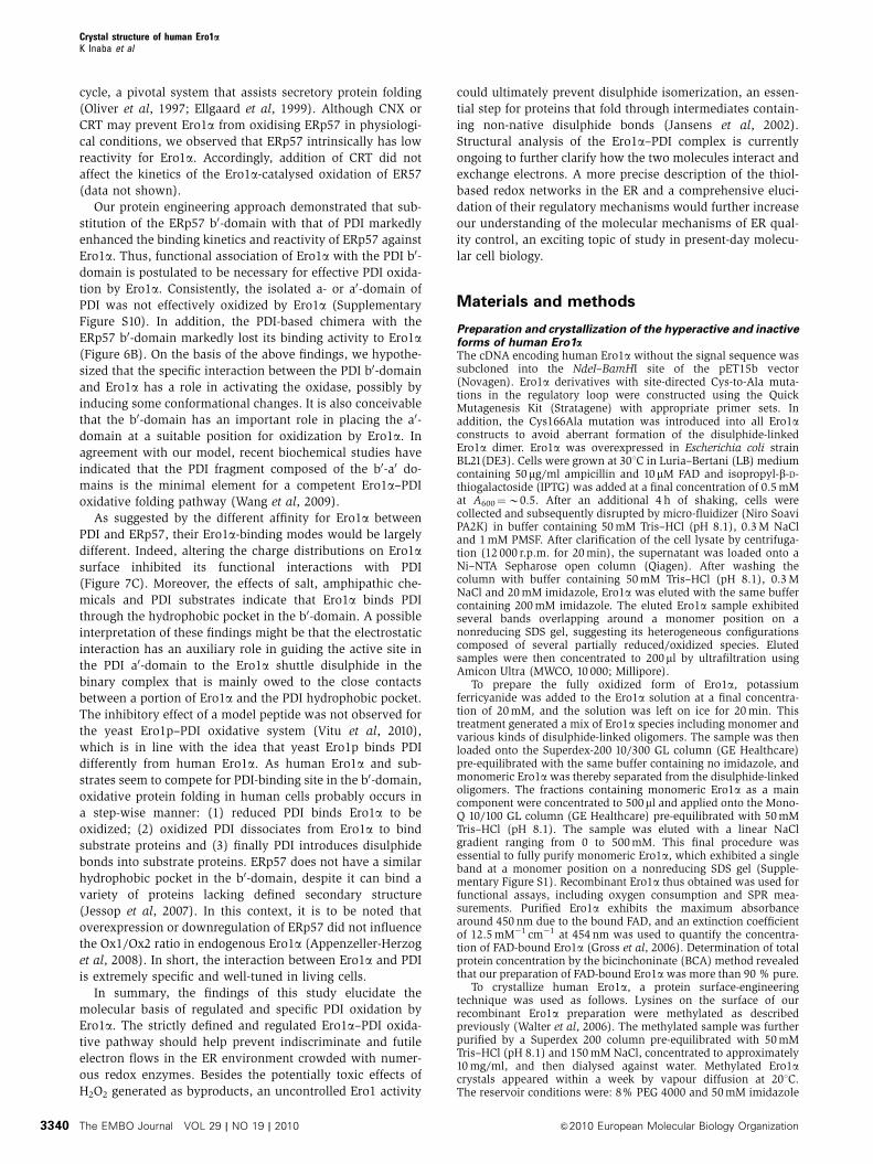
0
50
100
150
200
250
0 200 400 600 800 1000 1200 1400
O2
conc
entra
tion
(μM
)
Time (s)
Human MGRGWGFLFGLLGAVWLLSSG---HGEEQPPETAAQRCFCQVSG---YLDDCTCDVETIDRFNNYRLFPRLQKLLESDYFRYYKVNLKRP 84
Human CPFWNDISQCGRRDCAVKPCQSDEVPDGIKSASYKYSEEANNLIEECEQAERLGAVDESLSEETQKAVLQWTKHDDSSDNFCEADDIQSP 174
Human EAEYVDLLLNPERYTGYKGPDAWKIWNVIYEENCFKPQTIKRPLNPLASGQGTSEENTFYSWLEGLCVEKRAFYRLISGLHASINVHLSA 264
Human RYLLQETWLEKKWGHNITEFQQRFDGILTEGEGPRRLKNLYFLYLIELRALSKVLPFFERPDFQLFTGNKIQDEENKMLLLEILHEIKSF 354
Human PLHFDENSFFAGDKKEAHKLKEDFRLHFRNISRIMDCVGCFKCRLWGKLQTQGLGTALKILFSEKLIANMPESGPSYEFHLTRQEIVSLF 444
Human NAFGRISTSVKELENFRNLLQNIH------------------------------------------------------------------ 468
Human -------------------------------------------------------------------------
Yeast MRLRTAIATLCLTAFTSATSNNSYIATDQTQNAFNDTHFCKVDRNDHVSPSCNVTFNELNAINEN-IRDDLSALLKSDFFKYFRLDLYKQ 89
Yeast CSFWDAN----DGLCLNRACSVDVVEDWDTLPEYWQPEILGSFNNDTMKEADDSDDECKFLDQLCQTSKKPVDIEDTIN-YCDVNDFNGK 174
Yeast NAVLIDLTANPERFTGYGGKQAGQIWSTIYQDNCFTIG------------------------ETGESLAKDAFYRLVSGFHASIGTHLSK 240
Yeast EYLNTKTG---KWEPNLDLFMARIG------NFPDRVTNMYFNYAVVAKALWKIQPYLPEFSFCDLVNKEIKNKMDNVIS-------QLD 314
Yeast TKIFNEDLVFANDLS--LTLKDEFRSRFKNVTKIMDCVQCDRCRLWGKIQTTGYATALKILFEINDADEFTK--QHIVGKLTKYELIALL 400
Yeast QTFGRLSESIESVNMFEKMYGKRLNGSENRLSSFFQNNFFNILKEAGKSIRYTIENINSTKEGKKKTNNSQSHVFDDLKMPKAEIVPRPS 490
Yeast NGTVNKWKKAWNTEVNNVLEAFRFIYRSYLDLPRNIWELSLMKVYKFWNKFIGVADYVSEETREPISYKLDIQ 560
A
BhEro1 hyperactive(Cys104&131Ala)
94 99 104 131
90 100 105 143 150
295
349
yEro1p hyperactive(Cys150&295Ala)
Inactive
Hyper-active
Wild type
C
35 37 46 48
40 52
85 166
166
208 241
208
391 394 397
352 355
yEro1p inactive
hEro1 inactive(Cys99&104Ala)
–Ero1
Figure 1 Amino acid sequence, secondary structure and disulphide connectivity of human Ero1a and yeast Ero1p. (A) Sequence alignment ofhuman Ero1a and yeast Ero1p using Clustal W. Cylinders and arrows above the sequences represent determined a-helices and b-strands inhuman Ero1a, respectively. Helices constituting the four-helix bundle in the catalytic core region are shown by dark blue cylinders. Grey aminoacids denote loop regions with invisible electron density. Regulatory cysteines and active-site cysteines are shown in red and green,respectively. Dotted lines connecting yellow circles indicate the characterized structural disulphides in human Ero1a. The broken line atthe N-terminal region indicates the signal sequence. (B) Hyperactive and inactive mutants of human and yeast Ero1s. Cysteine residues areshown with yellow circles, the Cys-to-Ala mutation sites in the Ero1 mutants are represented by grey circles. Structural, regulatory and active-site disulphides, which are based on the previous studies (Sevier et al, 2007; Appenzeller-Herzog et al, 2008; Baker et al, 2008), are representedby black, red and green lines, respectively. In yeast Ero1p, Cys90–Cys349 also seems to function as a regulatory disulphide because thereduction of Cys90–Cys349 and Cys150–Cys295 was observed during the Ero1p catalysis (Sevier et al, 2007). (C) Oxygen consumption by wild-type Ero1a and its hyperactive and inactive mutants constructed in this study during the oxidation of human PDI in the presence of 10 mMreduced glutathione (GSH). A control reaction monitoring oxygen consumption in the absence of Ero1a (GSH/PDI only) is as indicated.
Crystal structure of human Ero1aK Inaba et al
&2010 European Molecular Biology Organization The EMBO Journal VOL 29 | NO 19 | 2010 3331
approximately 27% (128 residues; Figure 1A). To elucidate
the different functional regulation mechanisms in these two
homologous enzymes, we set out to obtain structural infor-
mation on human Ero1a.
In this study, we report the crystal structures of full-length
human Ero1a in both its hyperactive and inactive forms, and
we describe how human Ero1a limits its oxidative activity
using four regulatory cysteines properly positioned within a
critical loop that transfers electrons from PDI to the FAD-
containing active site. Our study provides structural and
further mechanistic insights into the internal disulphide re-
arrangement regulating Ero1a activity, originally proposed by
the preceding studies (Appenzeller-Herzog et al, 2008; Baker
et al, 2008). Our studies also suggest that the hydrophobic
pocket in the PDI b0-domain is an essential element for the
specific oxidation of PDI by Ero1a and electrostatic interac-
tions have an auxiliary role in the functional Ero1a–PDI
interaction. A detailed molecular view of the regulated
Ero1a catalysis of PDI oxidation is presented.
Results
Overall structure of human Ero1aHigh-quality crystals could not be obtained from recombinant
wild-type human Ero1a. This was probably due to its hetero-
geneous configurations, as suggested by non-reducing SDS–
PAGE (Supplementary Figure S1). Therefore, mutants were
produced in which two of the four regulatory cysteines were
mutated to alanines, so that only one disulphide bond can be
formed. Previous mutagenesis studies demonstrated that
Cys94–Cys99 is the essential disulphide to directly engage
in disulphide exchange with PDI, and the Cys104&131Ala
mutant that homogeneously forms the Cys94–Cys99 disul-
phide is hyperactive (Baker et al, 2008). Accordingly, this
mutant exhibited an even higher rate of oxygen consumption
(i.e., PDI oxidation) than wild-type Ero1a (Figure 1C) and
migrated as a single band with a slower electrophoretic
mobility than the inactive mutant as did Ox1 of cellular
Ero1a (Supplementary Figure S1; Benham et al, 2000).
Using this hyperactive Ero1a mutant, we obtained a well-
diffracting crystal of space group I222 with one molecule per
asymmetric unit. The X-ray diffraction data sets were col-
lected at the maximum resolution (2.35 A) using the SPring-8
beamline BL44XU. The structural model was refined at 2.35 A
to an R-factor of 0.237 (Rfree¼ 0.282) with very good stereo-
chemistry. According to PROCHECK analysis, 99.7% of the
residues were in the most favoured and the allowed regions
of the Ramachandran diagram, whereas no residues were in
the disallowed region (Table I).
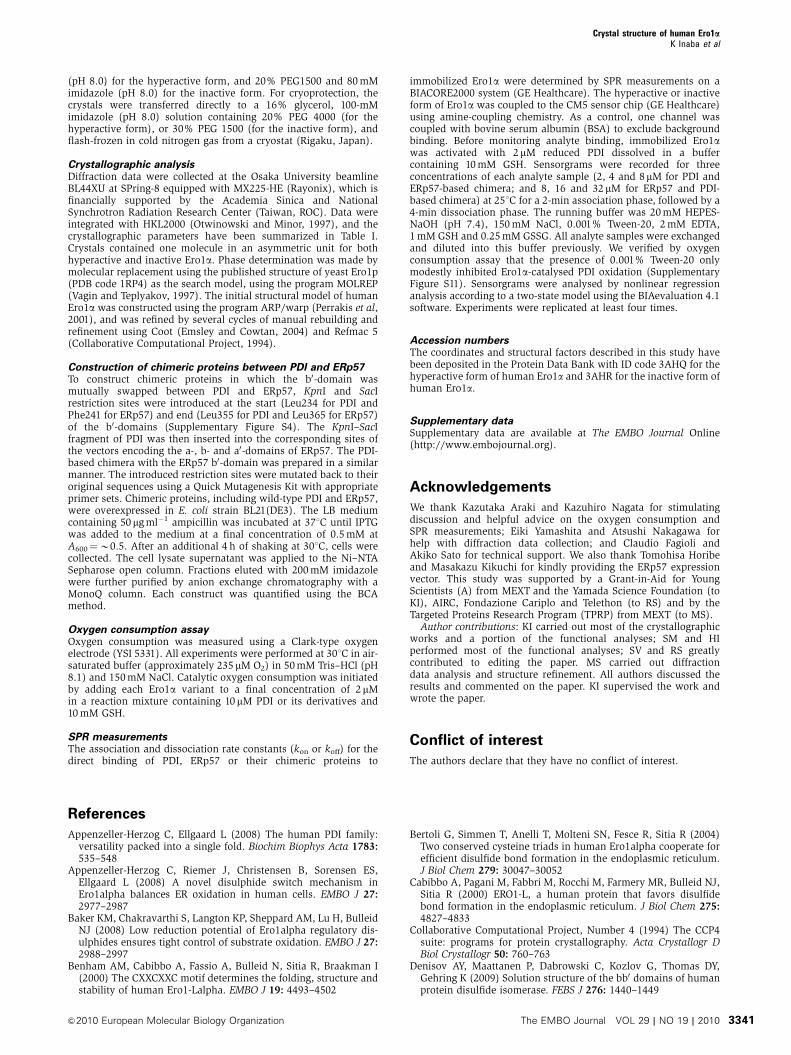
The entire human Ero1a chain formed a single globular
domain highly rich in a-helices, in which five intramolecular
disulphide bonds were identified (Cys35–Cys48, Cys37–
Cys46, Cys85–Cys391, Cys208–Cys241 and Cys394–Cys397;
Figure 2A and B). Thus, this crystallographic study confirms
the human Ero1a disulphide-bond pattern suggested by mass
spectrometric analyses (Appenzeller-Herzog et al, 2008). The
N-terminal segment, which had been deleted and therefore
was structurally uncharacterized in the crystallographic study
of yeast Ero1p (Gross et al, 2004), formed an anti-parallel
b-hairpin containing the Cys35–Cys48 and Cys37–Cys46
disulphides (Figure 2B). This region was located opposite to
the reaction centre, including the isoalloxazine ring of FAD
and its proximal disulphide Cys394–Cys397 (Figure 2A),
making it unlikely that the N-terminal region is directly
involved in PDI oxidation catalysis. Consistently, the removal
of this region did not inhibit Ero1a oxidative activity in vivo
(Bertoli et al, 2004).
Overall, the loop regions in human Ero1a were longer than
those of yeast Ero1p. In particular, three loop regions span-
ning from Asp90 to Cys131 (loop A,‘regulatory loop’), Cys166
to Gln172 (loop B) and Gln212 to Glu238 (loop C) lacked
electron density, probably due to their extreme flexibility
(Figure 2A). All of the regulatory cysteines were contained
in loop A, and therefore their locations could not be specified
in the hyperactive Ero1a. It may be that the intrinsically
flexible nature of loop A is essential for electron shuttling
from PDI to the FAD-proximal site of Ero1a.
Importantly, the catalytic core regions of human Ero1a and
yeast Ero1p were nearly superimposable, with an average
RMSD value for Ca atoms of 0.87 A (Figure 2C). The four-
helix bundle scaffold with an interior electron-accepting
cofactor is a hallmark of disulphide bond-generating enzymes
widely distributed from bacteria to eukaryotes (Sevier et al,
2005; Inaba and Ito, 2008). In contrast, there were large
structural differences between human and yeast Ero1s in
the other regions (Figure 2C). Noticeably, although the
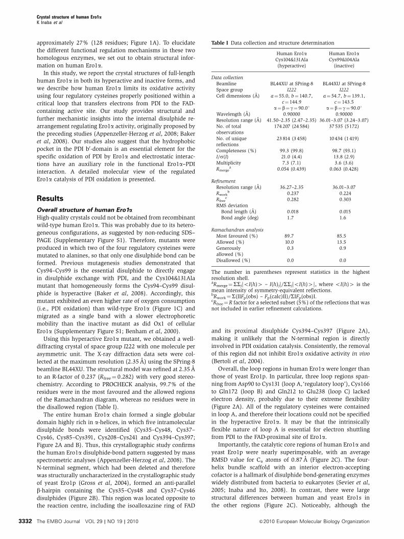
Table I Data collection and structure determination
Human Ero1aCys104&131Ala(hyperactive)
Human Ero1aCys99&104Ala
(inactive)
Data collectionBeamline BL44XU at SPring-8 BL44XU at SPring-8Space group I222 I222Cell dimensions (A) a¼ 55.0, b¼ 140.7,
c¼ 144.9a¼ 54.7, b¼ 139.1,
c¼ 143.5a¼ b¼ g¼ 90.01 a¼ b¼ g¼ 90.01
Wavelength (A) 0.90000 0.90000Resolution range (A) 41.50–2.35 (2.47–2.35) 36.01–3.07 (3.24–3.07)No. of totalobservations
174 207 (24 584) 37 535 (5172)
No. of uniquereflections
23 814 (3 458) 10 434 (1 419)
Completeness (%) 99.3 (99.8) 98.7 (93.1)I/s(I) 21.0 (4.4) 13.8 (2.9)Multiplicity 7.3 (7.1) 3.6 (3.6)Rmerge
a 0.054 (0.439) 0.063 (0.428)
RefinementResolution range (A) 36.27–2.35 36.01–3.07Rwork
b 0.237 0.224Rfree
c 0.282 0.303RMS deviation
Bond length (A) 0.018 0.015Bond angle (deg) 1.7 1.6
Ramachandran analysisMost favoured (%) 89.7 85.5Allowed (%) 10.0 13.5Generouslyallowed (%)
0.3 0.9
Disallowed (%) 0.0 0.0
The number in parentheses represent statistics in the highestresolution shell.aRmerge¼SSj|oI(h)4 – I(h)j|/SSj|oI(h)4|, where oI(h)4 is themean intensity of symmetry-equivalent reflections.bRwork¼S(IIFp(obs) – Fp(calc)II)/SIFp(obs)I.cRfree¼R factor for a selected subset (5%) of the reflections that wasnot included in earlier refinement calculations.
Crystal structure of human Ero1aK Inaba et al
The EMBO Journal VOL 29 | NO 19 | 2010 &2010 European Molecular Biology Organization3332
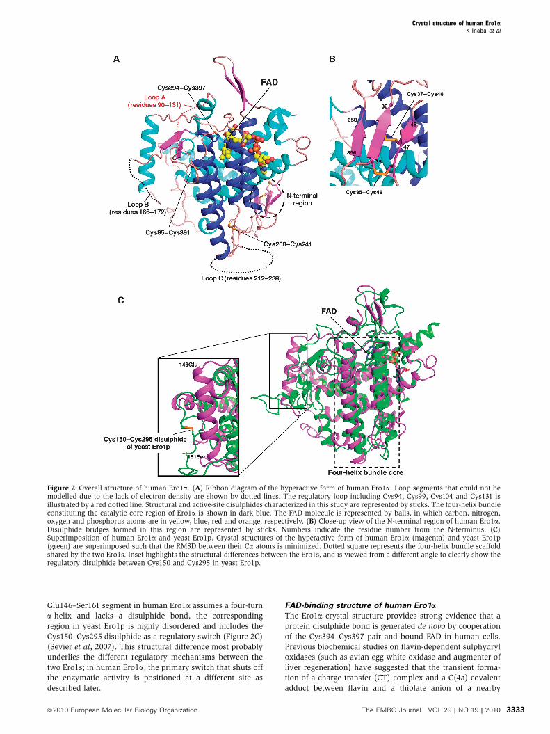
Glu146–Ser161 segment in human Ero1a assumes a four-turn
a-helix and lacks a disulphide bond, the corresponding
region in yeast Ero1p is highly disordered and includes the
Cys150–Cys295 disulphide as a regulatory switch (Figure 2C)
(Sevier et al, 2007). This structural difference most probably
underlies the different regulatory mechanisms between the
two Ero1s; in human Ero1a, the primary switch that shuts off
the enzymatic activity is positioned at a different site as
described later.
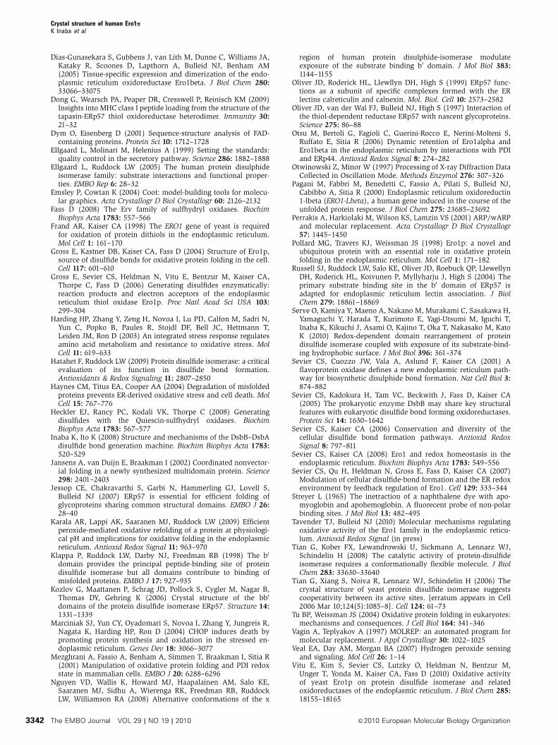
FAD-binding structure of human Ero1aThe Ero1a crystal structure provides strong evidence that a
protein disulphide bond is generated de novo by cooperation
of the Cys394–Cys397 pair and bound FAD in human cells.
Previous biochemical studies on flavin-dependent sulphydryl
oxidases (such as avian egg white oxidase and augmenter of
liver regeneration) have suggested that the transient forma-
tion of a charge transfer (CT) complex and a C(4a) covalent
adduct between flavin and a thiolate anion of a nearby
Figure 2 Overall structure of human Ero1a. (A) Ribbon diagram of the hyperactive form of human Ero1a. Loop segments that could not bemodelled due to the lack of electron density are shown by dotted lines. The regulatory loop including Cys94, Cys99, Cys104 and Cys131 isillustrated by a red dotted line. Structural and active-site disulphides characterized in this study are represented by sticks. The four-helix bundleconstituting the catalytic core region of Ero1a is shown in dark blue. The FAD molecule is represented by balls, in which carbon, nitrogen,oxygen and phosphorus atoms are in yellow, blue, red and orange, respectively. (B) Close-up view of the N-terminal region of human Ero1a.Disulphide bridges formed in this region are represented by sticks. Numbers indicate the residue number from the N-terminus. (C)Superimposition of human Ero1a and yeast Ero1p. Crystal structures of the hyperactive form of human Ero1a (magenta) and yeast Ero1p(green) are superimposed such that the RMSD between their Ca atoms is minimized. Dotted square represents the four-helix bundle scaffoldshared by the two Ero1s. Inset highlights the structural differences between the Ero1s, and is viewed from a different angle to clearly show theregulatory disulphide between Cys150 and Cys295 in yeast Ero1p.
Crystal structure of human Ero1aK Inaba et al
&2010 European Molecular Biology Organization The EMBO Journal VOL 29 | NO 19 | 2010 3333
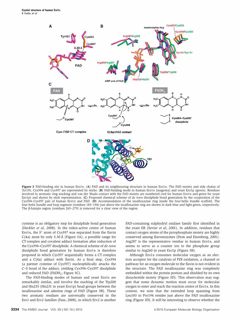
cysteine is an obligatory step for disulphide bond generation
(Heckler et al, 2008). In the redox-active centre of human
Ero1a, the Sg atom of Cys397 was separated from the flavin
C(4a) atom by only 3.30 A (Figure 3A), a possible range for
CT complex and covalent adduct formation after reduction of
the Cys394–Cys397 disulphide. A chemical scheme of de novo
disulphide bond generation by human Ero1a is therefore
proposed in which Cys397 sequentially forms a CT complex
and a C(4a) adduct with flavin. As a final step, Cys394
(a partner cysteine of Cys397) nucleophilically attacks the
C–S bond of the adduct, yielding Cys394–Cys397 disulphide
and reduced FAD (FADH2; Figure 3C).
The FAD-binding modes of human and yeast Ero1s are
remarkably similar, and involve the stacking of the Trp200
and His255 (His231 in yeast Ero1p) head groups between the
isoalloxazine and adenine rings of FAD (Figure 3B). These
two aromatic residues are universally conserved in the
Ero1 and Erv2 families (Fass, 2008), in which Erv2 is another
FAD-containing sulphydryl oxidase family first identified in
the yeast ER (Sevier et al, 2001). In addition, residues that
contact oxygen atoms of the pyrophosphate moiety are highly
conserved among flavoenzymes (Dym and Eisenberg, 2001).
Arg287 is the representative residue in human Ero1a, and
seems to serve as a counter ion to the phosphate group
similar to Arg260 in yeast Ero1p (Figure 3B).
Although Ero1a consumes molecular oxygen as an elec-
tron acceptor for the catalysis of PDI oxidation, a channel or
pathway for an oxygen molecule to the flavin is not evident in
the structure. The FAD isoalloxazine ring was completely
embedded within the protein portion and shielded by its own
dinucleotide moiety (Figure 3D). This observation may sug-
gest that some dynamic motion must occur for molecular
oxygen to enter and reach the reaction centre of Ero1a. In this
context, we note that the extended loop spanning from
Leu183 to Pro194 resides just above the FAD isoalloxazine
ring (Figure 3D). It will be interesting to observe whether the
Figure 3 FAD-binding site in human Ero1a. (A) FAD and its neighbouring structure in human Ero1a. The FAD moiety and side chains ofTyr191, Cys394 and Cys397 are represented by sticks. (B) FAD-binding mode in human Ero1a (magenta) and yeast Ero1p (green). Residuesinvolved in aromatic ring stacking and van der Waals contact with the FAD moiety are numbered (red for human Ero1a and green for yeastEro1p) and shown by stick representation. (C) Proposed chemical scheme of de novo disulphide bond generation by the cooperation of theCys394–Cys397 pair of human Ero1a and FAD. (D) Accommodation of the isoalloxazine ring inside the four-helix bundle scaffold. Thefour-helix bundle and loop segment (residues 183–194) just above the isoalloxazine ring are shown in dark blue and light green, respectively.The b-hairpin region (residues 265–279) is removed for a clear view of the region.
Crystal structure of human Ero1aK Inaba et al
The EMBO Journal VOL 29 | NO 19 | 2010 &2010 European Molecular Biology Organization3334
redox-dependent interaction between FAD and its neighbour-
ing polypeptide or the PDI binding to Ero1a modulates the
conformation and dynamism of the FAD-proximal region,
thereby facilitating molecular oxygen entry.
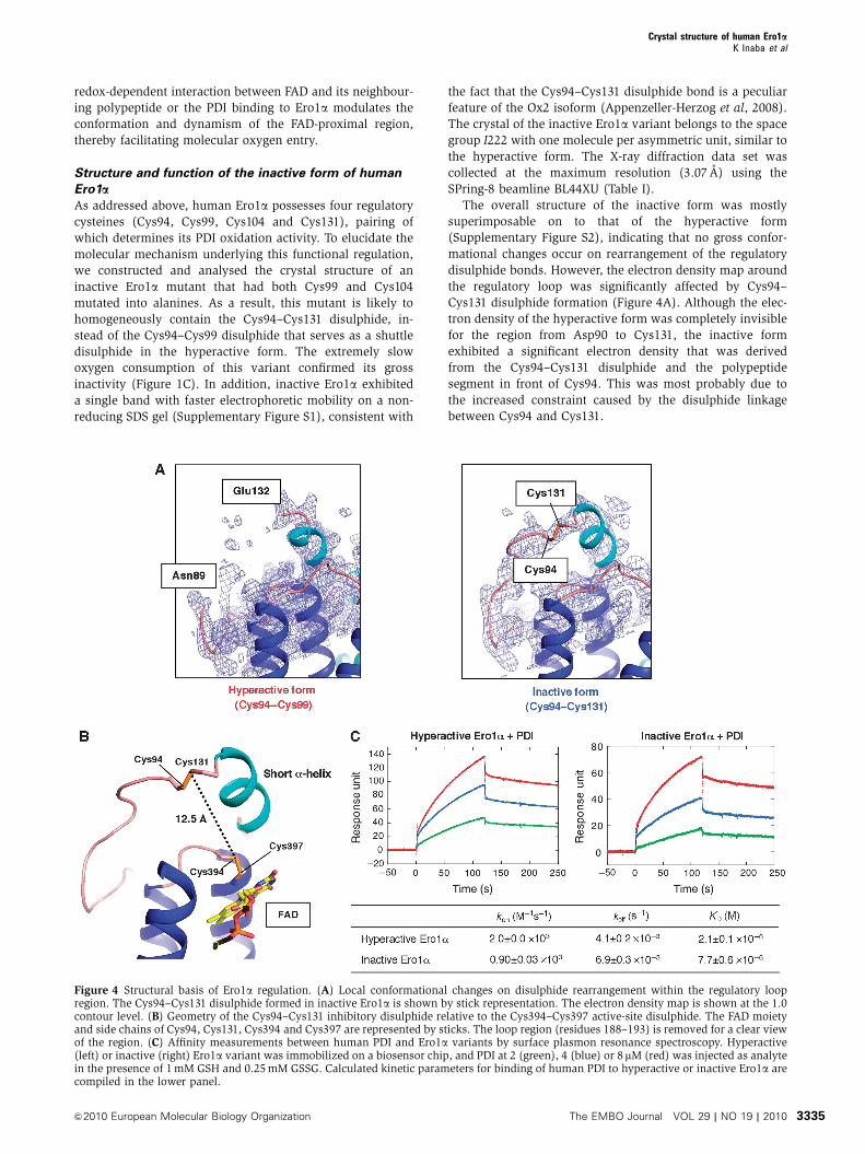
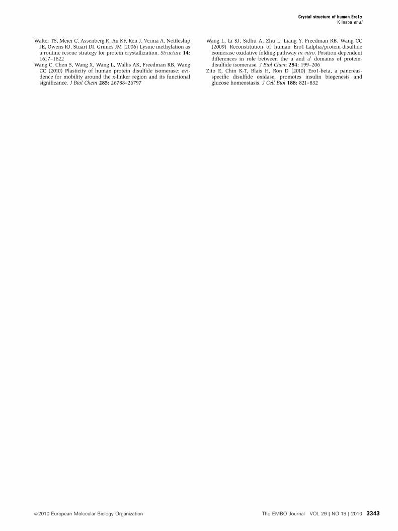
Structure and function of the inactive form of human
Ero1aAs addressed above, human Ero1a possesses four regulatory
cysteines (Cys94, Cys99, Cys104 and Cys131), pairing of
which determines its PDI oxidation activity. To elucidate the
molecular mechanism underlying this functional regulation,
we constructed and analysed the crystal structure of an
inactive Ero1a mutant that had both Cys99 and Cys104
mutated into alanines. As a result, this mutant is likely to
homogeneously contain the Cys94–Cys131 disulphide, in-
stead of the Cys94–Cys99 disulphide that serves as a shuttle
disulphide in the hyperactive form. The extremely slow
oxygen consumption of this variant confirmed its gross
inactivity (Figure 1C). In addition, inactive Ero1a exhibited
a single band with faster electrophoretic mobility on a non-
reducing SDS gel (Supplementary Figure S1), consistent with
the fact that the Cys94–Cys131 disulphide bond is a peculiar
feature of the Ox2 isoform (Appenzeller-Herzog et al, 2008).
The crystal of the inactive Ero1a variant belongs to the space
group I222 with one molecule per asymmetric unit, similar to
the hyperactive form. The X-ray diffraction data set was
collected at the maximum resolution (3.07 A) using the
SPring-8 beamline BL44XU (Table I).
The overall structure of the inactive form was mostly
superimposable on to that of the hyperactive form
(Supplementary Figure S2), indicating that no gross confor-
mational changes occur on rearrangement of the regulatory
disulphide bonds. However, the electron density map around
the regulatory loop was significantly affected by Cys94–
Cys131 disulphide formation (Figure 4A). Although the elec-
tron density of the hyperactive form was completely invisible
for the region from Asp90 to Cys131, the inactive form
exhibited a significant electron density that was derived
from the Cys94–Cys131 disulphide and the polypeptide
segment in front of Cys94. This was most probably due to
the increased constraint caused by the disulphide linkage
between Cys94 and Cys131.
Figure 4 Structural basis of Ero1a regulation. (A) Local conformational changes on disulphide rearrangement within the regulatory loopregion. The Cys94–Cys131 disulphide formed in inactive Ero1a is shown by stick representation. The electron density map is shown at the 1.0contour level. (B) Geometry of the Cys94–Cys131 inhibitory disulphide relative to the Cys394–Cys397 active-site disulphide. The FAD moietyand side chains of Cys94, Cys131, Cys394 and Cys397 are represented by sticks. The loop region (residues 188–193) is removed for a clear viewof the region. (C) Affinity measurements between human PDI and Ero1a variants by surface plasmon resonance spectroscopy. Hyperactive(left) or inactive (right) Ero1a variant was immobilized on a biosensor chip, and PDI at 2 (green), 4 (blue) or 8mM (red) was injected as analytein the presence of 1 mM GSH and 0.25 mM GSSG. Calculated kinetic parameters for binding of human PDI to hyperactive or inactive Ero1a arecompiled in the lower panel.
Crystal structure of human Ero1aK Inaba et al
&2010 European Molecular Biology Organization The EMBO Journal VOL 29 | NO 19 | 2010 3335
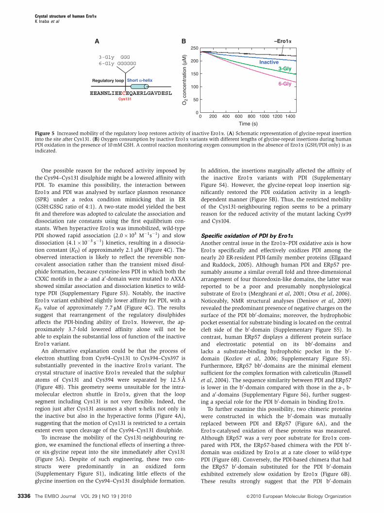
One possible reason for the reduced activity imposed by
the Cys94–Cys131 disulphide might be a lowered affinity with
PDI. To examine this possibility, the interaction between
Ero1a and PDI was analysed by surface plasmon resonance
(SPR) under a redox condition mimicking that in ER
(GSH:GSSG ratio of 4:1). A two-state model yielded the best
fit and therefore was adopted to calculate the association and
dissociation rate constants using the first equilibrium con-
stants. When hyperactive Ero1a was immobilized, wild-type
PDI showed rapid association (2.0�103 M�1s�1) and slow
dissociation (4.1�10�3 s�1) kinetics, resulting in a dissocia-
tion constant (KD) of approximately 2.1 mM (Figure 4C). The
observed interaction is likely to reflect the reversible non-
covalent association rather than the transient mixed disul-
phide formation, because cysteine-less PDI in which both the
CXXC motifs in the a- and a0-domain were mutated to AXXA
showed similar association and dissociation kinetics to wild-
type PDI (Supplementary Figure S3). Notably, the inactive
Ero1a variant exhibited slightly lower affinity for PDI, with a
KD value of approximately 7.7 mM (Figure 4C). The results
suggest that rearrangement of the regulatory disulphides
affects the PDI-binding ability of Ero1a. However, the ap-
proximately 3.7-fold lowered affinity alone will not be
able to explain the substantial loss of function of the inactive
Ero1a variant.
An alternative explanation could be that the process of
electron shuttling from Cys94–Cys131 to Cys394–Cys397 is
substantially prevented in the inactive Ero1a variant. The
crystal structure of inactive Ero1a revealed that the sulphur
atoms of Cys131 and Cys394 were separated by 12.5 A
(Figure 4B). This geometry seems unsuitable for the intra-
molecular electron shuttle in Ero1a, given that the loop
segment including Cys131 is not very flexible. Indeed, the
region just after Cys131 assumes a short a-helix not only in
the inactive but also in the hyperactive forms (Figure 4A),
suggesting that the motion of Cys131 is restricted to a certain
extent even upon cleavage of the Cys94–Cys131 disulphide.
To increase the mobility of the Cys131-neighbouring re-
gion, we examined the functional effects of inserting a three-
or six-glycine repeat into the site immediately after Cys131
(Figure 5A). Despite of such engineering, these two con-
structs were predominantly in an oxidized form
(Supplementary Figure S1), indicating little effects of the
glycine insertion on the Cys94–Cys131 disulphide formation.
In addition, the insertions marginally affected the affinity of
the inactive Ero1a variants with PDI (Supplementary
Figure S4). However, the glycine-repeat loop insertion sig-
nificantly restored the PDI oxidation activity in a length-
dependent manner (Figure 5B). Thus, the restricted mobility
of the Cys131-neighbouring region seems to be a primary
reason for the reduced activity of the mutant lacking Cys99
and Cys104.
Specific oxidation of PDI by Ero1a
Another central issue in the Ero1a–PDI oxidative axis is how
Ero1a specifically and effectively oxidizes PDI among the
nearly 20 ER-resident PDI-family member proteins (Ellgaard
and Ruddock, 2005). Although human PDI and ERp57 pre-
sumably assume a similar overall fold and three-dimensional
arrangement of four thioredoxin-like domains, the latter was
reported to be a poor and presumably nonphysiological
substrate of Ero1a (Mezghrani et al, 2001; Otsu et al, 2006).
Noticeably, NMR structural analyses (Denisov et al, 2009)
revealed the predominant presence of negative charges on the
surface of the PDI bb0-domains; moreover, the hydrophobic
pocket essential for substrate binding is located on the central
cleft side of the b0-domain (Supplementary Figure S5). In
contrast, human ERp57 displays a different protein surface
and electrostatic potential on its bb0-domains and
lacks a substrate-binding hydrophobic pocket in the b0-
domain (Kozlov et al, 2006; Supplementary Figure S5).
Furthermore, ERp57 bb0-domains are the minimal element
sufficient for the complex formation with calreticulin (Russell
et al, 2004). The sequence similarity between PDI and ERp57
is lower in the b0-domain compared with those in the a-, b-
and a0-domains (Supplementary Figure S6), further suggest-
ing a special role for the PDI b0-domain in binding Ero1a.
To further examine this possibility, two chimeric proteins
were constructed in which the b0-domain was mutually
replaced between PDI and ERp57 (Figure 6A), and the
Ero1a-catalysed oxidation of these proteins was measured.
Although ERp57 was a very poor substrate for Ero1a com-
pared with PDI, the ERp57-based chimera with the PDI b0-
domain was oxidized by Ero1a at a rate closer to wild-type
PDI (Figure 6B). Conversely, the PDI-based chimera that had
the ERp57 b0-domain substituted for the PDI b0-domain
exhibited extremely slow oxidation by Ero1a (Figure 6B).
These results strongly suggest that the PDI b0-domain
A
EEANNLIEECEQAERLGAVDESL
Short α-helix
Cys131
Regulatory loop
3-Gly GGG6-Gly GGGGGG
0
50
100
150
200
250
0 200 400 600 800 1000 1200 1400
O2
conc
entr
atio
n (μ
M)
Time (s)
B
6-Gly
3-GlyInactive
–Ero1�
Figure 5 Increased mobility of the regulatory loop restores activity of inactive Ero1a. (A) Schematic representation of glycine-repeat insertioninto the site after Cys131. (B) Oxygen consumption by inactive Ero1a variants with different lengths of glycine-repeat insertions during humanPDI oxidation in the presence of 10 mM GSH. A control reaction monitoring oxygen consumption in the absence of Ero1a (GSH/PDI only) is asindicated.
Crystal structure of human Ero1aK Inaba et al
The EMBO Journal VOL 29 | NO 19 | 2010 &2010 European Molecular Biology Organization3336
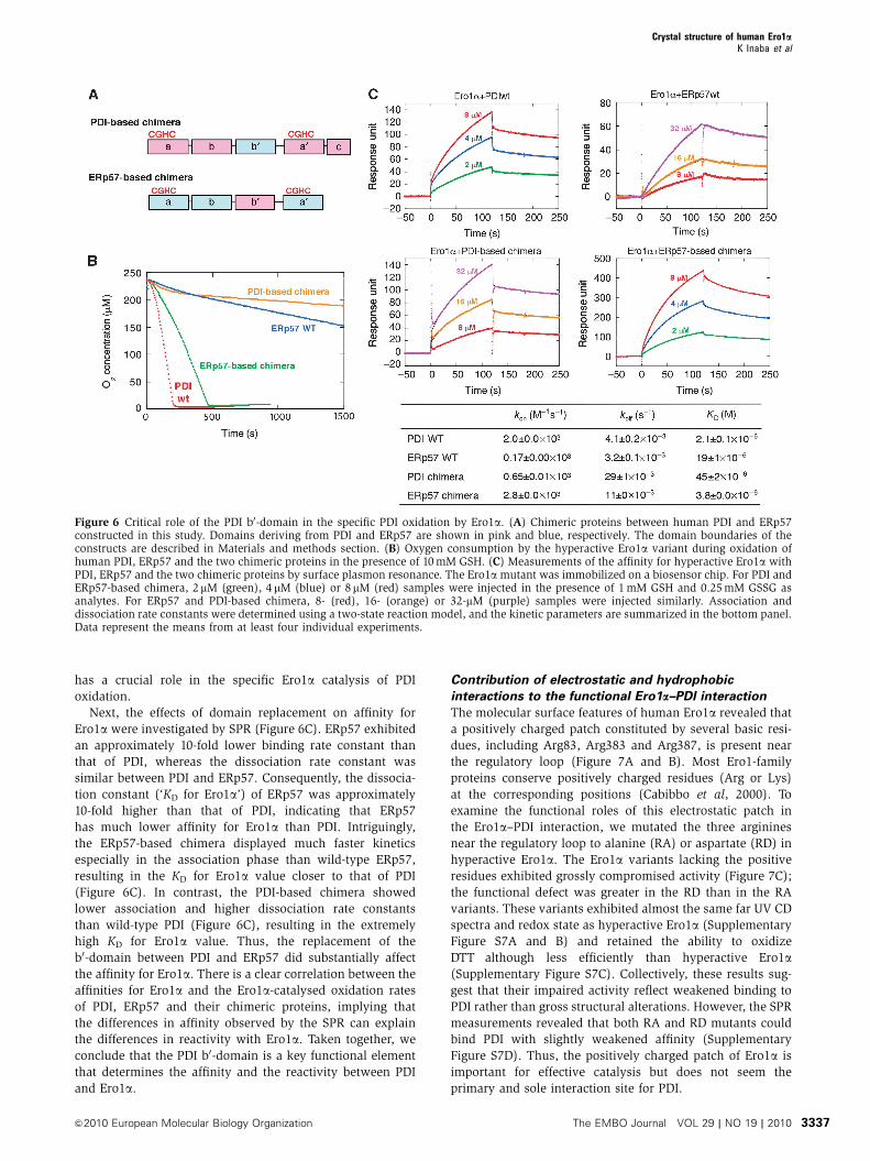
has a crucial role in the specific Ero1a catalysis of PDI
oxidation.
Next, the effects of domain replacement on affinity for
Ero1a were investigated by SPR (Figure 6C). ERp57 exhibited
an approximately 10-fold lower binding rate constant than
that of PDI, whereas the dissociation rate constant was
similar between PDI and ERp57. Consequently, the dissocia-
tion constant (‘KD for Ero1a’) of ERp57 was approximately
10-fold higher than that of PDI, indicating that ERp57
has much lower affinity for Ero1a than PDI. Intriguingly,
the ERp57-based chimera displayed much faster kinetics
especially in the association phase than wild-type ERp57,
resulting in the KD for Ero1a value closer to that of PDI
(Figure 6C). In contrast, the PDI-based chimera showed
lower association and higher dissociation rate constants
than wild-type PDI (Figure 6C), resulting in the extremely
high KD for Ero1a value. Thus, the replacement of the
b0-domain between PDI and ERp57 did substantially affect
the affinity for Ero1a. There is a clear correlation between the
affinities for Ero1a and the Ero1a-catalysed oxidation rates
of PDI, ERp57 and their chimeric proteins, implying that
the differences in affinity observed by the SPR can explain
the differences in reactivity with Ero1a. Taken together, we
conclude that the PDI b0-domain is a key functional element
that determines the affinity and the reactivity between PDI
and Ero1a.
Contribution of electrostatic and hydrophobic
interactions to the functional Ero1a–PDI interaction
The molecular surface features of human Ero1a revealed that
a positively charged patch constituted by several basic resi-
dues, including Arg83, Arg383 and Arg387, is present near
the regulatory loop (Figure 7A and B). Most Ero1-family
proteins conserve positively charged residues (Arg or Lys)
at the corresponding positions (Cabibbo et al, 2000). To
examine the functional roles of this electrostatic patch in
the Ero1a–PDI interaction, we mutated the three arginines
near the regulatory loop to alanine (RA) or aspartate (RD) in
hyperactive Ero1a. The Ero1a variants lacking the positive
residues exhibited grossly compromised activity (Figure 7C);
the functional defect was greater in the RD than in the RA
variants. These variants exhibited almost the same far UV CD
spectra and redox state as hyperactive Ero1a (Supplementary
Figure S7A and B) and retained the ability to oxidize
DTT although less efficiently than hyperactive Ero1a(Supplementary Figure S7C). Collectively, these results sug-
gest that their impaired activity reflect weakened binding to
PDI rather than gross structural alterations. However, the SPR
measurements revealed that both RA and RD mutants could
bind PDI with slightly weakened affinity (Supplementary
Figure S7D). Thus, the positively charged patch of Ero1a is
important for effective catalysis but does not seem the
primary and sole interaction site for PDI.
Figure 6 Critical role of the PDI b0-domain in the specific PDI oxidation by Ero1a. (A) Chimeric proteins between human PDI and ERp57constructed in this study. Domains deriving from PDI and ERp57 are shown in pink and blue, respectively. The domain boundaries of theconstructs are described in Materials and methods section. (B) Oxygen consumption by the hyperactive Ero1a variant during oxidation ofhuman PDI, ERp57 and the two chimeric proteins in the presence of 10 mM GSH. (C) Measurements of the affinity for hyperactive Ero1a withPDI, ERp57 and the two chimeric proteins by surface plasmon resonance. The Ero1a mutant was immobilized on a biosensor chip. For PDI andERp57-based chimera, 2mM (green), 4mM (blue) or 8mM (red) samples were injected in the presence of 1 mM GSH and 0.25 mM GSSG asanalytes. For ERp57 and PDI-based chimera, 8- (red), 16- (orange) or 32-mM (purple) samples were injected similarly. Association anddissociation rate constants were determined using a two-state reaction model, and the kinetic parameters are summarized in the bottom panel.Data represent the means from at least four individual experiments.
Crystal structure of human Ero1aK Inaba et al
&2010 European Molecular Biology Organization The EMBO Journal VOL 29 | NO 19 | 2010 3337
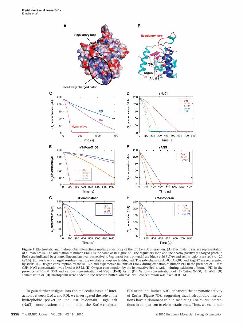
To gain further insights into the molecular basis of inter-
action between Ero1a and PDI, we investigated the role of the
hydrophobic pocket in the PDI b0-domain. High salt
(NaCl) concentrations did not inhibit the Ero1a-catalysed
PDI oxidation. Rather, NaCl enhanced the enzymatic activity
of Ero1a (Figure 7D), suggesting that hydrophobic interac-
tions have a dominant role in mediating Ero1a–PDI interac-
tions in comparison to electrostatic ones. Thus, we examined
Figure 7 Electrostatic and hydrophobic interactions mediate specificity of the Ero1a–PDI interaction. (A) Electrostatic surface representationof human Ero1a. The orientation of human Ero1a is the same as in Figure 2A. The regulatory loop and the nearby positively charged patch inEro1a are indicated by a dotted line and an oval, respectively. Regions of basic potential are blue (420 kBT/e) and acidic regions are red (o�20kBT/e). (B) Positively charged residues near the regulatory loop are highlighted. The side chains of Arg83, Arg383 and Arg387 are representedby sticks. (C) Oxygen consumption by the RD, RA and hyperactive mutants of Ero1a during oxidation of human PDI in the presence of 10 mMGSH. NaCl concentration was fixed at 0.3 M. (D) Oxygen consumption by the hyperactive Ero1a variant during oxidation of human PDI in thepresence of 10 mM GSH and various concentrations of NaCl. (E–H) As in (D). Various concentrations of (E) Triton X-100, (F) ANS, (G)somatostatin or (H) mastoparan were added to the reaction buffer, whereas NaCl concentration was fixed at 0.3 M.
Crystal structure of human Ero1aK Inaba et al
The EMBO Journal VOL 29 | NO 19 | 2010 &2010 European Molecular Biology Organization3338
effects of amphipathic chemicals or substrate proteins known
to interact with the b0-domain of PDI. Triton X-100 was
previously reported to substantially decrease the interactions
between the PDI b0-domain and model substrate peptides
(Klappa et al, 1998). This detergent inhibited the Ero1a-
catalysed oxidation of PDI (Figure 7E), without significantly
affecting DTT oxidation (Supplementary Figure S8). This
inhibitory effect was observed also at concentrations lower
than the crucial micellar concentration value (approximately
0.01%). Similar effects were obtained with 1-anilinonaphtha-
lene-8-sulphonate (ANS, Figure 7F), a fluorescence probe
that binds to hydrophobic patches on proteins (Streyer,
1965), de facto competing with Ero1 for hydrophobic pocket
of PDI. Remarkably, somatostatin and mastoparan, which
bind the hydrophobic pocket of the PDI b0-domain with KD
values of approximately 35 mM and 130 mM, respectively
(Klappa et al, 1998), also inhibited PDI oxidation in a dose-
dependent manner (Figure 7G and H). The greater effects of
somatostatin may reflect its higher affinity for PDI. These
results suggest that Ero1a competes with the model sub-
strates for PDI binding.
Discussion
Regulatory mechanism of Ero1a oxidative activity
In all eukaryotes, the ER redox must be tightly controlled.
This study elucidates the atomic resolution structures of full-
length human Ero1a in its active and inactive forms, reveal-
ing new insights into the mechanisms of disulphide-bond
formation in human cells. Although human Ero1a and yeast
Ero1p share a four-helix bundle scaffold in the catalytic core
region, and an intramolecular disulphide-shuttling mechan-
ism, there are remarkable differences in their regulation.
Thus, human Ero1a does not contain a regulatory disulphide
similar to the one (Cys150–Cys295) that in yeast Ero1p links
the nonhelical cap region, containing Cys100–Cys105 (shuttle
cysteines), to the helical core region, containing the active
site Cys352–Cys355 (see Figure 1; Supplementary Figure S9).
It is envisaged that reduction in the Cys150–Cys295 disul-
phide makes the shuttle cysteines even more mobile,
thereby increasing Ero1p activity (Sevier et al, 2007). Another
difference is observed in the disulphide that links the
first cysteines in each active triad (Cys90–349 in yeast
Ero1p and Cys85–Cys391 in human Ero1a). Loss of the
Cys90–Cys349 disulphide increased yeast Ero1p activity,
although to a lesser effect than deletion of the Cys150–
Cys295 pair (Sevier et al, 2007). In contrast, the Cys85–
Cys391 disulphide in human Ero1a resisted GSH-mediated
reduction in vivo (Appenzeller-Herzog et al 2008) and its
absence resulted in severe destabilization of Ero1a structure
and substantial decrease in Ero1a activity (K Inaba, unpub-
lished data; Benham et al, 2000; Bertoli et al, 2004; Tavender
and Bulleid, 2010).
These features imply that both human and yeast Ero1s
modulate their oxidative activity differently. In place of Ero1p
Cys150–Cys295, human Ero1a uses Cys104 and Cys131 to
regulate the mobility of the electron-shuttle loop, and hence
its overall activity. Our results confirm that the formation of
Cys94–Cys131 and possibly Cys99–Cys104 disulphides yields
an inactive form (Appenzeller-Herzog et al, 2008; Baker et al,
2008). Previous studies have suggested that Cys94 is the
primary residue to form mixed disulphides with PDI
(Bertoli et al, 2004). An important issue concerns the identity
of the partner cysteine of Cys94 during or after the disulphide
exchange with PDI. In the hyperactive form containing the
Cys94–Cys99 disulphide, Cys99 is unpaired during the redox
interaction with PDI and is probably located around the
middle of the regulatory loop, which seems to satisfactorily
accomplish the electron shuttle from Cys94–Cys99 to the
active-site disulphide Cys394–Cys397. In this regard, our
previous study showed that a Cys99Ala mutant displayed
weak dominant-negative activity in the in vivo oxidative
folding of JcM chains, possibly reflecting its impaired detach-
ment from PDI (Bertoli et al, 2004). It is inferred that Cys99
contributes to promoting the resolution of the PDI–Ero1a(Cys94) mixed disulphide presumably by transferring oxidis-
ing equivalents from FAD to PDI through the redox commu-
nications between the Cys394–Cys397 and Cys94–Cys99
pairs. In contrast, Cys131, bound to Cys94 in the inactive
isoform, is located at the edge of the regulatory loop and
juxtaposed to a short a-helix (Ala133–Leu136). The resultant
restricted mobility of Cys131 most probably underlies its low
efficiency of intramolecular electron transfer, due to its
inability to reach the inner active site. This idea is strongly
reinforced by the observation that the increased mobility of
the segment by glycine-repeat insertion after Cys131 signifi-
cantly restored the PDI oxidation activity of the mutant
(Figure 5B). Taken together, we propose that the oxidative
activity of human Ero1a is regulated by disulphide bond
rearrangements within the regulatory loop, which modulate
the ability to rapidly transfer reducing equivalents from PDI
to the FAD-containing active site.
It is interesting to question how the hyperactive (Ox1) and
inactive (Ox2) forms are interconverted in physiological
conditions. Previous pulse-chase experiments demonstrated
that the Ox2 form is intrinsically stable and predominant
in living cells (Benham et al, 2000). More recently, the
overexpression of wild-type PDI or cysteine-less (i.e. redox-
inactive) PDI was observed to markedly increase the Ox1
form (Otsu et al, 2006). Conversely, PDI knockdown clearly
diminished the Ox1/Ox2 ratio of endogenous Ero1a(Appenzeller-Herzog et al, 2008). These findings suggest
that PDI binding induces some conformational changes in
the Ero1a regulatory loop. Disulphide rearrangement within
the loop is thereby promoted, leading to the preferred occur-
rence of the Ox1 form. It is conceivable that PDI acts not
only as a substrate but also as a modulator that lowers the
energy barrier for the Ox2–Ox1 interconversion and stabilizes
Ox1 relative to Ox2. The physiological reductant essential
for this disulphide rearrangement remains an important
open question.
Molecular basis of the specific Ero1a–PDI oxidative
pathway
Another important insight gained in this study is the mole-
cular mechanism of effective and specific PDI oxidation by
Ero1a in the ER of human cells. ERp57 has a high similarity to
PDI in both amino-acid sequence (Supplementary Figure S6)
and three-dimensional structure (Dong et al, 2009;
Supplementary Figure S5). Nevertheless, Ero1a is unable to
effectively oxidize ERp57 (Figure 6B). ERp57 forms com-
plexes with calnexin (CNX) or calreticulin (CRT) in the ER
lumen (Oliver et al, 1999). It functions as a protein disulphide
isomerase or reductase for glycoproteins entering the CNX
Crystal structure of human Ero1aK Inaba et al
&2010 European Molecular Biology Organization The EMBO Journal VOL 29 | NO 19 | 2010 3339
cycle, a pivotal system that assists secretory protein folding
(Oliver et al, 1997; Ellgaard et al, 1999). Although CNX or
CRT may prevent Ero1a from oxidising ERp57 in physiologi-
cal conditions, we observed that ERp57 intrinsically has low
reactivity for Ero1a. Accordingly, addition of CRT did not
affect the kinetics of the Ero1a-catalysed oxidation of ER57
(data not shown).
Our protein engineering approach demonstrated that sub-
stitution of the ERp57 b0-domain with that of PDI markedly
enhanced the binding kinetics and reactivity of ERp57 against
Ero1a. Thus, functional association of Ero1a with the PDI b0-
domain is postulated to be necessary for effective PDI oxida-
tion by Ero1a. Consistently, the isolated a- or a0-domain of
PDI was not effectively oxidized by Ero1a (Supplementary
Figure S10). In addition, the PDI-based chimera with the
ERp57 b0-domain markedly lost its binding activity to Ero1a(Figure 6B). On the basis of the above findings, we hypothe-
sized that the specific interaction between the PDI b0-domain
and Ero1a has a role in activating the oxidase, possibly by
inducing some conformational changes. It is also conceivable
that the b0-domain has an important role in placing the a0-
domain at a suitable position for oxidization by Ero1a. In
agreement with our model, recent biochemical studies have
indicated that the PDI fragment composed of the b0-a0 do-
mains is the minimal element for a competent Ero1a–PDI
oxidative folding pathway (Wang et al, 2009).
As suggested by the different affinity for Ero1a between
PDI and ERp57, their Ero1a-binding modes would be largely
different. Indeed, altering the charge distributions on Ero1asurface inhibited its functional interactions with PDI
(Figure 7C). Moreover, the effects of salt, amphipathic che-
micals and PDI substrates indicate that Ero1a binds PDI
through the hydrophobic pocket in the b0-domain. A possible
interpretation of these findings might be that the electrostatic
interaction has an auxiliary role in guiding the active site in
the PDI a0-domain to the Ero1a shuttle disulphide in the
binary complex that is mainly owed to the close contacts
between a portion of Ero1a and the PDI hydrophobic pocket.
The inhibitory effect of a model peptide was not observed for
the yeast Ero1p–PDI oxidative system (Vitu et al, 2010),
which is in line with the idea that yeast Ero1p binds PDI
differently from human Ero1a. As human Ero1a and sub-
strates seem to compete for PDI-binding site in the b0-domain,
oxidative protein folding in human cells probably occurs in
a step-wise manner: (1) reduced PDI binds Ero1a to be
oxidized; (2) oxidized PDI dissociates from Ero1a to bind
substrate proteins and (3) finally PDI introduces disulphide
bonds into substrate proteins. ERp57 does not have a similar
hydrophobic pocket in the b0-domain, despite it can bind a
variety of proteins lacking defined secondary structure
(Jessop et al, 2007). In this context, it is to be noted that
overexpression or downregulation of ERp57 did not influence
the Ox1/Ox2 ratio in endogenous Ero1a (Appenzeller-Herzog
et al, 2008). In short, the interaction between Ero1a and PDI
is extremely specific and well-tuned in living cells.
In summary, the findings of this study elucidate the
molecular basis of regulated and specific PDI oxidation by
Ero1a. The strictly defined and regulated Ero1a–PDI oxida-
tive pathway should help prevent indiscriminate and futile
electron flows in the ER environment crowded with numer-
ous redox enzymes. Besides the potentially toxic effects of
H2O2 generated as byproducts, an uncontrolled Ero1 activity
could ultimately prevent disulphide isomerization, an essen-
tial step for proteins that fold through intermediates contain-
ing non-native disulphide bonds (Jansens et al, 2002).
Structural analysis of the Ero1a–PDI complex is currently
ongoing to further clarify how the two molecules interact and
exchange electrons. A more precise description of the thiol-
based redox networks in the ER and a comprehensive eluci-
dation of their regulatory mechanisms would further increase
our understanding of the molecular mechanisms of ER qual-
ity control, an exciting topic of study in present-day molecu-
lar cell biology.
Materials and methods
Preparation and crystallization of the hyperactive and inactiveforms of human Ero1aThe cDNA encoding human Ero1a without the signal sequence wassubcloned into the NdeI–BamHI site of the pET15b vector(Novagen). Ero1a derivatives with site-directed Cys-to-Ala muta-tions in the regulatory loop were constructed using the QuickMutagenesis Kit (Stratagene) with appropriate primer sets. Inaddition, the Cys166Ala mutation was introduced into all Ero1aconstructs to avoid aberrant formation of the disulphide-linkedEro1a dimer. Ero1a was overexpressed in Escherichia coli strainBL21(DE3). Cells were grown at 301C in Luria–Bertani (LB) mediumcontaining 50 mg/ml ampicillin and 10 mM FAD and isopropyl-b-D-thiogalactoside (IPTG) was added at a final concentration of 0.5 mMat A600¼B0.5. After an additional 4 h of shaking, cells werecollected and subsequently disrupted by micro-fluidizer (Niro SoaviPA2K) in buffer containing 50 mM Tris–HCl (pH 8.1), 0.3 M NaCland 1 mM PMSF. After clarification of the cell lysate by centrifuga-tion (12 000 r.p.m. for 20 min), the supernatant was loaded onto aNi–NTA Sepharose open column (Qiagen). After washing thecolumn with buffer containing 50 mM Tris–HCl (pH 8.1), 0.3 MNaCl and 20 mM imidazole, Ero1a was eluted with the same buffercontaining 200 mM imidazole. The eluted Ero1a sample exhibitedseveral bands overlapping around a monomer position on anonreducing SDS gel, suggesting its heterogeneous configurationscomposed of several partially reduced/oxidized species. Elutedsamples were then concentrated to 200ml by ultrafiltration usingAmicon Ultra (MWCO, 10 000; Millipore).
To prepare the fully oxidized form of Ero1a, potassiumferricyanide was added to the Ero1a solution at a final concentra-tion of 20 mM, and the solution was left on ice for 20 min. Thistreatment generated a mix of Ero1a species including monomer andvarious kinds of disulphide-linked oligomers. The sample was thenloaded onto the Superdex-200 10/300 GL column (GE Healthcare)pre-equilibrated with the same buffer containing no imidazole, andmonomeric Ero1a was thereby separated from the disulphide-linkedoligomers. The fractions containing monomeric Ero1a as a maincomponent were concentrated to 500ml and applied onto the Mono-Q 10/100 GL column (GE Healthcare) pre-equilibrated with 50 mMTris–HCl (pH 8.1). The sample was eluted with a linear NaClgradient ranging from 0 to 500 mM. This final procedure wasessential to fully purify monomeric Ero1a, which exhibited a singleband at a monomer position on a nonreducing SDS gel (Supple-mentary Figure S1). Recombinant Ero1a thus obtained was used forfunctional assays, including oxygen consumption and SPR mea-surements. Purified Ero1a exhibits the maximum absorbancearound 450 nm due to the bound FAD, and an extinction coefficientof 12.5 mM�1 cm�1 at 454 nm was used to quantify the concentra-tion of FAD-bound Ero1a (Gross et al, 2006). Determination of totalprotein concentration by the bicinchoninate (BCA) method revealedthat our preparation of FAD-bound Ero1a was more than 90 % pure.
To crystallize human Ero1a, a protein surface-engineeringtechnique was used as follows. Lysines on the surface of ourrecombinant Ero1a preparation were methylated as describedpreviously (Walter et al, 2006). The methylated sample was furtherpurified by a Superdex 200 column pre-equilibrated with 50 mMTris–HCl (pH 8.1) and 150 mM NaCl, concentrated to approximately10 mg/ml, and then dialysed against water. Methylated Ero1acrystals appeared within a week by vapour diffusion at 201C.The reservoir conditions were: 8% PEG 4000 and 50 mM imidazole
Crystal structure of human Ero1aK Inaba et al
The EMBO Journal VOL 29 | NO 19 | 2010 &2010 European Molecular Biology Organization3340
(pH 8.0) for the hyperactive form, and 20% PEG1500 and 80 mMimidazole (pH 8.0) for the inactive form. For cryoprotection, thecrystals were transferred directly to a 16% glycerol, 100-mMimidazole (pH 8.0) solution containing 20% PEG 4000 (for thehyperactive form), or 30% PEG 1500 (for the inactive form), andflash-frozen in cold nitrogen gas from a cryostat (Rigaku, Japan).
Crystallographic analysisDiffraction data were collected at the Osaka University beamlineBL44XU at SPring-8 equipped with MX225-HE (Rayonix), which isfinancially supported by the Academia Sinica and NationalSynchrotron Radiation Research Center (Taiwan, ROC). Data wereintegrated with HKL2000 (Otwinowski and Minor, 1997), and thecrystallographic parameters have been summarized in Table I.Crystals contained one molecule in an asymmetric unit for bothhyperactive and inactive Ero1a. Phase determination was made bymolecular replacement using the published structure of yeast Ero1p(PDB code 1RP4) as the search model, using the program MOLREP(Vagin and Teplyakov, 1997). The initial structural model of humanEro1a was constructed using the program ARP/warp (Perrakis et al,2001), and was refined by several cycles of manual rebuilding andrefinement using Coot (Emsley and Cowtan, 2004) and Refmac 5(Collaborative Computational Project, 1994).
Construction of chimeric proteins between PDI and ERp57To construct chimeric proteins in which the b0-domain wasmutually swapped between PDI and ERp57, KpnI and SacIrestriction sites were introduced at the start (Leu234 for PDI andPhe241 for ERp57) and end (Leu355 for PDI and Leu365 for ERp57)of the b0-domains (Supplementary Figure S4). The KpnI–SacIfragment of PDI was then inserted into the corresponding sites ofthe vectors encoding the a-, b- and a0-domains of ERp57. The PDI-based chimera with the ERp57 b0-domain was prepared in a similarmanner. The introduced restriction sites were mutated back to theiroriginal sequences using a Quick Mutagenesis Kit with appropriateprimer sets. Chimeric proteins, including wild-type PDI and ERp57,were overexpressed in E. coli strain BL21(DE3). The LB mediumcontaining 50 mg ml�1 ampicillin was incubated at 371C until IPTGwas added to the medium at a final concentration of 0.5 mM atA600¼B0.5. After an additional 4 h of shaking at 301C, cells werecollected. The cell lysate supernatant was applied to the Ni–NTASepharose open column. Fractions eluted with 200 mM imidazolewere further purified by anion exchange chromatography with aMonoQ column. Each construct was quantified using the BCAmethod.
Oxygen consumption assayOxygen consumption was measured using a Clark-type oxygenelectrode (YSI 5331). All experiments were performed at 301C in air-saturated buffer (approximately 235 mM O2) in 50 mM Tris–HCl (pH8.1) and 150 mM NaCl. Catalytic oxygen consumption was initiatedby adding each Ero1a variant to a final concentration of 2mMin a reaction mixture containing 10 mM PDI or its derivatives and10 mM GSH.
SPR measurementsThe association and dissociation rate constants (kon or koff) for thedirect binding of PDI, ERp57 or their chimeric proteins to
immobilized Ero1a were determined by SPR measurements on aBIACORE2000 system (GE Healthcare). The hyperactive or inactiveform of Ero1a was coupled to the CM5 sensor chip (GE Healthcare)using amine-coupling chemistry. As a control, one channel wascoupled with bovine serum albumin (BSA) to exclude backgroundbinding. Before monitoring analyte binding, immobilized Ero1awas activated with 2mM reduced PDI dissolved in a buffercontaining 10 mM GSH. Sensorgrams were recorded for threeconcentrations of each analyte sample (2, 4 and 8mM for PDI andERp57-based chimera; and 8, 16 and 32mM for ERp57 and PDI-based chimera) at 251C for a 2-min association phase, followed by a4-min dissociation phase. The running buffer was 20 mM HEPES-NaOH (pH 7.4), 150 mM NaCl, 0.001% Tween-20, 2 mM EDTA,1 mM GSH and 0.25 mM GSSG. All analyte samples were exchangedand diluted into this buffer previously. We verified by oxygenconsumption assay that the presence of 0.001% Tween-20 onlymodestly inhibited Ero1a-catalysed PDI oxidation (SupplementaryFigure S11). Sensorgrams were analysed by nonlinear regressionanalysis according to a two-state model using the BIAevaluation 4.1software. Experiments were replicated at least four times.
Accession numbersThe coordinates and structural factors described in this study havebeen deposited in the Protein Data Bank with ID code 3AHQ for thehyperactive form of human Ero1a and 3AHR for the inactive form ofhuman Ero1a.
Supplementary dataSupplementary data are available at The EMBO Journal Online(http://www.embojournal.org).
Acknowledgements
We thank Kazutaka Araki and Kazuhiro Nagata for stimulatingdiscussion and helpful advice on the oxygen consumption andSPR measurements; Eiki Yamashita and Atsushi Nakagawa forhelp with diffraction data collection; and Claudio Fagioli andAkiko Sato for technical support. We also thank Tomohisa Horibeand Masakazu Kikuchi for kindly providing the ERp57 expressionvector. This study was supported by a Grant-in-Aid for YoungScientists (A) from MEXT and the Yamada Science Foundation (toKI), AIRC, Fondazione Cariplo and Telethon (to RS) and by theTargeted Proteins Research Program (TPRP) from MEXT (to MS).
Author contributions: KI carried out most of the crystallographicworks and a portion of the functional analyses; SM and HIperformed most of the functional analyses; SV and RS greatlycontributed to editing the paper. MS carried out diffractiondata analysis and structure refinement. All authors discussed theresults and commented on the paper. KI supervised the work andwrote the paper.
Conflict of interest
The authors declare that they have no conflict of interest.
References
Appenzeller-Herzog C, Ellgaard L (2008) The human PDI family:versatility packed into a single fold. Biochim Biophys Acta 1783:535–548
Appenzeller-Herzog C, Riemer J, Christensen B, Sorensen ES,Ellgaard L (2008) A novel disulphide switch mechanism inEro1alpha balances ER oxidation in human cells. EMBO J 27:2977–2987
Baker KM, Chakravarthi S, Langton KP, Sheppard AM, Lu H, BulleidNJ (2008) Low reduction potential of Ero1alpha regulatory dis-ulphides ensures tight control of substrate oxidation. EMBO J 27:2988–2997
Benham AM, Cabibbo A, Fassio A, Bulleid N, Sitia R, Braakman I(2000) The CXXCXXC motif determines the folding, structure andstability of human Ero1-Lalpha. EMBO J 19: 4493–4502
Bertoli G, Simmen T, Anelli T, Molteni SN, Fesce R, Sitia R (2004)Two conserved cysteine triads in human Ero1alpha cooperate forefficient disulfide bond formation in the endoplasmic reticulum.J Biol Chem 279: 30047–30052
Cabibbo A, Pagani M, Fabbri M, Rocchi M, Farmery MR, Bulleid NJ,Sitia R (2000) ERO1-L, a human protein that favors disulfidebond formation in the endoplasmic reticulum. J Biol Chem 275:4827–4833
Collaborative Computational Project, Number 4 (1994) The CCP4suite: programs for protein crystallography. Acta Crystallogr DBiol Crystallogr 50: 760–763
Denisov AY, Maattanen P, Dabrowski C, Kozlov G, Thomas DY,Gehring K (2009) Solution structure of the bb0 domains of humanprotein disulfide isomerase. FEBS J 276: 1440–1449
Crystal structure of human Ero1aK Inaba et al
&2010 European Molecular Biology Organization The EMBO Journal VOL 29 | NO 19 | 2010 3341
Dias-Gunasekara S, Gubbens J, van Lith M, Dunne C, Williams JA,Kataky R, Scoones D, Lapthorn A, Bulleid NJ, Benham AM(2005) Tissue-specific expression and dimerization of the endo-plasmic reticulum oxidoreductase Ero1beta. J Biol Chem 280:33066–33075
Dong G, Wearsch PA, Peaper DR, Cresswell P, Reinisch KM (2009)Insights into MHC class I peptide loading from the structure of thetapasin-ERp57 thiol oxidoreductase heterodimer. Immunity 30:21–32
Dym O, Eisenberg D (2001) Sequence-structure analysis of FAD-containing proteins. Protein Sci 10: 1712–1728
Ellgaard L, Molinari M, Helenius A (1999) Setting the standards:quality control in the secretory pathway. Science 286: 1882–1888
Ellgaard L, Ruddock LW (2005) The human protein disulphideisomerase family: substrate interactions and functional proper-ties. EMBO Rep 6: 28–32
Emsley P, Cowtan K (2004) Coot: model-building tools for molecu-lar graphics. Acta Crystallogr D Biol Crystallogr 60: 2126–2132
Fass D (2008) The Erv family of sulfhydryl oxidases. BiochimBiophys Acta 1783: 557–566
Frand AR, Kaiser CA (1998) The ERO1 gene of yeast is requiredfor oxidation of protein dithiols in the endoplasmic reticulum.Mol Cell 1: 161–170
Gross E, Kastner DB, Kaiser CA, Fass D (2004) Structure of Ero1p,source of disulfide bonds for oxidative protein folding in the cell.Cell 117: 601–610
Gross E, Sevier CS, Heldman N, Vitu E, Bentzur M, Kaiser CA,Thorpe C, Fass D (2006) Generating disulfides enzymatically:reaction products and electron acceptors of the endoplasmicreticulum thiol oxidase Ero1p. Proc Natl Acad Sci USA 103:299–304
Harding HP, Zhang Y, Zeng H, Novoa I, Lu PD, Calfon M, Sadri N,Yun C, Popko B, Paules R, Stojdl DF, Bell JC, Hettmann T,Leiden JM, Ron D (2003) An integrated stress response regulatesamino acid metabolism and resistance to oxidative stress. MolCell 11: 619–633
Hatahet F, Ruddock LW (2009) Protein disulfide isomerase: a criticalevaluation of its function in disulfide bond formation.Antioxidants & Redox Signaling 11: 2807–2850
Haynes CM, Titus EA, Cooper AA (2004) Degradation of misfoldedproteins prevents ER-derived oxidative stress and cell death. MolCell 15: 767–776
Heckler EJ, Rancy PC, Kodali VK, Thorpe C (2008) Generatingdisulfides with the Quiescin-sulfhydryl oxidases. BiochimBiophys Acta 1783: 567–577
Inaba K, Ito K (2008) Structure and mechanisms of the DsbB–DsbAdisulfide bond generation machine. Biochim Biophys Acta 1783:520–529
Jansens A, van Duijn E, Braakman I (2002) Coordinated nonvector-ial folding in a newly synthesized multidomain protein. Science298: 2401–2403
Jessop CE, Chakravarthi S, Garbi N, Hammerling GJ, Lovell S,Bulleid NJ (2007) ERp57 is essential for efficient folding ofglycoproteins sharing common structural domains. EMBO J 26:28–40
Karala AR, Lappi AK, Saaranen MJ, Ruddock LW (2009) Efficientperoxide-mediated oxidative refolding of a protein at physiologi-cal pH and implications for oxidative folding in the endoplasmicreticulum. Antioxid Redox Signal 11: 963–970
Klappa P, Ruddock LW, Darby NJ, Freedman RB (1998) The b0
domain provides the principal peptide-binding site of proteindisulfide isomerase but all domains contribute to binding ofmisfolded proteins. EMBO J 17: 927–935
Kozlov G, Maattanen P, Schrag JD, Pollock S, Cygler M, Nagar B,Thomas DY, Gehring K (2006) Crystal structure of the bb0
domains of the protein disulfide isomerase ERp57. Structure 14:1331–1339
Marciniak SJ, Yun CY, Oyadomari S, Novoa I, Zhang Y, Jungreis R,Nagata K, Harding HP, Ron D (2004) CHOP induces death bypromoting protein synthesis and oxidation in the stressed en-doplasmic reticulum. Genes Dev 18: 3066–3077
Mezghrani A, Fassio A, Benham A, Simmen T, Braakman I, Sitia R(2001) Manipulation of oxidative protein folding and PDI redoxstate in mammalian cells. EMBO J 20: 6288–6296
Nguyen VD, Wallis K, Howard MJ, Haapalainen AM, Salo KE,Saaranen MJ, Sidhu A, Wierenga RK, Freedman RB, RuddockLW, Williamson RA (2008) Alternative conformations of the x
region of human protein disulphide-isomerase modulateexposure of the substrate binding b0 domain. J Mol Biol 383:1144–1155
Oliver JD, Roderick HL, Llewllyn DH, High S (1999) ERp57 func-tions as a subunit of specific complexes formed with the ERlectins calreticulin and calnexin. Mol. Biol. Cell 10: 2573–2582
Oliver JD, van der Wal FJ, Bulleid NJ, High S (1997) Interaction ofthe thiol-dependent reductase ERp57 with nascent glycoproteins.Science 275: 86–88
Otsu M, Bertoli G, Fagioli C, Guerini-Rocco E, Nerini-Molteni S,Ruffato E, Sitia R (2006) Dynamic retention of Ero1alpha andEro1beta in the endoplasmic reticulum by interactions with PDIand ERp44. Antioxid Redox Signal 8: 274–282
Otwinowski Z, Minor W (1997) Processing of X-ray Diffraction DataCollected in Oscillation Mode. Methods Enzymol 276: 307–326
Pagani M, Fabbri M, Benedetti C, Fassio A, Pilati S, Bulleid NJ,Cabibbo A, Sitia R (2000) Endoplasmic reticulum oxidoreductin1-lbeta (ERO1-Lbeta), a human gene induced in the course of theunfolded protein response. J Biol Chem 275: 23685–23692
Perrakis A, Harkiolaki M, Wilson KS, Lamzin VS (2001) ARP/wARPand molecular replacement. Acta Crystallogr D Biol Crystallogr57: 1445–1450
Pollard MG, Travers KJ, Weissman JS (1998) Ero1p: a novel andubiquitous protein with an essential role in oxidative proteinfolding in the endoplasmic reticulum. Mol Cell 1: 171–182
Russell SJ, Ruddock LW, Salo KE, Oliver JD, Roebuck QP, LlewellynDH, Roderick HL, Koivunen P, Myllyharju J, High S (2004) Theprimary substrate binding site in the b0 domain of ERp57 isadapted for endoplasmic reticulum lectin association. J BiolChem 279: 18861–18869
Serve O, Kamiya Y, Maeno A, Nakano M, Murakami C, Sasakawa H,Yamaguchi Y, Harada T, Kurimoto E, Yagi-Utsumi M, Iguchi T,Inaba K, Kikuchi J, Asami O, Kajino T, Oka T, Nakasako M, KatoK (2010) Redox-dependent domain rearrangement of proteindisulfide isomerase coupled with exposure of its substrate-bind-ing hydrophobic surface. J Mol Biol 396: 361–374
Sevier CS, Cuozzo JW, Vala A, Aslund F, Kaiser CA (2001) Aflavoprotein oxidase defines a new endoplasmic reticulum path-way for biosynthetic disulphide bond formation. Nat Cell Biol 3:874–882
Sevier CS, Kadokura H, Tam VC, Beckwith J, Fass D, Kaiser CA(2005) The prokaryotic enzyme DsbB may share key structuralfeatures with eukaryotic disulfide bond forming oxidoreductases.Protein Sci 14: 1630–1642
Sevier CS, Kaiser CA (2006) Conservation and diversity of thecellular disulfide bond formation pathways. Antioxid RedoxSignal 8: 797–811
Sevier CS, Kaiser CA (2008) Ero1 and redox homeostasis in theendoplasmic reticulum. Biochim Biophys Acta 1783: 549–556
Sevier CS, Qu H, Heldman N, Gross E, Fass D, Kaiser CA (2007)Modulation of cellular disulfide-bond formation and the ER redoxenvironment by feedback regulation of Ero1. Cell 129: 333–344
Streyer L (1965) The inetraction of a naphthalene dye with apo-myoglobin and apohemoglobin. A fluorecent probe of non-polarbinding sites. J Mol Biol 13: 482–495
Tavender TJ, Bulleid NJ (2010) Molecular mechanisms regulatingoxidative activity of the Ero1 family in the endoplasmic reticu-lum. Antioxid Redox Signal (in press)
Tian G, Kober FX, Lewandrowski U, Sickmann A, Lennarz WJ,Schindelin H (2008) The catalytic activity of protein-disulfideisomerase requires a conformationally flexible molecule. J BiolChem 283: 33630–33640
Tian G, Xiang S, Noiva R, Lennarz WJ, Schindelin H (2006) Thecrystal structure of yeast protein disulfide isomerase suggestscooperativity between its active sites. [erratum appears in Cell2006 Mar 10;124(5):1085–8]. Cell 124: 61–73
Tu BP, Weissman JS (2004) Oxidative protein folding in eukaryotes:mechanisms and consequences. J Cell Biol 164: 341–346
Vagin A, Teplyakov A (1997) MOLREP: an automated program formolecular replacement. J Appl Crystallogr 30: 1022–1025
Veal EA, Day AM, Morgan BA (2007) Hydrogen peroxide sensingand signaling. Mol Cell 26: 1–14
Vitu E, Kim S, Sevier CS, Lutzky O, Heldman N, Bentzur M,Unger T, Yonda M, Kaiser CA, Fass D (2010) Oxidative activityof yeast Ero1p on protein disulfide isomerase and relatedoxidoreductases of the endoplasmic reticulum. J Biol Chem 285:18155–18165
Crystal structure of human Ero1aK Inaba et al
The EMBO Journal VOL 29 | NO 19 | 2010 &2010 European Molecular Biology Organization3342
Walter TS, Meier C, Assenberg R, Au KF, Ren J, Verma A, NettleshipJE, Owens RJ, Stuart DI, Grimes JM (2006) Lysine methylation asa routine rescue strategy for protein crystallization. Structure 14:1617–1622
Wang C, Chen S, Wang X, Wang L, Wallis AK, Freedman RB, WangCC (2010) Plasticity of human protein disulfide isomerase: evi-dence for mobility around the x-linker region and its functionalsignificance. J Biol Chem 285: 26788–26797
Wang L, Li SJ, Sidhu A, Zhu L, Liang Y, Freedman RB, Wang CC(2009) Reconstitution of human Ero1-Lalpha/protein-disulfideisomerase oxidative folding pathway in vitro. Position-dependentdifferences in role between the a and a0 domains of protein-disulfide isomerase. J Biol Chem 284: 199–206
Zito E, Chin K-T, Blais H, Ron D (2010) Ero1-beta, a pancreas-specific disulfide oxidase, promotes insulin biogenesis andglucose homeostasis. J Cell Biol 188: 821–832
Crystal structure of human Ero1aK Inaba et al
&2010 European Molecular Biology Organization The EMBO Journal VOL 29 | NO 19 | 2010 3343
Top Related