X-ray micro-diffraction and its applications in material ... 2005 pdf/x-primer/dooryhee... · X-ray...
65
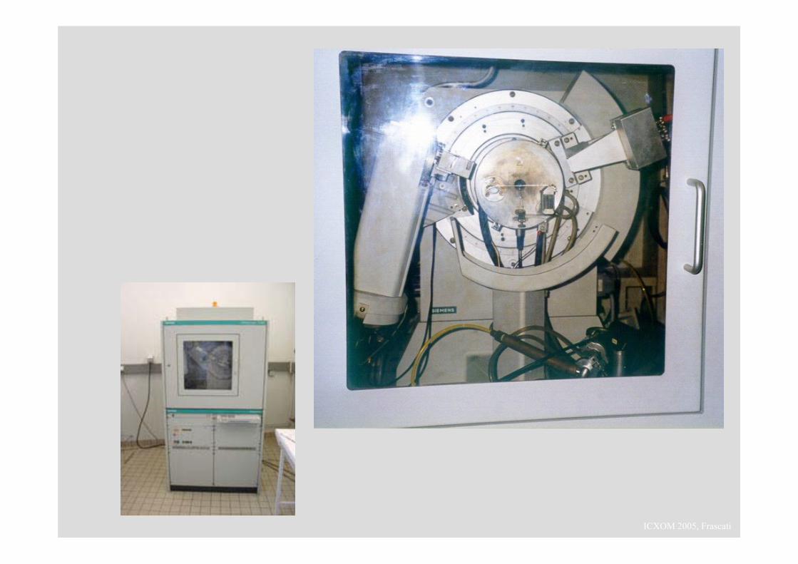
ICXOM 2005, Frascati X-ray micro-diffraction and its applications in material (archaeological) science Eric Dooryhée, CNRS Grenoble
Transcript of X-ray micro-diffraction and its applications in material ... 2005 pdf/x-primer/dooryhee... · X-ray...
ICXOM 2005, Frascati
X-ray micro-diffraction and its applications in material (archaeological) science
Eric Dooryhée, CNRS Grenoble
ICXOM 2005, Frascati
brief introduction to X-ray diffraction
ICXOM 2005, Frascati
Detector
crystal
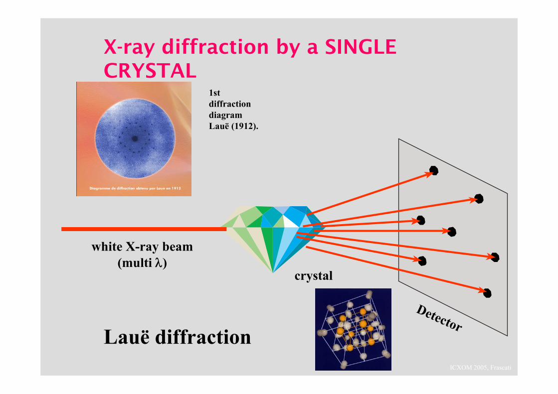
X-ray diffraction by a SINGLE CRYSTAL
1st diffraction diagramLauë (1912).
white X-ray beam(multi λ)
Lauë diffraction
ICXOM 2005, Frascati
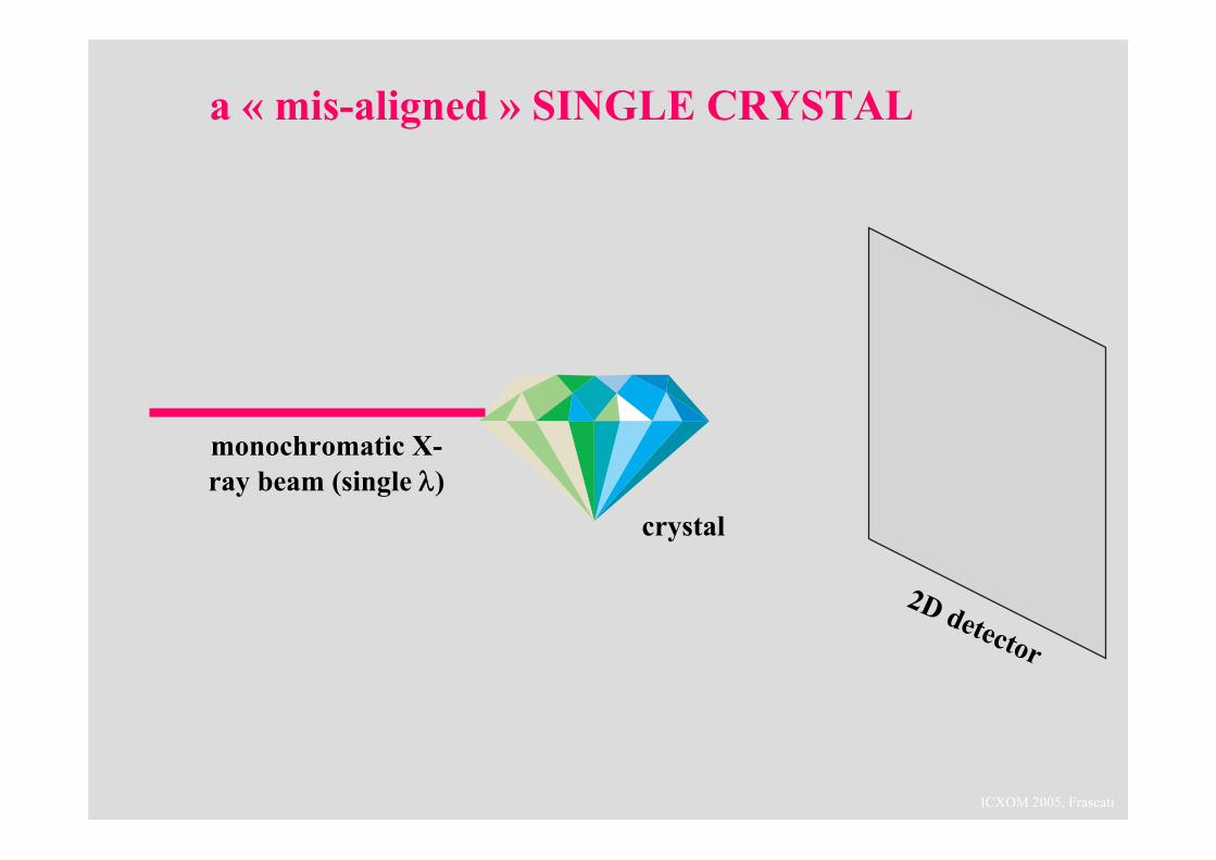
a « mis-aligned » SINGLE CRYSTAL
2D detector
crystal
monochromatic X-ray beam (single λ)
ICXOM 2005, Frascati
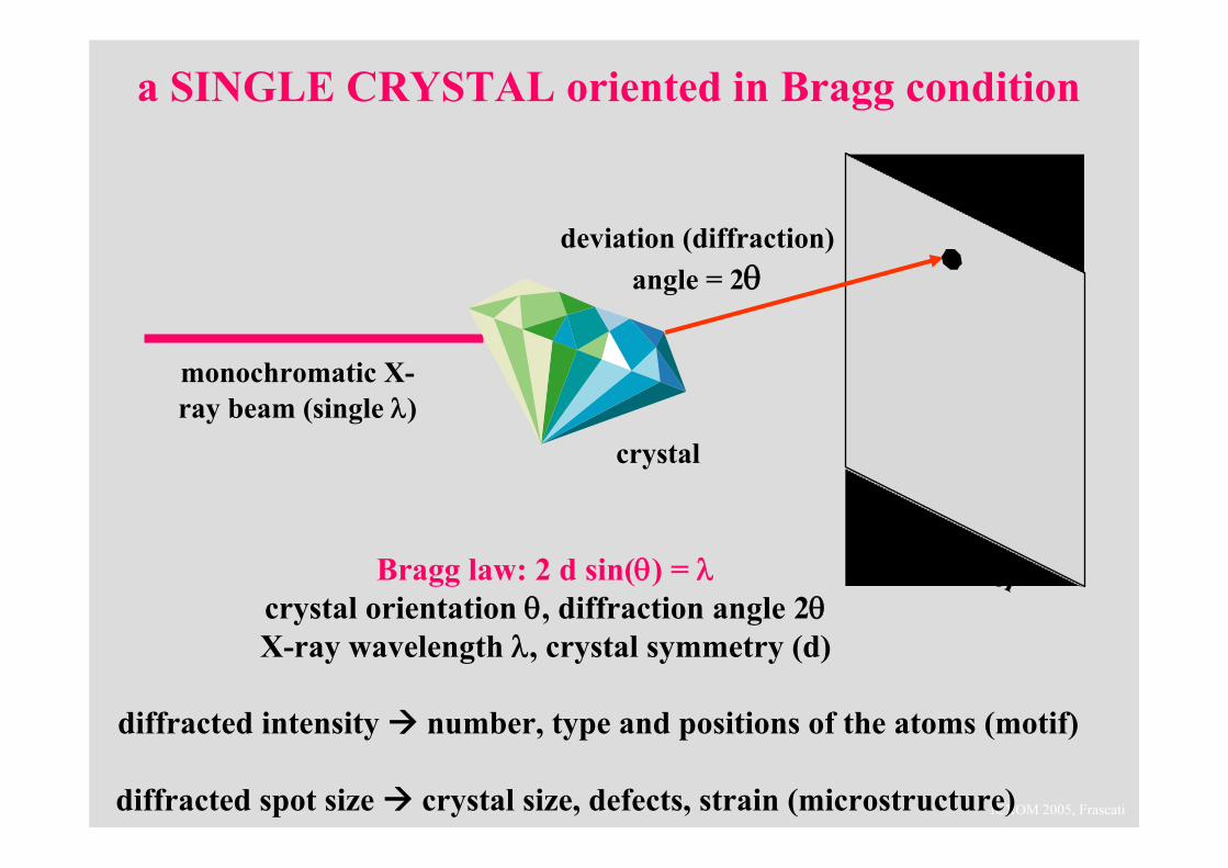
a SINGLE CRYSTAL oriented in Bragg condition
2D detector
crystal
monochromatic X-ray beam (single λ)
Bragg law: 2 d sin(θ) = λcrystal orientation θ, diffraction angle 2θX-ray wavelength λ, crystal symmetry (d)
diffracted intensity number, type and positions of the atoms (motif)
diffracted spot size crystal size, defects, strain (microstructure)
deviation (diffraction) angle = 2θ
ICXOM 2005, Frascati
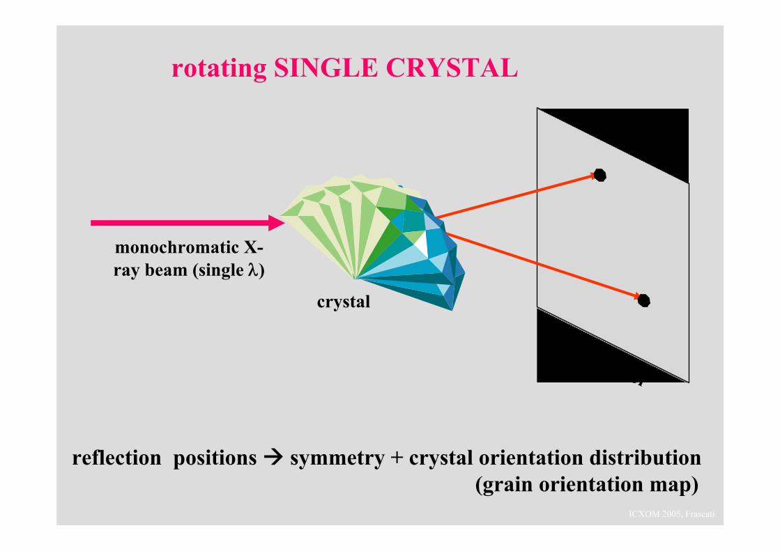
rotating SINGLE CRYSTAL
2D detector
crystal
monochromatic X-ray beam (single λ)
reflection positions symmetry + crystal orientation distribution(grain orientation map)
ICXOM 2005, Frascati
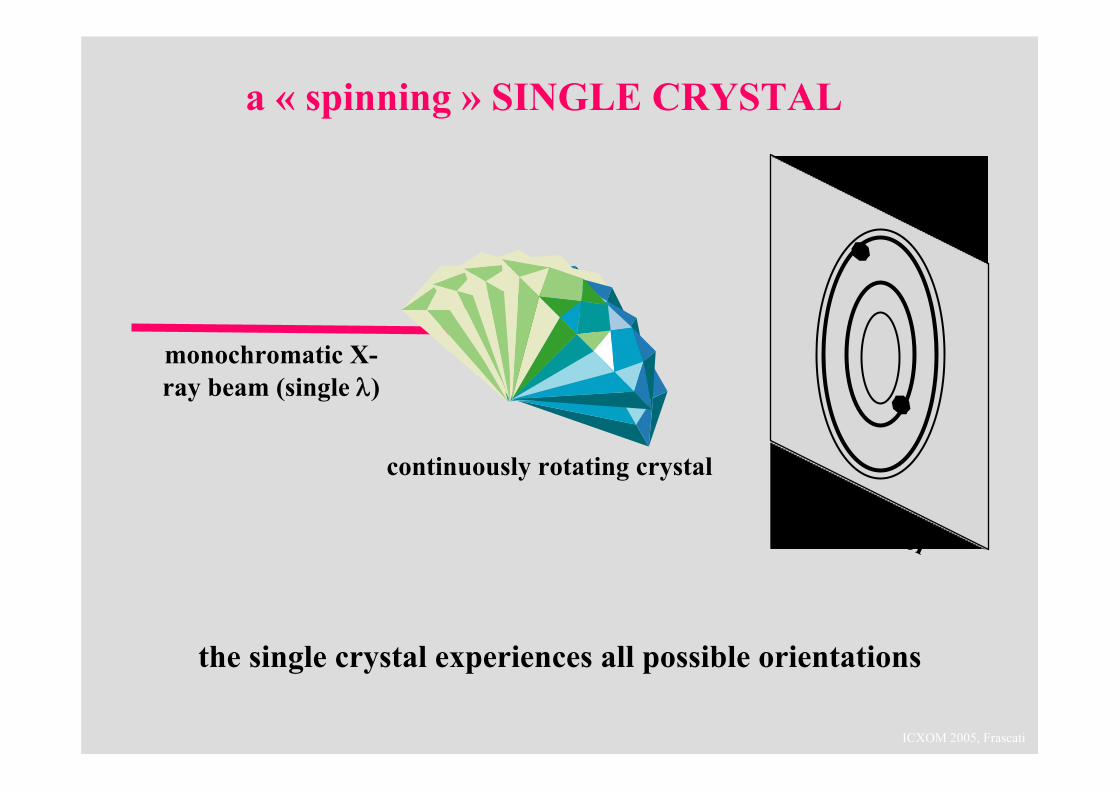
a « spinning » SINGLE CRYSTAL
2D detector
continuously rotating crystal
monochromatic X-ray beam (single λ)
the single crystal experiences all possible orientations
ICXOM 2005, Frascati
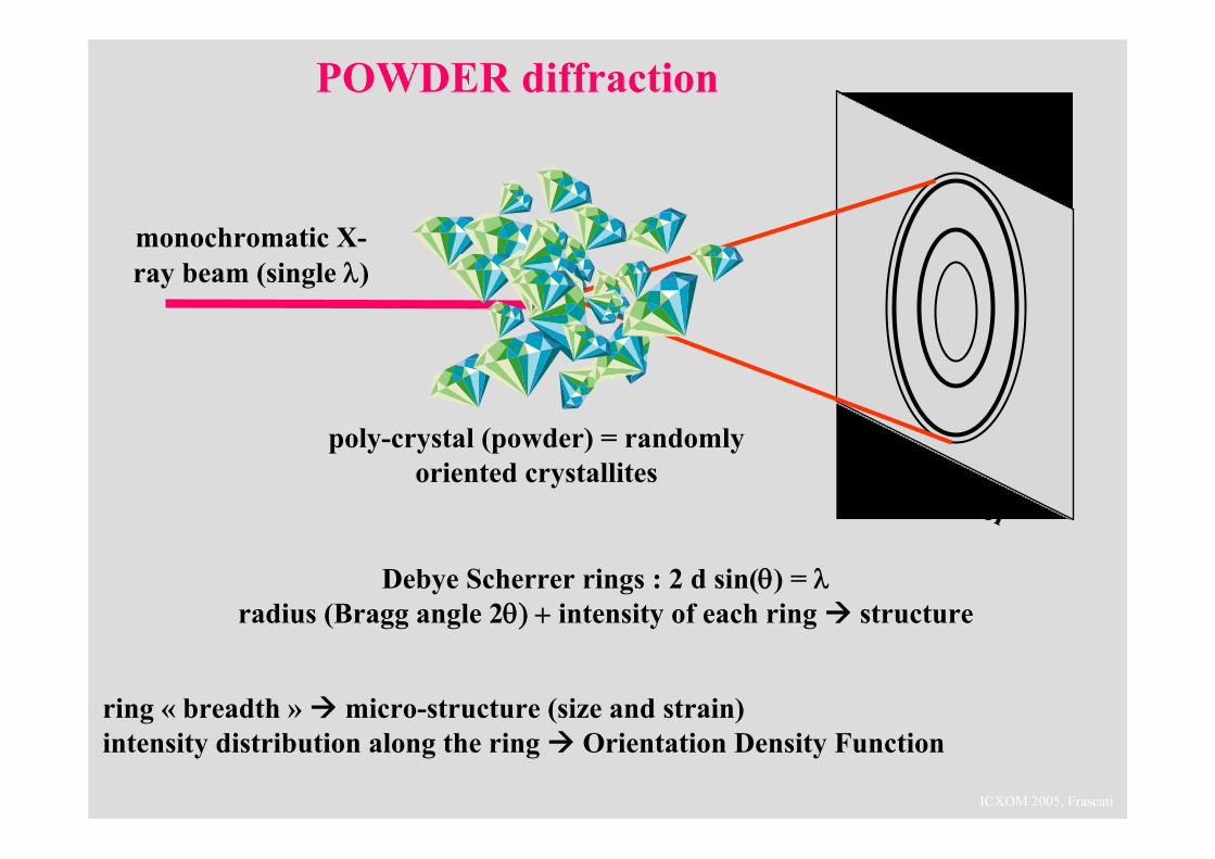
POWDER diffraction
2D detector
poly-crystal (powder) = randomlyoriented crystallites
monochromatic X-ray beam (single λ)
Debye Scherrer rings : 2 d sin(θ) = λradius (Bragg angle 2θ) + intensity of each ring structure
ring « breadth » micro-structure (size and strain)intensity distribution along the ring Orientation Density Function
ICXOM 2005, Frascati
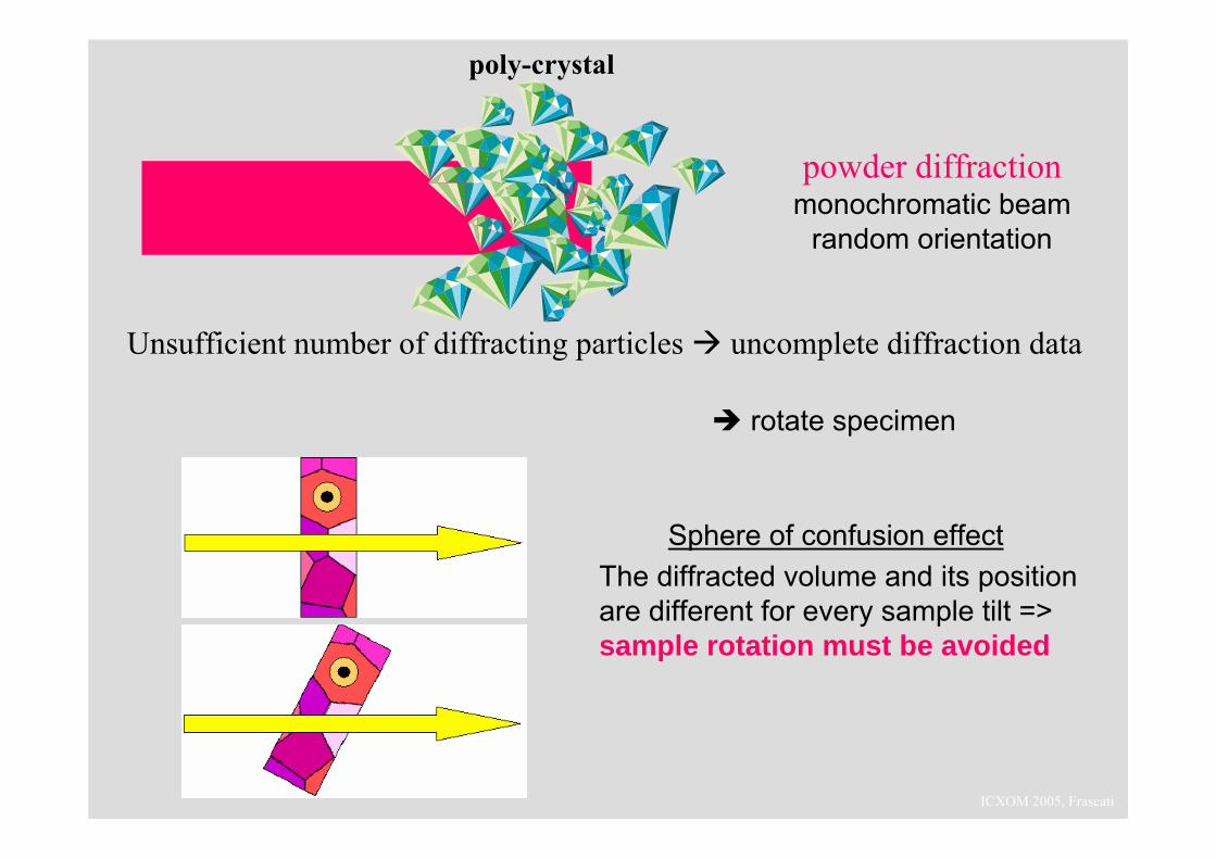
Unsufficient number of diffracting particles uncomplete diffraction data
powder diffractionmonochromatic beam
random orientation
poly-crystal
Sphere of confusion effectThe diffracted volume and its position are different for every sample tilt => sample rotation must be avoided
rotate specimen
ICXOM 2005, Frascati
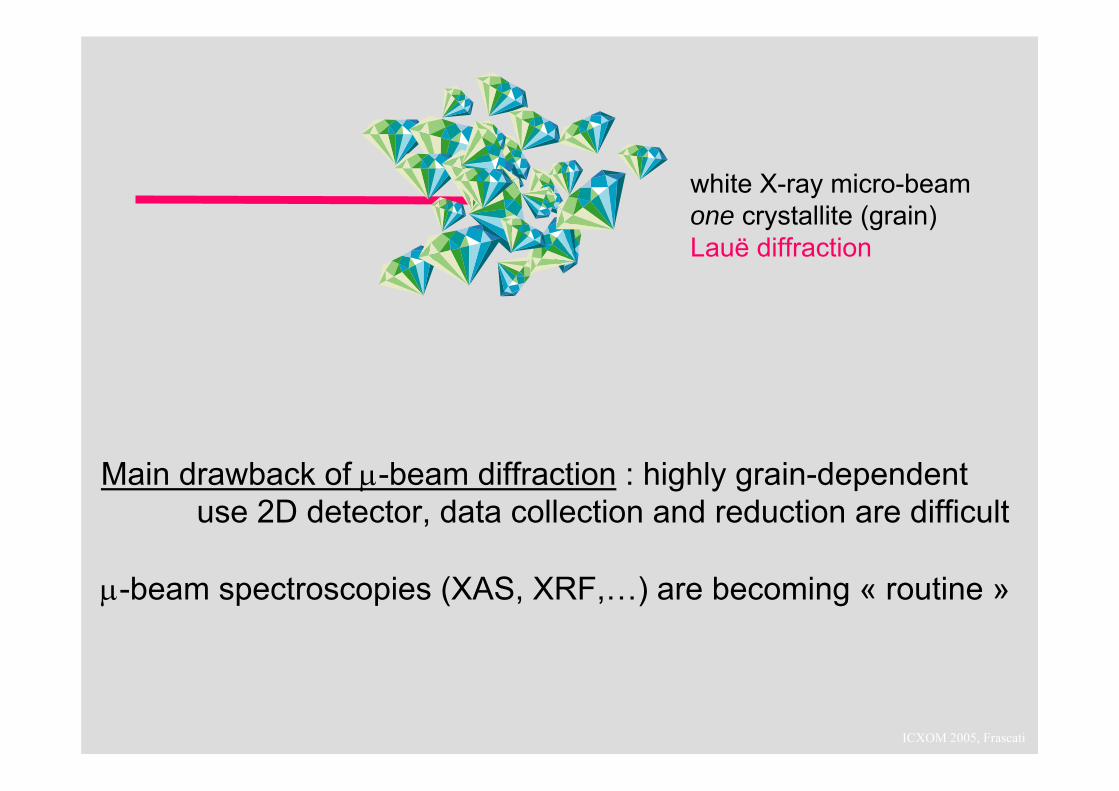
white X-ray micro-beamone crystallite (grain)Lauë diffraction
Main drawback of μ-beam diffraction : highly grain-dependentuse 2D detector, data collection and reduction are difficult
μ-beam spectroscopies (XAS, XRF,…) are becoming « routine »
ICXOM 2005, Frascati
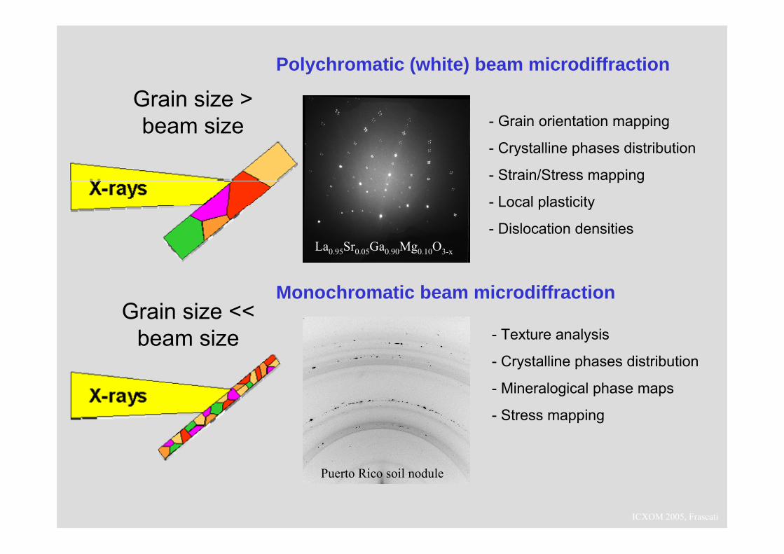
La0.95Sr0.05Ga0.90Mg0.10O3-x
Puerto Rico soil nodule
- Grain orientation mapping
- Crystalline phases distribution
- Strain/Stress mapping
- Local plasticity
- Dislocation densities
Polychromatic (white) beam microdiffraction
Monochromatic beam microdiffraction
Grain size > beam size
Grain size << beam size - Texture analysis
- Crystalline phases distribution
- Mineralogical phase maps
- Stress mapping
ICXOM 2005, Frascati
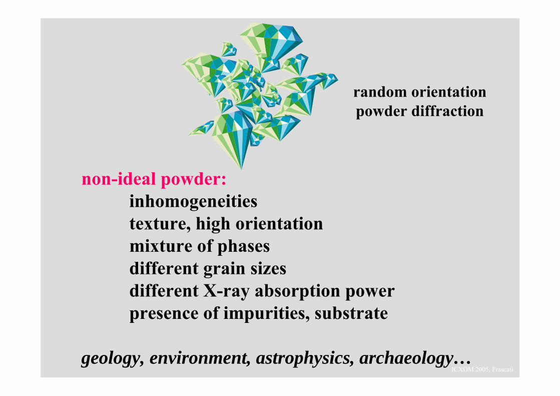
random orientationpowder diffraction
non-ideal powder:inhomogeneitiestexture, high orientationmixture of phasesdifferent grain sizesdifferent X-ray absorption powerpresence of impurities, substrate
geology, environment, astrophysics, archaeology…
ICXOM 2005, Frascati
brief introduction to X-ray diffraction
the possibility for μ-diffraction
ICXOM 2005, Frascati
ICXOM 2005, Frascati
ICXOM 2005, Frascati
ICXOM 2005, Frascati
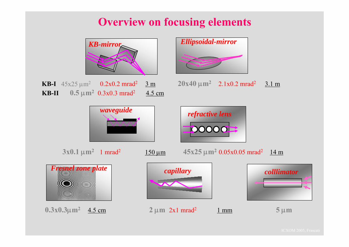
KB-mirror
refractive lenswaveguide
45x25 μm2
Overview on focusing elementsEllipsoidal-mirror
0.2x0.2 mrad2
0.3x0.3 mrad20.5 μm2KB-IKB-II
20x40 μm2 2.1x0.2 mrad23 m4.5 cm
3.1 m
3x0.1 μm2 1 mrad2 150 μm 45x25 μm2 0.05x0.05 mrad2 14 m
colllimatorFresnel zone plate
5 μm0.3x0.3μm2 4.5 cm
capillary
2 μm 1 mm2x1 mrad2
ICXOM 2005, Frascati
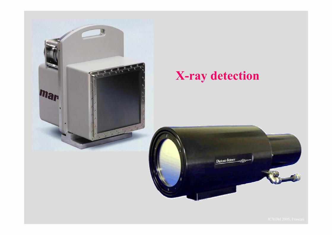
X-ray detection
ICXOM 2005, Frascati
brief introduction to X-ray diffraction
the need for μ-diffraction
ICXOM 2005, Frascati
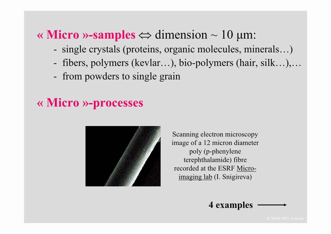
« Micro »-samples ⇔ dimension ~ 10 μm:- single crystals (proteins, organic molecules, minerals…)- fibers, polymers (kevlar…), bio-polymers (hair, silk…),… - from powders to single grain
« Micro »-processes
Scanning electron microscopyimage of a 12 micron diameter
poly (p-phenyleneterephthalamide) fibre
recorded at the ESRF Micro-imaging lab (I. Snigireva)
4 examples
ICXOM 2005, Frascati
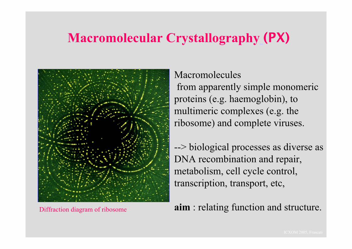
Macromolecular Crystallography (PX)
Diffraction diagram of ribosome
Macromoleculesfrom apparently simple monomericproteins (e.g. haemoglobin), to multimeric complexes (e.g. theribosome) and complete viruses.
--> biological processes as diverse as DNA recombination and repair, metabolism, cell cycle control, transcription, transport, etc,
aim : relating function and structure.
ICXOM 2005, Frascatistructure of a ribosome
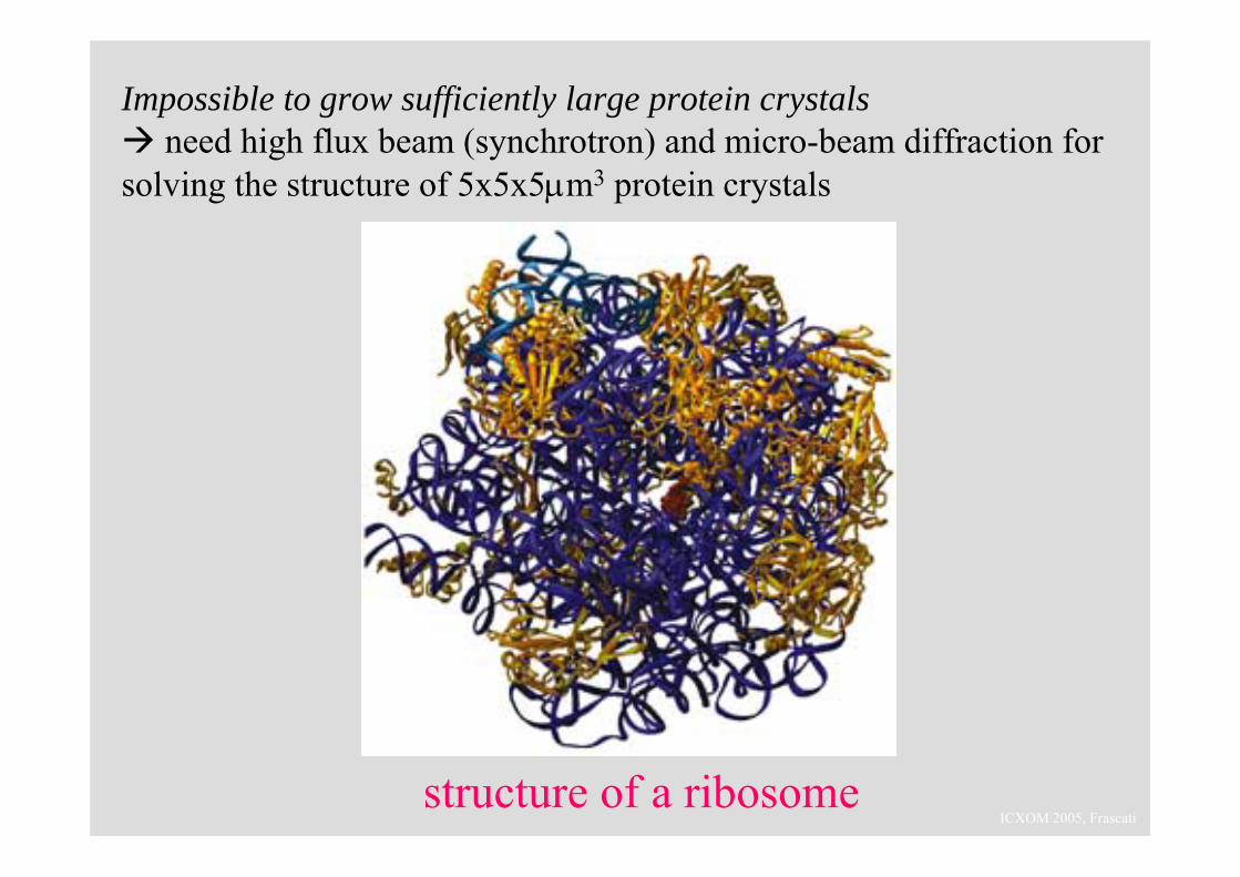
Impossible to grow sufficiently large protein crystalsneed high flux beam (synchrotron) and micro-beam diffraction for
solving the structure of 5x5x5μm3 protein crystals

ICXOM 2005, Frascati
Riekel, C., M. Müller and F. Vollrath (1999) Macromolecules 32(13).Riekel, C. and F. Vollrath (2001) Int. J. Biol. Macrom. 29(3).
Extrusion of a single Nephila silk fibre of about 5 micron diameter. In situ micro-diffraction during forced silking from live spiders.
ICXOM 2005, Frascati
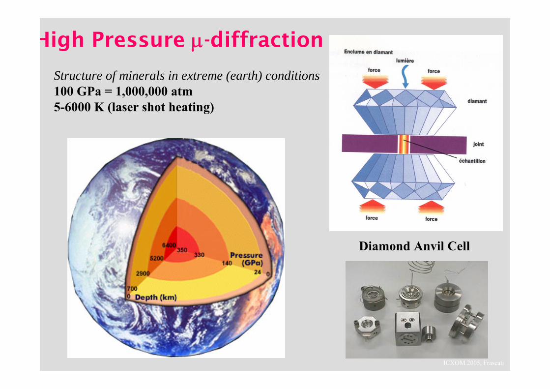
High Pressure μ-diffraction
Structure of minerals in extreme (earth) conditions100 GPa = 1,000,000 atm5-6000 K (laser shot heating)
Diamond Anvil Cell
ICXOM 2005, Frascati
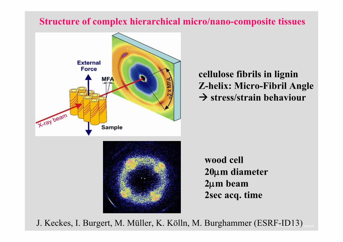
Structure of complex hierarchical micro/nano-composite tissues
J. Keckes, I. Burgert, M. Müller, K. Kölln, M. Burghammer (ESRF-ID13)
cellulose fibrils in ligninZ-helix: Micro-Fibril Angle
stress/strain behaviour
wood cell20μm diameter2μm beam2sec acq. time
ICXOM 2005, Frascati
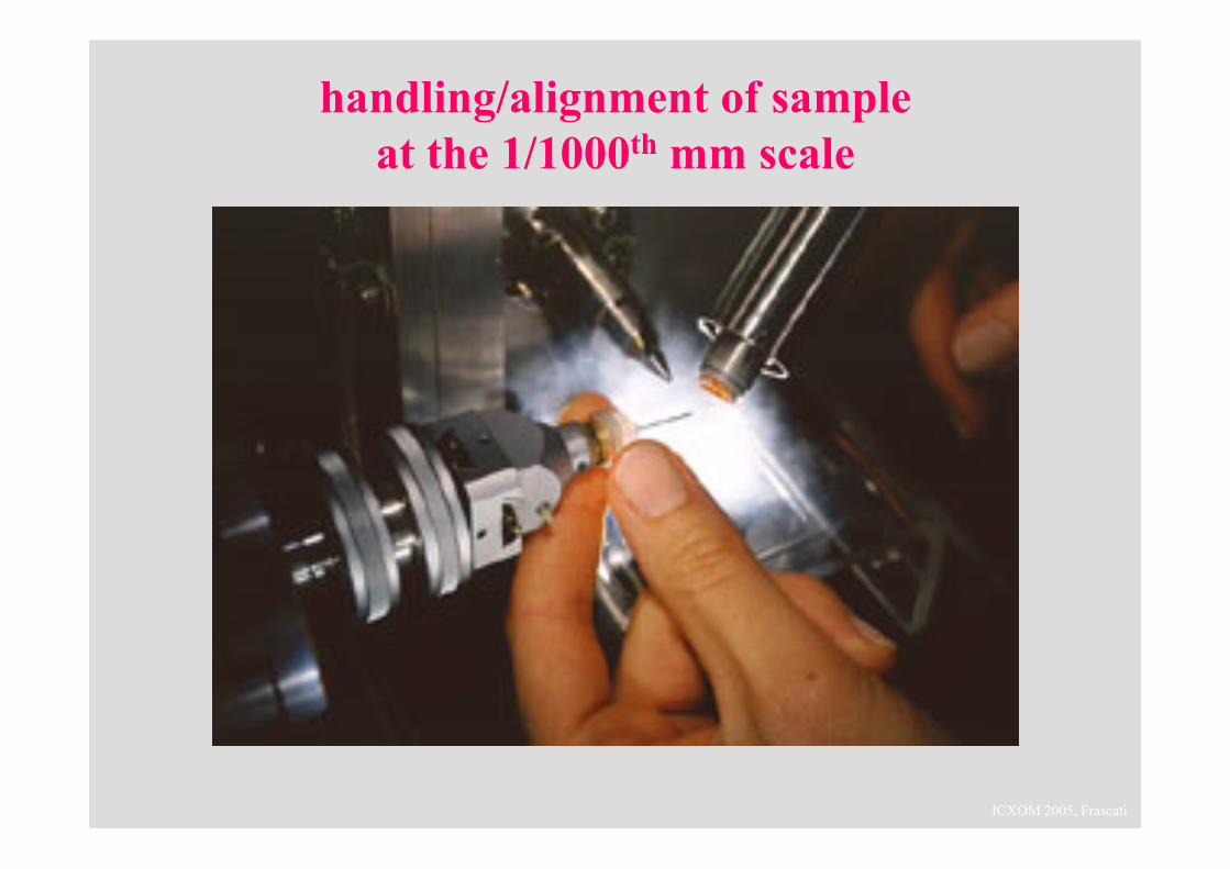
handling/alignment of sampleat the 1/1000th mm scale
ICXOM 2005, Frascati
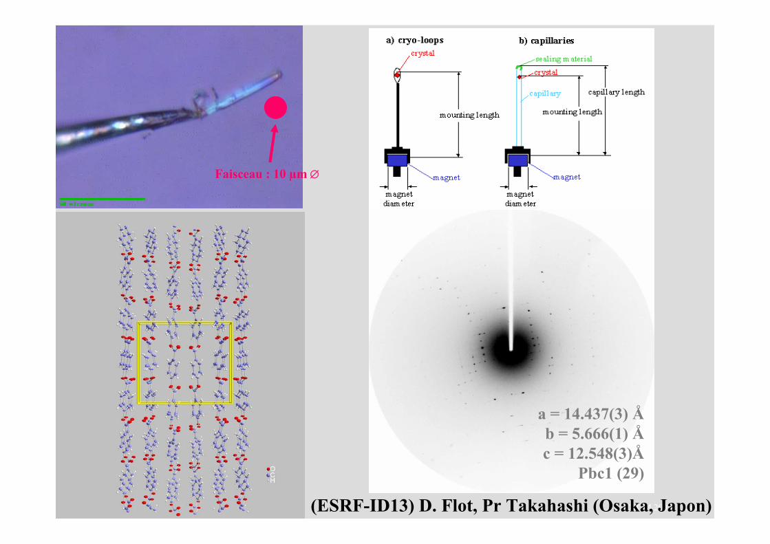
a = 14.437(3) Åb = 5.666(1) Åc = 12.548(3)Å
Pbc1 (29)
(ESRF-ID13) D. Flot, Pr Takahashi (Osaka, Japon)
Faisceau : 10 µm ∅
ICXOM 2005, Frascati
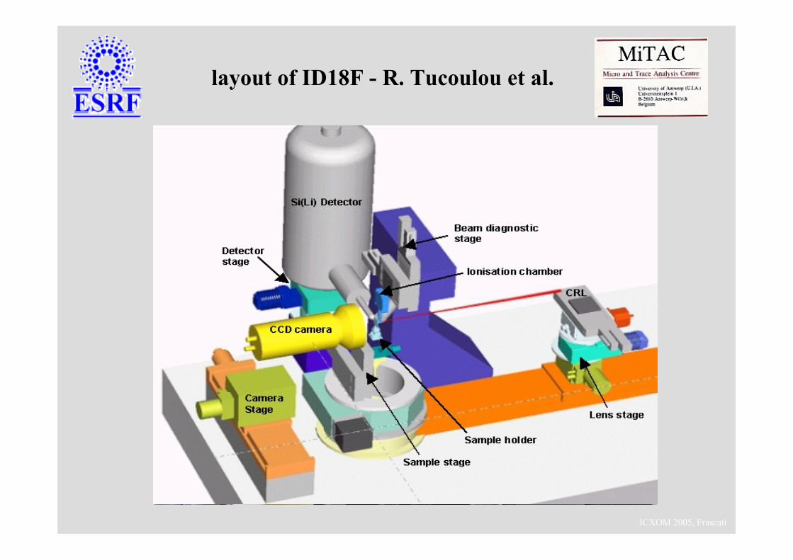
layout of ID18F - R. Tucoulou et al.
ICXOM 2005, Frascati
Materialsscience Biology
Environment
Physics
Medecine
ChemistryArt & Archaeology
© E. Chalmin
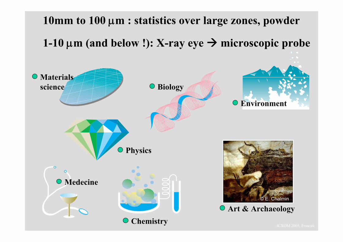
10mm to 100 μm : statistics over large zones, powder
1-10 μm (and below !): X-ray eye microscopic probe
ICXOM 2005, Frascati
brief introduction to X-ray diffraction
the need for micro-beam X-ray diffraction
applications to Cultural Heritage
ICXOM 2005, Frascati
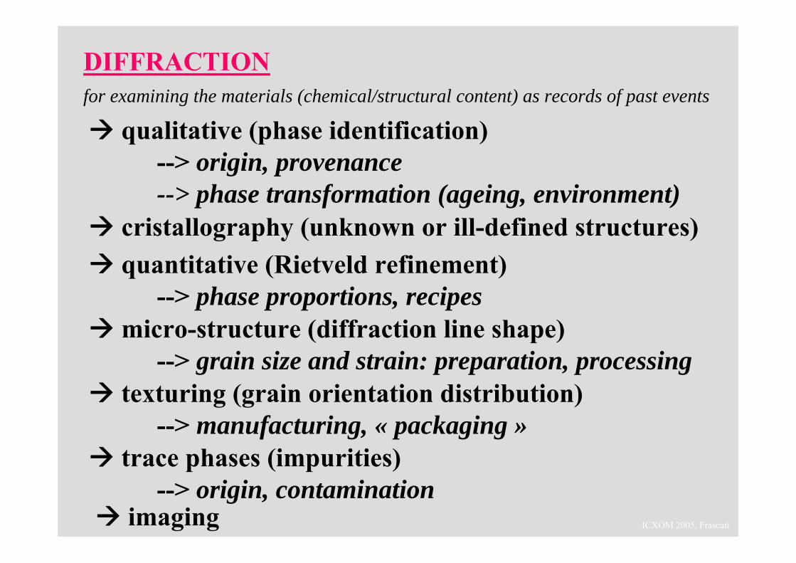
DIFFRACTIONfor examining the materials (chemical/structural content) as records of past events
quantitative (Rietveld refinement) --> phase proportions, recipes
cristallography (unknown or ill-defined structures)
qualitative (phase identification) --> origin, provenance--> phase transformation (ageing, environment)
imaging
micro-structure (diffraction line shape)--> grain size and strain: preparation, processing
texturing (grain orientation distribution)--> manufacturing, « packaging »
trace phases (impurities) --> origin, contamination
ICXOM 2005, Frascati
M. Müller, M.Z. Papiz, D.T. Clarke, M.A. Roberts, B.M. Murphy, M. Burghammer, C. Riekel, E. Pantos and J. Gunneweg
Identification of textiles from the Khirbet Qumran caves
The Dead Sea Scrolls (2nd cent. BC-1st cent. AD) : the 1st proof of the Old Testament ? a controversy…
To relate the chemical/structural information of the material (pottery, clothing) with the archaeological issues :Where/when do the fragments come from (Essenes) ?
μdiffraction on single fibrils tells the difference between cotton, linen (cellulose) and wool. Mixing wool (animal textile) with sacred parchments ? State of preservation.
To identify the colorants in the textiles: mineral/vegetal, organic/inorganic, non-crystalline dyes (provenance, date ?)
Also attached particles : soil contamination, green Cu corrosion
ICXOM 2005, Frascati
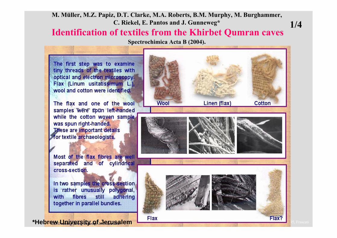
M. Müller, M.Z. Papiz, D.T. Clarke, M.A. Roberts, B.M. Murphy, M. Burghammer, C. Riekel, E. Pantos and J. Gunneweg*
Identification of textiles from the Khirbet Qumran cavesSpectrochimica Acta B (2004).
*Hebrew University of Jerusalem
1/4
ICXOM 2005, Frascati
ICXOM 2005, Frascati
ICXOM 2005, Frascati
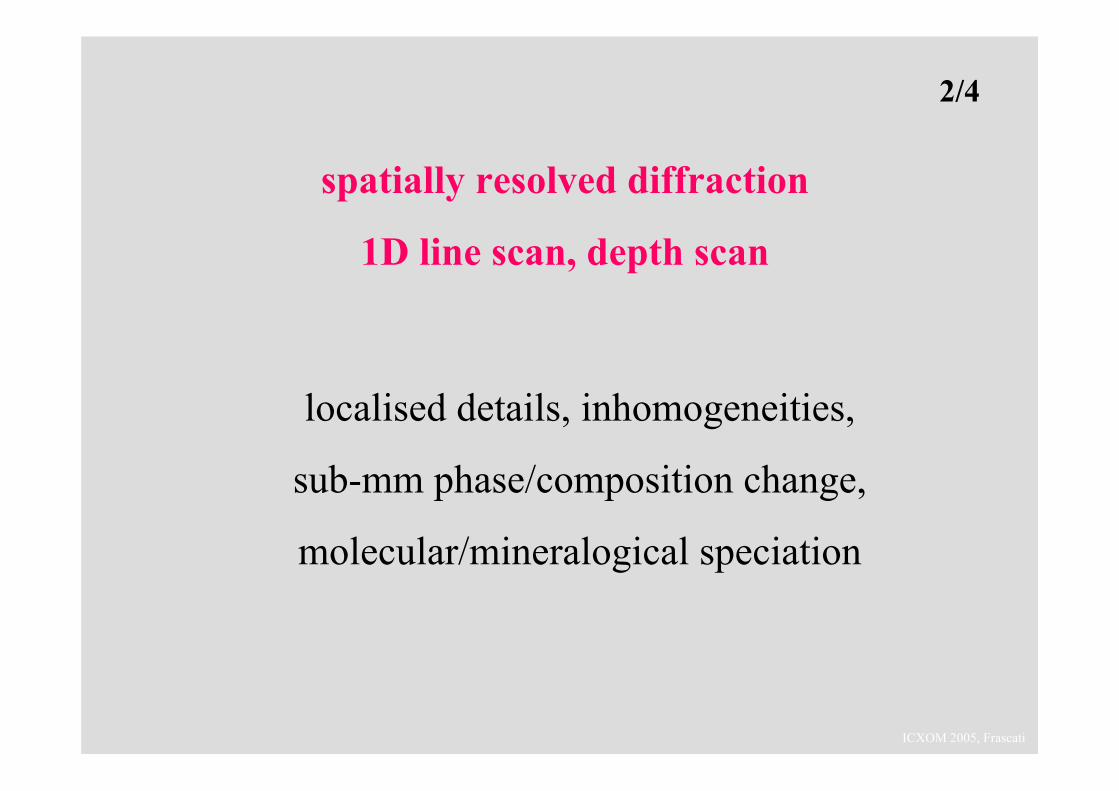
spatially resolved diffraction
1D line scan, depth scan
localised details, inhomogeneities,
sub-mm phase/composition change,
molecular/mineralogical speciation
2/4
ICXOM 2005, Frascati
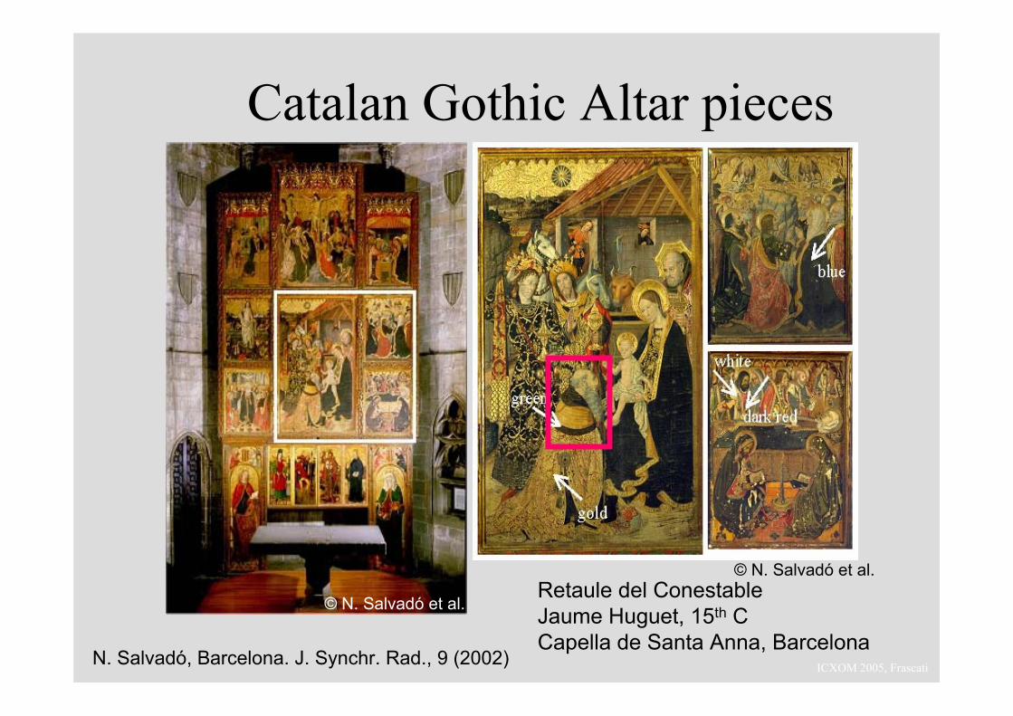
Retaule del ConestableJaume Huguet, 15th CCapella de Santa Anna, Barcelona
N. Salvadó, Barcelona. J. Synchr. Rad., 9 (2002)
Catalan Gothic Altar pieces
© N. Salvadó et al.
© N. Salvadó et al.
ICXOM 2005, Frascati
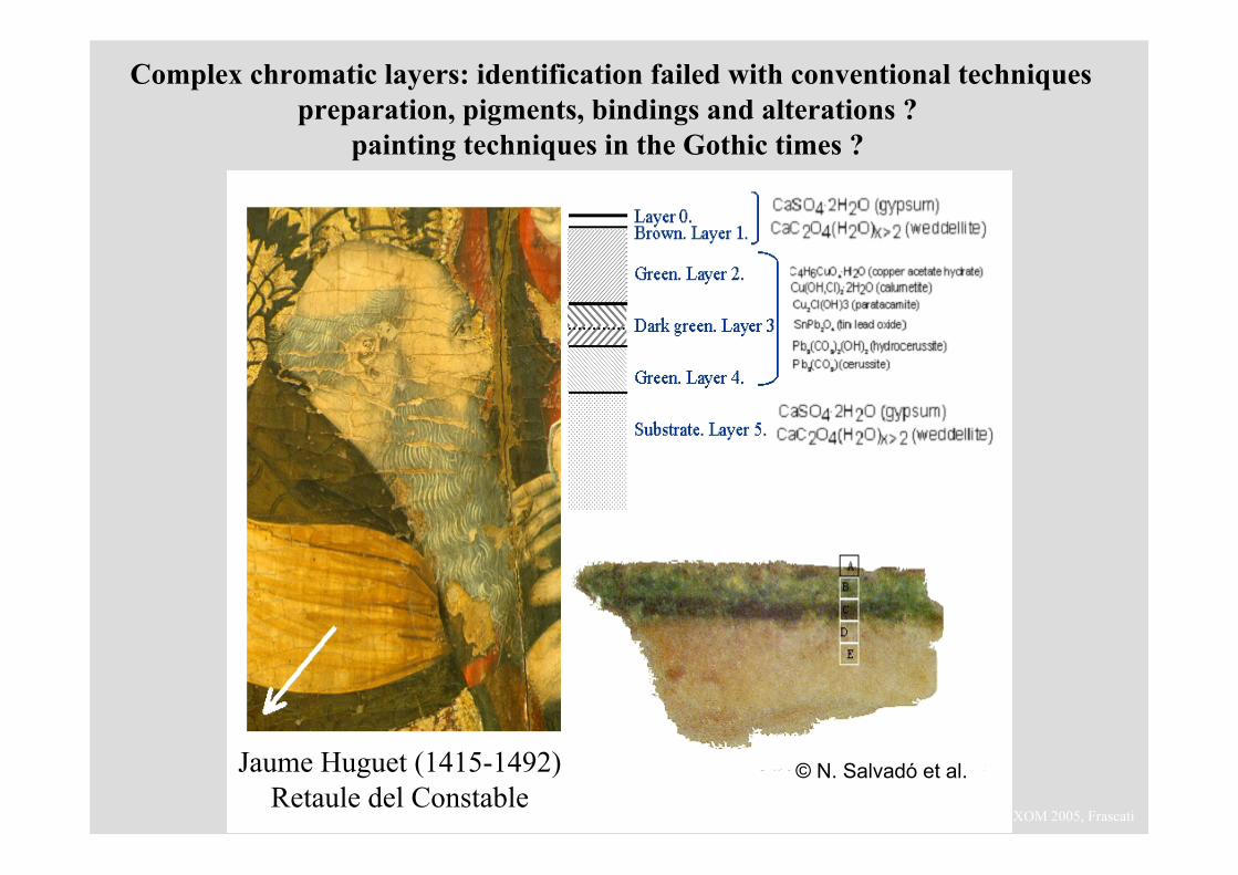
Complex chromatic layers: identification failed with conventional techniquespreparation, pigments, bindings and alterations ?
painting techniques in the Gothic times ?
© N. Salvadó et al.Jaume Huguet (1415-1492)Retaule del Constable
ICXOM 2005, Frascati
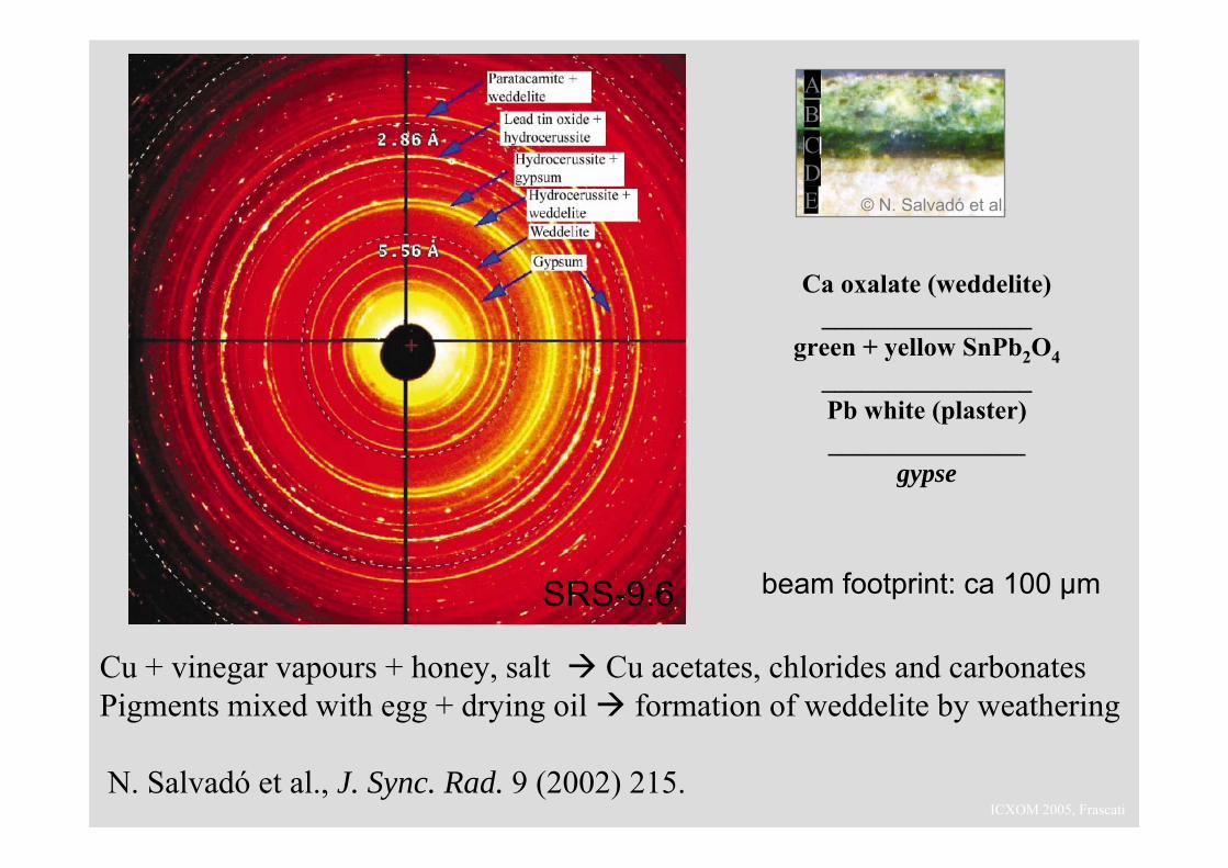
Cu + vinegar vapours + honey, salt Cu acetates, chlorides and carbonatesPigments mixed with egg + drying oil formation of weddelite by weathering
N. Salvadó et al., J. Sync. Rad. 9 (2002) 215.
beam footprint: ca 100 μmSRS-9.6
EDCBA
© N. Salvadó et al.
Ca oxalate (weddelite)________________
green + yellow SnPb2O4________________Pb white (plaster)_______________
gypse
ICXOM 2005, Frascati
spatially resolved diffraction
2D phase mapping
elemental and phase speciation
3/4
ICXOM 2005, Frascati
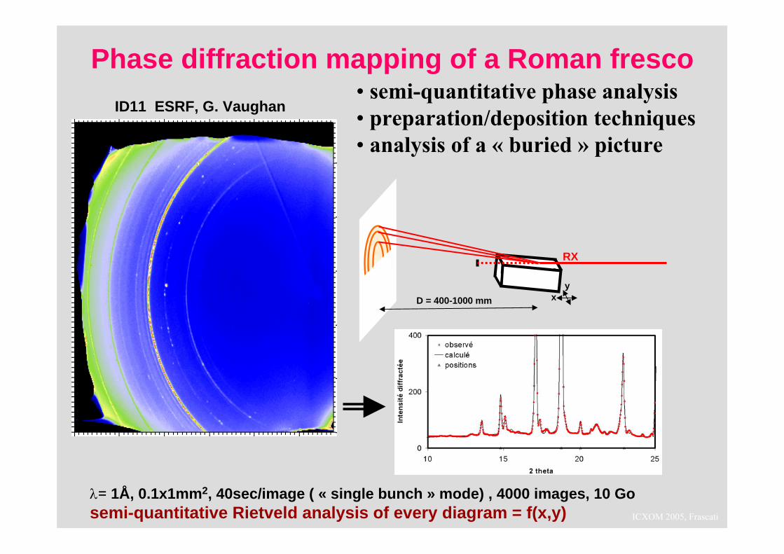
λ= 1Å, 0.1x1mm2, 40sec/image ( « single bunch » mode) , 4000 images, 10 Gosemi-quantitative Rietveld analysis of every diagram = f(x,y)
D = 400-1000 mm
RX
xy
ID11 ESRF, G. Vaughan
Phase diffraction mapping of a Roman fresco• semi-quantitative phase analysis• preparation/deposition techniques• analysis of a « buried » picture
ICXOM 2005, Frascati
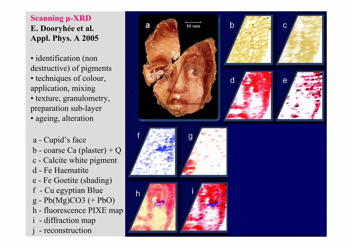
Scanning μ-XRDE. Dooryhée et al. Appl. Phys. A 2005
• identification (non destructive) of pigments • techniques of colour, application, mixing• texture, granulometry, preparation sub-layer• ageing, alteration
a - Cupid’s face b - coarse Ca (plaster) + Qc - Calcite white pigmentd - Fe Haematitee - Fe Goetite (shading)f - Cu egyptian Blueg - Pb(Mg)CO3 (+ PbO)h - fluorescence PIXE mapi - diffraction mapj - reconstruction

ICXOM 2005, Frascati
micro-structure
high resolution diffraction line shape
stress/strain, deformation field,
grain size distribution
defects
4/4
ICXOM 2005, Frascati
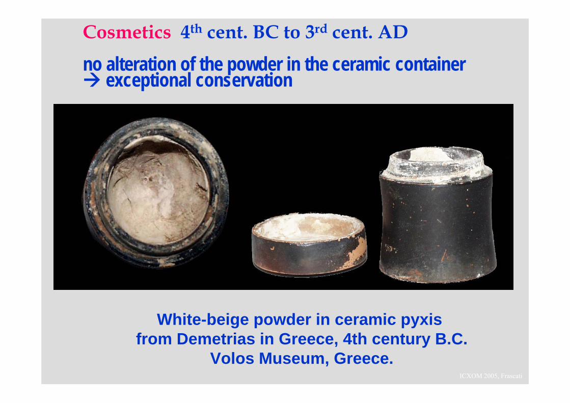
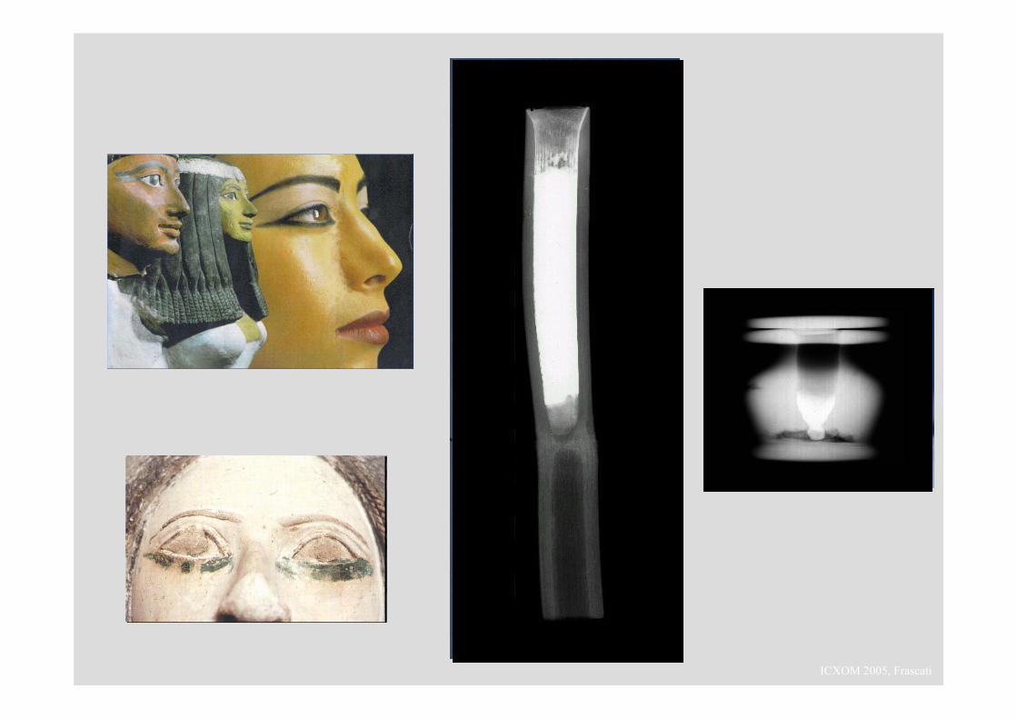
Cosmetics 4th cent. BC to 3rd cent. AD
no alteration of the powder in the ceramic container exceptional conservation
White-beige powder in ceramic pyxisfrom Demetrias in Greece, 4th century B.C.
Volos Museum, Greece.
ICXOM 2005, Frascati
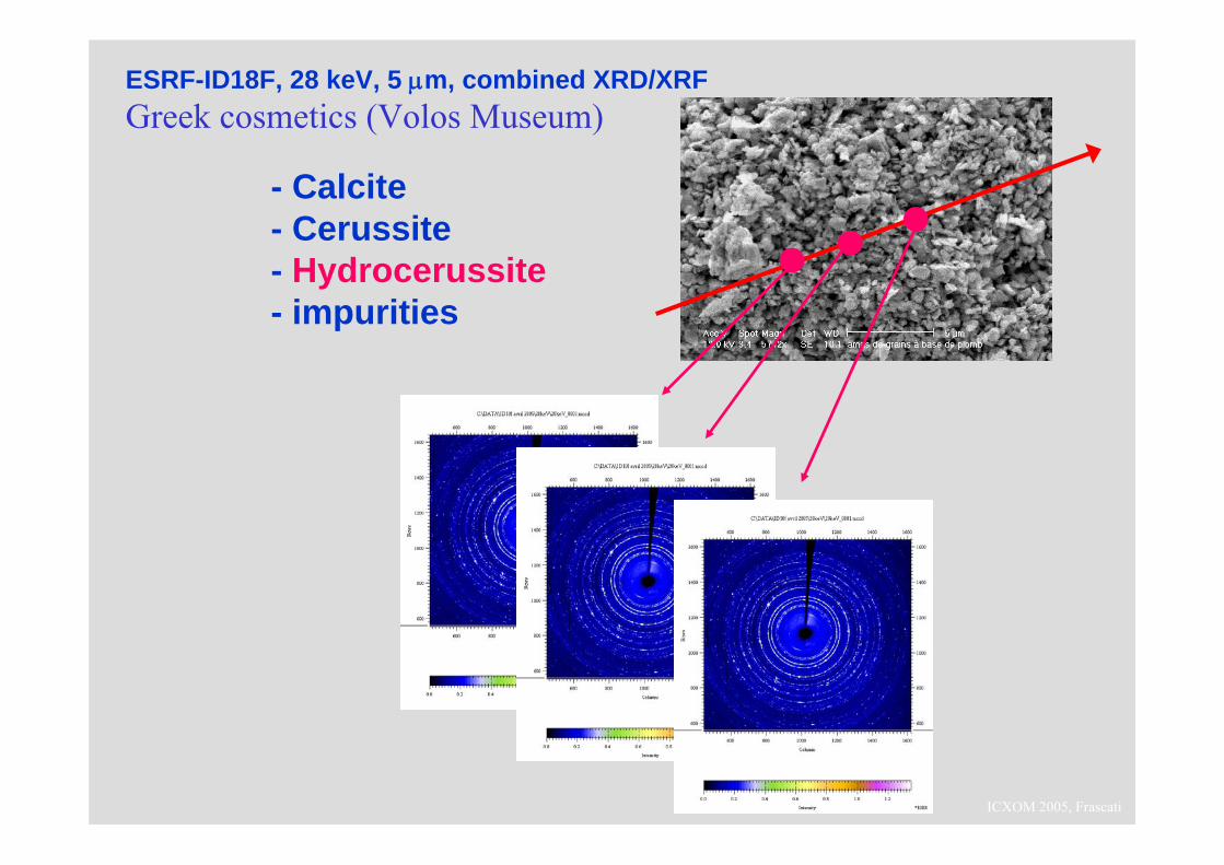
- Calcite- Cerussite- Hydrocerussite- impurities
ESRF-ID18F, 28 keV, 5 μm, combined XRD/XRFGreek cosmetics (Volos Museum)
ICXOM 2005, Frascati
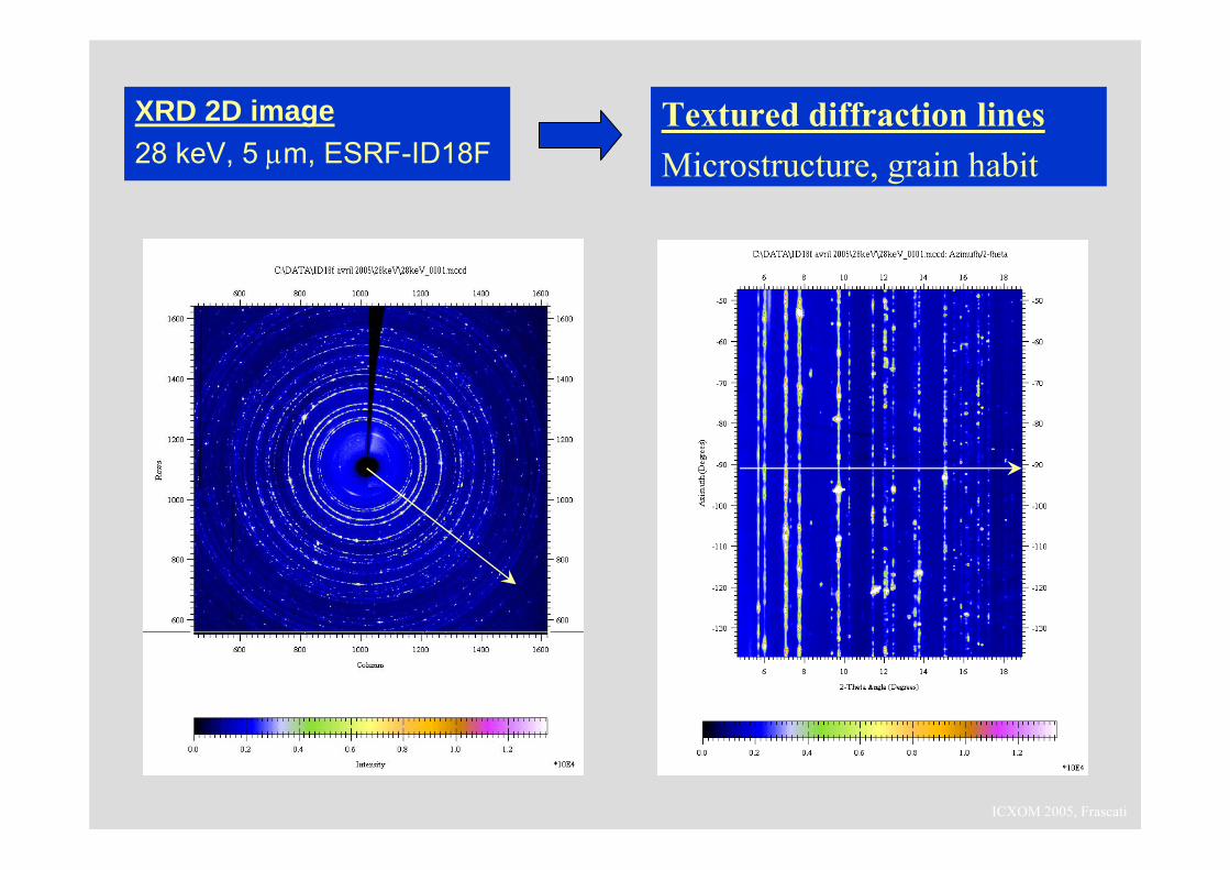
XRD 2D image28 keV, 5 μm, ESRF-ID18F
Textured diffraction linesMicrostructure, grain habit
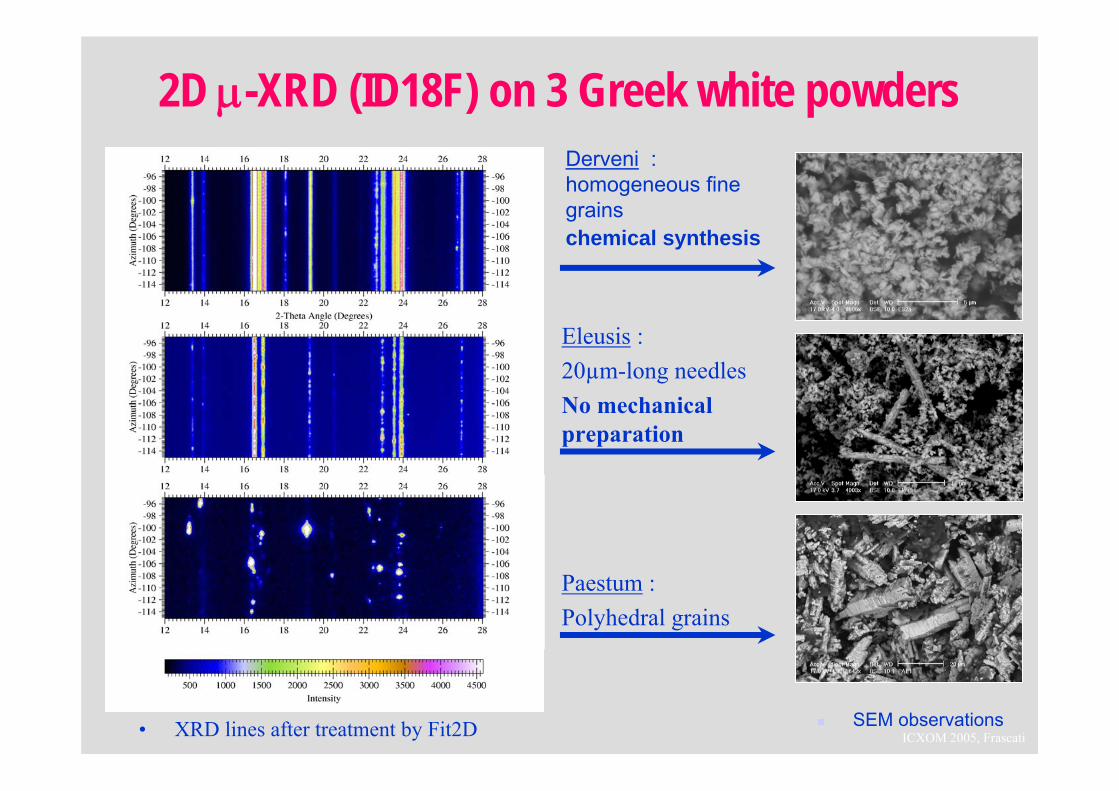
ICXOM 2005, Frascati• XRD lines after treatment by Fit2D
2D μ-XRD (ID18F) on 3 Greek white powdersDerveni : homogeneous fine grainschemical synthesis
SEM observations
Eleusis :20µm-long needlesNo mechanical preparation
Paestum :Polyhedral grains
ICXOM 2005, Frascati
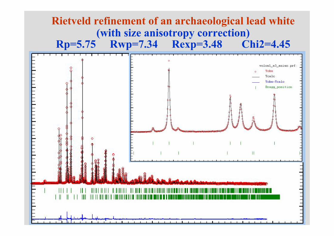
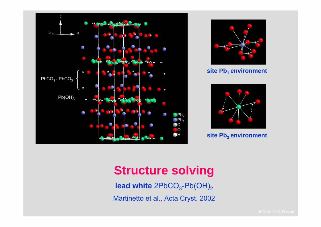
Rietveld refinement of an archaeological lead white(with size anisotropy correction)
Rp=5.75 Rwp=7.34 Rexp=3.48 Chi2=4.45
ICXOM 2005, Frascati
site Pb1 environment
site Pb2 environment
Structure solvinglead white 2PbCO3-Pb(OH)2
Martinetto et al., Acta Cryst. 2002
PbCO3 - PbCO3
Pb(OH)2
ICXOM 2005, Frascati

ICXOM 2005, Frascati
20 µm
1 mm
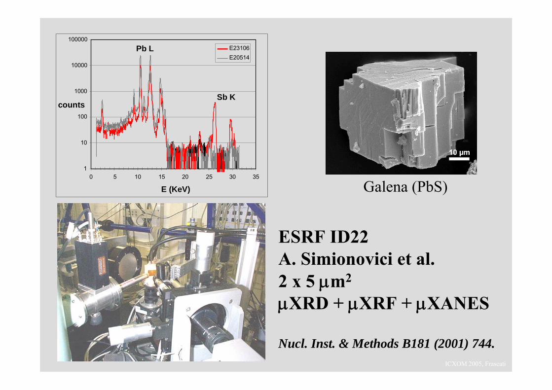
Powder processing in Ancient Egypt
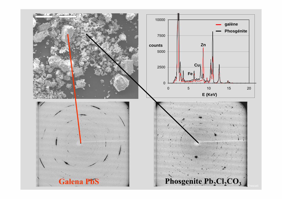
Galena (PbS)
Cerussite (PbCO3) synthetic Pb chlorides
ICXOM 2005, Frascati
1
10
100
1000
10000
100000
0 5 10 15 20 25 30 35
E (KeV)
counts
E23106E20514
Pb L
Sb K
ESRF ID22A. Simionovici et al.2 x 5 μm2
μXRD + μXRF + μXANES
Nucl. Inst. & Methods B181 (2001) 744.
Galena (PbS)
ICXOM 2005, Frascati
0
2500
5000
7500
10000
0 5 10 15 20
E (KeV)
counts
galènePhosgénite
Cu
Zn
Fe
Galena PbS Phosgenite Pb2Cl2CO3
ICXOM 2005, Frascati
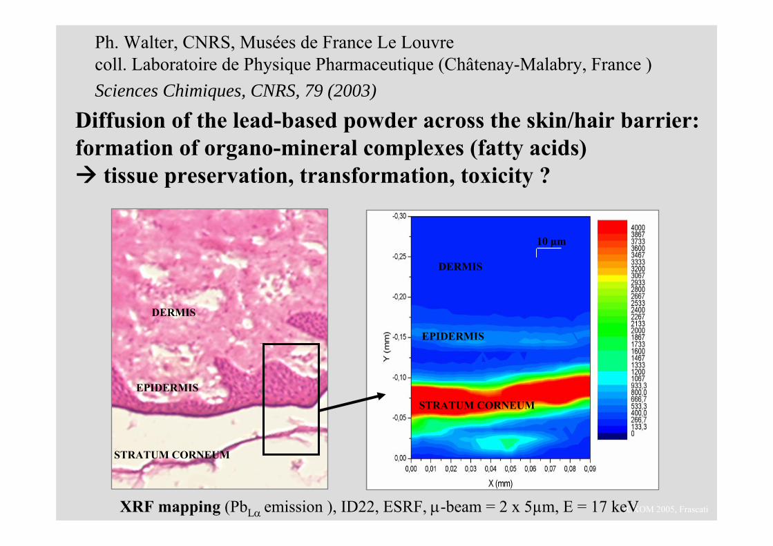
Ph. Walter, CNRS, Musées de France Le Louvre coll. Laboratoire de Physique Pharmaceutique (Châtenay-Malabry, France )Sciences Chimiques, CNRS, 79 (2003)
XRF mapping (PbLα emission ), ID22, ESRF, μ-beam = 2 x 5µm, E = 17 keV
Diffusion of the lead-based powder across the skin/hair barrier: formation of organo-mineral complexes (fatty acids)
tissue preservation, transformation, toxicity ?
STRATUM CORNEUM
EPIDERMIS
DERMIS
DERMIS
EPIDERMIS
STRATUM CORNEUM
10 µm
ICXOM 2005, Frascati
brief introduction to X-ray diffraction
the need for micro-beam X-ray diffraction
applications to Cultural Heritage- micro-analysis- line/depth scans- 2D phase/element imaging- micro-structure (strain, grain size)- white beam μdiffraction: the Terra Sigillata case
ICXOM 2005, Frascati
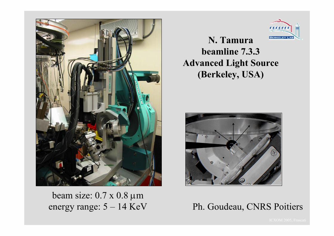
beam size: 0.7 x 0.8 μmenergy range: 5 – 14 KeV
N. Tamurabeamline 7.3.3
Advanced Light Source (Berkeley, USA)
Ph. Goudeau, CNRS Poitiers
ICXOM 2005, Frascati
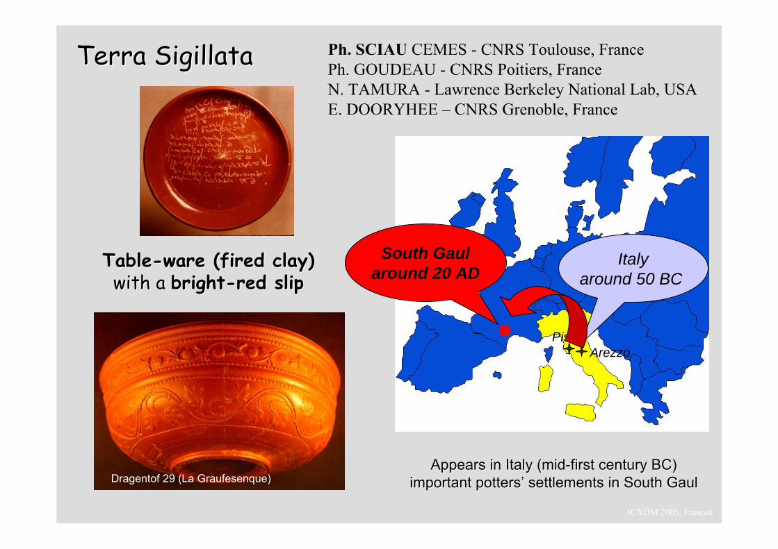
Terra Terra SigillataSigillata
South Gaularound 20 AD
Italyaround 50 BC
PiseArezzo
AppearsAppears in Italy (in Italy (midmid--first century BC)first century BC)important potters’ settlements in South Gaulimportant potters’ settlements in South Gaul
TableTable--ware (fired clay)ware (fired clay)with a with a brightbright--red slipred slip
Dragentof 29 (La Graufesenque)
Ph. SCIAU CEMES - CNRS Toulouse, France Ph. GOUDEAU - CNRS Poitiers, FranceN. TAMURA - Lawrence Berkeley National Lab, USAE. DOORYHEE – CNRS Grenoble, France
ICXOM 2005, Frascati
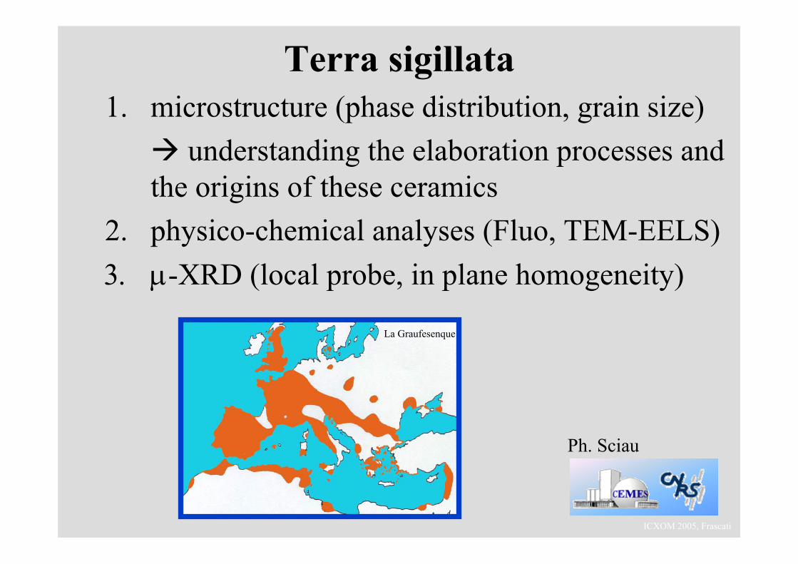
Terra sigillata1. microstructure (phase distribution, grain size)
understanding the elaboration processes and the origins of these ceramics
2. physico-chemical analyses (Fluo, TEM-EELS)3. μ-XRD (local probe, in plane homogeneity)
La Graufesenque
Ph. Sciau
ICXOM 2005, Frascati
(b)
2Θ
Χ
(a) (b)
2Θ
Χ
2Θ
Χ
(a)(a)
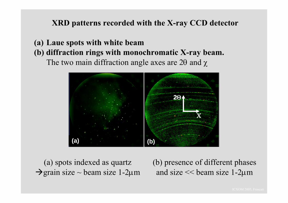
XRD patterns recorded with the X-ray CCD detector
(a) Laue spots with white beam(b) diffraction rings with monochromatic X-ray beam.
The two main diffraction angle axes are 2θ and χ
(a) spots indexed as quartz grain size ~ beam size 1-2μm
(b) presence of different phases and size << beam size 1-2μm
ICXOM 2005, Frascati
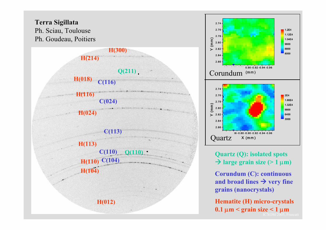
H(104)H(110)
H(113)
H(024)
H(116)
H(018)
H(214)H(300)
H(012)
C(104)C(110)
C(113)
C(024)
C(116)
Q(110)
Q(211)
Quartz (Q): isolated spots large grain size (> 1 μm)
Corundum (C): continuous and broad lines very fine grains (nanocrystals)
Hematite (H) micro-crystals 0.1 μm < grain size < 1 μm
Terra Terra SigillataSigillataPh. Sciau, ToulousePh. Goudeau, Poitiers
-0.84 -0 .86 -0.88 -0.90 -0.92 -0 .94 -0.96
2.86
2.84
2.82
2.80
2.78
2.76
2.74
X (m m )
Y (
mm
)
8000
8800
9600
1.04E4
1.12E4
1.2E4
-0.84 -0 .86 -0.88 -0.90 -0.92 -0 .94 -0.96
2.86
2.84
2.82
2.80
2.78
2.76
2.74
X (m m )
Y (
mm
)
3000
6400
9800
1.32E4
1.66E4
2E4
(b)
(d)
-0.84 -0 .86 -0.88 -0.90 -0.92 -0 .94 -0.96
2.86
2.84
2.82
2.80
2.78
2.76
2.74
X (m m )
Y (
mm
)
8000
8800
9600
1.04E4
1.12E4
1.2E4
-0.84 -0 .86 -0.88 -0.90 -0.92 -0 .94 -0.96
2.86
2.84
2.82
2.80
2.78
2.76
2.74
X (m m )
Y (
mm
)
3000
6400
9800
1.32E4
1.66E4
2E4
(b)
(d)
Corundum
Quartz
ICXOM 2005, Frascati
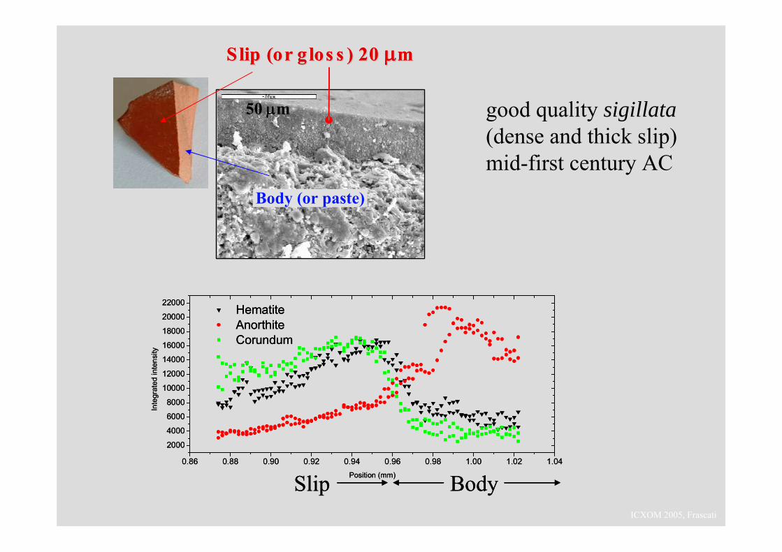
Slip (or S lip (or g los sg los s ) 20 ) 20 μμmm
50 μm
Body (or paste)
S lip (or S lip (or g los sg los s ) 20 ) 20 μμmm
50 μm
Body (or paste)
good quality sigillata(dense and thick slip) mid-first century AC
0.86 0.88 0.90 0.92 0.94 0.96 0.98 1.00 1.02 1.04
2000
4000
6000
8000
10000
12000
14000
16000
18000
20000
22000 Hematite Anorthite Corundum
Inte
grat
ed in
tens
ity
Position (mm)Slip Body0.86 0.88 0.90 0.92 0.94 0.96 0.98 1.00 1.02 1.04
2000
4000
6000
8000
10000
12000
14000
16000
18000
20000
22000 Hematite Anorthite Corundum
Inte
grat
ed in
tens
ity
Position (mm)Slip Body
ICXOM 2005, Frascati
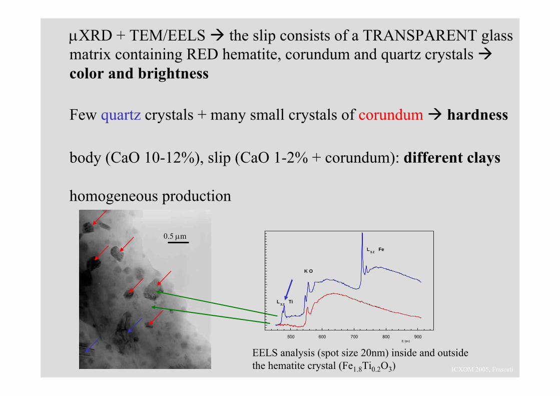
0.5 μm
EELS analysis (spot size 20nm) inside and outside the hematite crystal (Fe1.8Ti0.2O3)
500 600 700 800 900E (ev)
L3.2
Fe
K O
L3.2
Ti
μXRD + TEM/EELS the slip consists of a TRANSPARENT glass matrix containing RED hematite, corundum and quartz crystals color and brightness
Few quartz crystals + many small crystals of corundum hardness
body (CaO 10-12%), slip (CaO 1-2% + corundum): different clays
homogeneous production
ICXOM 2005, Frascati
brief introduction to X-ray diffraction
the need for micro-beam X-ray diffraction
applications to Cultural Heritage
white beam μdiffraction
μ−SAXS, μ−GISAXS, …
ICXOM 2005, Frascati
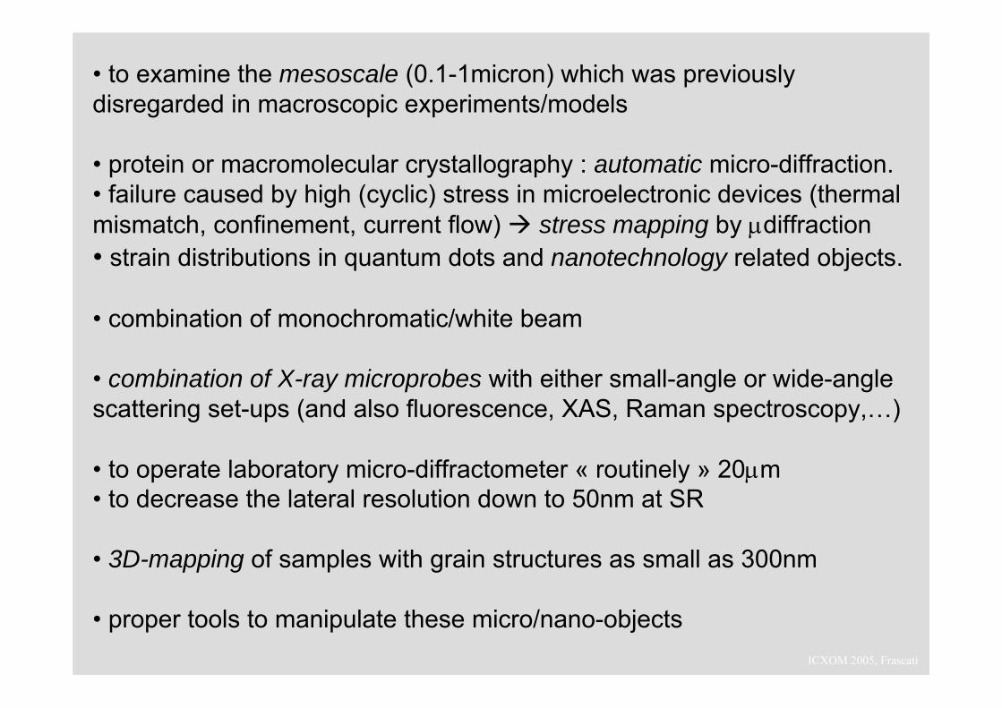
• to examine the mesoscale (0.1-1micron) which was previously disregarded in macroscopic experiments/models
• protein or macromolecular crystallography : automatic micro-diffraction.• failure caused by high (cyclic) stress in microelectronic devices (thermal mismatch, confinement, current flow) stress mapping by μdiffraction• strain distributions in quantum dots and nanotechnology related objects.
• combination of monochromatic/white beam
• combination of X-ray microprobes with either small-angle or wide-angle scattering set-ups (and also fluorescence, XAS, Raman spectroscopy,…)
• to operate laboratory micro-diffractometer « routinely » 20μm• to decrease the lateral resolution down to 50nm at SR
• 3D-mapping of samples with grain structures as small as 300nm
• proper tools to manipulate these micro/nano-objects
ICXOM 2005, Frascati
Acknowledgments
- C2RMF, Paris : Ph. Walter, G. Tsoucaris, C. Deeb, J. Castaing, E. Van Elslande, E. Welcomme, J. Salomon, …
- Laboratoire de cristallographie, CNRS Grenoble : P. Martinetto, M. Anne, J.L. Hodeau, …
- ESRF, Grenoble : J. Susini, A. Fitch, M. Cotte, A. Simionovici, …
and Ph. Sciau (Toulouse), Ph. Goudeau (Poitiers), N. Tamura (Berkeley), T. Ungar (Budapest), …