X-ray diffraction Biophysics. W. K. Röntgen (1845 – 1923) Wife’s hand.
26
X-ray diffraction Biophysics
-
Upload
quentin-long -
Category
Documents
-
view
214 -
download
0
Transcript of X-ray diffraction Biophysics. W. K. Röntgen (1845 – 1923) Wife’s hand.
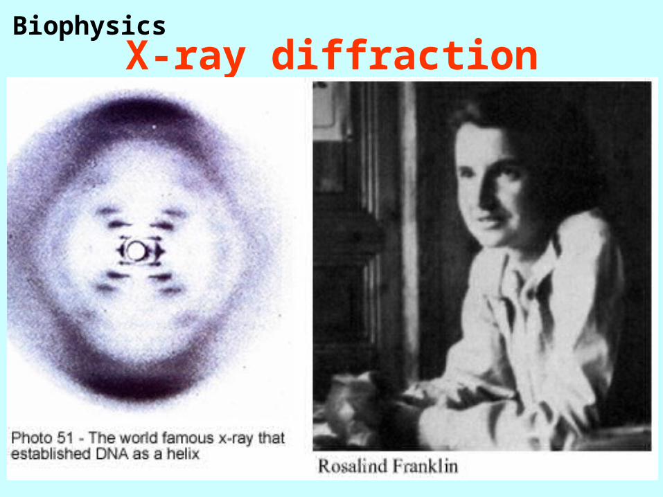
X-ray diffractionBiophysics
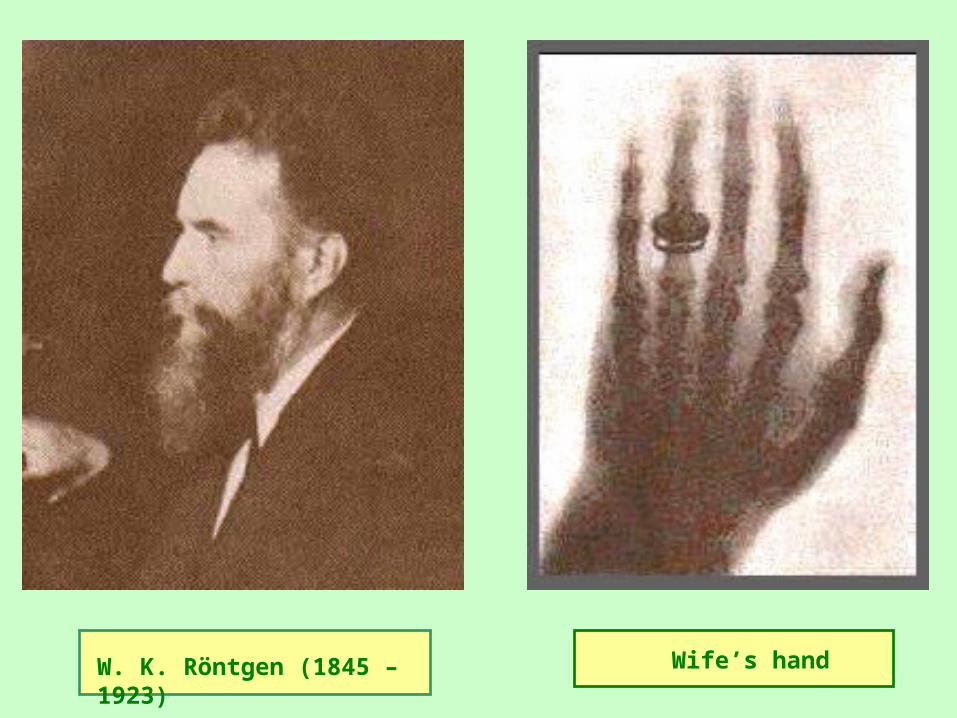
W. K. Röntgen (1845 – 1923) Wife’s hand
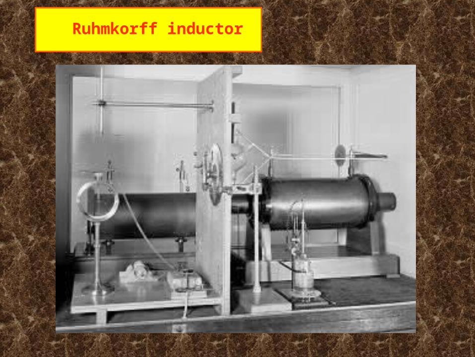
Ruhmkorff inductor

X-ray tube with rotating anode
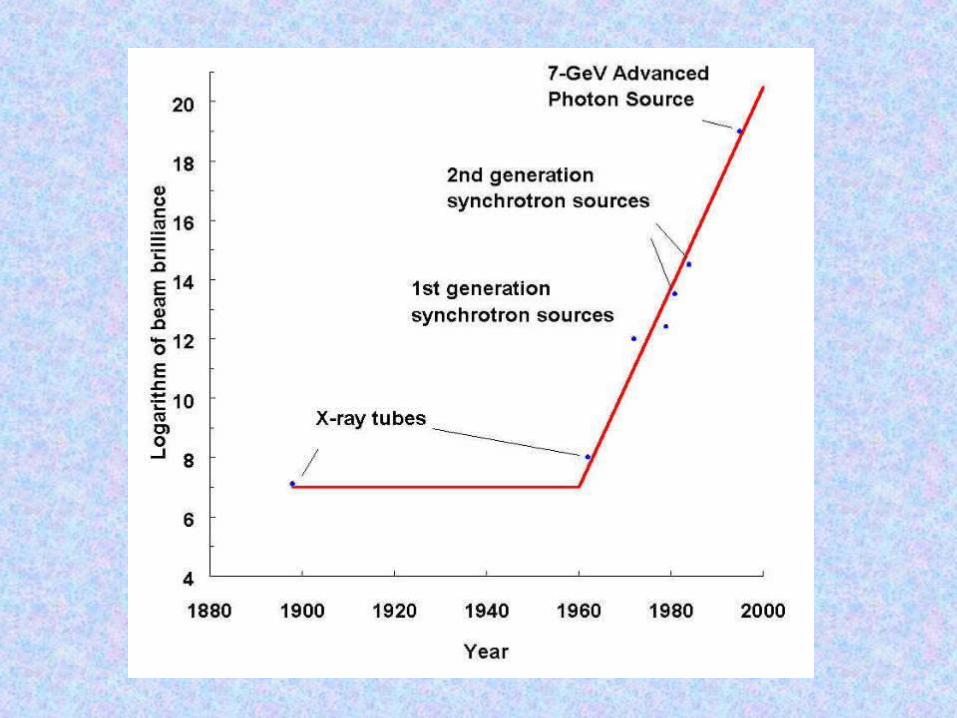

Syncrotron in Grenoble
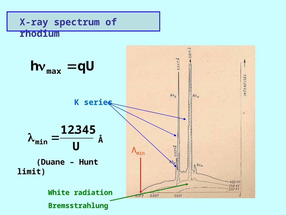
X-ray spectrum of rhodium
qUh max
Λmin
K series
U
345.12min Å
(Duane – Hunt limit)
White radiation
Bremsstrahlung
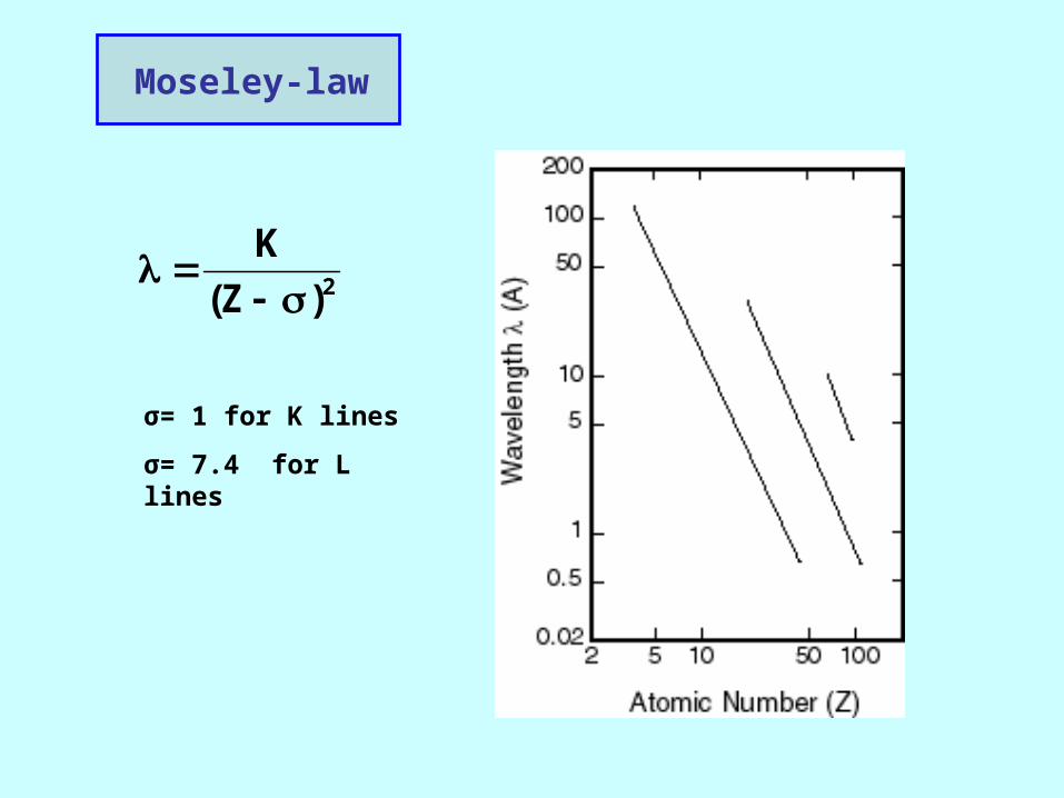
Moseley-law
σ= 1 for K lines
σ= 7.4 for L lines
2)Z(
K
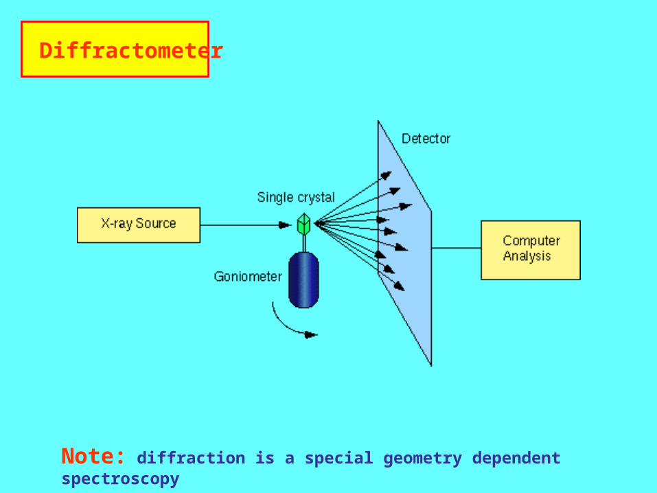
Diffractometer
Note: diffraction is a special geometry dependent spectroscopy
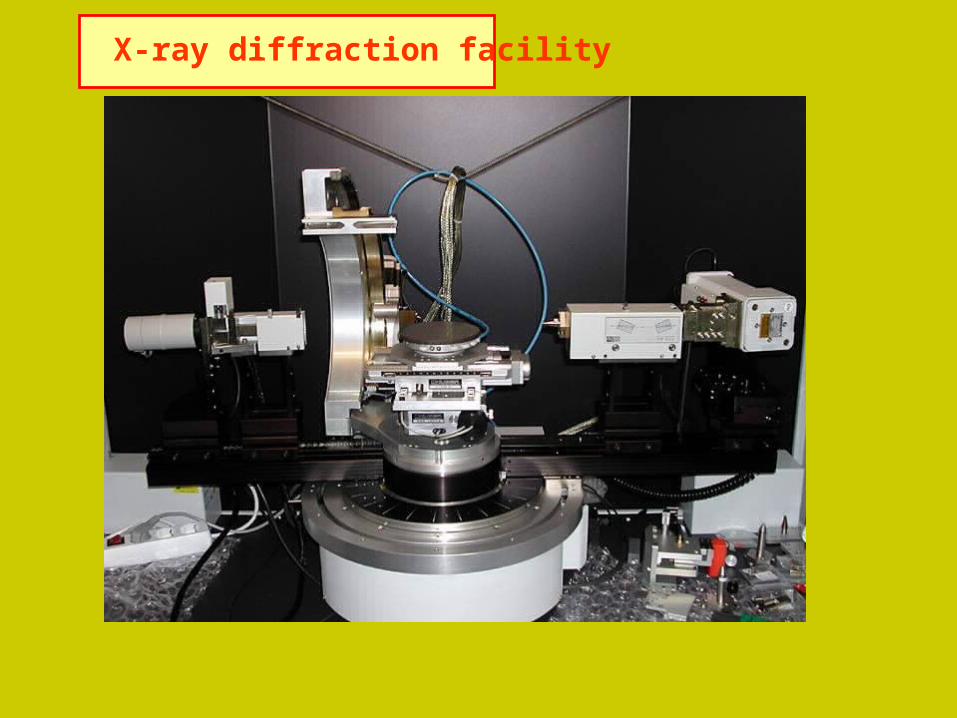
X-ray diffraction facility
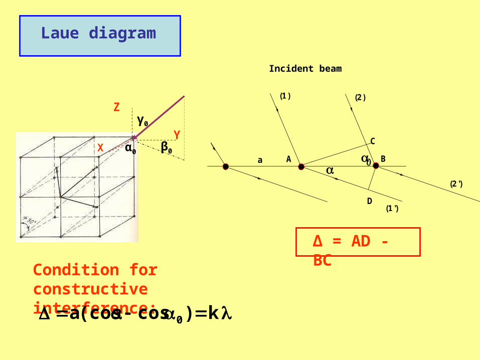
= AD - BC
(2')
(2)
(1')
(1)
a
D
C
BA
Laue diagram
Condition for constructive interference:
k)cos(cosa 0
Δ = AD - BC
Incident beam
X
Z
Yα0 β0
γ0
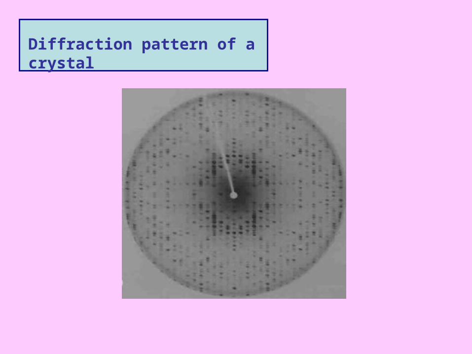
Diffraction pattern of a crystal
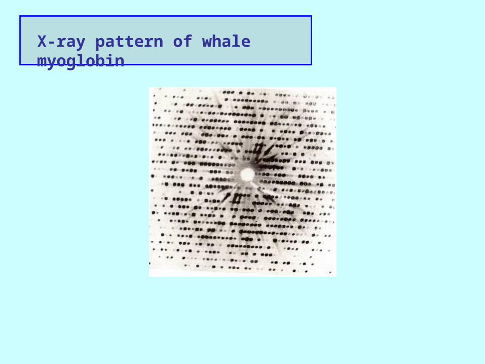
X-ray pattern of whale myoglobin
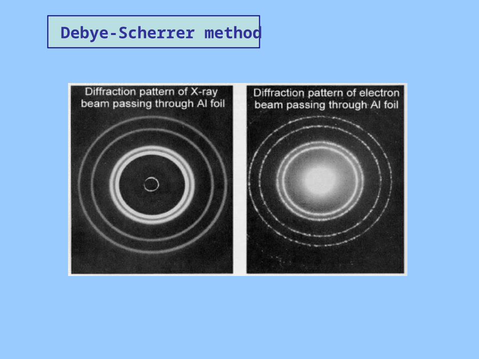
Debye-Scherrer method
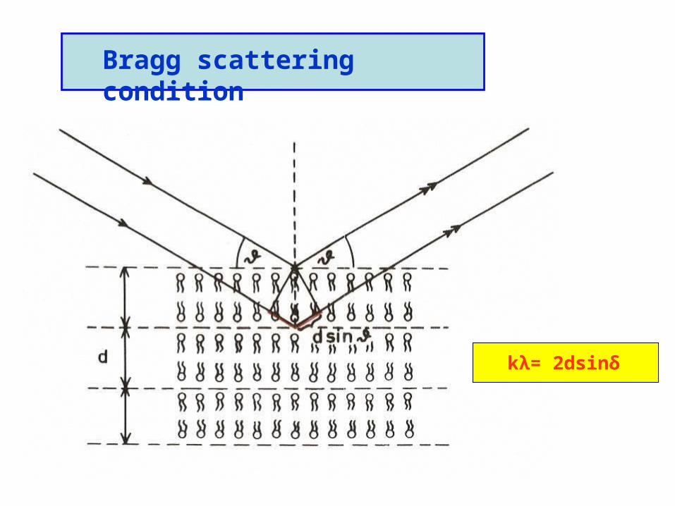
Bragg scattering condition
kλ= 2dsinδ
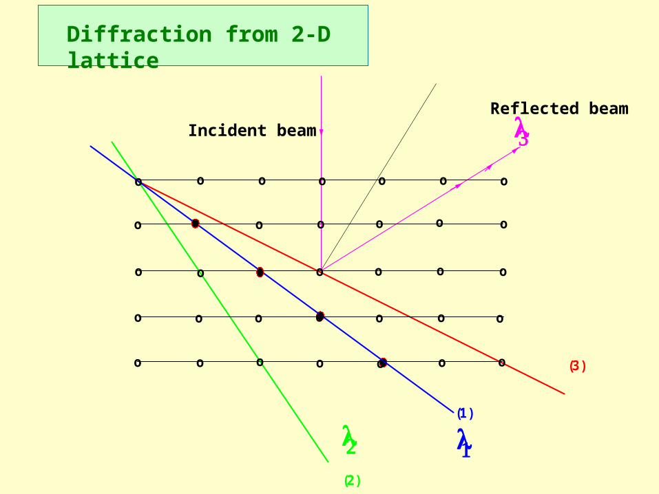
(3)
(2)
(1)
ooooooo
ooooooo
ooooooo
ooooooo
o oooooo
Diffraction from 2-D lattice
Incident beamReflected beam
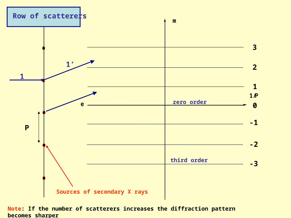
e1/P
m
Sources of secondary X rays
zero order 0
1
3
2
-1
-2
-3third order
P
Row of scatterers
1
1’
Note: If the number of scatterers increases the diffraction pattern becomes sharper
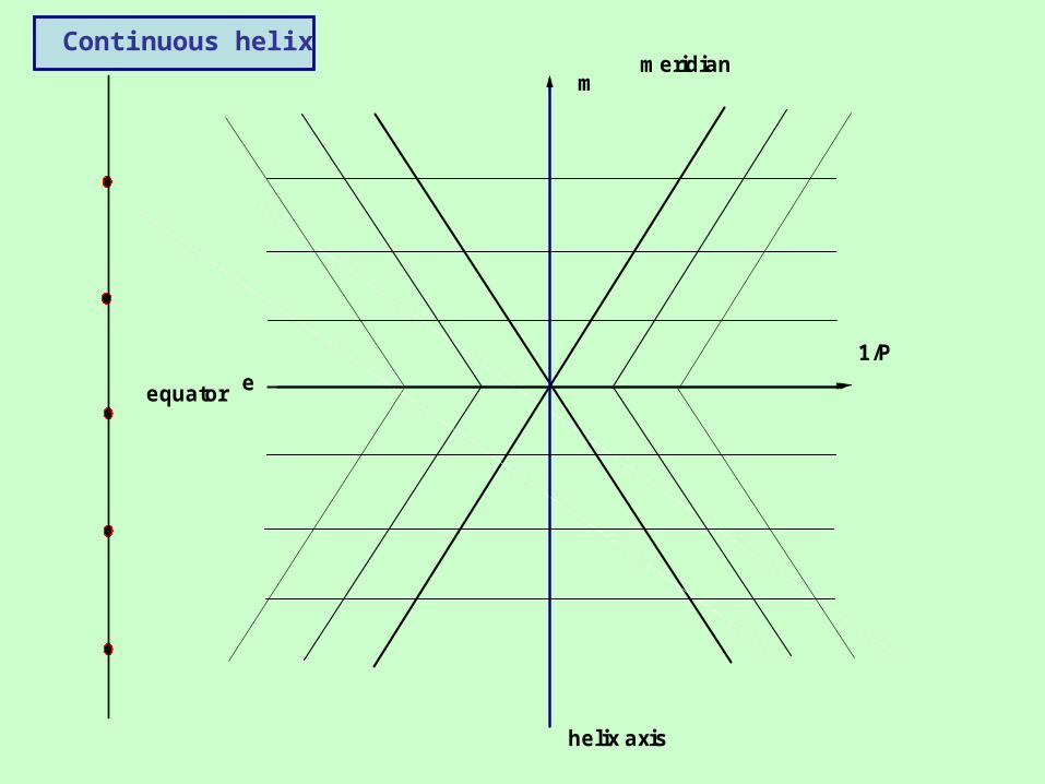
helix axis
equator
meridian
e1/P
m
Continuous helix
helix axis
equator
meridian
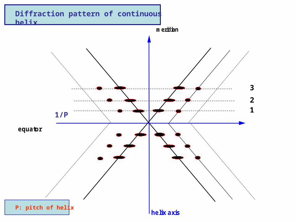
Diffraction pattern of continuous helix
12
3
1/P
P: pitch of helix
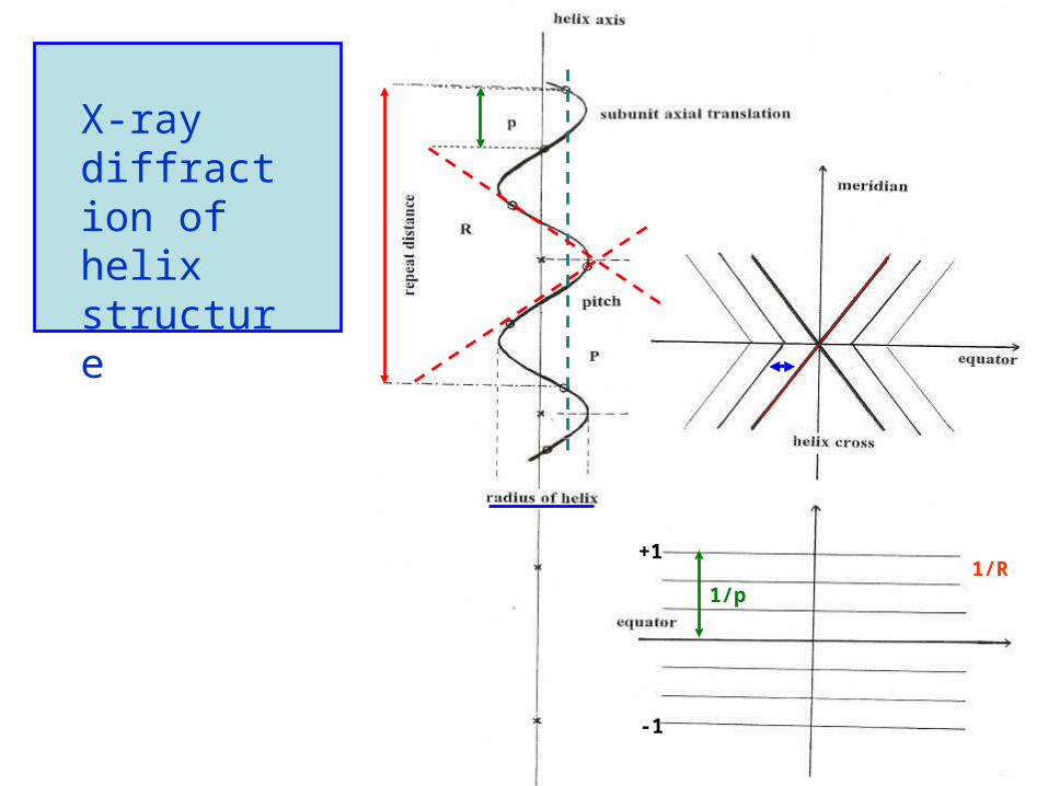
X-ray diffraction of helix structure
1/R
-1
+1
1/p
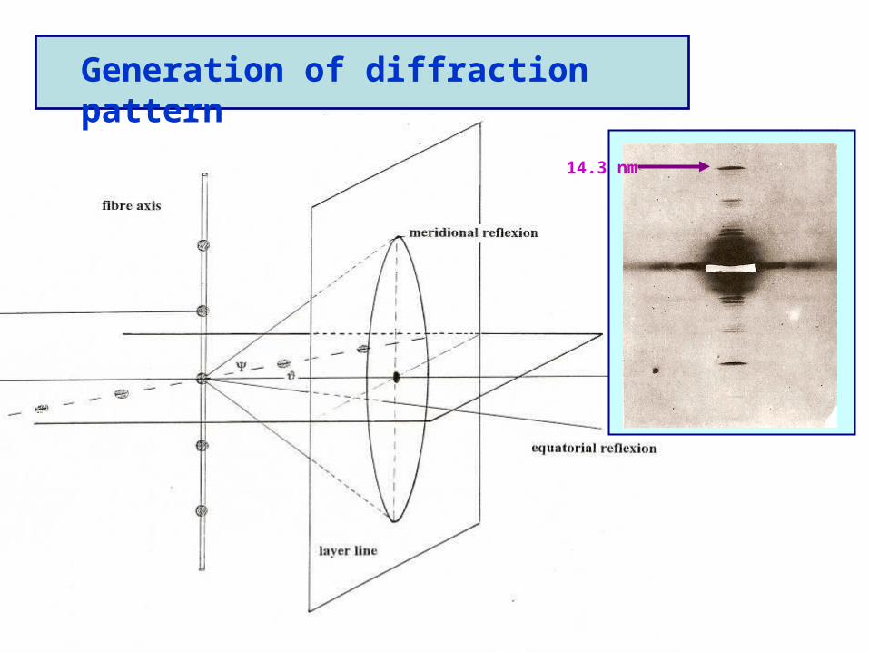
Generation of diffraction pattern
14.3 nm
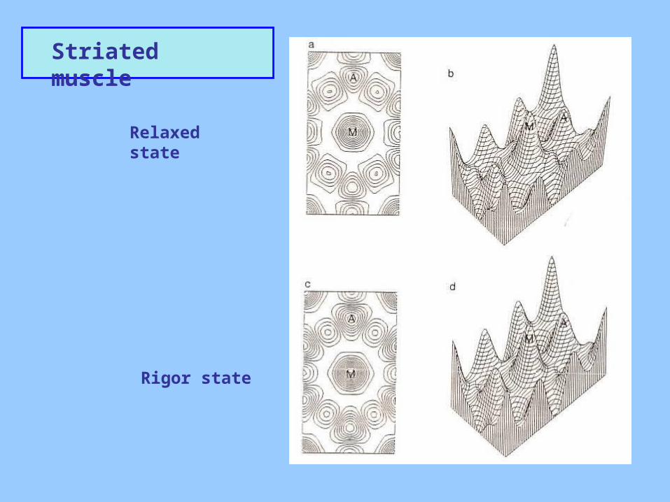
Striated muscle
Relaxed state
Rigor state
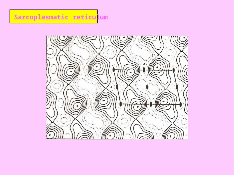
Sarcoplasmatic reticulum
I4-1 supernova