· Web viewAKT is a serine/threonine kinase that regulates tumor cell proliferation, survival,...
44
<Original article> GV1001 inhibits cell viability and induces apoptosis in castration-resistant prostate cancer cells through the AKT/NF-κB/VEGF pathway Running title: GV1001 in castration-resistant prostate cancer Yong Hyun Park 1,2 , Ae Ryang Jung 1,2 , Ga Eun Kim 1,2 , Mee Young Kim 1,2 , U- Syn Ha 1,2 , Sung-Hoo Hong 1,2 , Sae Woong Kim 1 , Ji Youl Lee 1,2 1 Department of Urology, Seoul St. Mary's Hospital, College of Medicine, The Catholic University of Korea 2 Cancer Research Institute, College of Medicine, The Catholic University of Korea
Transcript of · Web viewAKT is a serine/threonine kinase that regulates tumor cell proliferation, survival,...
<Original article>
GV1001 inhibits cell viability and induces apoptosis in castration-
resistant prostate cancer cells through the AKT/NF-κB/VEGF
pathway
Running title: GV1001 in castration-resistant prostate cancer
Yong Hyun Park1,2, Ae Ryang Jung1,2, Ga Eun Kim1,2, Mee Young Kim1,2, U-Syn Ha1,2, Sung-
Hoo Hong1,2, Sae Woong Kim1, Ji Youl Lee1,2
1 Department of Urology, Seoul St. Mary's Hospital, College of Medicine, The Catholic
University of Korea
2 Cancer Research Institute, College of Medicine, The Catholic University of Korea
* Corresponding author
Ji Youl Lee, M.D., Ph.D.
Department of Urology, Seoul St. Mary's Hospital, College of Medicine, The Catholic
University of Korea
222 Banpo-daero, Seocho-gu, Seoul 06591, Republic of Korea
TEL: +82-2-2258-6076
FAX: +82-2-599-7839
E-mail: [email protected]
Word count: abstract 205 words, article 2,850 words
Abstract
Purpose: We examined the effect of GV1001 in castration castration-resistant
prostate cancer (CRPC) cell growth and invasion and explored the potential
molecular mechanisms of action.
Materials and Methods: The in vitro anti-cancer effects of GV1001 in CRPC cells
were examined using cell viability assay, TUNEL assay, and flow cytometry analysis.
To evaluate the effects of GV1001 on different steps of angiogenesis, wound healing
assay, transwell invasion assay, endothelial cell tube formation assay, and western
blot analysis were performed. Finally, the anti-cancer effects of GV1001 on tumor
growth in vivo were examined in a CRPC xenograft model.
Results: GV1001 inhibited cell viability and induced apoptosis in CRPC cells in vitro,
accompanied by down-regulation of Bcl-2 and caspase-3. GV1001 also inhibited
different steps of angiogenesis, such as migration, invasion, and endothelial tube
formation, along with decreased expression of MMP-2, MMP-9, and CD31 and
increased expression of TIMP-1 and TIMP-2. Mechanistically, GV1001 significantly
decreased the levels of phosphorylated AKT, phosphorylated p65, and VEGF in
CRPC cells in a dose-dependent manner. GV1001 was effective in suppressing
tumor growth and inducing apoptosis in a CRPC xenograft mouse model.
Conclusions: Our data demonstrated that GV1001 inhibited cell viability, induced
apoptosis, and inhibited angiogenesis in CRPC cells by inhibition of the AKT/NF-
κB/VEGF signaling pathway.
Keywords: castration-resistant prostate cancer, GV1001, AKT/NF-κB/VEGF
signaling pathway

Introduction
Prostate cancer is the most common cancer in men in the United States, and the
incidence of prostate cancer has been rapidly increasing over the last few years in
Korea [1, 2]. Androgen deprivation therapy is the standard of care for advanced or
metastatic disease due to its good clinical response, however, this good response
only lasts on average 2-3 years, with eventual progression toward castration-
resistant prostate cancer (CRPC) [3, 4]. Since the publication of two pivotal trials,
docetaxel-based chemotherapy has been the standard treatment for patients with
CRPC [5, 6]. In addition to docetaxel-based chemotherapy, several new agents with
different mechanisms of action have shown favorable clinical outcomes compared
with standard of care or placebo in randomized controlled trials of CRPC. These
agents include the new taxane, cabazitaxel [7]; second-generation hormonal agents,
abiraterone acetate [8, 9] and enzalutamide [10]; the immunotherapeutic agent,
sipuleucel-T [11]; and the bone-targeting agent, radium-223 [12]. Despite these
improvements in the last 5 years, additional treatment options are still needed to
prolong the survival of patients with CRPC, especially those with resistance to these
existing treatments.
GV1001 consists of small 16-mer peptides that is derived from the human
telomerase enzyme (hTERT 611-626; EARPALLTSRLRFIPK), and is undergoing
clinical trials as a cancer vaccine [13]. GV1001 has been shown to induce specific
immune response in patients with pancreatic cancer, non-small cell lung cancer,
melanoma, and hepatocellular carcinoma in several phase I/II clinical trials. Although
all of these clinical trials have examined the anticancer effect of GV1001 as a
peptide vaccine, unexpected intracellular localization of GV1001 was observed.
Through its interaction with HSP70 and HSP90, GV1001 can penetrate the cell
membrane and localize in the cytoplasm. These findings suggest a possible role of
GV1001 as a cell-penetrating peptide [14].
Given the demonstrated ability of GV1001 to penetrate cancer cells and directly
interact with important signaling molecules, we speculated that GV1001 might have
anti-cancer activity in cancer cells, including prostate cancer cells. In this study, we
examined the effects of GV1001 on CRPC cell growth and invasion and explored the
potential molecular mechanisms of action. Furthermore, the in vivo efficacy of
GV1001 was investigated using a xenograft mouse model.
Materials and Methods
Cells and reagents
Human CRPC cell lines, DU145 and PC3, were purchased from the Korean Cell
Line Bank (Seoul, Korea) and cultured in Dulbecco’s Modified Eagle’s Medium
(DMEM) containing 10% fetal bovine serum (FBS), and penicillin (100 U/ml) at 37°C
in a humidified 5% CO2 incubator. Human umbilical vein endothelial cells (HUVECs)
were purchased from Life Technologies (Carlsbad, CA, USA) and cultured in Medium
200 (Invitrogen, Carlsbad, CA, USA) containing the LVES-supplement (Invitrogen).
GV1001 (molecular weight, 1,686 g/mol) was supplied as a freeze-dried peptide
produced under good manufacturing practice conditions by GemVax & Kael
(Seongnam, Korea). GV1001 was stored at -20°C and thawed in phosphate buffer
solution (maximum solubility: ~ 100 mg/mL in saline at ambient conditions).
Cell viability assay
DU145 (4 x 10³/well) and PC3 cells (5 x 10³/well) were seeded in 96-well plates.
After 24 h of incubation, cells were treated with GV1001 (0, 50, 100, 150, or 200
µM), and plates were incubated for 48 h at 37°C. The wells were washed once with
PBS, and then 90 µL of culture medium was added to each well. Next, 10 µl
PrestoBlue™ Cell Viability Reagent (Invitrogen) was added to each well, and the
plate was incubated for 2 h at 37°C. The optical density (OD) was measured with an
enzyme-linked immunosorbent assay (ELISA) plate reader and OD value of 570 nm
to 600 nm was calculated. Each experiment was performed in three wells and
repeated at least three times.
TUNEL assay
DU145 (3 x 10⁴/well) and PC3 cells (4 x 10⁴/well) were seeded into a Nunc Lab-
Tak chamber slide system (Thermo Scientific, Rockford, IL, USA). After 24 h of
incubation, cells were treated with GV1001 (0, 100, or 200 µM) for 48 h. Terminal
deoxynucleotidyl transferase-mediated deoxyuridine triphosphate nick end labeling
(TUNEL) (Roche Diagnostics, Mannheim, Germany) was used to identify apoptotic
cell death. Cells were fixed with 4% paraformaldehyde for 1 h at room temperature.
After washing with PBS, cells were permeabilized in PBS containing 0.1% Triton X-
100 and 0.1% sodium citrate for 5 min on ice and incubated with a mixture of TdT
solution and fluorescein isothiocyanate dUTP solution in a humidified chamber for 1
h at 37°C. Cells treated with DNase served as positive controls. The samples were
stained with 4′, 6-diamidino-2-phenylindole (DAPI; Vector, Peterborough, England) to
visualize cell nuclei, and stained cells were examined using an Olympus BX51
microscope (Olympus Optical Co. Ltd., Tokyo, Japan). Ten different areas were
randomly selected; the numbers of TUNEL-positive cells were counted, and the ratio
of TUNEL-positive cells to total cells was calculated.
Flow cytometry analysis
DU145 and PC3 cells were treated with GV1001 (0, 100, or 200 µM) for 48 h. After
harvesting, cells were resuspended in 500 µL 1X binding buffer and were stained
with Annexin V (5 µL) and PI (10 µL) (BD Biosciences, San Jose, CA, USA) for 15
min at 4°C in the dark. The samples were analyzed immediately by flow cytometry
(FACScanto II, Becton Dickinson, San Jose, CA, USA).
Wound healing assay
Cells were seeded in a six-well plate at a density of 5 x 10⁵ cells/well in culture
medium. After 24 h, the cell layer was scratched with a 200 µL pipette tip, and the
plates were washed with PBS twice to remove detached cells. Fresh culture medium
containing different concentrations of GV1001 (0, 100, or 200 µM) was added to
wells. At 0, 24, and 48 h later, the wound area was photographed using an Olympus
BX51 microscope (Olympus Optical Co. Ltd.).
Transwell invasion assay
Chemotactic motility of cells was determined using transwell chambers (6.5 mm
insert, 8.0 µm pore size; Corning, NY, USA). The upper chamber was coated with
Matrigel (1:9, Matrigel:DMEM) and dried at room temperature for 2 h. DU145 and
PC3 cells were then seeded into the upper chamber with serum-free-DMEM
containing GV1001 (0, 100, or 200 µM). Basal medium containing 10% FBS was
added to the lower chambers as a chemoattractant. After 48 h of incubation, non-
migrating cells were removed with cotton swabs, and migrating cells were stained
with 0.5% crystal violet solution (Sigma-Aldrich, St. Louis, MO, USA). Cells were
dissolved in 20% acetic acid, and solubilization-produced color was measured with
an ELISA plate reader at 570 nm.
Endothelial tube formation assay
HUVECs (5 x 10⁴cells/well) were seeded into 48-well plates pre-coated with
Matrigel in 150 µL EBM-2 medium (Lonza, Walkersville, MD, USA) containing low
serum (5% FBS), bFGF (35 ng/mL) and VEGF (1 ng/mL). After 24 h, cells were
treated with GV1001 (0, 100, or 200 µM) and incubated for 18 h at 37°C, and tube
formation was evaluated.
Western blot analysis
Treated cells were lysed in ice-cold RIPA buffer (Cell Signaling Technology,
Danvers, MA, USA) containing ethylenediamine tetraacetic acid-free protease
inhibitor cocktail (Roche Diagnostics). Equal amounts of samples (30 µg of protein)
were separated on a NuPAGE 4–12% bis-tris gel (Invitrogen) and then transferred
onto a nitrocellulose membrane. After blocking, membranes were incubated with
primary antibodies, including rabbit anti-MMP2 (1:1,000, Santa Cruz
Biotechnologies, Santa Cruz, CA, USA), mouse anti-MMP9 (1:1,000, Santa Cruz
Biotechnologies), rabbit anti-TIMP1 (1:2,000, Abcam, Cambridge, MA, USA), mouse
anti-TIMP2 (1:2,000, Abcam), rabbit anti-VEGF (1:1,000, Santa Cruz
Biotechnologies), rabbit anti-CD31 (1:1,000, Abcam), and mouse anti-β-actin
(1:1000, Abcam). After washing, the membrane was incubated with secondary
antibody, and bands were visualized using ECL methods (Amersham, Arlington
Height, IL, USA). Protein bands were detected using LAS-1000 (Fujifilm Medical
Systems USA, Stamford, CT, USA).
in vivo xenograft models of human CRPC
All animal experiments were approved by the Institutional Animal Care and Use
Committee (IACUC-2015-0129-01) at The Catholic University of Korea. Six-week-old
male BALB/c nude mice (n = 20, Orient Bio Co., Seongnam, Korea) were
subcutaneously injected with DU145 (4 x 10⁶ cells) and PC3 (5 x 10⁶ cells) into
mouse flanks (10 mice per cell line). When the tumors reached 10 mm in diameter,
subcutaneous injection with 100 µL PBS or 1 mg/kg GV1001 in 100 µL PBS (5 mice
per treatment group) was performed daily for 4 weeks. Tumor sizes were measured
three times a week, and tumor volume was calculated according to the following
formula: volume (mm³) = (length x width²)/2.
Immunohistochemistry
Tumor tissues were fixed in 4 % paraformaldehyde for 24 h at 4°C, and tissue
paraffin sections (5 µm) were incubated overnight. Slides were immunostained with
primary antibodies against vascular endothelial growth factor (VEGF; 1:200, Santa
Cruz Biotechnologies), CD31 (1:200, Abcam), and DAPI (Vector Labs, Burlingame,
CA, USA). Fluorescent imaging was performed with an Olympus BX51 fluorescence
microscope (magnification of x200), and images were analyzed using ZEN 2009
software (Carl Zeiss, Jena, Germany).
Statistical analysis
Statistical analyses were performed using SPSS® version 24.0 software (IBM,
Armonk, NY, USA). All experiments were repeated at least three times, and data are
expressed as mean ± standard deviation (SD). Statistical significance was
determined by independent T-test, and P-values ≤0.05 were considered statistically
significant.
Results
GV1001 inhibited cell viability and induced apoptosis in CRPC cells
We first examined the effect of GV1001 on cell viability of CRPC cells using
PrestoBlue™ Cell Viability Reagent. As shown in Fig. 1A, treatment with GV1001
induced a dose-dependent reduction of cell viability in both DU145 and PC3 cells
(P<0.05).
To determine whether the reduction of cell viability induced by GV1001 was
associated with apoptosis, we performed TUNEL assay and Annexin V-FITC/PI
staining performed in CRPC cells treated with various concentrations of GV1001 (0,
100, and 200 µM). Figure 1B shows representative images for each treatment group;
apoptotic cells were stained red, and cell nuclei were stained blue. The TUNEL-
positive DU145 cells in the 0, 100, and 200 µM samples represented 1±0.35,
10±0.44, and 18±6.23% of the total cells, respectively. Similarly, the TUNEL-positive
PC3 cells treated with 0, 100, and 200 µM GV1001 comprised 2±1.24, 8±1.46, and
20±2.32% of the total, respectively (Fig. 1C). TUNEL-positive cells were significantly
increased in response to GV1001 in a dose-dependent manner in both DU145 and
PC-3 cells (P<0.05). Furthermore, as shown in Fig. 1C, the proportions of early and
late apoptotic DU145 cells treated with 0, 100 and 200 0 µM GV1001 were
7.44±2.41, 9.89±1.08, and 11±2.42%, respectively (P<0.05). Similarly, the
proportions of early and late apoptotic cells in PC3 cells treated with 0, 100 and 200
0 µM GV1001 were 6.10±2.86, 8.24±1.56, and 9.66±1.56%, respectively (P<0.05).
Together, these results indicated that GV1001 significantly decreased cell viability
and induced apoptosis in CRPC cells in a dose-dependent manner.
We next examined the effects of GV1001 on apoptosis-related proteins Bcl-2 and
cleaved caspase-3 using western blot (Fig. 1D). GV1001 induced a dose-dependent
increase in the expression level of cleaved caspase-3 protein and led to a dose-
dependent decrease in the expression level of Bcl-2 protein.
GV1001 inhibited invasion and migration capacity of CRPC cells
Since invasion and migration capacity are closely associated with metastatic
potential, we next assessed the effects of GV1001 on invasion and migration of
CRPC cells using transwell invasion assay and wound healing assay. As shown in
Fig. 2A, GV1001 treatment (0, 100, and 200 µM) significantly inhibited the invasion
of DU145 (100±0%, 79.79±15.49%, and 71.23±11.30%) and PC3 cells (100±0%,
77.12±13.15%, and 54.62±26.07%) in a dose-dependent manner. In addition, the
migration capacity of DU145 and PC3 cells was reduced in a dose- and time-
dependent manner compared with control group (Fig. 2B). To confirm the above
results, western blot analysis was performed for expression of MMP and TIMP
proteins, which are important in cell invasion and migration. As shown in Fig. 2C,
GV1001 significantly decreased MMP-2 and MMP-9 expression in CRPC cells, and
increased TIMP-1 and TIMP-2 expression.
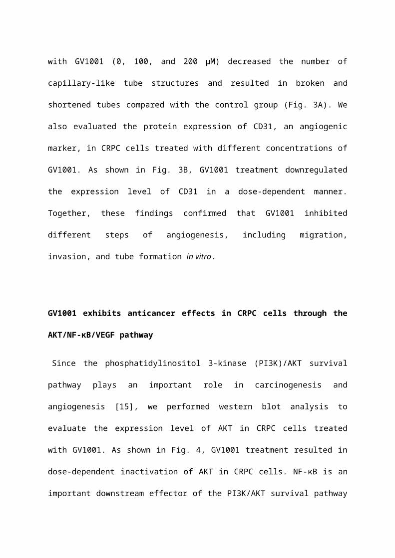
GV1001 inhibited angiogenesis
To determine the effect of GV1001 on angiogenesis in vitro, we performed tube
formation assays in HUVECs. Treatment of HUVECs with GV1001 (0, 100, and 200
µM) decreased the number of capillary-like tube structures and resulted in broken
and shortened tubes compared with the control group (Fig. 3A). We also evaluated
the protein expression of CD31, an angiogenic marker, in CRPC cells treated with
different concentrations of GV1001. As shown in Fig. 3B, GV1001 treatment
downregulated the expression level of CD31 in a dose-dependent manner. Together,
these findings confirmed that GV1001 inhibited different steps of angiogenesis,
including migration, invasion, and tube formation in vitro.
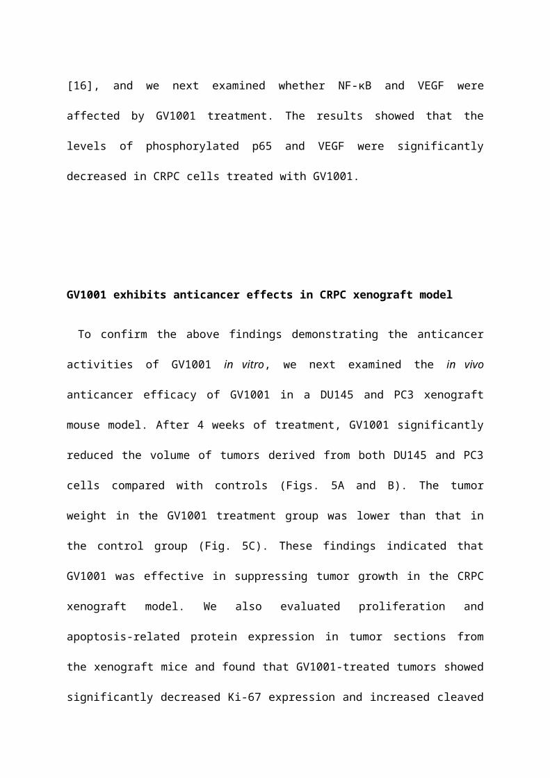
GV1001 exhibits anticancer effects in CRPC cells through the
AKT/NF-κB/VEGF pathway
Since the phosphatidylinositol 3-kinase (PI3K)/AKT survival pathway plays an
important role in carcinogenesis and angiogenesis [15], we performed western blot
analysis to evaluate the expression level of AKT in CRPC cells treated with GV1001.
As shown in Fig. 4, GV1001 treatment resulted in dose-dependent inactivation of
AKT in CRPC cells. NF-κB is an important downstream effector of the PI3K/AKT
survival pathway [16], and we next examined whether NF-κB and VEGF were
affected by GV1001 treatment. The results showed that the levels of phosphorylated
p65 and VEGF were significantly decreased in CRPC cells treated with GV1001.

GV1001 exhibits anticancer effects in CRPC xenograft model
To confirm the above findings demonstrating the anticancer activities of GV1001 in
vitro, we next examined the in vivo anticancer efficacy of GV1001 in a DU145 and
PC3 xenograft mouse model. After 4 weeks of treatment, GV1001 significantly
reduced the volume of tumors derived from both DU145 and PC3 cells compared
with controls (Figs. 5A and B). The tumor weight in the GV1001 treatment group was
lower than that in the control group (Fig. 5C). These findings indicated that GV1001
was effective in suppressing tumor growth in the CRPC xenograft model. We also
evaluated proliferation and apoptosis-related protein expression in tumor sections
from the xenograft mice and found that GV1001-treated tumors showed significantly
decreased Ki-67 expression and increased cleaved caspase-3 expression compared
with controls (Fig. 5D). In addition, phosphorylated p65 expression was significantly
decreased in GV1001-treated tumors.
Discussion
GV1001 was initially developed as a telomerase-specific peptide vaccine for
pancreas and lung cancer [17, 18]. Interestingly, Lee at al. found that GV1001 had
an unexpected function as a cell-penetrating peptide, and the delivered GV1001 was
predominantly located in the cytoplasm of the cells through its interaction with
HSP70 and HSP90 [14]. Several researchers later reported anti-cancer effects of
GV1001 in various cancer cells [14, 19, 20]. In the present study, we demonstrated
that GV1001 had potent effects in inhibition of cell viability, induction of apoptosis,
and inhibition of angiogenesis in CRPC cells in a dose-dependent manner. Our in
vivo study also demonstrated that GV1001 exhibited inhibitory effects on tumor
growth in CRPC xenograft-bearing mice. The anti-cancer effects of GV1001 in CRPC
cells may be mediated through inhibition of the AKT/NF-κB/VEGF pathway. To the
best of our knowledge, this is the first report of the anticancer effects and biological
mechanism of GV1001 in human CRPC cells.
Angiogenesis is an important step of tumorigenesis [21]. Thus, inhibition of
angiogenesis has become an effective treatment strategy for solid cancers. VEGF is
a key regulator of endothelial cell proliferation, migration, invasion, vascular
permeability, and vasodilation [22]. In this study, we examined the role of GV1001 in
tumor angiogenesis and found that GV1001 strongly inhibited tube formation.
GV1001 also inhibited cell invasion and migration capacity and resulted in dose-
dependent suppression of VEGF. To examine the underlying mechanisms of the anti-
angiogenic effects of GV1001, we examined AKT and NF-κB protein expression.
GV1001 markedly inhibited the expression of AKT and NF-κB in CRPC cells.

AKT is a serine/threonine kinase that regulates tumor cell proliferation, survival,
bio-energetics, and angiogenesis by phosphorylation of its molecular targets,
including factors in the NF-κB pathway [23]. Inflammatory stimuli can activate the
PI3K/Akt pathway in various cell types, leading to phosphorylation of IκBα.
Phosphorylation and nuclear translocation of p65 lead to activation of NF-κB and
production of VEGF [24]. This is an important mechanism of pathologic
angiogenesis, and GV1001 may inhibit the AKT/NF-κB/VEGF pathway and resulted
in decreased tumor angiogenesis.
The precise mechanisms underlying the anti-cancer effects of GV1001 have not
been completely elucidated. In addition to our data demonstrating effects on the
AKT/NF-κB/VEGF pathway, GV1001 exhibits several potential biological
mechanisms of action, with involvement in the TGF-β signaling pathway [25], the
hypoxia-induced HIF-1α-VEGF signaling axis [19], and the ERK and p38 MAP
kinase pathway [26]. In addition, GV1001 inhibits gonadotropin-releasing hormone
(GnRH) and 5α-reductase, which are important in prostate cancer [27, 28]. Kim et al.
reported that GV1001 can be a therapeutic agent for testosterone-induced benign
prostatic hyperplasia (BPH) [27]. After establishing BPH in castrated rats via daily
subcutaneous injections of testosterone propionate, GV1001 was administered for 4
weeks. The authors found that GV1001 significantly decreased prostate weight and
inhibited 5α-reductase activity in a dose-dependent manner. GV1001 is a ligand for
GnRH receptor to selectively stimulate the Gαs/cAMP pathway and antagonize Gαq-
coupled Ca2+ release by leuprolide acetate in prostate cancer [28]. GV1001 might
also exhibit anti-cancer effects in CRPC cells through these biological mechanisms.
In conclusions, we demonstrated that GV1001 inhibited cell viability, induced
apoptosis, and inhibited angiogenesis in CRPC cells by inhibition of the AKT/NF-
κB/VEGF signaling pathway. These results provide experimental evidence for
GV1001 as a potential therapeutic agent of CRPC.
Conflict of interest
No potential conflicts of interest are disclosed.
Acknowledgements
This work was supported by the National Research Foundation of Korea (NRF)
grant funded by the Korean government (MSIP) (No. 2017R1D1A1B03030051).

References
1. Siegel RL, Miller KD, Jemal A. Cancer statistics, 2016. CA Cancer J Clin. 2016;
66: 7-30.
2. Oh CM, Won YJ, Jung KW, Kong HJ, Cho H, Lee JK, et al. Cancer Statistics in
Korea: Incidence, Mortality, Survival, and Prevalence in 2013. Cancer Res Treat.
2016; 48: 436-50.
3. Culine S, Droz JP. Chemotherapy in advanced androgen-independent prostate
cancer 1990-1999: a decade of progress? Ann Oncol. 2000; 11: 1523-30.
4. Beltran H, Beer TM, Carducci MA, de Bono J, Gleave M, Hussain M, et al. New
therapies for castration-resistant prostate cancer: efficacy and safety. Eur Urol. 2011;
60: 279-90.
5. Tannock IF, de Wit R, Berry WR, Horti J, Pluzanska A, Chi KN, et al. Docetaxel
plus prednisone or mitoxantrone plus prednisone for advanced prostate cancer. N
Engl J Med. 2004; 351: 1502-12.
6. Petrylak DP, Tangen CM, Hussain MH, Lara PN, Jr., Jones JA, Taplin ME, et al.
Docetaxel and estramustine compared with mitoxantrone and prednisone for
advanced refractory prostate cancer. N Engl J Med. 2004; 351: 1513-20.
7. de Bono JS, Oudard S, Ozguroglu M, Hansen S, Machiels JP, Kocak I, et al.
Prednisone plus cabazitaxel or mitoxantrone for metastatic castration-resistant
prostate cancer progressing after docetaxel treatment: a randomised open-label trial.
Lancet. 2010; 376: 1147-54.
8. de Bono JS, Logothetis CJ, Molina A, Fizazi K, North S, Chu L, et al. Abiraterone
and increased survival in metastatic prostate cancer. N Engl J Med. 2011; 364: 1995-
2005.
9. Ryan CJ, Smith MR, de Bono JS, Molina A, Logothetis CJ, de Souza P, et al.
Abiraterone in metastatic prostate cancer without previous chemotherapy. N Engl J
Med. 2013; 368: 138-48.
10. Scher HI, Fizazi K, Saad F, Taplin ME, Sternberg CN, Miller K, et al. Increased
survival with enzalutamide in prostate cancer after chemotherapy. N Engl J Med.
2012; 367: 1187-97.
11. Kantoff PW, Higano CS, Shore ND, Berger ER, Small EJ, Penson DF, et al.
Sipuleucel-T immunotherapy for castration-resistant prostate cancer. N Engl J Med.
2010; 363: 411-22.
12. Parker C, Nilsson S, Heinrich D, Helle SI, O'Sullivan JM, Fossa SD, et al. Alpha
emitter radium-223 and survival in metastatic prostate cancer. N Engl J Med. 2013;
369: 213-23.
13. Shaw VE, Naisbitt DJ, Costello E, Greenhalf W, Park BK, Neoptolemos JP, et al.
Current status of GV1001 and other telomerase vaccination strategies in the
treatment of cancer. Expert Rev Vaccines. 2010; 9: 1007-16.
14. Lee SA, Kim BR, Kim BK, Kim DW, Shon WJ, Lee NR, et al. Heat shock protein-
mediated cell penetration and cytosolic delivery of macromolecules by a telomerase-
derived peptide vaccine. Biomaterials. 2013; 34: 7495-505.
15. Toren P, Zoubeidi A. Targeting the PI3K/Akt pathway in prostate cancer:
challenges and opportunities (review). Int J Oncol. 2014; 45: 1793-801.
16. Meng F, Henson R, Patel T. Chemotherapeutic stress selectively activates NF-
kappa B-dependent AKT and VEGF expression in liver cancer-derived endothelial
cells. Am J Physiol Cell Physiol. 2007; 293: C749-60.
17. Brunsvig PF, Aamdal S, Gjertsen MK, Kvalheim G, Markowski-Grimsrud CJ, Sve
I, et al. Telomerase peptide vaccination: a phase I/II study in patients with non-small
cell lung cancer. Cancer Immunol Immunother. 2006; 55: 1553-64.
18. Bernhardt SL, Gjertsen MK, Trachsel S, Moller M, Eriksen JA, Meo M, et al.
Telomerase peptide vaccination of patients with non-resectable pancreatic cancer: A
dose escalating phase I/II study. Br J Cancer. 2006; 95: 1474-82.
19. Kim BK, Kim BR, Lee HJ, Lee SA, Kim BJ, Kim H, et al. Tumor-suppressive
effect of a telomerase-derived peptide by inhibiting hypoxia-induced HIF-1alpha-
VEGF signaling axis. Biomaterials. 2014; 35: 2924-33.
20. Kim GE, Jung AR, Kim MY, Lee JB, Im JH, Lee KW, et al. GV1001 Induces
Apoptosis by Reducing Angiogenesis in Renal Cell Carcinoma Cells Both In Vitro
and In Vivo. Urology. 2018; 113: 129-37.
21. Folkman J. Role of angiogenesis in tumor growth and metastasis. Semin Oncol.
2002; 29: 15-8.
22. Ellis LM, Hicklin DJ. VEGF-targeted therapy: mechanisms of anti-tumour activity.
Nat Rev Cancer. 2008; 8: 579-91.
23. Mitsiades CS, Mitsiades N, Koutsilieris M. The Akt pathway: molecular targets for
anti-cancer drug development. Curr Cancer Drug Targets. 2004; 4: 235-56.
24. Ridiandries A, Tan JT, Bursill CA. The Role of CC-Chemokines in the Regulation
of Angiogenesis. Int J Mol Sci. 2016; 17: 1856.
25. Chen W, Shin KH, Kim S, Shon WJ, Kim RH, Park NH, et al. hTERT peptide
fragment GV1001 demonstrates radioprotective and antifibrotic effects through
suppression of TGFbeta signaling. Int J Mol Med. 2018; 41: 3211-20.
26. Ko YJ, Kwon KY, Kum KY, Lee WC, Baek SH, Kang MK, et al. The Anti-
Inflammatory Effect of Human Telomerase-Derived Peptide on P. gingivalis
Lipopolysaccharide-Induced Inflammatory Cytokine Production and Its Mechanism in
Human Dental Pulp Cells. Mediators Inflamm. 2015; 2015: 385127.
27. Kim KS, Yang HY, Chang SC, Kim YM, Lee KY, Lee BM, et al. Potential
repositioning of GV1001 as a therapeutic agent for testosteroneinduced benign
prostatic hyperplasia. Int J Mol Med. 2018; 42: 2260-8.
28. Kim JW, Yadav DK, Kim SJ, Lee MY, Park JM, Kim BS, et al. Anti-cancer effect of
GV1001 for prostate cancer; function as a ligand of GnRHR. Endocr Relat Cancer.
2018 [Epub ahead of print].

Figure 1. GV1001 inhibits cell viability and induces apoptosis in CRPC cells
(A) Human CRPC cell lines were treated with GV1001 (0, 50, 100, 150, and 200 µM)
for 48 h and cell viability was assessed. (B) Apoptotic cells were evaluated by
TUNEL assay in DU145 and PC3 cells treated as indicated. The ratio of TUNEL
positive cells was calculated as the number of cells with positive TUNEL staining
(red) divided by the number of cells with positive cells staining (blue). All images are
400x magnification. (C) Evaluation of GV1001-induced apoptosis in CRPC cells by
flow cytometry. The percentages of apoptotic cells were analyzed by dot plot. All data
were expressed as mean ± standard deviation. *P<0.05 compared with controls. (D)
Western blot analysis for Bcl-2 and cleaved caspase-3 in CRPC cells after GV1001
treatment. The β-actin housekeeping protein was used as an internal control.

Figure 2. GV1001 inhibits invasion and migration capacity of CRPC cells
(A) Representative microscopic images of cells that had migrated into the lower
chamber are shown at 200x magnification. The migrated cells were quantified using
a bar graph. Data are expressed as mean ± standard deviation. *P<0.05 compared
with controls. (B) At 0, 24, and 48 h after scratching in wound healing assays, cell
migration was assessed by microscopy (100x magnification). (C) Western blot
analysis for MMP-2, MMP-9, TIMP-1 and TIMP-2 in CRPC cells after GV1001
treatment. The β-actin housekeeping protein was used as an internal control.

Figure 3. GV1001 inhibits angiogenesis
(A) The endothelial tube formation assay was performed in CRPC cells treated with
various concentrations of GV1001 (0, 100, and 200 µM). Representative microscopic
images of changes in cell morphology are shown at 50x magnification. The number
of tube-like structures was counted, and tube formation was calculated as a
percentage of the control. Data are expressed as mean ± standard deviation.
*P<0.05 compared with controls. (B) Western blot analysis for CD31 protein in CRPC
cells treated with GV1001. The β-actin was used as a loading control.
Figure 4. GV1001 exhibits anticancer effects in CRPC cells through the
AKT/NF-κB/VEGF pathway
Figure 5. GV1001 exhibits anticancer effects in a CRPC xenograft model
(A) Representative images of tumors from the DU145 and PC3 xenograft mouse
model. (B, C) Mean tumor volume and weight are shown. (D) Tumors were excised
and processed for hematoxylin and eosin staining and immunohistochemical staining
for Ki-67, CD31, and p65. Representative images are shown at 400x.