Schwanniomyces occidentalis bbbb usage and directed...
34
New insights into the fructosyltransferase activity of Schwanniomyces occidentalis β-fructofuranosidase emerging from a non-conventional codon usage and directed mutation Miguel Álvaro-Benito 1 , Miguel de Abreu 1 , Francisco Portillo 2 , Julia Sánz-Aparicio 3 and María Fernández-Lobato 1,* 1 Centro de Biología Molecular Severo Ochoa. Departamento de Biología Molecular (UAM- CSIC), Universidad Autónoma Madrid, Cantoblanco, 28049 Madrid, Spain. 2 Departamento de Bioquímica, Facultad de Medicina, Instituto de Investigaciones Biomédicas Alberto Sols (UAM-CSIC), 28029 Madrid, Spain. 3 Grupo de Cristalografía Macromolecular y Biología Estructural, Instituto de Química-Física "Rocasolano", CSIC, Serrano 119, 28006 Madrid, Spain. * Correspondence to: M. Fernández-Lobato. Centro de Biología Molecular Severo Ochoa, Departamento de Biología Molecular (UAM-CSIC), Universidad Autónoma Madrid, Cantoblanco, 28049 Madrid, Spain. Phone: 34-91-1964492. Fax: 34-91-1964420. e-mail: [email protected] Running Title: Non-conventional CUG decoding improves Ffase properties Copyright © 2010, American Society for Microbiology and/or the Listed Authors/Institutions. All Rights Reserved. Appl. Environ. Microbiol. doi:10.1128/AEM.01614-10 AEM Accepts, published online ahead of print on 17 September 2010 on October 22, 2018 by guest http://aem.asm.org/ Downloaded from
Transcript of Schwanniomyces occidentalis bbbb usage and directed...
New insights into the fructosyltransferase activity of Schwanniomyces
occidentalis ββββ-fructofuranosidase emerging from a non-conventional codon
usage and directed mutation
Miguel Álvaro-Benito1, Miguel de Abreu1, Francisco Portillo2, Julia Sánz-Aparicio3 and
María Fernández-Lobato1,*
1Centro de Biología Molecular Severo Ochoa. Departamento de Biología Molecular (UAM-
CSIC), Universidad Autónoma Madrid, Cantoblanco, 28049 Madrid, Spain. 2Departamento
de Bioquímica, Facultad de Medicina, Instituto de Investigaciones Biomédicas Alberto Sols
(UAM-CSIC), 28029 Madrid, Spain. 3Grupo de Cristalografía Macromolecular y Biología
Estructural, Instituto de Química-Física "Rocasolano", CSIC, Serrano 119, 28006 Madrid,
Spain.
*Correspondence to: M. Fernández-Lobato. Centro de Biología Molecular Severo Ochoa,
Departamento de Biología Molecular (UAM-CSIC), Universidad Autónoma Madrid,
Cantoblanco, 28049 Madrid, Spain. Phone: 34-91-1964492. Fax: 34-91-1964420. e-mail:
Running Title: Non-conventional CUG decoding improves Ffase properties
Copyright © 2010, American Society for Microbiology and/or the Listed Authors/Institutions. All Rights Reserved.Appl. Environ. Microbiol. doi:10.1128/AEM.01614-10 AEM Accepts, published online ahead of print on 17 September 2010
on October 22, 2018 by guest
http://aem.asm
.org/D
ownloaded from
1
ABSTRACT 1
2
Schwanniomyces occidentalis β-fructofuranosidase (Ffase) releases β-fructose from the non-3
reducing end of β-fructans and synthesizes 6-kestose and 1-kestose, both considered as 4
prebiotic fructooligosaccharides. Analysing the amino acid sequence of this protein reveals 5
that it includes a serine instead of a leucine at position 196, caused by a non-universal 6
decoding of the unique mRNA leucine codon CUG. Substitution of Ser196 by leucine 7
dramatically lowers the apparent catalytic efficiency (kcat/Km) of the enzyme (approx. 1000-8
fold) but surprisingly, its transferase activity is enhanced by almost 3-fold, as is the enzymes’ 9
specificity for 6-kestose synthesis. The influence of six Ffase residues on enzyme activity was 10
analysed on both the Leu/Ser196 backgrounds (Trp47, Asn49, Asn52, Ser111, Lys181 and 11
Pro232). Only N52S and P232V mutations improved the transferase activity of the wild-type 12
enzyme (about 1.6-fold). Modelling the transfructosylation products into the active site, in 13
combination with an analysis of the kinetics and transfructosylation reactions defines a new 14
region responsible for the transferase specificity of this enzyme. 15
16
17
18
19
Keywords: yeasts; fructosyltransferase; 6-kestose; non-conventional codon usage; 20
fructooligosaccharides. 21
on October 22, 2018 by guest
http://aem.asm
.org/D
ownloaded from
2
INTRODUCTION 1
β-fructofuranosidases (EC 3.2.1.26) are enzymes of biotechnological interest that 2
catalyse the release of β-fructose from the non-reducing terminus of various β-D-3
fructofuranoside substrates. In general, they exhibit a high degree of sequence homology and 4
based on their amino acid sequences, they fall into family 32 of the glycosyl-hydrolases (GH), 5
along with invertases, inulinases and fructosyltransferases (http:/afmb.cnrs-mrs.fr/CAZY). 6
The GH32 family has been studied intensely and some three-dimensional structures are now 7
available, such as that of inulinase from Aspergillus awamorii (26), fructan-exohydrolase 8
from Cichorium intybus (34), or invertase from Thermotoga maritima (2) and Arabidopsis 9
thaliana (35). These proteins contain a five-blade β-propeller N-terminal catalytic module and 10
a C-terminal β-sandwich domain (19). Multiple-sequence alignment of GH32 proteins, which 11
are included into the GH-J clan together with the GH68 proteins of the inulosucrase family, 12
reveals the presence of three conserved motifs each containing a key acidic residue (in bold) 13
implicated in substrate binding and hydrolysis: Asn-Asp-Pro-Asn-Gly (NDPNG), Arg-Asp-14
Pro (RDP) and Glu-Cys (EC) (28). These conserved residues are implicated in a double 15
displacement reaction in which a covalent glycosyl-enzyme intermediate is formed. Thus, the 16
catalytic mechanism proposed for the Saccharomyces cerevisiae invertase implies that Asp23 17
(NDPNG) acts as a nucleophile and Glu204 (EC) as the acid/base catalyst (29), whereas 18
Asp309 (RDP) of Acetobacter diazotropicus levansucrase influences the efficiency of sucrose 19
hydrolysis (7) and Arg188 and Asp189 of this latter motif define the substrate binding and 20
specificity of exoinulinase from A. awamorii toward fructopyranosyl residues (26). 21
As well as hydrolysing sucrose, β-fructofuranosidases may also catalyze the synthesis 22
of short-chain fructooligosaccharides (FOS), in which one to three fructosyl moieties are 23
linked to the sucrose skeleton by different glycosidic bonds depending on the source of the 24
on October 22, 2018 by guest
http://aem.asm
.org/D
ownloaded from

3
enzyme (12, 21, 31). FOS act as prebiotics and they exert a beneficial effect on human health, 1
participating in the prevention of cardiovascular diseases, colon cancer or osteoporosis (16). 2
Currently, FOS are mainly produced by Aspergillus fructosyltransferase in industry (10, 31), 3
providing a mixture of FOS with an inulin-type structure that contains β-(2→1)-linked 4
fructose-oligomers (1F-FOS: 1-kestose, nystose). Curiously, when the link between two 5
fructose units (6F-FOS: 6-kestose), or between fructose and glucosyl moiety (6G-FOS: 6
neokestose), involves a β-(2→6)-link, the prebiotic properties of the FOS may be enhanced 7
beyond that of commercial FOS (23). 8
The yeast Schwanniomyces occidentalis (also Debaryomyces occidentalis) produces a 9
number of extracellular enzymes that make it of interest in biotechnology. Several of its 10
amylolytic enzymes have been characterized, including amylases and glucoamylase (1, 9), as 11
well as an invertase (17). In addition, we also characterized an extracellular β-12
fructofuranosidase (Ffase) from this yeast that hydrolyzes sucrose, 1-kestose and nystose (5). 13
This enzyme exhibited a transfructosylating activity that efficiently produces the 14
trisaccharides 6-kestose and 1-kestose in the ratio 3:1, generating the highest 6-kestose yield 15
yet reported as far as we know. The Ffase three-dimensional structure has recently been 16
resolved (6) and represented as bi-modular homodimer arranged like other GH32 enzymes. 17
The Asp50 (NDPNG) and Glu230 (EC) located at the centre of the propeller are the catalytic 18
residues implicated in substrate binding and hydrolysis, whereas Arg178 and Asp179 form 19
the RDP motif (6). 20
The genetic code of some yeasts incorporates certain variation. For example, while 21
CUG was believed to be an universal codon for leucine, in the cytoplasm of certain species of 22
the Candida genus (15) it encodes a serine, as in Pichia farinosa (33). The reassignment of 23
on October 22, 2018 by guest
http://aem.asm
.org/D
ownloaded from
4
this codon is mediated by a novel serine-tRNA that acquired a leucine 5’-CAG-3’ anticodon 1
(25). 2
Here we show that deviation from the standard use of the CUG leucine codon to 3
encode serine was correlated with the transferase capacity and specificity of the Ffase 4
enzyme. Indeed, the S196L substitution enhanced the transferase activity of the enzyme 3-5
fold. Several site-directed mutants were generated and characterised, studying their 6
transferase capacity. These results are considered on the basis of the enzymes’ three-7
dimensional structure, which enables a novel putative binding site of sucrose to be identified 8
that serves as a water substitute donor in the hydrolytic reaction yielding the tranglycosylation 9
product 6-kestose. 10
11
MATERIALS AND METHODS 12
Materials, organisms, transformations and growth conditions. 1-kestose [α-D-13
glucopyranosyl-(1→2)-β-D-fructofuranosyl-(1→2)-β-D-fructofuranose] and nystose [α-D-14
glucopyranosyl-(1→2)-β-D-fructofuranosyl-(1→2)-β-D-fructofuranosyl-(1→2)-β-D-15
fructofuranose] were obtained from TCI Europe (Zwijndrecht, Belgium). The 16
Schwanniomyces occidentalis strains employed were ATCC26077, ATCC20499 and 17
ATCC26076. Saccharomyces cerevisiae EUROSCARF Y02321 [BY4741; Mat a; his3∆1; 18
leu2∆0; met15∆0; ura3∆0; YIL162w(SUC2)::kanMX4] (accession code Y02321) was used as 19
the expression host and it was transformed by the standard lithium acetate method. Yeasts 20
were grown at 29ºC on YEPD (1% yeast extract, 2% peptone, 2% glucose) or Inulin-based 21
medium (2% yeast extract, 1.5% inulin). SC(U)D or SC(U)S media (0.67% YNB, 0.1% 22
leucine, 0.05% histidine and methionine, 2% glucose (D) or 2% sucrose (S)) were used to 23
select transformants and YPGal (1% yeast extract, 1% peptone, 2% galactose) was used to 24
on October 22, 2018 by guest
http://aem.asm
.org/D
ownloaded from
5
induce protein expression in S. cerevisiae. Growth was monitored spectrophotometrically at a 1
wavelength of 600 nm (A600nm). Escherichia coli DH5α [(F´I EndA1, hsdR17 (rk-mk-) 2
supE44 thi-1 recA1 gyrA (Naf) relA1 ∆(lacIZYA-argF) U169 deoR (φ80dlac∆(lacZ)M15)] 3
was used for DNA manipulation and amplification by standard techniques. 4
Plasmids, cloning and mutagenesis. Genomic DNA was isolated from yeast using 5
standard techniques. The Sw. occidentalis INV (X17604) gene was obtained by PCR using 6
genomic DNA and the primers SOINVatg (+1 to +22), 5’-7
CGGGATCCATGGTACAAGTTTTAAGTGTAT-3’ and SOINVter (+1586 to +1608) 5’-8
CCTCGAGCTACTTATTTAGTTCTCTAATGA-3’, which include BamHI and XhoI 9
recognition sequences (in bold), respectively. The 1.6 kb PCR products were treated with 10
BamHI-XhoI, inserted into the pST-Blue1 vector (Perfectly Blunt® Cloning Kit Novagen), 11
and sequenced (SIDI, Universidad Autónoma de Madrid, Spain). Plasmids for sequence 12
analysis were purified with the Wizard Plus SV Minipreps kit (Promega) according to the 13
manufacturer’s protocol. Some sequence changes were founded in the INV gene and the new 14
sequence was denominated as Ffase. For heterologous expression, the Ffase gene from Sw. 15
occidentalis ATCC26077 was amplified using the FfpYES-B (+1 +25) 5’-16
TAGGATCCAACATGGTACAAGTTTTAAGTGTATTAG-3’ and FfpYES-X (+1591 17
+1614) 5’-CATCTCTAGACTAGCCCTACTTATTTAGTTCTCT-3’ primers. Restriction 18
sites for BamHI and XbaI (shown in bold) were included in these primers to clone the PCR 19
product into the pYES2.0 shuttle vector (Invitrogen) under the control of the GAL1 promoter, 20
thereby generating the Ffase-pYES construction. This plasmid was used as a template for 21
L196S, L196E, W47Y, N49S, N52S, S111T, K181F, P232V site directed mutagenesis using 22
specific primers (Table 1) and the method described previously (6). The PCR product was 23
incubated with DpnI for 2 hours to digest the parental DNA and 5 µl of this reaction was used 24
on October 22, 2018 by guest
http://aem.asm
.org/D
ownloaded from
6
directly to transform E. coli. DNA sequencing was used to verify that only the desire mutation 1
was present in the amplified products. 2
Protein purification, detection and quantification. β-Fructofuranosidase from Sw. 3
occidentalis ATCC26077 (Ffase) was purified as described elsewhere (27). Basically, yeast 4
was grown in inulin-based medium (1L) and the culture filtrate (1.4 x10³ U ml-1; 8.9 mg ml-1) 5
was concentrated, fractioned and dialyzed (against buffer A: 20 mM Tris-HCl pH 7.0) using a 6
VivaFlow 50 system (Vivascience). The resulting fraction (60.7x10³ U ml-1; 54.7 mg ml-1) 7
was applied on a DEAE-Sephacel column equilibrated with buffer A and the proteins were 8
eluted with a 0-0.5 M NaCl gradient in buffer A. The active fractions eluting in 0.15 M NaCl 9
were pooled, dialyzed in buffer A and concentrated (105.8 x10³ U ml-1; 1 mg ml-1). For 10
proteins expressed in S. cerevisiae optimum conditions for expression were defined in a time 11
course and the heterologous proteins was analysed measuring the fructofuranosidase activity 12
(sucrose hydrolysis) and/or in Western blots. Yeasts were pre-grown in SC(U)D medium and 13
they were then grown in YPGal medium (1 L) to the beginning of the stationary phase 14
(A600=6-7). The culture filtrates (0.6-12 U ml-1; about 15 mg ml-1) were then concentrated 15
(20-360 U ml-1; about 6 mg ml-1) and applied to a DEAE-Sephacel chromatography column 16
(see above). The active fractions eluted in 0.1 M NaCl were pooled, dialyzed, concentrated 17
(0.05-0.8 U ml-1; about 0.7 mg ml-1) and stored at -70ºC as described elsewhere (6). 18
Coomassie-stained SDS-PAGE (8%) of the samples confirmed the purity of the protein 19
fraction. 20
The polyclonal invertase antiserum used for Western blotting was generated by 21
injecting rabbits with commercially available S. cerevisiae invertase (Novozymes), using 1 22
mg of protein in Freund’s complete adjuvant for the initial injection. Decreasing amounts of 23
protein were subsequently injected in Freund’s incomplete adjuvant at the following intervals: 24
on October 22, 2018 by guest
http://aem.asm
.org/D
ownloaded from
7
2 week, 500 µg; 1 month, 200 µg; 2 month 100 µg. The immune serum was clarified using an 1
extracellular protein extract from S. cerevisiae Y02321 including the pYES2.0 vector. The 2
anti-INV antibody obtained was used at a dilution of 1:2000. For immunoblots, extracellular 3
proteins were prepared from cultures growing at 6-7 A600, which were then concentrated and 4
partly fractionated by filtration through Microcon YM-10 membranes. The proteins were 5
resolved electrophoretically and transferred to Immobilon-P membranes (Millipore) that were 6
probed with the invertase antibody. Binding of the antibody was detected using a secondary 7
goat anti-rabbit IgG coupled to horseradish peroxidase (GE Healthcare, Amersham) used as 8
indicated by the manufacturer. The Ffase associated with the cellular fraction was assayed 9
after the addition of glass beads and after five cycles of agitation in a vortex mixer for 1 min 10
as indicated (21). Protein concentrations were measured photometrically at 280 nm 11
(NanoDrop Spectrophotometer ND-1000). 12
MALDI-TOF analysis. Proteins were excised from SDS-PAGE gels, digested with 13
trypsin and identified by matrix-assisted laser desorption ionization-time-of-flight-mass 14
spectrometry (MALDI-TOF; Autoflex, Bruker, Bremen, Germany) at the Proteomic Service 15
of the “Centro de Biología Molecular Severo Ochoa” (Madrid). The tryptic peptide map 16
obtained was assigned by comparing their masses with thoses calculated from theoretical 17
tryptic digestion. The assignment was verified by analyzing the peptide by RP-LC/MS 18
(reverse-phase LC coupled to MS) using a Deca XP mass spectrometer and a ThermoHypersil 19
(0.18 x 150 mm) C18 column. The mass spectrometer was operated in the selected MS/MS 20
ion monitoring mode and the spectra from the peptide were analysed by assigning the 21
fragments to the candidate sequence after calculating the series of theoretical fragmentations. 22
Enzyme and kinetic analysis. Ffase (hydrolytic activity) was assayed by the 23
dinitrosalicylic acid (DNS) method adapted to a 96-well microplate and the optimal 24
on October 22, 2018 by guest
http://aem.asm
.org/D
ownloaded from
8
parameters were determined as described elsewhere(5). Standard Ffase activity was measured 1
using sucrose as a substrate and 1 unit (U) of activity was defined as that capable of 2
catalysing the formation of 1 µmol of reducing sugar per minute. For all kinetic analyses, the 3
velocity was measured in triplicate with 5 x 10⁻² to 5 x 10⁻⁴ mg ml-1 of enzyme and 0-500 mM 4
substrate. Reactions were performed over 20 min in 100 mM sodium acetate at the optimum 5
pH and temperature defined for each variant. The plotting and analysis of the curves was 6
carried out using the SigmaPlot software (version 7.101), and the kinetic parameters were 7
calculated fitting the initial rate values to the Michaelis-Menten equation. Ffase activity was 8
also detected by Zymogram analysis using non-denaturing gradient gels (4-15%, BioRad), 9
stained with 1% (w/v) 2,3,5-triphenyltetrazolium chloride (5). FOS production was analysed 10
by high-performance liquid chromatography after incubating 0.3 U of enzyme and 1 M (342 g 11
L-1) sucrose as indicated previously (5). 12
Computer analysis and molecular modelling. The nucleotide and amino acid 13
sequences were analysed using programs in the GCG Sequence Analysis Software Package 14
(available from the University of Wisconsin) in conjunction with sequence data from Swiss-15
Prot, GeneBank and EMBL databases. The sequence reported here was submitted to the 16
EMBL database (GenBank accession nº CQ890277). The structural analysis was carried out 17
using the O program (14) and the figures were obtained with PYMOL 18
(http://pymol.sourceforge.net/). The transfructosylating product 6-kestose was modelled from 19
the experimental 6-kestose crystal structure coordinates extracted from the Cambridge 20
Structural Database (CSD Refcode CELGIJ), superimposing its terminal fructose moiety onto 21
the fructose found in the Ffase crystal. The 1-kestose substrate was modelled into the Ffase 22
active-site as inferred from structural superimposition of the CiFEH-1Kestose coordinates 23
(PDB code 2AEZ) onto the Ffase-fructose complex (PDB code 3KF3). 24
on October 22, 2018 by guest
http://aem.asm
.org/D
ownloaded from
9
1
RESULTS 2
Sequence of the ββββ-fructofuranosidase from Schwanniomyces occidentalis contains 3
some unexpected changes. Initial studies on an invertase from Sw. occidentalis showed that 4
the active form of this enzyme was a homodimer consisting of two subunits, each containing 5
533 amino acids, whose sequence was deduced after analysis of the INV gene (18). We used 6
PCR to isolate this gene and found that the sequence amplified showed marked differences 7
from that previously reported, including four changes in the amino acid sequence encoded 8
(Fig. 1A). Identical results were found in two other Sw. occidentalis strains (see Materials and 9
Methods). The new INV gene isolated encoded a putative protein of 535 amino acids (instead 10
of 533) and it included the three conserved acidic residues in the motifs shared by all proteins 11
of the GH32 family (19, 28), NDPNG, RDP and EC.(Asp50, Asp179 and Glu230, 12
respectively). To verify that the β-fructofuranosidase from Sw. occidentalis (Ffase) with 13
transfructosylating activity that we had characterized previously (5) was encoded by the 14
sequence analysed, the enzyme was purified and analysed by MALDI-TOF and finger-15
printing (data not shown). Most of the masses retrieved (42% coverage) coincided with the 16
translation of the new DNA sequence. However, when an unexpected peptide of 2005 m/z 17
was analysed using microspray-ion trap M/S (data not shown), the Ffase sequence included a 18
serine instead of a leucine residue at position 196 (Fig. 1B). This change was caused by non-19
universal decoding of the unique mRNA CUG leucine codon, corresponding to 586CTG588 in 20
the DNA sequence analysed. 21
The non-universal decoding of the leucine CUG codon affects Ffase activity. The 22
functionality of the gene isolated here (hereafter Ffase) was verified through its heterologous 23
expression using a S. cerevisiae strain that was unable to use sucrose as a carbon source. 24
on October 22, 2018 by guest
http://aem.asm
.org/D
ownloaded from
10
Although the Ser196 residue (CUG=Ser) is included in a non-conserved region of the GH32 1
family, we mutated the Ffase triplet 586CTG588 to TCA that encodes serine in the standard 2
code in order to obtain a heterologously expressed protein with the wild-type amino acid 3
sequence (Ffase-Ser196). Genes encoding Ffase-Leu196 and Ffase-Ser196 were included in 4
S. cerevisiae and as expected, both complemented the lack of growth phenotype in sucrose of 5
the host strain. Maximum levels of hydrolytic activity (approximately 12 U ml-1) were 6
detected in the Ffase-Ser196 culture filtrates at the beginning of the stationary phase (12-16 h 7
of growth; A600=6), whereas activity fell to near 30% in cultures older than 30 h. Slightly 8
lower activity (≤8 U) was found in the cell-associated fraction during these phases. However, 9
replacing the Ser196 residue by leucine promoted a severe decrease in the levels of hydrolytic 10
activity in both fractions analysed (≤1 U ml-1), and it was not detected in non-denaturing 11
condition gels (Fig. 2A.a). A priori, these results suggest that Ffase-Leu196 would have 12
weaker activity, secretion or stability, or a combination of these characteristics. Analysis of 13
the extracellular Ffase using an anti-INV antibody that recognized this protein in its native 14
and heterologously produced forms (Fig. 2A.b) showed that they were secreted in similar 15
amounts. As expected, glycosylation is not the same in both yeast species (6) and the proteins 16
expressed in S. cerevisiae had a molecular mass of 95 kDa, at least 10 kDa higher than the 17
protein expressed in Sw. occidentalis. Moreover, there was a marked difference in their 18
estimated molecular mass on non-denaturing condition gels (Fig. 2A.a). 19
The Sw. occidentalis enzyme and the two proteins expressed in S. cerevisiae were 20
highly purified (Fig. 2A.c) and some of their biochemical characteristics were studied. In 21
general and as expected, the enzymes displayed classical Michaelis-Menten kinetics (data not 22
shown), and no significant changes were found in the kinetic behaviour of the wild-type and 23
the heterologously expressed Ffase-Ser196 (Table 2). However, the Ffase-Leu196 had a 24
on October 22, 2018 by guest
http://aem.asm
.org/D
ownloaded from
11
drastically lowered apparent catalytic efficiency relative to that of the wild-type enzyme 1
(kcat/Km of approx. 1/1000) and the difference was greater as the size of the substrate analysed 2
increased (sucrose to nystose). In addition, replacing the Ser196 with leucine affected other 3
biochemical characteristics of the enzyme’s hydrolytic activity, such as the pH and 4
temperature dependence. Thus, while no changes were found in the optimal pH (5.5 units) 5
and temperature (50-55 ºC) of the Ffase-Ser196 expressed in both yeast species, the optimal 6
temperature of the Ffase-Leu196 fell by 10 ºC (40-45 ºC) and he pH by 0.5 units (5.0; data not 7
shown). 8
Substitution at Ser196 modifies the transferase activity of Ffase. The Ffase from 9
Sw. occidentalis presents a transfructosylating activity that produces mainly 6-kestose (6F-10
FOS) and then 1-kestose (1F-FOS) in a 3:1 ratio (5). Therefore, we decided to examine the 11
transferase capacity of the two heterologously expressed enzymes (Fig. 2B). After a 6-12 h 12
reaction, a maximum FOS concentration of about 25 g L-1 was obtained using the enzyme 13
containing Ser196 expressed in both yeast species (S. cerevisiae or Sw. occidentalis), 14
corresponding to approximately 7% (w/w) of the total sugar composition in the mixture. 15
Surprisingly, the Ffase-Leu196 showed enhanced transferase capacity (almost 3-fold) and 39 16
g L-1 FOS was produced after a 12 h reaction that corresponded to 11.5% (w/w) of the total 17
sugar composition. The maximum concentration of FOS was reached in 72 h and it was 70.3 18
g L-1, which corresponded to almost 21% (w/w) of the sugar in the mixture. In addition, the 19
substitution of Ser196 by leucine increased the 6-kestose:1-kestose ratio about 7-fold (β-20
(2→6)-:β-(2→1)-linked FOS ratio), which meant that there was virtually no significant 21
production of 1-kestose (4.6 g L-1; Fig. 2C and D). 22
Levansucrases from Bacillus subtilis (24) and Gluconoazetobater diazotrophicus (22) 23
synthesize levans directly from sucrose, and these polymers are basically formed by β-(2→6)-24
on October 22, 2018 by guest
http://aem.asm
.org/D
ownloaded from
12
linked fructose units. An equivalent position to Ser196 in Ffase is occupied by glutamate in 1
these two levansucrases and thus, we decided to evaluate the effect of the S196E substitution 2
on Ffase activity. Although extracellular Ffase-Glu196 protein was evident in immunoblots 3
probed with the anti-INV antibodies (data not shown), no Ffase activity was detected in any 4
of the cellular fractions analysed. 5
As mentioned above, the catalytic domain of each subunit folds into a β-propeller that 6
is assembled from five blades (I-V) each composed of four antiparallel β-strands. Given the 7
position of Ser196 in the Ffase dimer (Figure 3A), the Ser196 residue is located in blade III, 8
forming a hydrogen bond with Arg178 from the RDP motif (Fig. 3B). 9
Improving the transferase activity by site-directed mutagenesis. A multiple 10
alignment of invertases and fructosyltransferases from different organisms available in the 11
EMBL/SWISS-PROT databases clearly revealed the presence of some well conserved 12
regions, enabling the positions of residues that could be related to their activity to be 13
established (Fig. 4). Indeed, variations in the amino acid sequence adjacent to the active 14
nucleophile site ((WM)NDPNG) have been associated with the transglycosylation capacity of 15
some invertases from plants such as onion (30) and wheat (32). Accordingly, the residues 16
Trp47, Asn49 and Asn52 from the Sw. occidentalis enzyme were mutated and the activity of 17
the new enzymes generated was analysed. Mutant enzymes with a W47Y, N49S or N52S 18
substitution had significantly higher Km values for sucrose on both a Leu and Ser196 protein 19
background (Table 3). In addition, the transfructosylating activity of W47Y and N49S 20
mutants was greatly reduced (Fig. 5), possibly as a consequence of the strong fall in catalytic 21
efficiency (of approx. 300-5000 fold for Ffase-Leu196 and approx. 11-78 fold for Ffase-22
Ser196; Table 3). By contrast, the N52S mutation increased the transfructosylating activity of 23
the Ffase-Ser196 variant by approx. 1.8-fold. 24
on October 22, 2018 by guest
http://aem.asm
.org/D
ownloaded from
13
Different plant invertases are characterized by single amino acid substitutions in the 1
conserved EC(P/V) sequence, which includes the acid/base catalyst (11). A proline is present 2
in extracellular invertases and exoinulinases, whereas a valine occupies this position in 3
vacuolar invertases (4) and fructosyltransferases (Fig. 4). The Pro238Val substitution in the 4
extracellular invertase from Chenopodium rubrum produced a decrease (28%) in the rate of 5
raffinose cleavage (4). Similarly, the P232V substitution also produced a notable reduction in 6
the catalytic efficiency of Ffase (approx. 32% on a Leu196 and 98% on a Ser196 7
background), although interestingly it increased the transfructosylase activity of the Ser196 8
variant by 1.6-fold (62%). Finally, positions Ser111 and Lys181 were mutated in Ffase 9
according to the homologous residues in Aspergillus spp. enzymes (Fig. 4), currently the main 10
industrial FOS producers (10, 31). However, substitutions S111T and K181F produced 11
mutant enzymes with reduced (K181F) or unaffected (S111T) catalytic efficiency (Table 3) 12
without improving the transferase activity (Fig. 5). 13
DISCUSSION 14
One aim of this study was to identify amino acid residues involved in the transferase 15
capacity of Ffase, as well as to generate improved enzymes with potential applications in FOS 16
production. Accordingly, we show that a single amino acid substitution involving the non-17
universal decoding of the leucine CUG codon as serine (S196L) switches the enzymatic 18
activity of the β-fructofuranosydase from Sw. occidentalis, reducing the hydrolase activity 19
and increasing the transferase capacity approximately 3-fold. We also show that the N52S and 20
P232V substitutions improve the transferase activity of the wild-type enzyme by about 1.6-21
fold. 22
A remarkable feature of yeasts is the wide variation in their genomic G+C content. It 23
has been proposed that the codon-usage bias is generally correlated with the overall genome 24
on October 22, 2018 by guest
http://aem.asm
.org/D
ownloaded from
14
G+C content (8), and that the codons favoured are due to the tDNA gene copy number (20). 1
CUG is a rare leucine codon in S. cerevisiae (G+C: 40%) and it is also a rare codon for serine 2
in Sw. occidentalis (G+C: 36%; 13). The decoding of the leucine CUG codon as serine has 3
been reported previously in the cytoplasm of some Candida species (15) and in Pichia 4
farinosa (33). However, our study of the Ffase sequence provides the first direct evidence for 5
CUG decoding as serine in Sw. occidentalis. In this context, it has not been possible to 6
express the β-glucuronidase (gusA), β-lactamase (bla) or the phleomycin resistance-7
conferring gene (Tn5ble) from Escherichia coli in Sw. occidentalis (13). Thus, an appropriate 8
DNA composition and codon-usage bias might be important parameters for efficient 9
expression of heterologous genes in this host. However, these three genes contain various 10
CUG codons (19 in gusA, 7 in bla, and 11 in Tn5ble) and their alternative decoding as serine 11
could be relevant in the functioning of these proteins when expressed in Sw. occidentalis. 12
In terms of Ffase activity, the S196L substitution might be expected to position the 13
longer leucine side-chain (β-propeller, blade III) in a hydrophobic pocket surrounded by 14
Ile203 (also in blade III), and Tyr229 and Pro232 from blade IV (Fig. 3B). This modification 15
may produce a local rearrangement that would not only affect the position of Arg178 from the 16
RDP motif but also, the conformation of the first strand of blade IV (Tyr229-Pro238) where 17
the catalytic Glu230 is located. Consequently, such modifications may be deleterious for 18
hydrolytic activity. However, the improvement in the Ffase transferase:hydrolase ratio 19
observed upon Ser/Leu replacement indicates that this region might influence the retention 20
time of sucrose in the enzyme active-site, and the subsequently higher probability of finding a 21
sucrose acceptor instead of a water molecule, leading to transglycosylation. 22
When we consider all the residues and positions investigated in this study (Fig. 3 and 23
4), it is noteworthy that the mutations affecting the biosynthetic activity of Ffase are situated 24
on October 22, 2018 by guest
http://aem.asm
.org/D
ownloaded from
15
in two regions. The N52S mutant is located in the (WM)NDPNG motif, a segment that 1
includes the nucleophile that is involved in developing transfructosylating activity in plant 2
enzymes. The mutations involving the Ser196 residue lie in a second region situated at the 3
opposite side of the active-site cleft, in the environment of the acid/base catalyst within the 4
EC(P/V) motif (Fig. 3B). Independently, the S196L and P232V replacements, introduce some 5
structural changes in this area leading to a general increase in FOS production. Moreover, the 6
specificity towards 6-kestose is enhanced by these substitutions and consequently, this region 7
may be envisaged as a potential novel acceptor site for binding sugar instead of water, 8
promoting subsequent transferase activity. 9
In an attempt to understand the possible molecular basis for this particular behaviour, 10
active-sites from Ffase and other structurally known GH32 enzymes were analysed, and the 11
transfructosylating products 1-kestose and 6-kestose were modelled into the active enzyme 12
site (Fig. 6A left and right). On inspection, one wall of the active-site cleft (at the right) is 13
hydrophobic and it is contoured by aromatic residues that are conserved among structurally 14
known GH32 enzymes. The role of these residues in substrate binding can be illustrated by 15
the fact that the terminal sugar unit of raffinose and 1-kestose stack against the Phe46 and 16
Trp41 in the plant and bacterial enzymes (Fig. 6b and c). By contrast, there is more variation 17
in the opposite wall containing the acid/base catalyst Glu230, which in the case of Ffase is 18
also shaped by the contiguous subunit within the dimer. The residues from the environment of 19
the EC motif, such as Asn227 and Gln228, are in close proximity to the sucrose moiety of the 20
putative 6-kestose molecule. Therefore, they appear to be properly positioned to influence 21
sucrose binding and consequently, the enhanced specificity towards 6-kestose upon changes 22
in this region could be understood. 23
on October 22, 2018 by guest
http://aem.asm
.org/D
ownloaded from
16
In summary, our results suggest a novel substrate donor-binding site located near the 1
EC motif that would yield the β-(2→6)-linked transfructosylation product. This novel site 2
may contribute to the fact that the enzyme from Sw. occidentalis is the highest producer of 6-3
kestose known. 4
ACKNOWLEDGEMENTS 5
This work was supported by grants from the Spanish Ministry of Education and 6
Science (BIO2007-67708-C04-03 and -04) and by an institutional grant from the Fundación 7
Ramón Areces to the Centro de Biología Molecular Severo Ochoa. M.A-B. and M.A. were 8
supported by the Spanish Comunidad de Madrid programme and a FPU fellowship from the 9
Spanish Ministry of Education and Science, respectively. We thank Dr Ivana Janatova, for 10
help during the start of this research and Dr Francisco J. Plou for his assistance with the in 11
HPLC analysis. 12
13
REFERENCES 14
1. Abarca, D., M. Fernández-Lobato, L. del Pozo, and A. Jiménez. 1991. Isolation of a 15
new gene (SWA2) encoding an alpha amylase from Schwanniomyces occidentalis and its 16
expression in Saccharomyces cerevisiae. FEBS Lett. 279:41-44. 17
2. Alberto, F., C. Bignon, G. Sulzenbacher, B. Henrissat, and M. Czjzek. 2004. The three-18
dimenisonal structure of invertase (β-fructosidase) from Thermotoga maritima reveals a 19
bimodular arrangement and an evolutionary relationship between retaining and inverting 20
glycosidases. J. Biol. Chem. 279:18903-18910. 21
3. Alberto, F., E. Jordi, B. Henrissat, and M. Czjzek. 2006. Crystal structure of inactivated 22
Thermotoga maritima invertase in complex with the trisaccharide substrate raffinose. 23
Biochem. J. 395:457-462. 24
on October 22, 2018 by guest
http://aem.asm
.org/D
ownloaded from
17
4. Altenbach, D., E. Rudiño-Pineira, C. Olvera, T. Boller, A. Wiemken, and T. Ritsema. 1
2008. An acceptor-substrate binding site determining glycosyl transfer emerges from 2
mutant analysis of a plant vacuolar invertase and a fructosyltransferase. Plant. Mol. Biol. 3
69:47-56. 4
5. Álvaro-Benito, M., M. de Abreu, L. Fernández-Arrojo, F.J. Plou, J. Jiménez-Barbero, 5
A. Ballesteros, J. Polaina, and M. Fernández-Lobato. 2007. Characterization of a β-6
fructofuranosidase from Schwanniomyces occidentalis with transfructosylating activity 7
yielding the prebiotic 6-kestose. J. Biotechnol. 132:75-81. 8
6. Álvaro-Benito, M., A. Polo, B. González, M. Fernández-Lobato, and J. Sanz-Aparicio. 9
2010. Structural and kinetic analysis of Schwanniomyces occidentalis invertase reveals a 10
new oligomerization pattern and the role of its supplementary domain in substrate 11
binding. J. Biol. Chem. 285:13930-13941. 12
7. Batista, F.R., L. Hernández, J.R. Fernández, J. Arrieta, C. Menéndez, R. Gómez, Y. 13
Támbara, and T. Pons. 1999. Substitution of Asp-309 by Asn in the Arg-Asp-Pro 14
(RDP) motif of Acetobacter diazotropicus levansucrase affects sucrose hydrolisis, but not 15
enzyme specifity. Biochem. J. 337:503-506. 16
8. Chen, S.L., W. Lee, A.K. Hottes, L. Shapiro, and H.H. McAdams. 2004. Codon usage 17
between genomes is constrained by genome-wide mutational processes. Proc. Natl. Acad. 18
Sci. USA. 101:3480-3485. 19
9. Dowhanick, T.M., I. Russell, S.W. Scherer, G.G. Stewart, and V.L. Seligy. 1990. 20
Expression and regulation of glucoamylase from the yeast Schwanniomyces castelli. J. 21
Bacteriol. 172:2360-2366. 22
on October 22, 2018 by guest
http://aem.asm
.org/D
ownloaded from
18
10. Ghazi, I., L. Fernández-Arrojo, H. Garcia-Arellano, M. Ferrer, A. Ballesteros, and 1
F.J. Plou. 2007. Purification and kinetic characterizaton of a fructosyltransferase from 2
Aspergillus aculeatus. J. Biotechnol. 128:204-211. 3
11. Göetz, M., and T. Roitsch. 1999. The different pH optima and substrate specificities of 4
extracellular and vacuolar invertases from plants are determined by a single amino-acid 5
substitution. Plant J. 20:707-711. 6
12. Gutiérrez-Alonso, P., L. Fernández -Arrojo, F.J. Plou, and M. Fernández-Lobato. 7
2009. Biochemical characterization of a β-fructofuranosidase from Rhodotorula 8
dairenensis with transfructosylating activity. FEMS Yeast Res. 9:768-773. 9
13. Janatova, I., P. Costaglioli, J. Wesche, J.M. Masson, and E. Meilhoc. 2003. 10
Development of a reporter system for the yeast Schwanniomyces occidentalis: influence 11
of DNA composition and codon usage. Yeast 20:687-701. 12
14. Jones, T.A., J.Y. Zou, S.W. Cowan, and M. Kjeldgaard. 1991. Improved methods for 13
building protein models in electron density maps and the location of errors in these 14
models. Acta Crystallogr. A. 47:110-119. 15
15. Jukes, T.H., and S. Osawa. 1996. CUG codons in Candida spp. J. Mol. Evol. 42:321-16
322. 17
16. Kaur, N., and A.K. Gupta. 2002. Applications of inulin and oligofructose in health and 18
nutrition. J. Biosci. 27:703-714. 19
17. Klein, R.D., M.R. Deibel, Jr., J.L. Sarcich, H.A. Zurcher-Neely, I.M. Reardon and 20
R.L. Heinrikson. 1989a. Purification and characterization of invertase from a novel 21
insustrial yeast, Schwanniomyces occidentalis. Prep. Biochem. 19:293-319. 22
on October 22, 2018 by guest
http://aem.asm
.org/D
ownloaded from
19
18. Klein, R.D., R.A. Poorman, M.A. Favreau, M.H. Shea, N.T. Hatzenbuhler and S.C. 1
Nulf. 1989b. Cloning and sequence analysis of the gene encoding invertase from the 2
yeast Schwanniomyces occidentalis. Curr. Genet. 16:145-152. 3
19. Lammens, W., K. Le Roy, L. Schroeven, A. Van Laere, A. Rabijns, and W. Van den 4
Ende. 2009. Structural insights into glycoside hydrolase family 32 and 68 enzymes: 5
functional implications. J. Exp. Bot. 60:727-740. 6
20. Lin, S.Y., J.K. Byrnes, J.K. Hwang, and W.H. Li. 2006. Codon-usage bias versus gene 7
conversion in the evolution of yeast duplicate genes. Proc. Natl. Acad. Sci. USA. 8
103:14412-14416. 9
21. Linde, D., I. Macias, L. Fernández-Arrojo, F.J. Plou, A. Jiménez, and M. Fernández-10
Lobato, M. 2009. Molecular and biochemical characterization of a β-fructofuranosidase 11
from Xanthophyllomyces dendrorhous. Appl. Environ. Microbiol. 75:1065-1073. 12
22. Martínez-Fleites, C., M. Ortíz-Lombardía, T. Pons, N. Tarbouriech, E.J. Taylor, 13
J.G. Arrieta, L. Hernández, and G.J. Davies. 2005. Crystal structure of levansucrase 14
from the Gram-negative bacterium Gluconoazetobacter diazotrophicus. Biochem. J. 15
390:19-27. 16
23. Marx, S.P., S. Winkler, and W. Hartmeier. 2000. Metabolization of β-(2,6)-linked 17
fructose-oligosaccharides by different bifidobateria. FEMS Microbiol. Lett. 182:163-169. 18
24. Meng, G., and K. Fütterer. 2003. Structural framework of fructosyltransfer in Bacillus 19
subtilis levansucrase. Nat. Struct. Biol. 10:935-941. 20
25. Miranda, I., R. Silva, and M.A.S. Santos. 2006. Evolution of the genetic code in yeasts. 21
Yeast 23:203-213. 22
26. Nagem, R.A.P., A.L. Rojas, A.M. Golubev, O.S. Korneeva, E.V. Eneyskaya, A.A. 23
Kulminskaya, K.N. Neustroev, and I. Polikarpov. 2004. Crystal structure of exo-24
on October 22, 2018 by guest
http://aem.asm
.org/D
ownloaded from
20
inulinase from Aspergillus awamori: the enzyme fold and structural determinants of 1
substrate recognition. J. Mol. Biol. 344:471-480. 2
27. Polo, A., M. Álvaro-Benito, M. Fernández-Lobato, and J. Sanz-Aparicio. 2009. 3
Crystallization and preliminary X-ray diffraction analysis of the fructofuranosidase from 4
Schwanniomyces occidenalis. Acta Crystallogr. Sect. F Struct. Biol. Cryst. Commun. 5
65:1162-1165. 6
28. Pons, T., D.G. Naumoff, C. Martínez-Fleites, and L. Hernández. 2004. Three acidic 7
residues are at the active site of a β-propeller architecture in glycoside hydrolase families 8
32, 43, 62 and 68. Proteins 54:424-432. 9
29. Reddy, A., and F. Maley. 1996. Studies on identifying the catalytic role of Glu-204 in 10
the active site of yeast invertase. J. Biol. Chem. 271:13953-13958. 11
30. Ritsema, T., L. Hernández, A. Verhaar, D. Altenbach, T. Boller, A Wiemken, and S. 12
Smeekens. 2006. Developing fructan-synthesizing capability in a plant invertase via 13
mutations in the sucrose-binding box. Plant J. 48:228-237. 14
31. Sangeetha, P.T., M.N. Ramesh, and S.G. Prapulla. 2005. Fructooligosaccharide 15
production using fructosyl transferase obtained from recycling culture of Aspergillus 16
oryzae CFR 202. Process Biochem. 40:1085-1088. 17
32. Schroeven, L., W. Lammens, A. Van Laere, and W. Van den Ende. 2008. 18
Transforming wheat vacuolar invertase into a high affinity sucrose:sucrose 1-19
fructosyltransferase. New Phytol. 178:572-580 20
33. Suzuki, C., T. Kashiwagi, and K. Hirayama. 2002. Alternative CUG codon usage (Ser 21
for Leu) in Pichia farinosa and the effect of a mutated killer gene in Saccharomyces 22
cerevisiae. Protein Eng. 15:251-255. 23
on October 22, 2018 by guest
http://aem.asm
.org/D
ownloaded from
21
34. Verhaest, M., W. Van den Ende, K. Le Roy, C.J. De Ranter, A. Van Laere, and A. 1
Rabijns. 2005. X-ray diffraction structure of a plant glycosyl hydrolase family 32 2
protein: fructan 1-exohydrolase IIa of Cichorium intybus. Plant J. 41:400-411. 3
35. Verhaest, M., W. Lammens, K. Le Roy, B. De Conick, C.J. De Ranter, A. Van Laere, 4
W. Van den Ende, and A. Rabijns. 2006. X-ray difraction structure of a cell-wall 5
invertase from Arabidopsis thaliana. Acta Crystallogr. D Biol. Crystallogr. 62:1555-6
1563. 7
36. Verhaest, M., W. Lammens, K. Le Roy, C.J. De Ranter, A. Van Laere, A. Rabijns, 8
and W. Van den Ende. 2007. Insights into the fine architecture of the active site of 9
chycory fructan 1-exohydrolase: 1-kestose as substrate vs sucrose as inhibitor. New 10
Phytol. 174:90-100. 11
12
on October 22, 2018 by guest
http://aem.asm
.org/D
ownloaded from
22
FIGURE LEGENDS 1
2
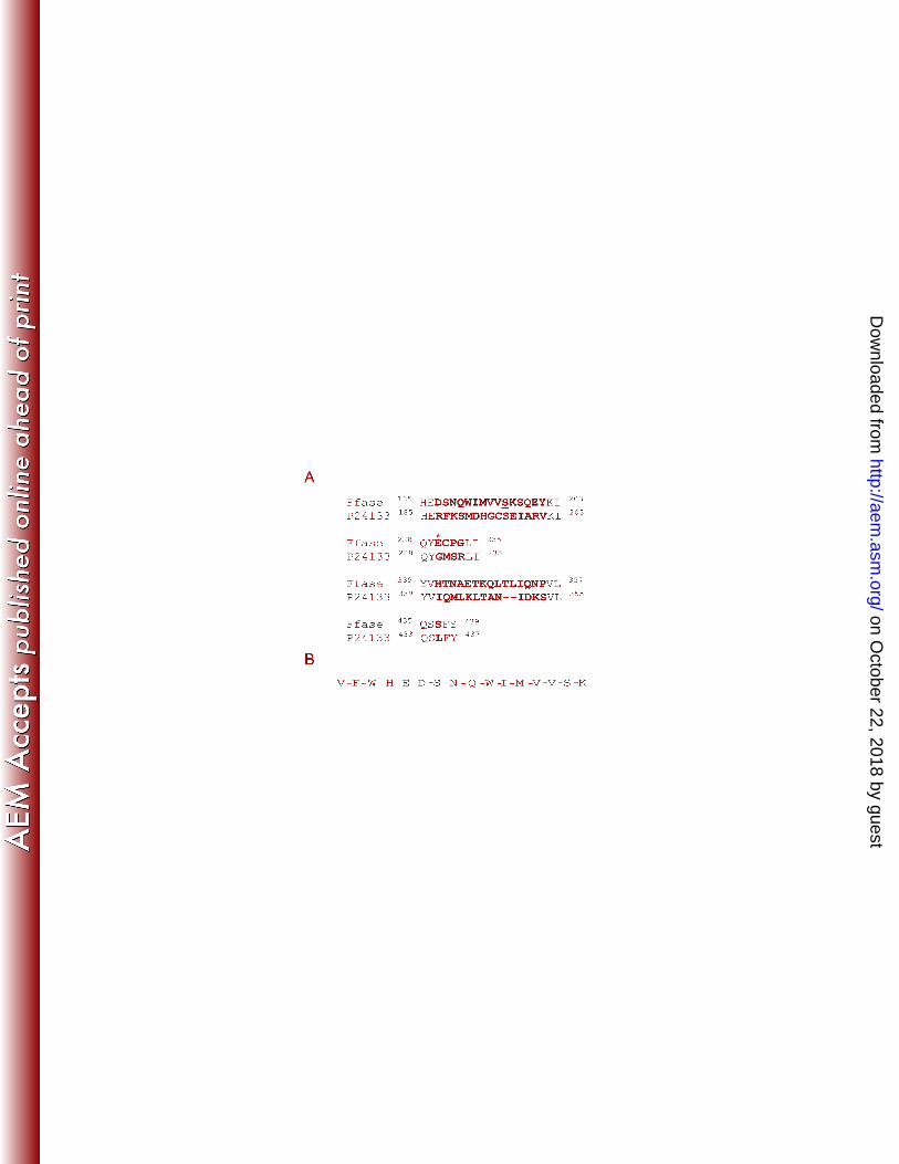
FIG. 1. (A) Alignment of the regions showing differences between the β-fructofuranosidase 3
from Sw. occidentalis described here (Ffase) and that previously reported (P24133). The 4
differences are indicated in bold. The Glu230 involved in sucrose hydrolysis, that is not 5
present in P24133 is marked with and asterisk and the Ser196 modified to Leu196 in S. 6
cerevisiae is underlined. The superscript numbers indicate the amino acid positions. (B) Ffase 7
was purified from Sw. occidentalis and digested with trypsin. The sequence of the peptide 8
2005 m/z identified by MALDI-TOF and generated by Microspray-ion trap MS analysis is 9
indicated. 10
11
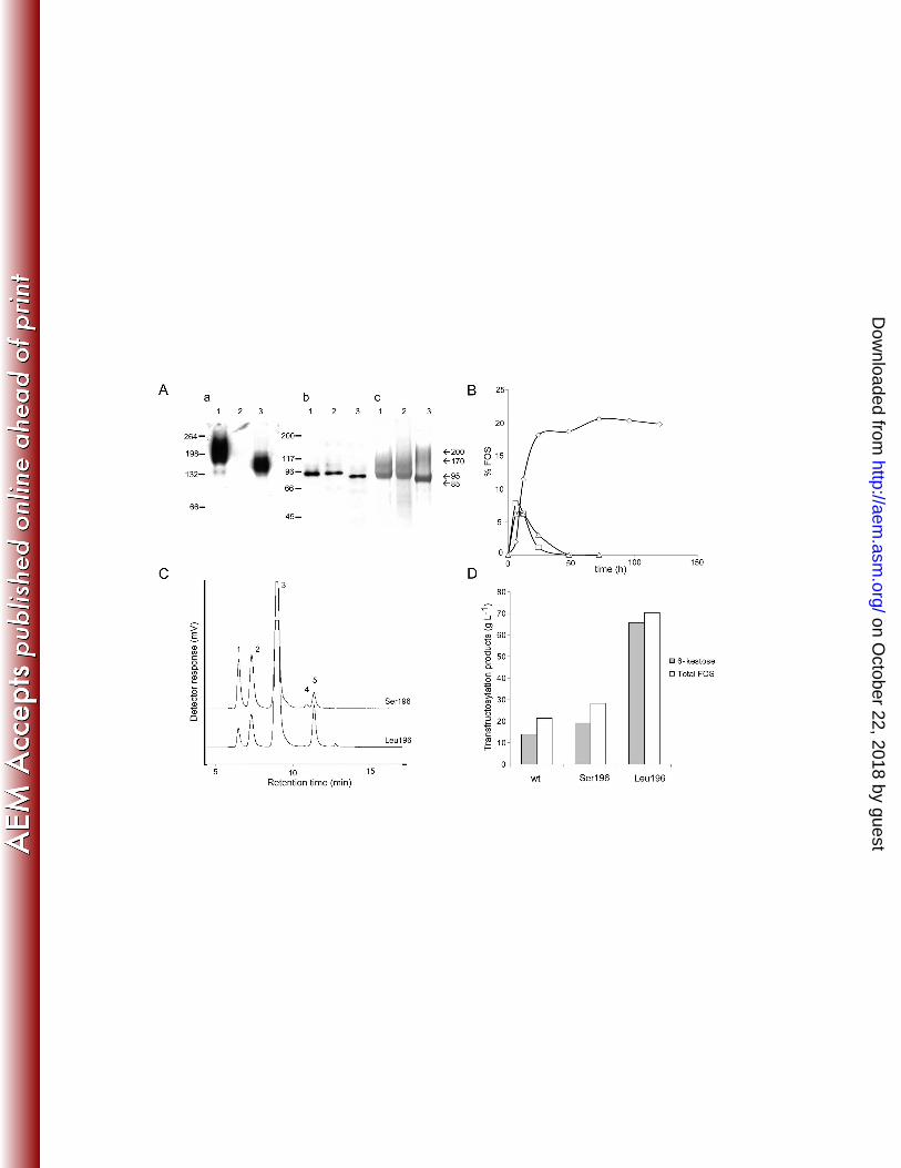
FIG. 2. Analysis of the enzymes expressed in Sw. occidentalis (wild-type, wt) or in S. 12
cerevisiae (Ffase-Serl96 and Ffase LeuI96). (A) Zymogram (a), Western blot (b) and 13
purification of proteins from S. cerevisiae expressing Ffase-Serl96 (lane1) or Ffase-Leul96 14
(lane2), and Sw. occidentalis (lane3). Hydrolase activity of 10 µg of total extracellular 15
proteins was revealed in situ using sucrose as the substrate (a), and 1 µg of each protein was 16
immunoblotted and probed with anti-INV antibodies (b). The purified proteins (10 µg) were 17
subjected to SDS-PAGE and Coomassie-stained (c). Numbers on the left of (a) and (b) 18
indicate the position of molecular mass standards in kDa, and the numbers on the right of (c) 19
indicate the molecular masses assigned to the Ffase in the different conditions assayed. (B) 20
Time course of FOS production catalyzed by the Ffase expressed in Sw. occidentalis 21
(triangles), and in S. cerevisiae containing Ser (squares) or Leu (rhombus) at position 196. 22
Data are represented in % FOS (w/w) of the total sugar composition in the reaction mixture. 23
The total reaction volume was 2 ml and 0.3 U of purified enzyme in 0.2 M sodium acetate 24
on October 22, 2018 by guest
http://aem.asm
.org/D
ownloaded from
23
buffer pH 5.6 were used. The reaction temperature was 50 ºC for the wild-type and the Ffase-1
Serl96 enzymes, and 45 ºC for the Ffase-Leul96 variant. (C) HPLC chromatogram 2
corresponding to the reactions of maximum FOS production obtained with the Ffase-Serl96 3
(6h) and Ffase-Leul96 (72h) expressed in S. cerevisiae. The detector response scale for both 4
chromatograms was the same: (1) Fructose, (2) Glucose, (3) Sucrose, (4) l-kestose, and (5) 6-5
kestose. (D) Maximum FOS and 6-kestose concentrations (g L-1) produced by the wild-type 6
(wt), Ffase-Serl96 (Ser196) and Ffase-Leul96 (Leu196) enzymes. 7
8
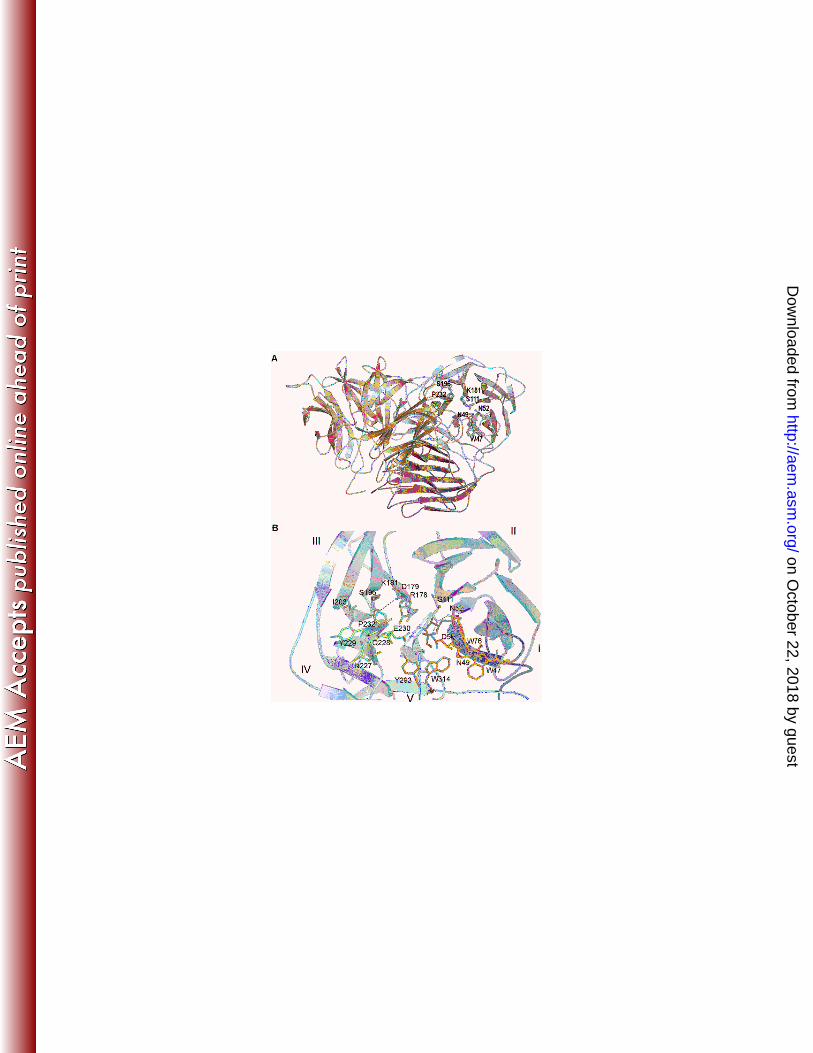
FIG. 3. (A) A cartoon of the three-dimensional structure of the Sw. occidentalis Ffase. Two 9
monomers (blue and orange) associate to form a tight dimer through mainly polar and 10
hydrophobic interactions. The two domains within each monomer are shaded distinctly. The 11
residues investigated in this work are represented as sticks. (B) The catalytic domain folds 12
into a propeller made up of five blades (I-V), each represented in a different colour. The 13
residues mentioned in the text are shown as sticks with the same colour code. Fructose in the 14
Ffase active-site is represented in grey. Asp50 (NDPNG), Asp179 (RDP) and Glu230 (EC) 15
are the catalytic residues. The putative position of a modelled Leu196 is represented in grey. 16
17
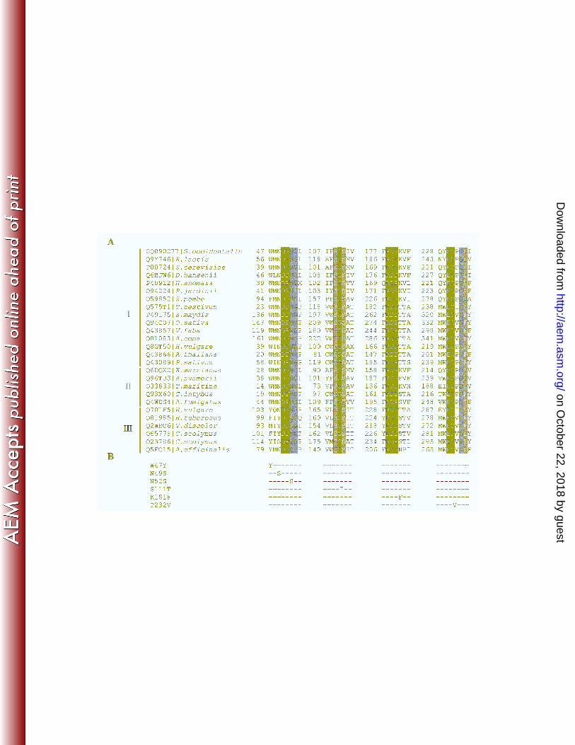
FIG. 4. (A) Multiple alignments of Ffase and several GH32 proteins in the regions 18
surrounding the sites mutated. The accession code and organism source are indicated in the 19
first column. Identical residues are indicated in black, whereas conserved and semi-conserved 20
residues are marked in dark and light grey, respectively. I: β-fructofuranosidase, EC3.2.1.26; 21
II: β-fructosidase, EC3.2.1.80; III fructosyltransferases 2.4.1.(99/100/243). (B) Amino acid 22
changes included in the Leu or Ser196 background are shown. 23
on October 22, 2018 by guest
http://aem.asm
.org/D
ownloaded from
24
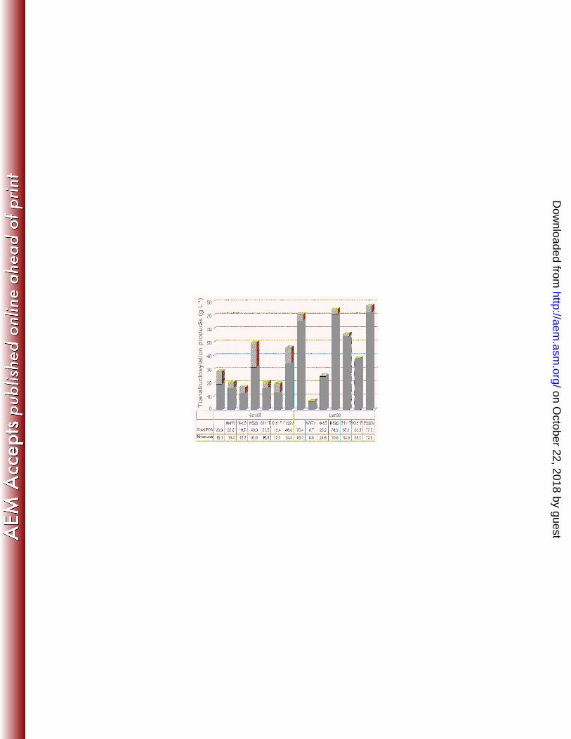
FIG. 5. FOS produced by Ffase-Ser196 and Ffase-Leu196 mutants. Reactions were carried 1
out on sucrose. The concentration of the total FOS (light grey) and 6-kestose (dark grey) 2
obtained are indicated in g L-1. 3
4
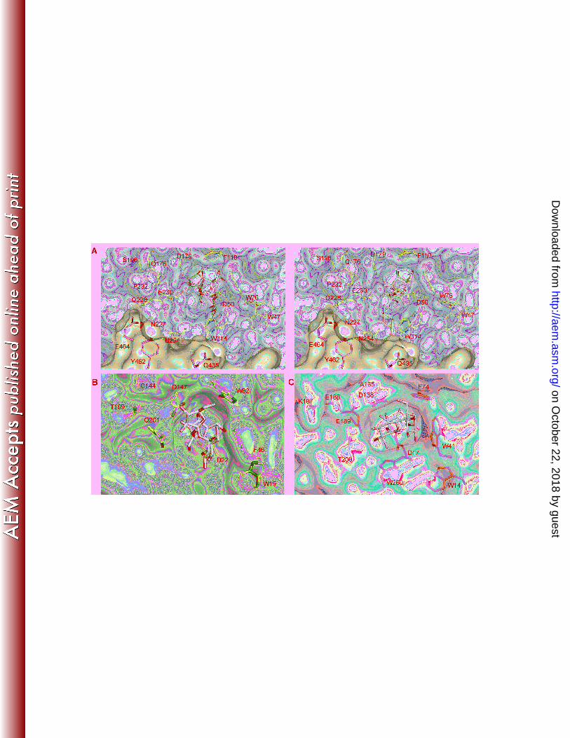
FIG. 6. (A) Close up view of the active site in Sw. occidentalis Ffase in a complex with 5
fructose (PDBcode 3KF3). Putative positions for 1-kestose (green; left) and the 6-kestose 6
(yellow; right) transfructosylation products have been modelled into the active site of Ffase. 7
(B) The fructan 1-exohydrolase IIa from Cichorium intybus (CiFEH) complexed with 1-8
kestose (PDBcode 2AEZ) and (C) the invertase from Thermotoga maritima complexed with 9
raffinose (PDBcode 1W2T). The ligands found in the crystals are represented as white sticks. 10
As can be seen in (A), the active-site of Ffase (blue) is also shaped by the adjacent subunit 11
(orange) and it defines additional subsites to those found in the bacterial and plant enzymes. 12
on October 22, 2018 by guest
http://aem.asm
.org/D
ownloaded from
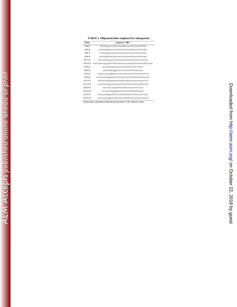
TABLE 1. Oligonucleotides employed for mutagenesis
Name Sequence 5´����3´
196S F GTTGTTTCAAAATCGCAAGAGTACAAAATTCAAATTTTTGG
196S R CGATTTTGAAACAACCATGATCCATTGATTTGAATCTTCATGC
196E F GTTGTTGAAAAATCGCAAGAGTACAAAATTCAAATTTTTGG
196E R CGATTTTTCAACAACCATGATCCATTGATTTGAATCTTCATGC
W47Y F GAAAAAGGATATATGAATGATCGAATGGTCTATTTCTACGATAA
W47Y R GATCATTCATATATCCTTTTTCCGGAGTAAAATGAATTAATGGCCGGTTATAC
N49S F GGATGGATGTCAGATCCGAATGGTCTATTCTACGAT
N49S R ATTCGGATCTGACATCCATCCTTTTTCCGGAGTAAA
N52S F GAATGATCCGAGTGGTCTATTCTACGATAAAACTGCTAAGCTTTG
N52S R GAATAGACCACTCGGATCATTCATCCATCCTTTTCCGGAGTAAATAC
S111T F GGTATCTTTACTGGTAGTATTGTCGTTGACCATAATAATACCTCT
S111T R AATACTACCAGTAAAGATACCTTCATTATCGTGTTCAGGTCCAAT
K181F F CGTGATCCATTCGTTTTCTGGCATGAAGATTCAAAT
K181F R CCAGAAAACGAATGGATCACGGAATTGGTTTGAGGA
P232V F TATGAATGTGTCGGTTTAATTGAAGTTCCTATTGAGAATTCAGAC
P232V R AATTAAACCGACACATTCATACTGATTTCCGTAATAACCAGAAGA
Nucleotides underlined indicate the position of the altered codon.
on October 22, 2018 by guest
http://aem.asm
.org/D
ownloaded from
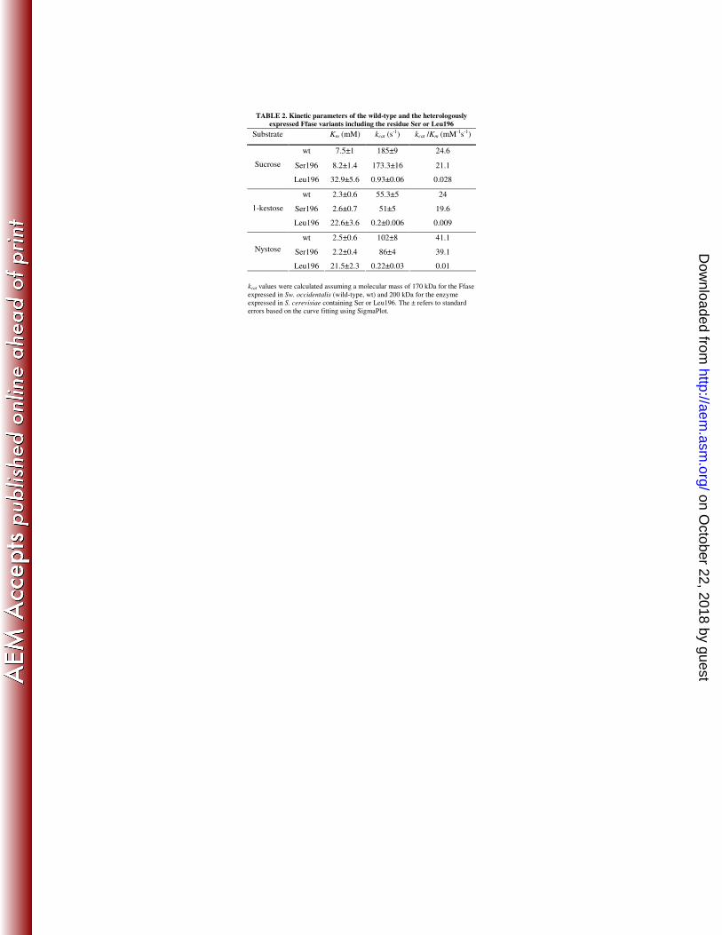
TABLE 2. Kinetic parameters of the wild-type and the heterologously
expressed Ffase variants including the residue Ser or Leu196
kcat values were calculated assuming a molecular mass of 170 kDa for the Ffase
expressed in Sw. occidentalis (wild-type, wt) and 200 kDa for the enzyme
expressed in S. cerevisiae containing Ser or Leu196. The ± refers to standard
errors based on the curve fitting using SigmaPlot.
Substrate Km (mM) kcat (s-1) kcat /Km (mM-1s-1)
wt 7.5±1 185±9 24.6
Ser196 8.2±1.4 173.3±16 21.1 Sucrose
Leu196 32.9±5.6 0.93±0.06 0.028
wt 2.3±0.6 55.3±5 24
Ser196 2.6±0.7 51±5 19.6 1-kestose
Leu196 22.6±3.6 0.2±0.006 0.009
wt 2.5±0.6 102±8 41.1
Ser196 2.2±0.4 86±4 39.1 Nystose
Leu196 21.5±2.3 0.22±0.03 0.01
on October 22, 2018 by guest
http://aem.asm
.org/D
ownloaded from
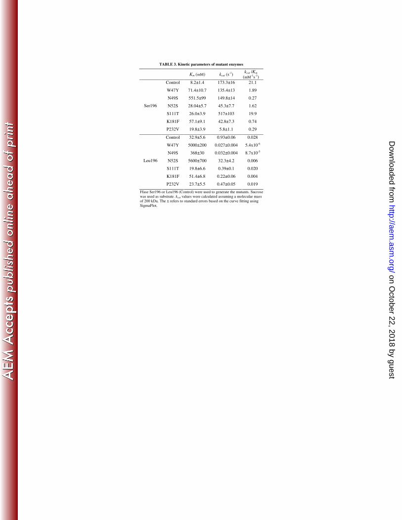
TABLE 3. Kinetic parameters of mutant enzymes
Km (mM) kcat (s-1)
kcat /Km
(mM-1s-1)
Control 8.2±1.4 173.3±16 21.1
W47Y 71.4±10.7 135.4±13 1.89
N49S 551.5±99 149.8±14 0.27
N52S 28.04±5.7 45.3±7.7 1.62
S111T 26.0±3.9 517±103 19.9
K181F 57.1±9.1 42.8±7.3 0.74
Ser196
P232V 19.8±3.9 5.8±1.1 0.29
Control 32.9±5.6 0.93±0.06 0.028
W47Y 5000±200 0.027±0.004 5.4x10⁻⁶
N49S 368±30 0.032±0.004 8.7x10⁻⁵
N52S 5600±700 32.3±4.2 0.006
S111T 19.8±6.6 0.39±0.1 0.020
K181F 51.4±6.8 0.22±0.06 0.004
Leu196
P232V 23.7±5.5 0.47±0.05 0.019
Ffase Ser196 or Leu196 (Control) were used to generate the mutants. Sucrose
was used as substrate. kcat values were calculated assuming a molecular mass
of 200 kDa. The ± refers to standard errors based on the curve fitting using
SigmaPlot.
on October 22, 2018 by guest
http://aem.asm
.org/D
ownloaded from