Use of the Hepatitis B Virus Recombinant Baculovirus-HepG2 System to Study the Effects of
10
ANTIMICROBIAL AGENTS AND CHEMOTHERAPY, 0066-4804/99/$04.0010 Aug. 1999, p. 2017–2026 Vol. 43, No. 8 Copyright © 1999, American Society for Microbiology. All Rights Reserved. Use of the Hepatitis B Virus Recombinant Baculovirus-HepG2 System to Study the Effects of (2)-b-29,39-Dideoxy-39- Thiacytidine on Replication of Hepatitis B Virus and Accumulation of Covalently Closed Circular DNA WILLIAM E. DELANEY IV, 1,2 THOMAS G. MILLER, 1 AND HARRIET C. ISOM 1,2,3 * Department of Microbiology and Immunology, 1 Cell and Molecular Biology Graduate Program, 2 and Department of Pathology, 3 Milton S. Hershey Medical Center, The Penn State University College of Medicine, Hershey, Pennsylvania 17033 Received 7 December 1998/Returned for modification 2 February 1999/Accepted 13 May 1999 (2)-b-2*,3*-Dideoxy-3*-thiacytidine (lamivudine [3TC]) is a nucleoside analog which effectively interferes with the replication of hepatitis B virus (HBV) DNA in vitro and in vivo. We have investigated the antiviral properties of 3TC in vitro in HepG2 cells infected with recombinant HBV baculovirus. Different types of information can be obtained with the HBV baculovirus-HepG2 system because (i) experiments can be carried out at various levels of HBV replication including levels significantly higher than those that can be obtained from conventional HBV-expressing cell lines, (ii) cultures can be manipulated and/or treated prior to or during the initiation of HBV expression, and (iii) high levels of HBV replication allow the rapid detection of HBV products including covalently closed circular (CCC) HBV DNA from low numbers of HepG2 cells. The treatment of HBV baculovirus-infected HepG2 cells with 3TC resulted in an inhibition of HBV replication, evidenced by reductions in the levels of both extracellular HBV DNA and intracellular replicative intermedi- ates. The effect of 3TC on HBV replication was both dose and time dependent, and the reductions in extracellular HBV DNA that we observed agreed well with the previously reported efficacy of 3TC in vitro. As expected, levels of HBV transcripts and extracellular hepatitis B surface antigen and e antigen were not affected by 3TC. Importantly, the HBV baculovirus-HepG2 system made it possible to observe for the first time that CCC HBV DNA levels are lower in cells treated with 3TC than in control cells. We also observed that the treatment of HepG2 cells prior to HBV baculovirus infection resulted in a slight increase in the efficacy of 3TC compared to treatments starting 24 h postinfection. The treatment of HepG2 cells with the highest concen- tration of 3TC tested in this study (2 mM) prior to the initiation of HBV replication markedly inhibited the accumulation of CCC DNA, whereas treatment with the same concentration of 3TC at a time when CCC HBV DNA pools were established within the cells was considerably less effective. In addition, our results suggest that in HepG2 cells, non-protein-associated relaxed circular HBV DNA and particularly CCC HBV DNA are considerably more resistant to 3TC treatment than other forms of HBV DNA, including replicative interme- diates and extracellular DNA. We conclude from these studies that the HBV baculovirus-HepG2 system has specific advantages for drug studies and can be used to complement other in vitro model systems currently used for testing antiviral compounds. Hepatitis B virus (HBV) is a hepatotropic DNA virus capa- ble of causing both acute and chronic hepatitis in man. The World Health Organization estimates that over 350 million people are chronically infected with HBV worldwide. Those persistently infected with HBV serve as a reservoir for the horizontal and vertical transmission of the virus and are also at increased risk of developing further liver disease (2). Approx- imately one of every four HBV carriers will eventually suc- cumb to chronic active hepatitis, cirrhosis, or hepatocellular carcinoma. HBV is believed to cause between 60 and 80% of the world’s primary liver cancer (37). Although effective vac- cines against HBV exist (17), vaccination is expensive and not readily available in all parts of the world and not all individuals develop immunity following vaccination. Therefore, research must also focus on developing effective treatments for the millions of people who remain persistently infected, as well as the population who will become infected despite the existence of vaccines. Currently, the most commonly used treatment for chronic HBV infection is the cytokine alpha interferon (IFN- a). Long-term studies on IFN-a therapy indicate that treat- ment can lead to the loss of circulating HBV antigens and improved survival rates but only in about 30% of patients receiving treatment (13, 26, 36). IFN also must be adminis- tered by injection and can have undesirable side effects which limit dosage. Alternative treatment options which are effective alone or in combination with IFN-a must be explored. (2)-b-29,39-Dideoxy-39-thiacytidine (lamivudine [3TC]) is a nucleoside analog originally described as an agent capable of inhibiting the replication of human immunodeficiency virus type 1 and type 2 (8). It was subsequently reported that 3TC was also effective at inhibiting HBV replication in vitro (12, 20, 22) and at reducing the level of HBV DNA in vivo in the sera of some animal models (33). The use of 3TC to treat chronic HBV infection has recently been approved by the Federal Drug Administration. Treatment with 3TC appears to be well tolerated and effective at reducing or clearing HBV DNA from * Corresponding author. Mailing address: Department of Microbi- ology and Immunology, Milton S. Hershey Medical Center, The Penn State University College of Medicine, 500 University Dr., Hershey, PA 17033. Phone: (717) 531-8609. Fax: (717) 531-4133. E-mail: hisom @psu.edu. 2017 Downloaded from https://journals.asm.org/journal/aac on 21 October 2021 by 208.102.62.251.
Transcript of Use of the Hepatitis B Virus Recombinant Baculovirus-HepG2 System to Study the Effects of
ANTIMICROBIAL AGENTS AND CHEMOTHERAPY,0066-4804/99/$04.0010
Aug. 1999, p. 2017–2026 Vol. 43, No. 8
Copyright © 1999, American Society for Microbiology. All Rights Reserved.
Use of the Hepatitis B Virus Recombinant Baculovirus-HepG2System to Study the Effects of (2)-b-29,39-Dideoxy-39-Thiacytidine on Replication of Hepatitis B Virus and
Accumulation of Covalently Closed Circular DNAWILLIAM E. DELANEY IV,1,2 THOMAS G. MILLER,1 AND HARRIET C. ISOM1,2,3*
Department of Microbiology and Immunology,1 Cell and Molecular Biology Graduate Program,2 andDepartment of Pathology,3 Milton S. Hershey Medical Center, The Penn State University
College of Medicine, Hershey, Pennsylvania 17033
Received 7 December 1998/Returned for modification 2 February 1999/Accepted 13 May 1999
(2)-b-2*,3*-Dideoxy-3*-thiacytidine (lamivudine [3TC]) is a nucleoside analog which effectively interfereswith the replication of hepatitis B virus (HBV) DNA in vitro and in vivo. We have investigated the antiviralproperties of 3TC in vitro in HepG2 cells infected with recombinant HBV baculovirus. Different types ofinformation can be obtained with the HBV baculovirus-HepG2 system because (i) experiments can be carriedout at various levels of HBV replication including levels significantly higher than those that can be obtainedfrom conventional HBV-expressing cell lines, (ii) cultures can be manipulated and/or treated prior to or duringthe initiation of HBV expression, and (iii) high levels of HBV replication allow the rapid detection of HBVproducts including covalently closed circular (CCC) HBV DNA from low numbers of HepG2 cells. Thetreatment of HBV baculovirus-infected HepG2 cells with 3TC resulted in an inhibition of HBV replication,evidenced by reductions in the levels of both extracellular HBV DNA and intracellular replicative intermedi-ates. The effect of 3TC on HBV replication was both dose and time dependent, and the reductions inextracellular HBV DNA that we observed agreed well with the previously reported efficacy of 3TC in vitro. Asexpected, levels of HBV transcripts and extracellular hepatitis B surface antigen and e antigen were not affectedby 3TC. Importantly, the HBV baculovirus-HepG2 system made it possible to observe for the first time thatCCC HBV DNA levels are lower in cells treated with 3TC than in control cells. We also observed that thetreatment of HepG2 cells prior to HBV baculovirus infection resulted in a slight increase in the efficacy of 3TCcompared to treatments starting 24 h postinfection. The treatment of HepG2 cells with the highest concen-tration of 3TC tested in this study (2 mM) prior to the initiation of HBV replication markedly inhibited theaccumulation of CCC DNA, whereas treatment with the same concentration of 3TC at a time when CCC HBVDNA pools were established within the cells was considerably less effective. In addition, our results suggest thatin HepG2 cells, non-protein-associated relaxed circular HBV DNA and particularly CCC HBV DNA areconsiderably more resistant to 3TC treatment than other forms of HBV DNA, including replicative interme-diates and extracellular DNA. We conclude from these studies that the HBV baculovirus-HepG2 system hasspecific advantages for drug studies and can be used to complement other in vitro model systems currently usedfor testing antiviral compounds.
Hepatitis B virus (HBV) is a hepatotropic DNA virus capa-ble of causing both acute and chronic hepatitis in man. TheWorld Health Organization estimates that over 350 millionpeople are chronically infected with HBV worldwide. Thosepersistently infected with HBV serve as a reservoir for thehorizontal and vertical transmission of the virus and are also atincreased risk of developing further liver disease (2). Approx-imately one of every four HBV carriers will eventually suc-cumb to chronic active hepatitis, cirrhosis, or hepatocellularcarcinoma. HBV is believed to cause between 60 and 80% ofthe world’s primary liver cancer (37). Although effective vac-cines against HBV exist (17), vaccination is expensive and notreadily available in all parts of the world and not all individualsdevelop immunity following vaccination. Therefore, researchmust also focus on developing effective treatments for the
millions of people who remain persistently infected, as well asthe population who will become infected despite the existenceof vaccines. Currently, the most commonly used treatment forchronic HBV infection is the cytokine alpha interferon (IFN-a). Long-term studies on IFN-a therapy indicate that treat-ment can lead to the loss of circulating HBV antigens andimproved survival rates but only in about 30% of patientsreceiving treatment (13, 26, 36). IFN also must be adminis-tered by injection and can have undesirable side effects whichlimit dosage. Alternative treatment options which are effectivealone or in combination with IFN-a must be explored.
(2)-b-29,39-Dideoxy-39-thiacytidine (lamivudine [3TC]) is anucleoside analog originally described as an agent capable ofinhibiting the replication of human immunodeficiency virustype 1 and type 2 (8). It was subsequently reported that 3TCwas also effective at inhibiting HBV replication in vitro (12, 20,22) and at reducing the level of HBV DNA in vivo in the seraof some animal models (33). The use of 3TC to treat chronicHBV infection has recently been approved by the FederalDrug Administration. Treatment with 3TC appears to be welltolerated and effective at reducing or clearing HBV DNA from
* Corresponding author. Mailing address: Department of Microbi-ology and Immunology, Milton S. Hershey Medical Center, The PennState University College of Medicine, 500 University Dr., Hershey, PA17033. Phone: (717) 531-8609. Fax: (717) 531-4133. E-mail: [email protected].
2017
Dow
nloa
ded
from
http
s://j
ourn
als.
asm
.org
/jour
nal/a
ac o
n 21
Oct
ober
202
1 by
208
.102
.62.
251.
the sera of patients (11, 16, 23, 25). A major concern with 3TCtherapy is that cessation of drug treatment results in the rapidreappearance of HBV DNA in serum, and the level and ra-pidity of rebound depends upon the length of 3TC treatment(11, 23, 25). The reason for this rebound is postulated to be thepersistence of a covalently closed circular (CCC) form of theHBV genome which resides in the nuclei of infected hepato-cytes (32). Although replicative forms of HBV DNA can beprematurely terminated by the incorporation of 3TC, there islittle or no evidence to suggest that existing CCC DNA poolscan be affected by treatment with 3TC or other nucleosideanalogs. However, Moraleda et al. (24) have reported that thetreatment of primary woodchuck hepatocytes with 3TC priorto infection with woodchuck hepatitis virus (WHV) can inhibitthe formation of WHV CCC DNA in vitro. Recently, 3TC hasalso been used as a prophylactic agent during orthotopic livertransplants, when HBV reinfection is a risk (1, 3, 14). Short-term results from these trials indicate that 3TC suppressesHBV DNA production by the donor liver in some patients;however, the prevention of reinfection and long-term efficacyhave yet to be determined. It is unknown if preemptive treat-ment with nucleosides such as 3TC prior to a transplant wouldbe sufficient to prevent the accumulation of CCC HBV DNAin new liver tissue and potential reactivation of the virus.
We have recently developed a novel in vitro system for thestudy of HBV (10). This system is based on the use of thebaculovirus Autographa californica as a vector for the efficientdelivery of a replication-competent HBV genome into HepG2cells. The advantages of the HBV baculovirus-HepG2 systeminclude (i) the ability to initiate extremely high levels of HBVexpression, (ii) a reproducible and precise control over thelevel of HBV expression, and (iii) the ability to rapidly detectHBV antigens, RNA, and both intracellular and extracellularDNA from low numbers of HepG2 cells. The HBV baculovi-rus-HepG2 cell system also allows the detection of CCC HBVDNA, which can be difficult to detect or undetectable in stablytransfected HBV-expressing cell lines such as HepG2 2.2.15and HB611 (28, 31). Unlike stable cell lines, the time of infec-tion can also be controlled, allowing the manipulation or treat-ment of cells prior to or during the initiation of HBV expres-sion. The primary goals of the studies described here were (i)to evaluate the utility of the HBV baculovirus-HepG2 systemas a tool for antiviral research using 3TC, an established in-hibitor of HBV replication, (ii) to provide further data on thein vitro efficacy of 3TC by investigating its effects on variouslevels of HBV replication, and (iii) to examine the effect ofadministering 3TC prior to the initiation of HBV expression,shortly after the initiation of HBV replication, and after theestablishment of intracellular CCC HBV DNA pools inHepG2 cells.
MATERIALS AND METHODS
Cell culture. The HepG2 cell line was maintained at 37°C in humidifiedincubators at 5% CO2 (18). HepG2 cells were fed minimal essential medium(Gibco BRL, Gaithersburg, Md.) supplemented with 10% heat-inactivated fetalbovine serum.
HBV baculovirus production and infection of HepG2 cells. The generation,amplification, and purification of the HBV baculovirus encoding a 1.3-unit lengthreplication-competent HBV genome has been previously described (10). Themechanism of baculovirus uptake by mammalian cells is currently unknown.Here, we use the term “infection” to describe the exposure and uptake ofbaculovirus particles by HepG2 cells. This process is not the same as a true viralinfection but rather is a mechanism for the transduction of HBV DNA into thecell. The infection procedure for HepG2 cells has been previously described (10).
3TC treatment. 3TC was a gift from BioChem Therapeutic Inc. (Laval, Que-bec, Canada). 3TC was resuspended in sterile water, aliquoted, and frozen at220°C to avoid repeated freezing and thawing of the drug. Medium containing3TC was prepared daily as needed with fresh aliquots of 3TC. In experiments inwhich 3TC treatment was initiated after viral infection, HepG2 cells were ex-
posed to the indicated concentration of 3TC 24 h postinfection (p.i.) or 4 days p.i.In experiments utilizing pretreatment with 3TC, cells were fed a medium con-taining 3TC 16 h prior to HBV baculovirus infection, HBV baculovirus infectionwas carried out in a medium containing 3TC, and cells were refed a freshmedium containing 3TC immediately after completion of the infection andwashing procedures.
Analysis of RNA. Total RNA was isolated from HepG2 cells by the single-stepacid guanidium method (6). Northern blot analysis was performed with 20 mg oftotal RNA as described previously (9). Nucleic acid hybridization was performedas described previously (27). A full-length double-stranded (DS) HBV genomewas used as a template to generate 32P-radiolabeled probes by using a Boehr-inger Mannheim (Indianapolis, Ind.) random prime DNA labeling kit.
Analysis of intracellular replicative intermediates. Cytoplasmic preparationscontaining HBV core particles were isolated from HepG2 cells as describedpreviously (15). Unprotected DNA was removed by adjusting cytoplasmic prep-arations so that they contained 10 mM MgCl2 and 500 mg of DNase I (Boehr-inger Mannheim) per ml followed by a 1-h incubation at 37°C. Replicativeintermediates were then isolated by proteinase K digestion, sequential phenoland chloroform extractions, and isopropanol precipitation as described previ-ously (10). Precipitated nucleic acids were resuspended in a small volume of TE(10 mM Tris, 1 mM EDTA), normalized by measurement of optical density at260 nm, and digested with 100 mg of RNase (Boehringer Mannheim) for 1 h at37°C. Replicative intermediates were then analyzed by electrophoresis in 1%agarose gels followed by Southern blotting as described previously (27). Nucleicacid hybridization was performed as described above for Northern blotting. Amodel 100A laser densitometer (Molecular Dynamics, Sunnyvale, Calif.)equipped with Quantity One software (Protein Databases Inc., Huntington Sta-tion, N.Y.) was used to analyze suitable exposures of Southern blots.
Detection of extracellular HBV DNA. Conditioned medium was collected fromHepG2 cells, centrifuged at 10,000 3 g for 10 min, and transferred to clean tubesto remove cellular debris. HBV particles were precipitated from medium sam-ples with polyethylene glycol 8000 (Sigma Chemical Co., St. Louis, Mo.) asdescribed previously (35). Viral pellets were resuspended in phosphate-bufferedsaline, and DNA was extracted by proteinase K digestion, sequential phenol andchloroform extractions, and isopropanol precipitation as described above. Tenmicrograms of tRNA (Boehringer Mannheim) was added as a carrier duringprecipitation. Nucleic acid pellets were resuspended in a small volume of TE anddigested with 500 mg of RNase per ml prior to analysis by electrophoresis andSouthern blotting.
Detection of CCC HBV DNA. Non-protein-associated circular HBV DNA wasextracted from HepG2 cells essentially as described previously (30). Extractednucleic acids were resuspended in water and normalized by measurement ofoptical density at 260 nm prior to digestion with 100 mg of RNase per ml and 30U of Plasmid-Safe ATP-dependent DNase (Epicenter Technologies, Madison,Wis.) for 3 h at 37°C. Samples were analyzed by electrophoresis and Southernblotting.
Analysis of secreted HBV antigens. The detection of hepatitis B surface anti-gen (HBsAg) was carried out by using a radioimmunoassay kit according to themanufacturer’s instructions (Abbott Laboratories, Abbott Park, Ill.). Hepatitis Be antigen (HBeAg) was also detected with a radioimmunoassay kit according tothe manufacturer’s instructions (Sorin Biomedica, Saluggia, Italy). Medium sam-ples collected from HepG2 cells were centrifuged at 6,000 3 g to remove cellulardebris, transferred to clean tubes, and stored at 220°C until analyzed.
RESULTS
Effect of 3TC on extracellular HBV DNA. Previous resultshave indicated that 3TC concentrations of approximately 0.2mM are effective at downregulating HBV replication and/orvirion secretion by 50 to 90% in vitro in HBV-expressing celllines (12, 20, 22). We therefore chose to examine the effects of3TC on HBV expression and replication mediated by HBVbaculovirus by using 0.02, 0.2, and 2.0 mM concentrations of3TC. HepG2 cells were infected with 50 PFU of HBV bacu-lovirus/cell because our previous findings have indicated thatcells infected at a multiplicity of infection (MOI) of 50 main-tain high levels of HBV replication for more than 10 days (10).For each concentration of 3TC tested in initial studies, drugtreatment starting 16 h prior to baculovirus infection (pretreat-ment) and a treatment starting 24 h after baculovirus infectionwere used (Fig. 1). It is important to note that 24 h p.i. (50 PFUof HBV baculovirus/cell) HBV replication has begun but thepredominant forms of DNA in cytoplasmic nucleocapsids aresingle-stranded (SS) HBV DNA molecules (10). Little matureDS HBV DNA appears in core particles and no extracellularHBV DNA or CCC HBV DNA can be detected at this time.
2018 DELANEY ET AL. ANTIMICROB. AGENTS CHEMOTHER.
Dow
nloa
ded
from
http
s://j
ourn
als.
asm
.org
/jour
nal/a
ac o
n 21
Oct
ober
202
1 by
208
.102
.62.
251.
At 48 h p.i., DS replicative intermediates as well as extracel-lular HBV DNA and non-protein-associated relaxed circular(RC) and CCC HBV DNA are readily detectable. Therefore,pretreatment with 3TC introduces the drug to the cell prior tothe presence of a replication-competent HBV genome, whiletreatments starting 24 h p.i. introduce the drug when both SSand DS HBV DNA are actively being synthesized.
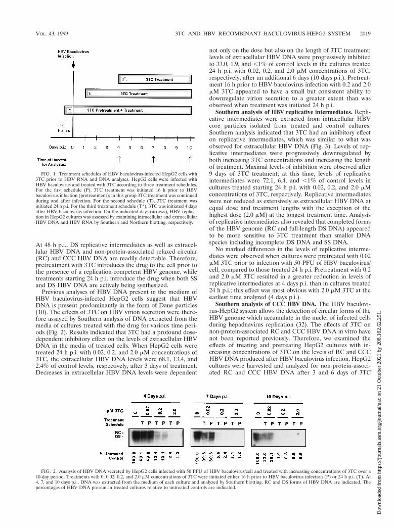
Previous analyses of HBV DNA present in the medium ofHBV baculovirus-infected HepG2 cells suggest that HBVDNA is present predominantly in the form of Dane particles(10). The effects of 3TC on HBV virion secretion were there-fore assayed by Southern analysis of DNA extracted from themedia of cultures treated with the drug for various time peri-ods (Fig. 2). Results indicated that 3TC had a profound dose-dependent inhibitory effect on the levels of extracellular HBVDNA in the media of treated cells. When HepG2 cells weretreated 24 h p.i. with 0.02, 0.2, and 2.0 mM concentrations of3TC, the extracellular HBV DNA levels were 68.1, 13.4, and2.4% of control levels, respectively, after 3 days of treatment.Decreases in extracellular HBV DNA levels were dependent
not only on the dose but also on the length of 3TC treatment;levels of extracellular HBV DNA were progressively inhibitedto 33.0, 1.9, and ,1% of control levels in the cultures treated24 h p.i. with 0.02, 0.2, and 2.0 mM concentrations of 3TC,respectively, after an additional 6 days (10 days p.i.). Pretreat-ment 16 h prior to HBV baculovirus infection with 0.2 and 2.0mM 3TC appeared to have a small but consistent ability todownregulate virion secretion to a greater extent than wasobserved when treatment was initiated 24 h p.i.
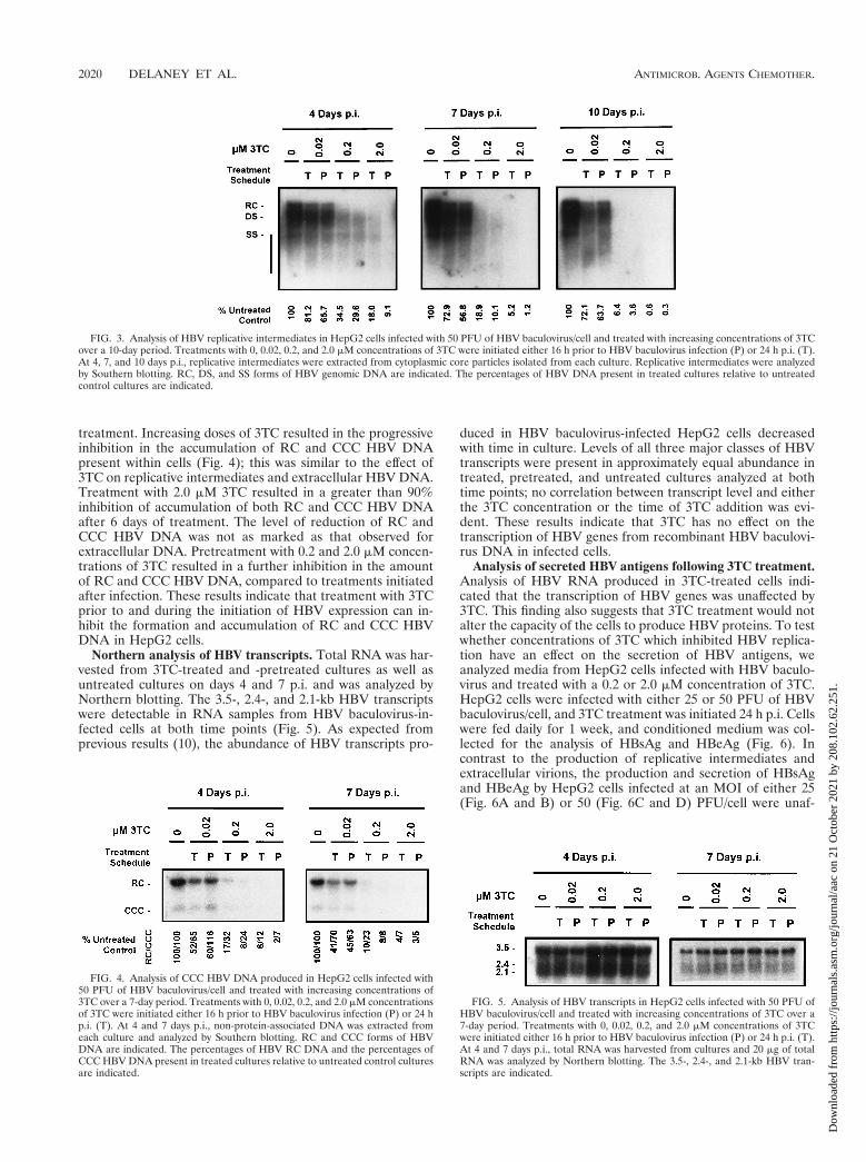
Southern analysis of HBV replicative intermediates. Repli-cative intermediates were extracted from intracellular HBVcore particles isolated from treated and control cultures.Southern analysis indicated that 3TC had an inhibitory effecton replicative intermediates, which was similar to what wasobserved for extracellular HBV DNA (Fig. 3). Levels of rep-licative intermediates were progressively downregulated byboth increasing 3TC concentrations and increasing the lengthof treatment. Maximal levels of inhibition were observed after9 days of 3TC treatment; at this time, levels of replicativeintermediates were 72.1, 6.4, and ,1% of control levels incultures treated starting 24 h p.i. with 0.02, 0.2, and 2.0 mMconcentrations of 3TC, respectively. Replicative intermediateswere not reduced as extensively as extracellular HBV DNA atequal dose and treatment lengths with the exception of thehighest dose (2.0 mM) at the longest treatment time. Analysisof replicative intermediates also revealed that completed formsof the HBV genome (RC and full-length DS DNA) appearedto be more sensitive to 3TC treatment than smaller DNAspecies including incomplete DS DNA and SS DNA.
No marked differences in the levels of replicative interme-diates were observed when cultures were pretreated with 0.02mM 3TC prior to infection with 50 PFU of HBV baculovirus/cell, compared to those treated 24 h p.i. Pretreatment with 0.2and 2.0 mM 3TC resulted in a greater reduction in levels ofreplicative intermediates at 4 days p.i. than in cultures treated24 h p.i.; this effect was most obvious with 2.0 mM 3TC at theearliest time analyzed (4 days p.i.).
Southern analysis of CCC HBV DNA. The HBV baculovi-rus-HepG2 system allows the detection of circular forms of theHBV genome which accumulate in the nuclei of infected cellsduring hepadnavirus replication (32). The effects of 3TC onnon-protein-associated RC and CCC HBV DNA in vitro havenot been reported previously. Therefore, we examined theeffects of treating and pretreating HepG2 cultures with in-creasing concentrations of 3TC on the levels of RC and CCCHBV DNA produced after HBV baculovirus infection. HepG2cultures were harvested and analyzed for non-protein-associ-ated RC and CCC HBV DNA after 3 and 6 days of 3TC
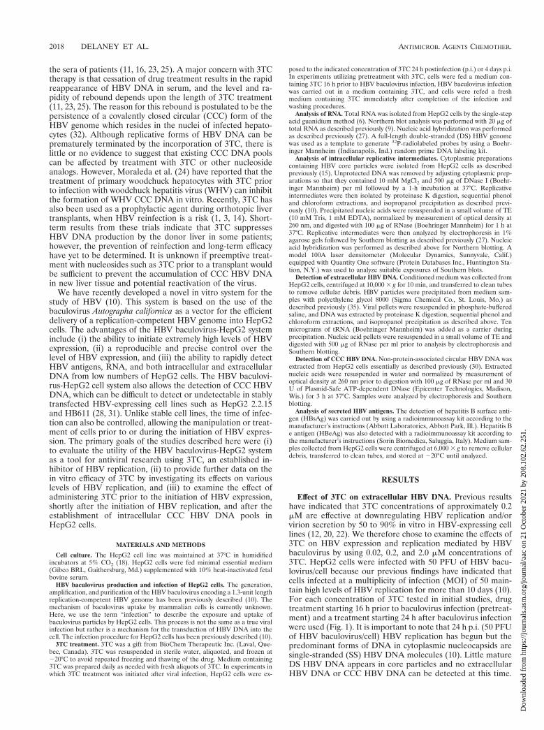
FIG. 1. Treatment schedules of HBV baculovirus-infected HepG2 cells with3TC prior to HBV RNA and DNA analyses. HepG2 cells were infected withHBV baculovirus and treated with 3TC according to three treatment schedules.For the first schedule (P), 3TC treatment was initiated 16 h prior to HBVbaculovirus infection (pretreatment); in this group 3TC treatment was continuedduring and after infection. For the second schedule (T), 3TC treatment wasinitiated 24 h p.i. For the third treatment schedule (T*), 3TC was initiated 4 daysafter HBV baculovirus infection. On the indicated days (arrows), HBV replica-tion in HepG2 cultures was assessed by examining intracellular and extracellularHBV DNA and HBV RNA by Southern and Northern blotting, respectively.
FIG. 2. Analysis of HBV DNA secreted by HepG2 cells infected with 50 PFU of HBV baculovirus/cell and treated with increasing concentrations of 3TC over a10-day period. Treatments with 0, 0.02, 0.2, and 2.0 mM concentrations of 3TC were initiated either 16 h prior to HBV baculovirus infection (P) or 24 h p.i. (T). At4, 7, and 10 days p.i., DNA was extracted from the medium of each culture and analyzed by Southern blotting. RC and DS forms of HBV DNA are indicated. Thepercentages of HBV DNA present in treated cultures relative to untreated controls are indicated.
VOL. 43, 1999 3TC AND HBV RECOMBINANT BACULOVIRUS-HEPG2 SYSTEM 2019
Dow
nloa
ded
from
http
s://j
ourn
als.
asm
.org
/jour
nal/a
ac o
n 21
Oct
ober
202
1 by
208
.102
.62.
251.
treatment. Increasing doses of 3TC resulted in the progressiveinhibition in the accumulation of RC and CCC HBV DNApresent within cells (Fig. 4); this was similar to the effect of3TC on replicative intermediates and extracellular HBV DNA.Treatment with 2.0 mM 3TC resulted in a greater than 90%inhibition of accumulation of both RC and CCC HBV DNAafter 6 days of treatment. The level of reduction of RC andCCC HBV DNA was not as marked as that observed forextracellular DNA. Pretreatment with 0.2 and 2.0 mM concen-trations of 3TC resulted in a further inhibition in the amountof RC and CCC HBV DNA, compared to treatments initiatedafter infection. These results indicate that treatment with 3TCprior to and during the initiation of HBV expression can in-hibit the formation and accumulation of RC and CCC HBVDNA in HepG2 cells.
Northern analysis of HBV transcripts. Total RNA was har-vested from 3TC-treated and -pretreated cultures as well asuntreated cultures on days 4 and 7 p.i. and was analyzed byNorthern blotting. The 3.5-, 2.4-, and 2.1-kb HBV transcriptswere detectable in RNA samples from HBV baculovirus-in-fected cells at both time points (Fig. 5). As expected fromprevious results (10), the abundance of HBV transcripts pro-
duced in HBV baculovirus-infected HepG2 cells decreasedwith time in culture. Levels of all three major classes of HBVtranscripts were present in approximately equal abundance intreated, pretreated, and untreated cultures analyzed at bothtime points; no correlation between transcript level and eitherthe 3TC concentration or the time of 3TC addition was evi-dent. These results indicate that 3TC has no effect on thetranscription of HBV genes from recombinant HBV baculovi-rus DNA in infected cells.
Analysis of secreted HBV antigens following 3TC treatment.Analysis of HBV RNA produced in 3TC-treated cells indi-cated that the transcription of HBV genes was unaffected by3TC. This finding also suggests that 3TC treatment would notalter the capacity of the cells to produce HBV proteins. To testwhether concentrations of 3TC which inhibited HBV replica-tion have an effect on the secretion of HBV antigens, weanalyzed media from HepG2 cells infected with HBV baculo-virus and treated with a 0.2 or 2.0 mM concentration of 3TC.HepG2 cells were infected with either 25 or 50 PFU of HBVbaculovirus/cell, and 3TC treatment was initiated 24 h p.i. Cellswere fed daily for 1 week, and conditioned medium was col-lected for the analysis of HBsAg and HBeAg (Fig. 6). Incontrast to the production of replicative intermediates andextracellular virions, the production and secretion of HBsAgand HBeAg by HepG2 cells infected at an MOI of either 25(Fig. 6A and B) or 50 (Fig. 6C and D) PFU/cell were unaf-
FIG. 3. Analysis of HBV replicative intermediates in HepG2 cells infected with 50 PFU of HBV baculovirus/cell and treated with increasing concentrations of 3TCover a 10-day period. Treatments with 0, 0.02, 0.2, and 2.0 mM concentrations of 3TC were initiated either 16 h prior to HBV baculovirus infection (P) or 24 h p.i. (T).At 4, 7, and 10 days p.i., replicative intermediates were extracted from cytoplasmic core particles isolated from each culture. Replicative intermediates were analyzedby Southern blotting. RC, DS, and SS forms of HBV genomic DNA are indicated. The percentages of HBV DNA present in treated cultures relative to untreatedcontrol cultures are indicated.
FIG. 4. Analysis of CCC HBV DNA produced in HepG2 cells infected with50 PFU of HBV baculovirus/cell and treated with increasing concentrations of3TC over a 7-day period. Treatments with 0, 0.02, 0.2, and 2.0 mM concentrationsof 3TC were initiated either 16 h prior to HBV baculovirus infection (P) or 24 hp.i. (T). At 4 and 7 days p.i., non-protein-associated DNA was extracted fromeach culture and analyzed by Southern blotting. RC and CCC forms of HBVDNA are indicated. The percentages of HBV RC DNA and the percentages ofCCC HBV DNA present in treated cultures relative to untreated control culturesare indicated.
FIG. 5. Analysis of HBV transcripts in HepG2 cells infected with 50 PFU ofHBV baculovirus/cell and treated with increasing concentrations of 3TC over a7-day period. Treatments with 0, 0.02, 0.2, and 2.0 mM concentrations of 3TCwere initiated either 16 h prior to HBV baculovirus infection (P) or 24 h p.i. (T).At 4 and 7 days p.i., total RNA was harvested from cultures and 20 mg of totalRNA was analyzed by Northern blotting. The 3.5-, 2.4-, and 2.1-kb HBV tran-scripts are indicated.
2020 DELANEY ET AL. ANTIMICROB. AGENTS CHEMOTHER.
Dow
nloa
ded
from
http
s://j
ourn
als.
asm
.org
/jour
nal/a
ac o
n 21
Oct
ober
202
1 by
208
.102
.62.
251.
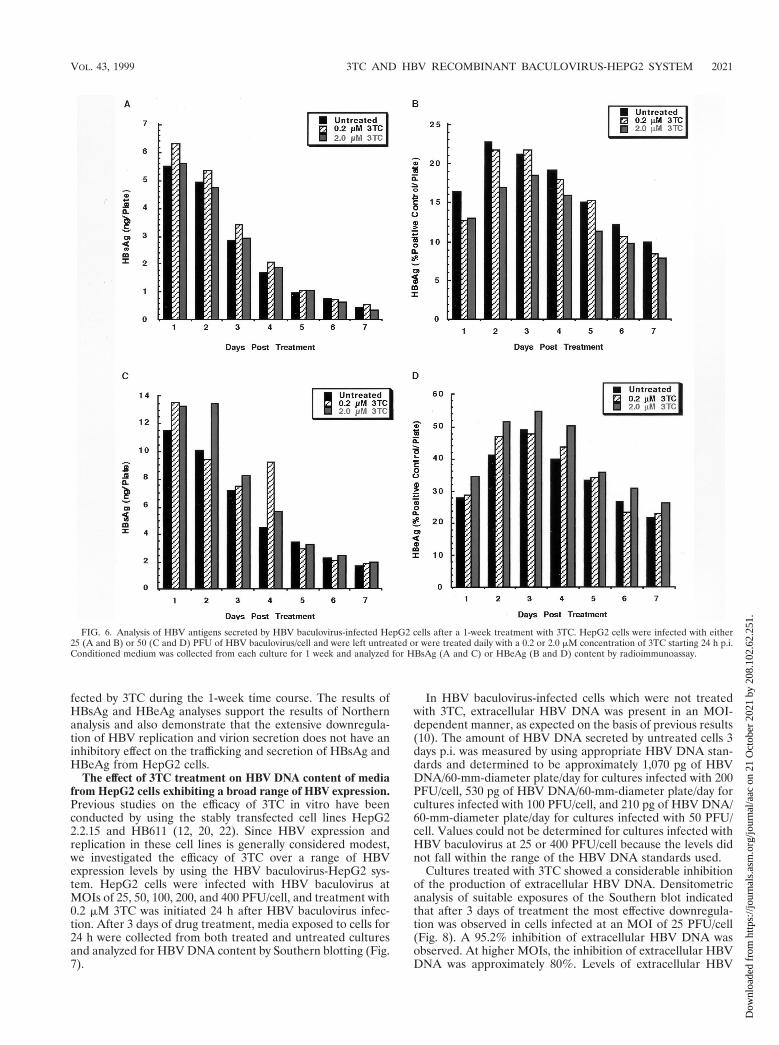
fected by 3TC during the 1-week time course. The results ofHBsAg and HBeAg analyses support the results of Northernanalysis and also demonstrate that the extensive downregula-tion of HBV replication and virion secretion does not have aninhibitory effect on the trafficking and secretion of HBsAg andHBeAg from HepG2 cells.
The effect of 3TC treatment on HBV DNA content of mediafrom HepG2 cells exhibiting a broad range of HBV expression.Previous studies on the efficacy of 3TC in vitro have beenconducted by using the stably transfected cell lines HepG22.2.15 and HB611 (12, 20, 22). Since HBV expression andreplication in these cell lines is generally considered modest,we investigated the efficacy of 3TC over a range of HBVexpression levels by using the HBV baculovirus-HepG2 sys-tem. HepG2 cells were infected with HBV baculovirus atMOIs of 25, 50, 100, 200, and 400 PFU/cell, and treatment with0.2 mM 3TC was initiated 24 h after HBV baculovirus infec-tion. After 3 days of drug treatment, media exposed to cells for24 h were collected from both treated and untreated culturesand analyzed for HBV DNA content by Southern blotting (Fig.7).
In HBV baculovirus-infected cells which were not treatedwith 3TC, extracellular HBV DNA was present in an MOI-dependent manner, as expected on the basis of previous results(10). The amount of HBV DNA secreted by untreated cells 3days p.i. was measured by using appropriate HBV DNA stan-dards and determined to be approximately 1,070 pg of HBVDNA/60-mm-diameter plate/day for cultures infected with 200PFU/cell, 530 pg of HBV DNA/60-mm-diameter plate/day forcultures infected with 100 PFU/cell, and 210 pg of HBV DNA/60-mm-diameter plate/day for cultures infected with 50 PFU/cell. Values could not be determined for cultures infected withHBV baculovirus at 25 or 400 PFU/cell because the levels didnot fall within the range of the HBV DNA standards used.
Cultures treated with 3TC showed a considerable inhibitionof the production of extracellular HBV DNA. Densitometricanalysis of suitable exposures of the Southern blot indicatedthat after 3 days of treatment the most effective downregula-tion was observed in cells infected at an MOI of 25 PFU/cell(Fig. 8). A 95.2% inhibition of extracellular HBV DNA wasobserved. At higher MOIs, the inhibition of extracellular HBVDNA was approximately 80%. Levels of extracellular HBV
FIG. 6. Analysis of HBV antigens secreted by HBV baculovirus-infected HepG2 cells after a 1-week treatment with 3TC. HepG2 cells were infected with either25 (A and B) or 50 (C and D) PFU of HBV baculovirus/cell and were left untreated or were treated daily with a 0.2 or 2.0 mM concentration of 3TC starting 24 h p.i.Conditioned medium was collected from each culture for 1 week and analyzed for HBsAg (A and C) or HBeAg (B and D) content by radioimmunoassay.
VOL. 43, 1999 3TC AND HBV RECOMBINANT BACULOVIRUS-HEPG2 SYSTEM 2021
Dow
nloa
ded
from
http
s://j
ourn
als.
asm
.org
/jour
nal/a
ac o
n 21
Oct
ober
202
1 by
208
.102
.62.
251.
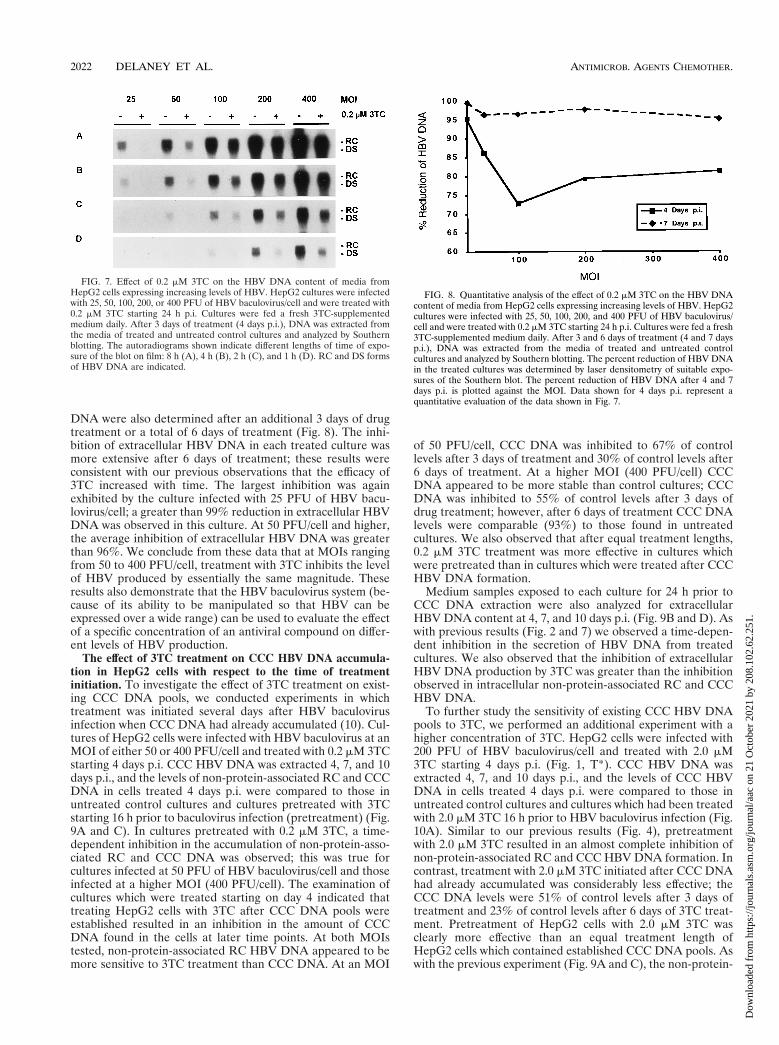
DNA were also determined after an additional 3 days of drugtreatment or a total of 6 days of treatment (Fig. 8). The inhi-bition of extracellular HBV DNA in each treated culture wasmore extensive after 6 days of treatment; these results wereconsistent with our previous observations that the efficacy of3TC increased with time. The largest inhibition was againexhibited by the culture infected with 25 PFU of HBV bacu-lovirus/cell; a greater than 99% reduction in extracellular HBVDNA was observed in this culture. At 50 PFU/cell and higher,the average inhibition of extracellular HBV DNA was greaterthan 96%. We conclude from these data that at MOIs rangingfrom 50 to 400 PFU/cell, treatment with 3TC inhibits the levelof HBV produced by essentially the same magnitude. Theseresults also demonstrate that the HBV baculovirus system (be-cause of its ability to be manipulated so that HBV can beexpressed over a wide range) can be used to evaluate the effectof a specific concentration of an antiviral compound on differ-ent levels of HBV production.
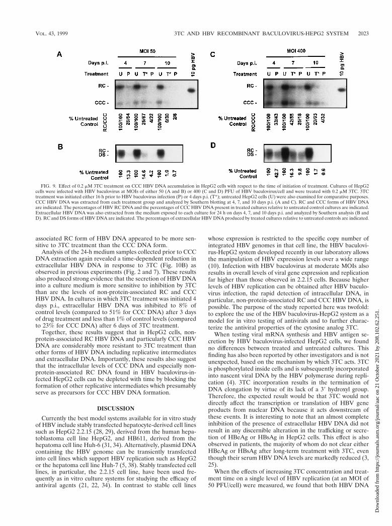
The effect of 3TC treatment on CCC HBV DNA accumula-tion in HepG2 cells with respect to the time of treatmentinitiation. To investigate the effect of 3TC treatment on exist-ing CCC DNA pools, we conducted experiments in whichtreatment was initiated several days after HBV baculovirusinfection when CCC DNA had already accumulated (10). Cul-tures of HepG2 cells were infected with HBV baculovirus at anMOI of either 50 or 400 PFU/cell and treated with 0.2 mM 3TCstarting 4 days p.i. CCC HBV DNA was extracted 4, 7, and 10days p.i., and the levels of non-protein-associated RC and CCCDNA in cells treated 4 days p.i. were compared to those inuntreated control cultures and cultures pretreated with 3TCstarting 16 h prior to baculovirus infection (pretreatment) (Fig.9A and C). In cultures pretreated with 0.2 mM 3TC, a time-dependent inhibition in the accumulation of non-protein-asso-ciated RC and CCC DNA was observed; this was true forcultures infected at 50 PFU of HBV baculovirus/cell and thoseinfected at a higher MOI (400 PFU/cell). The examination ofcultures which were treated starting on day 4 indicated thattreating HepG2 cells with 3TC after CCC DNA pools wereestablished resulted in an inhibition in the amount of CCCDNA found in the cells at later time points. At both MOIstested, non-protein-associated RC HBV DNA appeared to bemore sensitive to 3TC treatment than CCC DNA. At an MOI
of 50 PFU/cell, CCC DNA was inhibited to 67% of controllevels after 3 days of treatment and 30% of control levels after6 days of treatment. At a higher MOI (400 PFU/cell) CCCDNA appeared to be more stable than control cultures; CCCDNA was inhibited to 55% of control levels after 3 days ofdrug treatment; however, after 6 days of treatment CCC DNAlevels were comparable (93%) to those found in untreatedcultures. We also observed that after equal treatment lengths,0.2 mM 3TC treatment was more effective in cultures whichwere pretreated than in cultures which were treated after CCCHBV DNA formation.
Medium samples exposed to each culture for 24 h prior toCCC DNA extraction were also analyzed for extracellularHBV DNA content at 4, 7, and 10 days p.i. (Fig. 9B and D). Aswith previous results (Fig. 2 and 7) we observed a time-depen-dent inhibition in the secretion of HBV DNA from treatedcultures. We also observed that the inhibition of extracellularHBV DNA production by 3TC was greater than the inhibitionobserved in intracellular non-protein-associated RC and CCCHBV DNA.
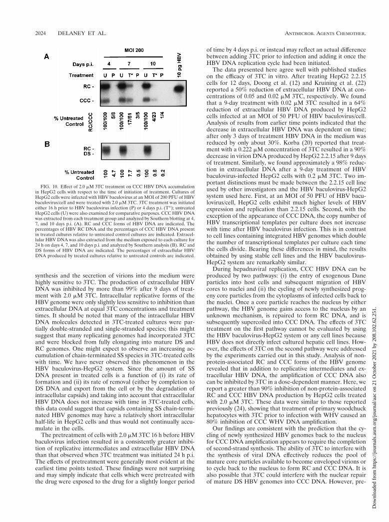
To further study the sensitivity of existing CCC HBV DNApools to 3TC, we performed an additional experiment with ahigher concentration of 3TC. HepG2 cells were infected with200 PFU of HBV baculovirus/cell and treated with 2.0 mM3TC starting 4 days p.i. (Fig. 1, T*). CCC HBV DNA wasextracted 4, 7, and 10 days p.i., and the levels of CCC HBVDNA in cells treated 4 days p.i. were compared to those inuntreated control cultures and cultures which had been treatedwith 2.0 mM 3TC 16 h prior to HBV baculovirus infection (Fig.10A). Similar to our previous results (Fig. 4), pretreatmentwith 2.0 mM 3TC resulted in an almost complete inhibition ofnon-protein-associated RC and CCC HBV DNA formation. Incontrast, treatment with 2.0 mM 3TC initiated after CCC DNAhad already accumulated was considerably less effective; theCCC DNA levels were 51% of control levels after 3 days oftreatment and 23% of control levels after 6 days of 3TC treat-ment. Pretreatment of HepG2 cells with 2.0 mM 3TC wasclearly more effective than an equal treatment length ofHepG2 cells which contained established CCC DNA pools. Aswith the previous experiment (Fig. 9A and C), the non-protein-
FIG. 7. Effect of 0.2 mM 3TC on the HBV DNA content of media fromHepG2 cells expressing increasing levels of HBV. HepG2 cultures were infectedwith 25, 50, 100, 200, or 400 PFU of HBV baculovirus/cell and were treated with0.2 mM 3TC starting 24 h p.i. Cultures were fed a fresh 3TC-supplementedmedium daily. After 3 days of treatment (4 days p.i.), DNA was extracted fromthe media of treated and untreated control cultures and analyzed by Southernblotting. The autoradiograms shown indicate different lengths of time of expo-sure of the blot on film: 8 h (A), 4 h (B), 2 h (C), and 1 h (D). RC and DS formsof HBV DNA are indicated.
FIG. 8. Quantitative analysis of the effect of 0.2 mM 3TC on the HBV DNAcontent of media from HepG2 cells expressing increasing levels of HBV. HepG2cultures were infected with 25, 50, 100, 200, and 400 PFU of HBV baculovirus/cell and were treated with 0.2 mM 3TC starting 24 h p.i. Cultures were fed a fresh3TC-supplemented medium daily. After 3 and 6 days of treatment (4 and 7 daysp.i.), DNA was extracted from the media of treated and untreated controlcultures and analyzed by Southern blotting. The percent reduction of HBV DNAin the treated cultures was determined by laser densitometry of suitable expo-sures of the Southern blot. The percent reduction of HBV DNA after 4 and 7days p.i. is plotted against the MOI. Data shown for 4 days p.i. represent aquantitative evaluation of the data shown in Fig. 7.
2022 DELANEY ET AL. ANTIMICROB. AGENTS CHEMOTHER.
Dow
nloa
ded
from
http
s://j
ourn
als.
asm
.org
/jour
nal/a
ac o
n 21
Oct
ober
202
1 by
208
.102
.62.
251.
associated RC form of HBV DNA appeared to be more sen-sitive to 3TC treatment than the CCC DNA form.
Analysis of the 24-h medium samples collected prior to CCCDNA extraction again revealed a time-dependent reduction inextracellular HBV DNA in response to 3TC (Fig. 10B) asobserved in previous experiments (Fig. 2 and 7). These resultsalso produced strong evidence that the secretion of HBV DNAinto a culture medium is more sensitive to inhibition by 3TCthan are the levels of non-protein-associated RC and CCCHBV DNA. In cultures in which 3TC treatment was initiated 4days p.i., extracellular HBV DNA was inhibited to 8% ofcontrol levels (compared to 51% for CCC DNA) after 3 daysof drug treatment and less than 1% of control levels (comparedto 23% for CCC DNA) after 6 days of 3TC treatment.
Together, these results suggest that in HepG2 cells, non-protein-associated RC HBV DNA and particularly CCC HBVDNA are considerably more resistant to 3TC treatment thanother forms of HBV DNA including replicative intermediatesand extracellular DNA. Importantly, these results also suggestthat the intracellular levels of CCC DNA and especially non-protein-associated RC DNA found in HBV baculovirus-in-fected HepG2 cells can be depleted with time by blocking theformation of other replicative intermediates which presumablyserve as precursors for CCC HBV DNA formation.
DISCUSSION
Currently the best model systems available for in vitro studyof HBV include stably transfected hepatocyte-derived cell linessuch as HepG2 2.2.15 (28, 29), derived from the human hepa-toblastoma cell line HepG2, and HB611, derived from thehepatoma cell line Huh-6 (31, 34). Alternatively, plasmid DNAcontaining the HBV genome can be transiently transfectedinto cell lines which support HBV replication such as HepG2or the hepatoma cell line Huh-7 (5, 38). Stably transfected celllines, in particular, the 2.2.15 cell line, have been used fre-quently as in vitro culture systems for studying the efficacy ofantiviral agents (21, 22, 34). In contrast to stable cell lines
whose expression is restricted to the specific copy number ofintegrated HBV genomes in that cell line, the HBV baculovi-rus-HepG2 system developed recently in our laboratory allowsthe manipulation of HBV expression levels over a wide range(10). Infection with HBV baculovirus at moderate MOIs alsoresults in overall levels of viral gene expression and replicationfar higher than those observed in 2.2.15 cells. Because higherlevels of HBV replication can be obtained after HBV baculo-virus infection, the rapid detection of intracellular DNA, inparticular, non-protein-associated RC and CCC HBV DNA, ispossible. The purpose of the study reported here was twofold:to explore the use of the HBV baculovirus-HepG2 system as amodel for in vitro testing of antivirals and to further charac-terize the antiviral properties of the cytosine analog 3TC.
When testing viral mRNA synthesis and HBV antigen se-cretion by HBV baculovirus-infected HepG2 cells, we foundno differences between treated and untreated cultures. Thisfinding has also been reported by other investigators and is notunexpected, based on the mechanism by which 3TC acts. 3TCis phosphorylated inside cells and is subsequently incorporatedinto nascent viral DNA by the HBV polymerase during repli-cation (4). 3TC incorporation results in the termination ofDNA elongation by virtue of its lack of a 39 hydroxyl group.Therefore, the expected result would be that 3TC would notdirectly affect the transcription or translation of HBV geneproducts from nuclear DNA because it acts downstream ofthese events. It is interesting to note that an almost completeinhibition of the presence of extracellular HBV DNA did notresult in any discernible alteration in the trafficking or secre-tion of HBeAg or HBsAg in HepG2 cells. This effect is alsoobserved in patients, the majority of whom do not clear eitherHBeAg or HBsAg after long-term treatment with 3TC, eventhough their serum HBV DNA levels are markedly reduced (3,25).
When the effects of increasing 3TC concentration and treat-ment time on a single level of HBV replication (at an MOI of50 PFU/cell) were measured, we found that both HBV DNA
FIG. 9. Effect of 0.2 mM 3TC treatment on CCC HBV DNA accumulation in HepG2 cells with respect to the time of initiation of treatment. Cultures of HepG2cells were infected with HBV baculovirus at MOIs of either 50 (A and B) or 400 (C and D) PFU of HBV baculovirus/cell and were treated with 0.2 mM 3TC. 3TCtreatment was initiated either 16 h prior to HBV baculovirus infection (P) or 4 days p.i. (T*); untreated HepG2 cells (U) were also examined for comparative purposes.CCC HBV DNA was extracted from each treatment group and analyzed by Southern blotting at 4, 7, and 10 days p.i. (A and C). RC and CCC forms of HBV DNAare indicated. The percentages of HBV RC DNA and the percentages of CCC HBV DNA present in treated cultures relative to untreated control cultures are indicated.Extracellular HBV DNA was also extracted from the medium exposed to each culture for 24 h on days 4, 7, and 10 days p.i. and analyzed by Southern analysis (B andD). RC and DS forms of HBV DNA are indicated. The percentages of extracellular HBV DNA produced by treated cultures relative to untreated controls are indicated.
VOL. 43, 1999 3TC AND HBV RECOMBINANT BACULOVIRUS-HEPG2 SYSTEM 2023
Dow
nloa
ded
from
http
s://j
ourn
als.
asm
.org
/jour
nal/a
ac o
n 21
Oct
ober
202
1 by
208
.102
.62.
251.
synthesis and the secretion of virions into the medium werehighly sensitive to 3TC. The production of extracellular HBVDNA was inhibited by more than 99% after 9 days of treat-ment with 2.0 mM 3TC. Intracellular replicative forms of theHBV genome were only slightly less sensitive to inhibition thanextracellular DNA at equal 3TC concentrations and treatmenttimes. It should be noted that many of the intracellular HBVDNA molecules detected in 3TC-treated cultures were par-tially double-stranded and single-stranded species; this mightsuggest that many replicating genomes had incorporated 3TCand were blocked from fully elongating into mature DS andRC genomes. One might expect to observe an increasing ac-cumulation of chain-terminated SS species in 3TC-treated cellswith time. We have never observed this phenomenon in theHBV baculovirus-HepG2 system. Since the amount of SSDNA present in treated cells is a function of (i) its rate offormation and (ii) its rate of removal (either by completion toDS DNA and export from the cell or by the degradation ofintracellular capsids) and taking into account that extracellularHBV DNA does not increase with time in 3TC-treated cells,this data could suggest that capsids containing SS chain-termi-nated HBV genomes may have a relatively short intracellularhalf-life in HepG2 cells and thus would not continually accu-mulate in the cells.
The pretreatment of cells with 2.0 mM 3TC 16 h before HBVbaculovirus infection resulted in a consistently greater inhibi-tion of replicative intermediates and extracellular HBV DNAthan that observed when 3TC treatment was initiated 24 h p.i.The effects of pretreatment were generally most evident at theearliest time points tested. These findings were not surprisingand may simply indicate that cells which were pretreated withthe drug were exposed to the drug for a slightly longer period
of time by 4 days p.i. or instead may reflect an actual differencebetween adding 3TC prior to infection and adding it once theHBV DNA replication cycle had been initiated.
The data presented here agree well with published studieson the efficacy of 3TC in vitro. After treating HepG2 2.2.15cells for 12 days, Doong et al. (12) and Kruining et al. (22)reported a 50% reduction of extracellular HBV DNA at con-centrations of 0.05 and 0.02 mM 3TC, respectively. We foundthat a 9-day treatment with 0.02 mM 3TC resulted in a 64%reduction of extracellular HBV DNA produced by HepG2cells infected at an MOI of 50 PFU of HBV baculovirus/cell.Analysis of results from earlier time points indicated that thedecrease in extracellular HBV DNA was dependent on time;after only 3 days of treatment HBV DNA in the medium wasreduced by only about 30%. Korba (20) reported that treat-ment with a 0.222 mM concentration of 3TC resulted in a 90%decrease in virion DNA produced by HepG2 2.2.15 after 9 daysof treatment. Similarly, we found approximately a 98% reduc-tion in extracellular DNA after a 9-day treatment of HBVbaculovirus-infected HepG2 cells with 0.2 mM 3TC. Two im-portant distinctions must be made between the 2.2.15 cell lineused by other investigators and the HBV baculovirus-HepG2system used here. First, at an MOI of 50 PFU of HBV bacu-lovirus/cell, HepG2 cells exhibit much higher levels of HBVexpression and replication than 2.2.15 cells. Second, with theexception of the appearance of CCC DNA, the copy number ofHBV transcriptional templates per culture does not increasewith time after HBV baculovirus infection. This is in contrastto cell lines containing integrated HBV genomes which doublethe number of transcriptional templates per culture each timethe cells divide. Bearing these differences in mind, the resultsobtained by using stable cell lines and the HBV baculovirus-HepG2 system are remarkably similar.
During hepadnaviral replication, CCC HBV DNA can beproduced by two pathways: (i) the entry of exogenous Daneparticles into host cells and subsequent migration of HBVcores to nuclei and (ii) the cycling of newly synthesized prog-eny core particles from the cytoplasms of infected cells back tothe nuclei. Once a core particle reaches the nucleus by eitherpathway, the HBV genome gains access to the nucleus by anunknown mechanism, is repaired to form RC DNA, and issubsequently supercoiled into CCC DNA. The effects of 3TCtreatment on the first pathway cannot be evaluated by usingthe HBV baculovirus-HepG2 system or any cell lines becauseHBV does not directly infect cultured hepatic cell lines. How-ever, the effects of 3TC on the second pathway were addressedby the experiments carried out in this study. Analysis of non-protein-associated RC and CCC forms of the HBV genomerevealed that in addition to replicative intermediates and ex-tracellular HBV DNA, the amplification of CCC DNA alsocan be inhibited by 3TC in a dose-dependent manner. Here, wereport a greater than 90% inhibition of non-protein-associatedRC and CCC HBV DNA production by HepG2 cells treatedwith 2.0 mM 3TC. These data were similar to those reportedpreviously (24), showing that treatment of primary woodchuckhepatocytes with 3TC prior to infection with WHV caused an80% inhibition of CCC WHV DNA amplification.
Our findings are consistent with the prediction that the cy-cling of newly synthesized HBV genomes back to the nucleusfor CCC DNA amplification appears to require the completionof second-strand synthesis. The ability of 3TC to interfere withthe synthesis of viral DNA effectively reduces the pool ofmature core particles available to become enveloped virions orto cycle back to the nucleus to form RC and CCC DNA. It isalso possible that 3TC could interfere with the nuclear repairof mature DS HBV genomes into CCC DNA. However, pre-
FIG. 10. Effect of 2.0 mM 3TC treatment on CCC HBV DNA accumulationin HepG2 cells with respect to the time of initiation of treatment. Cultures ofHepG2 cells were infected with HBV baculovirus at an MOI of 200 PFU of HBVbaculovirus/cell and were treated with 2.0 mM 3TC. 3TC treatment was initiatedeither 16 h prior to HBV baculovirus infection (P) or 4 days p.i. (T*); untreatedHepG2 cells (U) were also examined for comparative purposes. CCC HBV DNAwas extracted from each treatment group and analyzed by Southern blotting at 4,7, and 10 days p.i. (A). RC and CCC forms of HBV DNA are indicated. Thepercentages of HBV RC DNA and the percentages of CCC HBV DNA presentin treated cultures relative to untreated control cultures are indicated. Extracel-lular HBV DNA was also extracted from the medium exposed to each culture for24 h on days 4, 7, and 10 days p.i. and analyzed by Southern analysis (B). RC andDS forms of HBV DNA are indicated. The percentages of extracellular HBVDNA produced by treated cultures relative to untreated controls are indicated.
2024 DELANEY ET AL. ANTIMICROB. AGENTS CHEMOTHER.
Dow
nloa
ded
from
http
s://j
ourn
als.
asm
.org
/jour
nal/a
ac o
n 21
Oct
ober
202
1 by
208
.102
.62.
251.
vious studies (19) have suggested that this repair is most likelycarried out by host polymerases which are not sensitive to theconcentrations of 3TC used in our experiments. RC and par-ticularly CCC HBV DNA were not reduced to the same extentas extracellular HBV DNA when the same 3TC protocols wereused. These findings could suggest that when very few matureHBV cores are present in the cytoplasm, there is a tendency forthose cores to enter the CCC amplification pathway instead ofacquiring an envelope and exiting the cell. Indeed, the findingin this study that extracellular HBV DNA levels were sup-pressed to a greater extent than intracellular replicative inter-mediates provides support for this hypothesis. This finding in3TC-treated cells would not be unlike the natural early stagesof hepadnaviral replication when the initial cores producedafter infection are believed to cycle back to the nucleus toallow an amplification of CCC DNA before the secretion ofvirions takes place (32).
While studying the effects of initiating 3TC treatment onHBV baculovirus-infected HepG2 cells which had already ac-cumulated CCC DNA, we made several interesting observa-tions. The treatment of cultures which had already accumu-lated CCC DNA with 3TC did result in a reduction in the levelsof CCC DNA. We believe that this reduction is occurring as aresult of the block of HBV replication in the cytoplasm, whichlimits the number of mature genomes potentially available forcycling back to the nucleus to replenish CCC DNA pools. Notsurprisingly, treating cultures with existing CCC DNA was notas effective as treating HepG2 cells prior to the initiation ofHBV replication. Using the highest 3TC concentration that wetested (0.2 mM 3TC), we observed that CCC DNA was at 51%of control levels after 3 days of treatment and 23% of controllevels after 6 days of treatment. The data we have obtainedsuggest that, at least under the conditions used, the half-life ofCCC HBV DNA in HepG2 cells is roughly 3 days. This num-ber would agree well with the previously reported 3- to 5-dayhalf-life of duck hepatitis virus CCC in primary duck hepato-cytes (7). However, it is necessary to take into considerationthat the half-life of CCC DNA in an intact liver in which theCCC HBV DNA resides in nonreplicating hepatocytes maydiffer substantially from that in the in vitro HBV baculovirus-HepG2 system. It is also important to note that the productionof extracellular HBV DNA was consistently more sensitive toequal 3TC concentrations and treatment lengths than those ofreplicative intermediates and particularly CCC HBV DNA.
The initiation of 3TC treatment in HBV-positive patientsreceiving orthotopic liver transplants prior to transplantationmay have short-term benefits. First, administering 3TC beforesurgery should markedly lower the level of circulating virionscapable of infecting new liver tissue. Second, in new hepato-cytes which do become infected, a sufficient dose of 3TC mayblock or at least delay the onset of CCC DNA amplification bysuppressing HBV replication. Depending on the stability ofCCC DNA formed following infection, it is likely that someamplification will occur, albeit at a reduced rate, in 3TC-treated cells. Although it is unlikely that 3TC alone couldprevent liver reinfection, it is possible that continual treatmentwith sufficient doses may result in a significant delay in theaccumulation of CCC DNA within new tissue. Ultimately, acure for HBV will likely require the elucidation of a methodfor eliminating episomal HBV DNA in the nuclei of infectedcells. Whether this can be accomplished by an exogenous agentor by the induction of an existing cellular pathway remains tobe seen. Although the initial formation of CCC HBV DNAdue to viral entry may not be prevented by 3TC, the amplifi-cation of CCC DNA could potentially be blocked or delayedsufficiently to increase the efficacy of other antiviral agents.
One limitation of using stable cell lines, such as HepG22.2.15 cells, for evaluating the efficacy of an antiviral on HBVreplication is that the magnitude of virus replication is at astatic level predetermined by the number of integrated HBVgenome copies. This limitation does not exist when HBV rep-lication in HepG2 cells is mediated by recombinant HBV bac-ulovirus, because it is possible to modulate the level of pro-duction of HBV virions over several magnitudes simply byaltering the baculovirus MOI. In this study, we examined theeffects of 3TC on HBV replication by using input MOIs ofrecombinant HBV baculovirus that varied over a 16-foldrange. We found that the largest reduction of extracellularHBV DNA (.99% reduction after 6 days of treatment) oc-curred in cells infected with 25 PFU of baculovirus, the lowestMOI tested. Cultures infected at MOIs ranging from 50 PFU/cell to as high as 400 PFU/cell showed an average reduction ofextracellular HBV DNA of greater than 96% after 6 days of3TC treatment. This finding was somewhat unexpected andclearly indicated that 0.2 mM 3TC was highly effective at in-hibiting HBV replication even in the presence of largeamounts of the virus. However, it is also important to note thateven a 96% reduction in HBV replication still allowed highlevels of HBV virions to be secreted from 3TC-treated cellswhich were replicating very high levels of HBV (i.e., cellsinfected with HBV baculovirus at a high MOI). We estimatedthat cells infected with 200 PFU of HBV baculovirus/cell weresecreting approximately 1,070 pg of HBV DNA/60-mm-diam-eter plate/day at 4 days p.i. Cultures infected with 200 PFU/celland treated with 0.2 mM 3TC for 3 days exhibited a 79.3%reduction in extracellular HBV DNA; however, even after thisreduction, the cells were still secreting approximately 220 pg ofHBV DNA/60-mm-diameter plate/day at this time.
We conclude from these studies that the HBV baculovirus-HepG2 system has specific advantages for drug studies and canserve as a complement to other in vitro model systems cur-rently used for testing antiviral compounds. The results pre-sented here agree well with previous reports of the efficacy of3TC in reducing levels of extracellular HBV DNA in vitro.Different types of information can be obtained by using theHBV baculovirus-HepG2 system because experiments can becarried out at various levels of HBV replication, includinglevels significantly higher than those that can be obtained fromconventional HBV-expressing cell lines. The ability to manip-ulate and treat cells prior to HBV infection should also aid instudying the properties and potential efficacy of antivirals asprophylactic agents. Finally, the enhanced ability to detectCCC HBV DNA in the HBV baculovirus-HepG2 system fa-cilitates the in vitro study of a crucial form of the HBV ge-nome, which has to be evaluated in developing any treatmentprotocols for curing HBV infection.
ACKNOWLEDGMENTS
We thank Chris Tseng (NIH, NIAID Antiviral Research and Anti-microbial Chemistry Program) and Robert Rando of BioChem Ther-apeutic Inc. for providing the 3TC used in these studies. We also thankTim Grierson for photographic assistance.
This work was supported in part by research grants from the Na-tional Institutes of Health (CA73045 and CA23931 to H.C.I.).
REFERENCES1. Bartholomew, M. M., R. W. Jansen, L. J. Jeffers, K. R. Reddy, L. C. Johnson,
H. Bunzendahl, L. D. Condreay, A. G. Tzakis, E. R. Schiff, and N. A. Brown.1997. Hepatitis-B-virus resistance to lamivudine given for recurrent infectionafter orthotopic liver transplantation. Lancet 349:20–22. (Comment, 349:3–4.)
2. Beasley, R. P., L. Y. Hwang, C. C. Lin, and C. S. Chien. 1981. Hepatocellularcarcinoma and hepatitis B virus. A prospective study of 22 707 men inTaiwan. Lancet ii:1129–1133.
VOL. 43, 1999 3TC AND HBV RECOMBINANT BACULOVIRUS-HEPG2 SYSTEM 2025
Dow
nloa
ded
from
http
s://j
ourn
als.
asm
.org
/jour
nal/a
ac o
n 21
Oct
ober
202
1 by
208
.102
.62.
251.

3. Ben-Ari, Z., D. Shmueli, E. Mor, E. Shaharabani, N. Bar-Nathan, Z. Sha-pira, and R. Tur-Kaspa. 1997. Beneficial effect of lamivudine pre- and post-liver transplantation for hepatitis B infection. Transplant. Proc. 29:2687–2688.
4. Cammack, N., P. Rouse, C. L. Marr, P. J. Reid, R. E. Boehme, J. A. Coates,C. R. Penn, and J. M. Cameron. 1992. Cellular metabolism of (2) enantio-meric 29-deoxy-39-thiacytidine. Biochem. Pharmacol. 43:2059–2064.
5. Chang, C. M., K. S. Jeng, C. P. Hu, S. J. Lo, T. S. Su, L. P. Ting, C. K. Chou,S. H. Han, E. Pfaff, J. Salfeld, et al. 1987. Production of hepatitis B virus invitro by transient expression of cloned HBV DNA in a hepatoma cell line.EMBO J. 6:675–680.
6. Chomczynski, P., and N. Sacchi. 1987. Single-step method of RNA isolationby acid guanidinium thiocyanate-phenol-chloroform extraction. Anal. Bio-chem. 162:156–159.
7. Civitico, G. M., and S. A. Locarnini. 1994. The half-life of duck hepatitis Bvirus supercoiled DNA in congenitally infected primary hepatocyte cultures.Virology 203:81–89.
8. Coates, J. A. V., N. Cammack, H. J. Jenkinson, A. J. Jowett, M. I. Jowett,B. A. Pearson, C. R. Penn, P. L. Rouse, K. C. Viner, and J. M. Cameron.1992. (2)-29-Deoxy-39-thiacytidine is a potent, highly selective inhibitor ofhuman immunodeficiency virus type 1 and type 2 replication in vitro. Anti-microb. Agents Chemother. 36:733–739.
9. Davis, L. G., M. D. Dibner, and J. F. Battey. 1986. Preparation and analysisof RNA from eukaryotic cells, p. 129–156. In Basic methods in molecularbiology. Elsevier Science Publishing, Co., Inc., New York, N.Y.
10. Delaney, W. E., IV, and H. C. Isom. 1998. Hepatitis B virus replication inhuman HepG2 cells mediated by hepatitis B virus recombinant baculovirus.Hepatology 28:1134–1145.
11. Dienstag, J. L., R. P. Perrillo, E. R. Schiff, M. Bartholomew, C. Vicary, andM. Rubin. 1995. A preliminary trial of lamivudine for chronic hepatitis Binfection. N. Engl. J. Med. 333:1657–1661. (Comment, 333:1704–1705.)
12. Doong, S. L., C. H. Tsai, R. F. Schinazi, D. C. Liotta, and Y. C. Cheng. 1991.Inhibition of the replication of hepatitis B virus in vitro by 29,39-dideoxy-39-thiacytidine and related analogues. Proc. Natl. Acad. Sci. USA 88:8495–8499.
13. Evans, A. A., M. Fine, and W. T. London. 1997. Spontaneous seroconversionin hepatitis B e antigen-positive chronic hepatitis B: implications for inter-feron therapy. J. Infect. Dis. 176:845–850.
14. Grellier, L., D. Mutimer, M. Ahmed, D. Brown, A. K. Burroughs, K. Rolles,P. McMaster, P. Beranek, F. Kennedy, H. Kibbler, P. McPhillips, E. Elias,and G. Dusheiko. 1996. Lamivudine prophylaxis against reinfection in livertransplantation for hepatitis B cirrhosis. Lancet 348:1212–1215. (Erratum,349:364, 1997.)
15. Hirsch, R., R. Colgrove, and D. Ganem. 1988. Replication of duck hepatitisB virus in two differentiated human hepatoma cell lines after transfectionwith cloned viral DNA. Virology 167:136–142.
16. Honkoop, P., R. A. de Man, and H. G. Niesters. 1998. Quantitative assess-ment of hepatitis B virus DNA during a 24-week course of lamivudinetherapy. Ann. Intern. Med. 128:697. (Letter.)
17. Katkov, W. N. 1996. Hepatitis vaccines. Med. Clin. North Am. 80:1189–1200.(Review.)
18. Knowles, B. B., C. C. Howe, and D. P. Aden. 1980. Human hepatocellularcarcinoma cell lines secrete the major plasma proteins and hepatitis B sur-face antigen. Science 209:497–499.
19. Kock, J., and H. J. Schlicht. 1993. Analysis of the earliest steps of hepad-navirus replication: genome repair after infectious entry into hepatocytesdoes not depend on viral polymerase activity. J. Virol. 67:4867–4874.
20. Korba, B. E. 1996. In vitro evaluation of combination therapies againsthepatitis B virus replication. Antivir. Res. 29:49–51.
21. Korba, B. E., and J. L. Gerin. 1992. Use of a standardized cell culture assayto assess activities of nucleoside analogs against hepatitis B virus replication.Antivir. Res. 19:55–70.
22. Kruining, J., R. A. Heijtink, and S. W. Schalm. 1995. Antiviral agents inhepatitis B virus transfected cell lines: inhibitory and cytotoxic effect relatedto time of treatment. J. Hepatol. 22:263–267.
23. Lai, C. L., C. K. Ching, A. K. Tung, E. Li, J. Young, A. Hill, B. C. Wong, J.Dent, and P. C. Wu. 1997. Lamivudine is effective in suppressing hepatitis Bvirus DNA in Chinese hepatitis B surface antigen carriers: a placebo-con-trolled trial. Hepatology 25:241–244.
24. Moraleda, G., J. Saputelli, C. E. Aldrich, D. Averett, L. Condreay, and W. S.Mason. 1997. Lack of effect of antiviral therapy in nondividing hepatocytecultures on the closed circular DNA of woodchuck hepatitis virus. J. Virol.71:9392–9399.
25. Nevens, F., J. Main, P. Honkoop, D. L. Tyrrell, J. Barber, M. T. Sullivan, J.Fevery, R. A. de Man, and H. C. Thomas. 1997. Lamivudine therapy forchronic hepatitis B: a six-month randomized dose-ranging study. Gastroen-terology 113:1258–1263.
26. Niederau, C., T. Heintges, S. Lange, G. Goldmann, C. M. Niederau, L. Mohr,and D. Haussinger. 1996. Long-term follow-up of HBeAg-positive patientstreated with interferon alfa for chronic hepatitis B. N. Engl. J. Med. 334:1422–1427. (Comment, 334:1470–1471.)
27. Sambrook, J., E. F. Fritsch, and T. Maniatis. 1989. Analysis and cloning ofeukaryotic genomic DNA, p. 9.1–9.62. In N. Ford, C. Nolan, and M. Fergu-son (ed.), Molecular cloning. Cold Spring Harbor Laboratory Press, ColdSpring Harbor, N.Y.
28. Sells, M. A., M. L. Chen, and G. Acs. 1987. Production of hepatitis B virusparticles in Hep G2 cells transfected with cloned hepatitis B virus DNA.Proc. Natl. Acad. Sci. USA 84:1005–1009.
29. Sells, M. A., A. Z. Zelent, M. Shvartsman, and G. Acs. 1988. Replicativeintermediates of hepatitis B virus in HepG2 cells that produce infectiousvirions. J. Virol. 62:2836–2844.
30. Summers, J., P. M. Smith, and A. L. Horwich. 1990. Hepadnavirus envelopeproteins regulate covalently closed circular DNA amplification. J. Virol.64:2819–2824.
31. Tsurimoto, T., A. Fujiyama, and K. Matsubara. 1987. Stable expression andreplication of hepatitis B virus genome in an integrated state in a humanhepatoma cell line transfected with the cloned viral DNA. Proc. Natl. Acad.Sci. USA 84:444–448.
32. Tuttleman, J. S., C. Pourcel, and J. Summers. 1986. Formation of the poolof covalently closed circular viral DNA in hepadnavirus-infected cells. Cell47:451–460.
33. Tyrrell, D. L. J., K. Fischer, K. Savani, W. Tan, and L. Jewell. 1993. Treat-ment of chimpanzees and ducks with lamivudine, 29-39-dideoxy-39thiacyti-dine, results in a rapid suppression of hepadnaviral DNA in sera. Clin.Investig. Med. 16:B77. (Abstract.)
34. Ueda, K., T. Tsurimoto, T. Nagahata, O. Chisaka, and K. Matsubara. 1989.An in vitro system for screening anti-hepatitis B virus drugs. Virology 169:213–216.
35. Wei, Y., J. E. Tavis, and D. Ganem. 1996. Relationship between viral DNAsynthesis and virion envelopment in hepatitis B viruses. J. Virol. 70:6455–6458.
36. Wong, D. K., A. M. Cheung, K. O’Rourke, C. D. Naylor, A. S. Detsky, andJ. Heathcote. 1993. Effect of alpha-interferon treatment in patients withhepatitis B e antigen-positive chronic hepatitis B. A meta-analysis. Ann.Intern. Med. 119:312–323. (Comment, 120(Suppl. 1):12, 1994.)
37. World Health Organization. 1996. Fighting disease, fostering development.World health report. (Executive summary.) World Health Organization,Geneva, Switzerland.
38. Yaginuma, K., Y. Shirakata, M. Kobayashi, and K. Koike. 1987. Hepatitis Bvirus (HBV) particles are produced in a cell culture system by transientexpression of transfected HBV DNA. Proc. Natl. Acad. Sci. USA 84:2678–2682.
2026 DELANEY ET AL. ANTIMICROB. AGENTS CHEMOTHER.
Dow
nloa
ded
from
http
s://j
ourn
als.
asm
.org
/jour
nal/a
ac o
n 21
Oct
ober
202
1 by
208
.102
.62.
251.