Unveiling the role of the pesticides paraquat and rotenone on α-synuclein fibrillation in vitro
9
1 2 Unveiling the role of the pesticides paraquat and rotenone 3 on a-synuclein fibrillation in vitro 4 Maurı ´cio Valente Maturana Q1 a,1 , Anderson Sa ´ Pinheiro b,1 , 5 Theo Luiz Ferraz de Souza c , Cristian Follmer a, * 6 a Department of Physical Chemistry, Institute of Chemistry, Federal University of Rio de Janeiro, Rio de Janeiro 21941-909, Brazil 7 b Department of Biochemistry, Institute of Chemistry, Federal University of Rio de Janeiro, Rio de Janeiro 21941-909, Brazil 8 c Faculty of Pharmacy, Federal University of Rio de Janeiro, Rio de Janeiro 21941-909, Brazil 9 10 1. Introduction 11 Parkinson’s disease (PD) is the second most common age- 12 related disorder after Alzheimer’s disease and effects 1–2% of the 13 population over 65 years of age (Langston, 2006). The clinical 14 symptoms (resting tremor, bradykinesia, rigidity and postural 15 dysfunction) are due to a loss of dopaminergic neurons in the 16 substantia nigra pars compacta and a resultant dopamine (DA) 17 deficiency in the striatum (Langston et al., 1983; Langston, 2006; 18 Sulzer, 2007). Although the etiology of PD remains unknown, both 19 the formation of potentially toxic intracellular a-synuclein (aS) 20 deposits (called Lewy bodies and Lewy neurites) (Spillantini et al., 21 1998) and exposure to environmental toxins (Petrovitch et al., 22 2002; Kamel et al., 2007) are believed to correlative of 23 neuropathogenesis. 24 Epidemiological studies have indicated an increased incidence 25 of PD within populations that have been exposed to agrochemicals, 26 including herbicides and insecticides (Priyadarshi et al., 2000). 27 These compounds are also capable of inducing Parkinsonism in 28 different animal models via the inhibition of mitochondrial 29 complex I, which results in increased reactive oxygen species 30 (ROS) production as well as selective degeneration of nigrostriatal 31 neurons (Hartley et al., 1994). Furthermore, it has been reported 32 that certain agrochemicals might affect aS aggregation directly or 33 indirectly. For instance, exposing rodents to MPTP (1-methyl-4- 34 phenyl-1,2,3,6-tetrahydropyridine), paraquat or rotenone leads to 35 the up-regulation of aS (Manning-Bog et al., 2002; Chorfa et al., 36 2013). Moreover, a direct binding of aS to agrochemicals has been 37 reported to increase the rate of aS fibrillation in vitro, thereby 38 contributing to PD pathogenesis (Uversky et al., 2001a,b; Silva 39 et al., 2013a,b). Among these compounds are herbicides (paraquat, NeuroToxicology xxx (2014) xxx–xxx A R T I C L E I N F O Article history: Received 17 September 2014 Accepted 11 November 2014 Available online xxx Keywords: a-Synuclein Fibrils Parkinson’s Q2 disease Pesticides Paraquat Rotenone A B S T R A C T Epidemiological data have suggested that exposure to environmental toxins might be associated with the etiology of Parkinson’s disease (PD). In this context, certain agrochemicals are able to induce Parkinsonism in different animal models via the inhibition of mitochondrial complex I, which leads to an increase in both oxidative stress and the death of nigrostriatal neurons. Additionally, in vitro experiments have indicated that pesticides are capable of accelerating the fibrillation of the presynaptic protein a-synuclein (aS) by binding directly to the protein. However, the molecular details of these interactions are poorly understood. In the present work we demonstrate that paraquat and rotenone, two agrochemicals that lead to a Parkinsonian phenotype in vivo, bind to aS via solvent effects rather than through specific interactions. In fact, these compounds produced no significant effects on aS fibrillation under physiological concentrations of NaCl. NMR data suggest that paraquat interacts with the C-terminal domain of the disordered aS monomer. This interaction was markedly reduced in the presence of NaCl, presumably due to the disruption of electrostatic interactions between the protein and paraquat. Interestingly, the effects produced by short-term incubation of paraquat with aS on the protein conformation resembled those produced by incubating the protein with NaCl alone. Taken together, our data indicate that the effects of these agrochemicals on PD cannot be explained via direct interactions with aS, reinforcing the idea that the role of these compounds in PD is limited to the inhibition of mitochondrial complex I and/or the up-regulation of aS. ß 2014 Elsevier Inc. All rights reserved. * Corresponding author. Tel.: +55 21 2562 7752; fax: +55 21 2562 7265. E-mail address: [email protected] (C. Follmer). 1 These authors contributed equally to this work. G Model NEUTOX 1762 1–9 Please cite this article in press as: Maturana MV, et al. Unveiling the role of the pesticides paraquat and rotenone on a-synuclein fibrillation in vitro. Neurotoxicology (2014), http://dx.doi.org/10.1016/j.neuro.2014.11.006 Contents lists available at ScienceDirect NeuroToxicology http://dx.doi.org/10.1016/j.neuro.2014.11.006 0161-813X/ß 2014 Elsevier Inc. All rights reserved.
Transcript of Unveiling the role of the pesticides paraquat and rotenone on α-synuclein fibrillation in vitro
1
2
3
4 Q1
5
678
910
11
12
13
14
15
16
17
18
19
20
21
NeuroToxicology xxx (2014) xxx–xxx
Q2
G Model
NEUTOX 1762 1–9
Unveiling the role of the pesticides paraquat and rotenoneon a-synuclein fibrillation in vitro
Maurıcio Valente Maturana a,1, Anderson Sa Pinheiro b,1,Theo Luiz Ferraz de Souza c, Cristian Follmer a,*a Department of Physical Chemistry, Institute of Chemistry, Federal University of Rio de Janeiro, Rio de Janeiro 21941-909, Brazilb Department of Biochemistry, Institute of Chemistry, Federal University of Rio de Janeiro, Rio de Janeiro 21941-909, Brazilc Faculty of Pharmacy, Federal University of Rio de Janeiro, Rio de Janeiro 21941-909, Brazil
A R T I C L E I N F O
Article history:
Received 17 September 2014
Accepted 11 November 2014
Available online xxx
Keywords:
a-Synuclein
Fibrils
Parkinson’s disease
Pesticides
Paraquat
Rotenone
A B S T R A C T
Epidemiological data have suggested that exposure to environmental toxins might be associated with
the etiology of Parkinson’s disease (PD). In this context, certain agrochemicals are able to induce
Parkinsonism in different animal models via the inhibition of mitochondrial complex I, which leads to an
increase in both oxidative stress and the death of nigrostriatal neurons. Additionally, in vitro experiments
have indicated that pesticides are capable of accelerating the fibrillation of the presynaptic protein
a-synuclein (aS) by binding directly to the protein. However, the molecular details of these interactions
are poorly understood. In the present work we demonstrate that paraquat and rotenone, two
agrochemicals that lead to a Parkinsonian phenotype in vivo, bind to aS via solvent effects rather than
through specific interactions. In fact, these compounds produced no significant effects on aS fibrillation
under physiological concentrations of NaCl. NMR data suggest that paraquat interacts with the
C-terminal domain of the disordered aS monomer. This interaction was markedly reduced in the
presence of NaCl, presumably due to the disruption of electrostatic interactions between the protein and
paraquat. Interestingly, the effects produced by short-term incubation of paraquat with aS on the protein
conformation resembled those produced by incubating the protein with NaCl alone. Taken together, our
data indicate that the effects of these agrochemicals on PD cannot be explained via direct interactions
with aS, reinforcing the idea that the role of these compounds in PD is limited to the inhibition of
mitochondrial complex I and/or the up-regulation of aS.
� 2014 Elsevier Inc. All rights reserved.
Contents lists available at ScienceDirect
NeuroToxicology
22232425262728293031323334
1. Introduction
Parkinson’s disease (PD) is the second most common age-related disorder after Alzheimer’s disease and effects 1–2% of thepopulation over 65 years of age (Langston, 2006). The clinicalsymptoms (resting tremor, bradykinesia, rigidity and posturaldysfunction) are due to a loss of dopaminergic neurons in thesubstantia nigra pars compacta and a resultant dopamine (DA)deficiency in the striatum (Langston et al., 1983; Langston, 2006;Sulzer, 2007). Although the etiology of PD remains unknown, boththe formation of potentially toxic intracellular a-synuclein (aS)deposits (called Lewy bodies and Lewy neurites) (Spillantini et al.,1998) and exposure to environmental toxins (Petrovitch et al.,
3536373839
* Corresponding author. Tel.: +55 21 2562 7752; fax: +55 21 2562 7265.
E-mail address: [email protected] (C. Follmer).1 These authors contributed equally to this work.
Please cite this article in press as: Maturana MV, et al. Unveiling thfibrillation in vitro. Neurotoxicology (2014), http://dx.doi.org/10.101
http://dx.doi.org/10.1016/j.neuro.2014.11.006
0161-813X/� 2014 Elsevier Inc. All rights reserved.
2002; Kamel et al., 2007) are believed to correlative ofneuropathogenesis.
Epidemiological studies have indicated an increased incidenceof PD within populations that have been exposed to agrochemicals,including herbicides and insecticides (Priyadarshi et al., 2000).These compounds are also capable of inducing Parkinsonism indifferent animal models via the inhibition of mitochondrialcomplex I, which results in increased reactive oxygen species(ROS) production as well as selective degeneration of nigrostriatalneurons (Hartley et al., 1994). Furthermore, it has been reportedthat certain agrochemicals might affect aS aggregation directly orindirectly. For instance, exposing rodents to MPTP (1-methyl-4-phenyl-1,2,3,6-tetrahydropyridine), paraquat or rotenone leads tothe up-regulation of aS (Manning-Bog et al., 2002; Chorfa et al.,2013). Moreover, a direct binding of aS to agrochemicals has beenreported to increase the rate of aS fibrillation in vitro, therebycontributing to PD pathogenesis (Uversky et al., 2001a,b; Silvaet al., 2013a,b). Among these compounds are herbicides (paraquat,
e role of the pesticides paraquat and rotenone on a-synuclein6/j.neuro.2014.11.006
40 di41 (r42
43 in44 (F45 ol46 fo47 fib48 eq49 m50 pr51 th52 pa53 of54 ro
55 2.
56
57 pe58 et59 de60 ex61 ou62 ph63 Na64 un65 m66 NB67 (b68 m69 po70 Ec71 ex72 4873 3574 ro75 DM76 Co77 ac78 w79 pr80 ev81 pa82 Lo83
84 se85 ph86 w87 aS88 in89 m90 pr91 fib92 m93 pr94
95 Av96 te97 pr98 so99 ab100 co101 pl102 ag103 bo
104105106107108109110111112
113
114115116117118119120121122123124125126127128129130131132133134135
136
137138139140141142143144145146147148149150151152153154155156157158159160161162163164165
M.V. Maturana et al. / NeuroToxicology xxx (2014) xxx–xxx2
G Model
NEUTOX 1762 1–9
uron, trifularin), fungicides (dithiocarbamate), and botanicalotenone) or synthetic (kepone, DDT) pesticides.
There exists compelling evidence indicating that oligomerictermediates are the toxic entity generated during aS fibrillationollmer, 2014). In this context, compounds that stabilize aSigomers rather than fibrils have been associated with thermation of toxic aS aggregates (Winner et al., 2011). If aSrillation represents a non-toxic pathway by shifting theuilibrium toward the consumption of potentially toxic oligo-ers, the fact that agrochemicals accelerate the aS fibrillationocess argues against the hypothesis that direct interaction ofese compounds with aS contributes to aS-mediated PDthogenesis. To clarify this inconsistency, the molecular details
the interactions between aS and the agrochemicals paraquat andtenone were investigated.
Experimental
Fibrillation of aS. The expression and purification of aS wasrformed as previously described by our group (Coelho-Cerqueira
al., 2013). The molar concentration of the aS monomer wastermined by measuring absorbance at 276 nm using a molartinction coefficient of 5600 M�1 cm�1. aS fibrillation was carriedt by incubating 35 mM purified aS monomer in 10 mM sodiumosphate, pH 7.5, in the presence of varying concentrations ofCl (0–200 mM). The aggregation assay was performed at 37 8Cder agitation (350 rpm) using a Thermomixer Comfort equip-
ent (Eppendorf, Hamburg, Germany) in a 96-well plate (CorningS 96-well white plate) which contained a single 3 mm glass
orosilicate) bead per well. Fibril formation was monitored byeasuring Thioflavin-T (ThT) fluorescence using a single-timeint dilution protocol. ThT fluorescence was evaluated in a Carylypse Fluorimeter (Agilent Technologies, Santa Clara, USA) bycitation at 446 nm and collection of fluorescence emission at5 nm. To investigate the effect of rotenone on aS fibrillation, mM aS monomer was incubated in the presence of 100 mMtenone, dissolved in 100% DMSO. An equivalent amount of pure
SO (5% as final concentration) was used as a control.ncentrations of rotenone higher than 100 mM were nothievable in aqueous solution with 5% DMSO, a condition inhich DMSO did not interfere significantly in the fibrillationocess; however, paraquat is largely soluble in water and wasaluated in concentrations ranging from 0.1 to 1 mM. Bothraquat and rotenone were purchased from Sigma–Aldrich Co (St.uis, USA).Nucleation-dependent fibrillation of aS. For the fibrillation in
eding conditions, 35 mM aS monomer in 10 mM sodiumosphate, pH 7.5, in the presence or absence of 100 mM NaCl,
as incubated with aS seeds (5%, w/w) at 37 8C without agitation. seeds were prepared by allowing 100 mM of aS monomer tocubate for 8–10 days at 37 8C under agitation (350 rpm) untilature fibrils were created. Fragmented fibrils were thenoduced via ultrasound sonication (60 min; 40 kHz). To inducerillation induced via sodium dodecyl sulfate (SDS), 50 mM aS
onomer was incubated at 37 8C, without agitation, in theesence of 600 mM of SDS (12 molecules of SDS per aS monomer).NMR. All NMR experiments were performed at 15 8C on a Bruker
ance III 800 MHz spectrometer (Bruker Biospin GmbH, Reinstet-n, Germany) using an inverse-detection triple resonancez-gradientobe. The sample contained 200 mM purified 15N-aS in 10 mMdium phosphate, pH 7.5, [10% D2O (v/v)], in the presence orsence of 100 mM NaCl. Agrochemicals were evaluated atncentrations of either 100 mM rotenone or 1 mM paraquat. aSus 5% DMSO was used as a control for rotenone. Insolublegregates were removed via centrifugation. Spectra were acquiredth before and after incubation of the samples at 37 8C with
Please cite this article in press as: Maturana MV, et al. Unveiling
fibrillation in vitro. Neurotoxicology (2014), http://dx.doi.org/10.10
agitation (350 rmp) for 24 h. Phase-sensitive two-dimensional1H–15N Heteronuclear Single Quantum Coherence (HSQC) spectrawere recorded using Echo-anti Echo gradient selection. TopSpin3.1 was used for data acquisition. All spectra were processed withNMRPipe (Delaglio et al., 1995) and analyzed with NMRViewJ(Johnson and Blevins, 1994). Amide resonance assignments weremade according to previously reported chemical shift assignmentsfor the unfolded aS monomer (Rao et al., 2009) and chemical shiftperturbations (CSP) were calculated as follows:
CSP ¼
ffiffiffiffiffiffiffiffiffiffiffiffiffiffiffiffiffiffiffiffiffiffiffiffiffiffiffiffiffiffidH2 þ dN
10
� �2s
where dH and dN refer to the chemical shift of 1H and 15N,respectively.
Isothermal titration calorimetry (ITC). ITC measurements wereperformed using a VP-ITC calorimeter from MicroCal, Llc (North-ampton, USA). The titration of 50 mM aS with rotenone (100 mM)or paraquat (2 mM) involved 10 injections (10 � 25 mL or10 � 10 mL, respectively) at 5-min intervals with constant stirringat 307 rpm. The temperature was set at 37 8C. The reference cellwas filled with Milli-Q water. For the assays using rotenone,exactly the same concentration of DMSO (5%) was added to both aSsolution (or buffer alone) and rotenone to minimize the thermaleffects of dilution of DMSO in water. Any heat produced by thestudy compounds following dilution into buffer was subtractedfrom the raw data obtained with aS.
TEM. 10 mL aliquots of aS samples were deposited on a carbon-stabilized, parlodion-coated copper grid (300 Mesh). Samples weredried at room temperature. Images of Energy Filtered TransmissionElectron Microscopy (EFTEM) were acquired at the Institute ofChemistry, University of Campinas (Unicamp), Sao Paulo, using aCarl Zeiss Libra 120 transmission electron microscope operated at80 kV.
3. Results
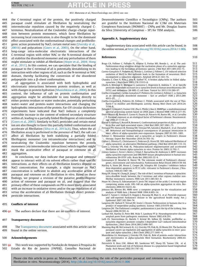
Rotenone and paraquat fail to accelerate aS fibrillation under
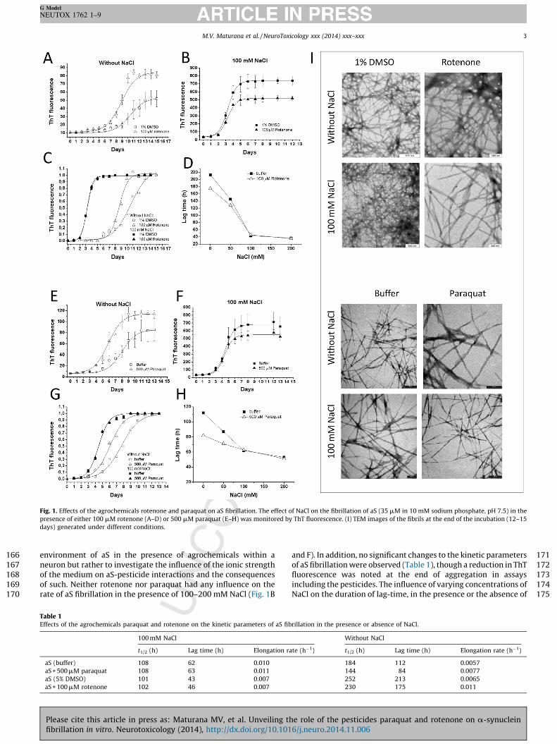
physiological concentrations of NaCl. aS fibrillation can be describedas a nucleated polymerization process in which the unfoldedmonomer undergoes self-assembly to form oligomeric intermedi-ates (nuclei) at the onset of aggregation, followed by monomeraccretion and fibril growth. Thus, aS fibrillation kinetics displaysigmoidal behavior with an initial lag-time phase (nucleation)followed by an exponential-growth phase (elongation) thatevolves until a plateau is reached (formation of mature amy-loid-fibrils). Fig. 1A and E indicates that both agrochemicals(100 mM rotenone or 500 mM paraquat) accelerate aS fibrillationin the presence of sodium phosphate, pH 7.4, which is inaccordance with previous reports (Uversky et al., 2001a,b; Silvaet al., 2013a,b). By analyzing the kinetic parameters extracted fromthe normalized curves displayed in Fig. 1C and G we verified thatthe duration of the lag-time and the t½ (time required to reach 50%of maximum fibrillation) significantly decreased in the presence ofthe pesticides, while the elongation rates increased (Table 1). Ourassays utilized a molar ratio of aS monomer:pesticide of �1:3 (forrotenone) and �1:15 (for paraquat). Rotenone did not producesignificant effects on aS fibrillation at concentrations below100 mM and higher concentrations were not evaluated due tothe very low solubility of this compound in aqueous medium.However, paraquat is highly soluble in water and a concentration-dependent effect on aS fibrillation was observed in the absence ofNaCl (Supplementary Fig. S1 A).
Next, we investigated the manner in which NaCl concentrationeffected the interaction of aS with paraquat or rotenone. Theintention of this assay was not to reproduce the physiological
the role of the pesticides paraquat and rotenone on a-synuclein16/j.neuro.2014.11.006
166
167
168
169
170
171172173174175
Fig. 1. Effects of the agrochemicals rotenone and paraquat on aS fibrillation. The effect of NaCl on the fibrillation of aS (35 mM in 10 mM sodium phosphate, pH 7.5) in the
presence of either 100 mM rotenone (A–D) or 500 mM paraquat (E–H) was monitored by ThT fluorescence. (I) TEM images of the fibrils at the end of the incubation (12–15
days) generated under different conditions.
M.V. Maturana et al. / NeuroToxicology xxx (2014) xxx–xxx 3
G Model
NEUTOX 1762 1–9
environment of aS in the presence of agrochemicals within aneuron but rather to investigate the influence of the ionic strengthof the medium on aS-pesticide interactions and the consequencesof such. Neither rotenone nor paraquat had any influence on therate of aS fibrillation in the presence of 100–200 mM NaCl (Fig. 1B
Table 1Effects of the agrochemicals paraquat and rotenone on the kinetic parameters of aS fib
100 mM NaCl
t1/2 (h) Lag time (h) Elongation ra
aS (buffer) 108 62 0.010
aS + 500 mM paraquat 108 63 0.011
aS (5% DMSO) 101 43 0.007
aS + 100 mM rotenone 102 46 0.007
Please cite this article in press as: Maturana MV, et al. Unveiling thfibrillation in vitro. Neurotoxicology (2014), http://dx.doi.org/10.101
and F). In addition, no significant changes to the kinetic parametersof aS fibrillation were observed (Table 1), though a reduction in ThTfluorescence was noted at the end of aggregation in assaysincluding the pesticides. The influence of varying concentrations ofNaCl on the duration of lag-time, in the presence or the absence of
rillation in the presence or absence of NaCl.
Without NaCl
te (h�1) t1/2 (h) Lag time (h) Elongation rate (h�1)
184 112 0.0057
144 84 0.0077
252 213 0.0065
230 175 0.011
e role of the pesticides paraquat and rotenone on a-synuclein6/j.neuro.2014.11.006
176 th177 th178 or179 w180 Ev181 ki182 (S183 (T184 an185 ly186 w187
188 as189 m190 ad191 an192 re193 re194 et195 in196 th197 m198 ph199 do200
201 of
202 ab203 th204 ab205 w206 so207 sh208 ac209 of210 Th211 of212 ab213 ag
214215216217218219220221222223224225226227228229230231232233234235236237238239240241242243244245246247248249250251
C
Figab
inc
M.V. Maturana et al. / NeuroToxicology xxx (2014) xxx–xxx4
G Model
NEUTOX 1762 1–9
e pesticides, is presented in Fig. 1D and H. We can clearly verifyat the duration of the lag-time is nearly identical in the presence the absence of the agrochemical when the aggregation assayas performed using NaCl concentrations higher than 100 mM.en at a high paraquat concentration (1 mM), aS fibrillationnetics were unaffected in the presence of 100 mM NaClupplementary Fig. S1 A). Transmission electron microscopyEM) images indicated that neither paraquat nor rotenone causedy apparent alterations to fibril morphology (Fig. 1I). Additional-
, these agrochemicals did not increase aS oligomer content evenhen in the presence of NaCl.
The concentrations of both paraquat and rotenone used in oursays are remarkable superior than those detected in animalodels exposed to these pesticides. For instance, the subcutaneousministration of 20 mg/kg of paraquat in rats resulted in a serumd striatal extracellular concentration of �8 and �0.5 mM,spectively (Shimizu et al., 2001). Rotenone concentrationsached in animal models are in the nM range (Greenamyre
al., 2003). Considering that the physiological concentration of aS brain has been estimated to be around 1 mM (Seo et al., 2002),e molar ratio of pesticides to aS monomer in our experimentsight be considered hypothetically higher than that reachedysiologically, which reinforces the idea of these pesticides likely
not have any effect aS fibrillation via a direct interaction.Paraquat and rotenone do not affect aS fibrillation in the presence
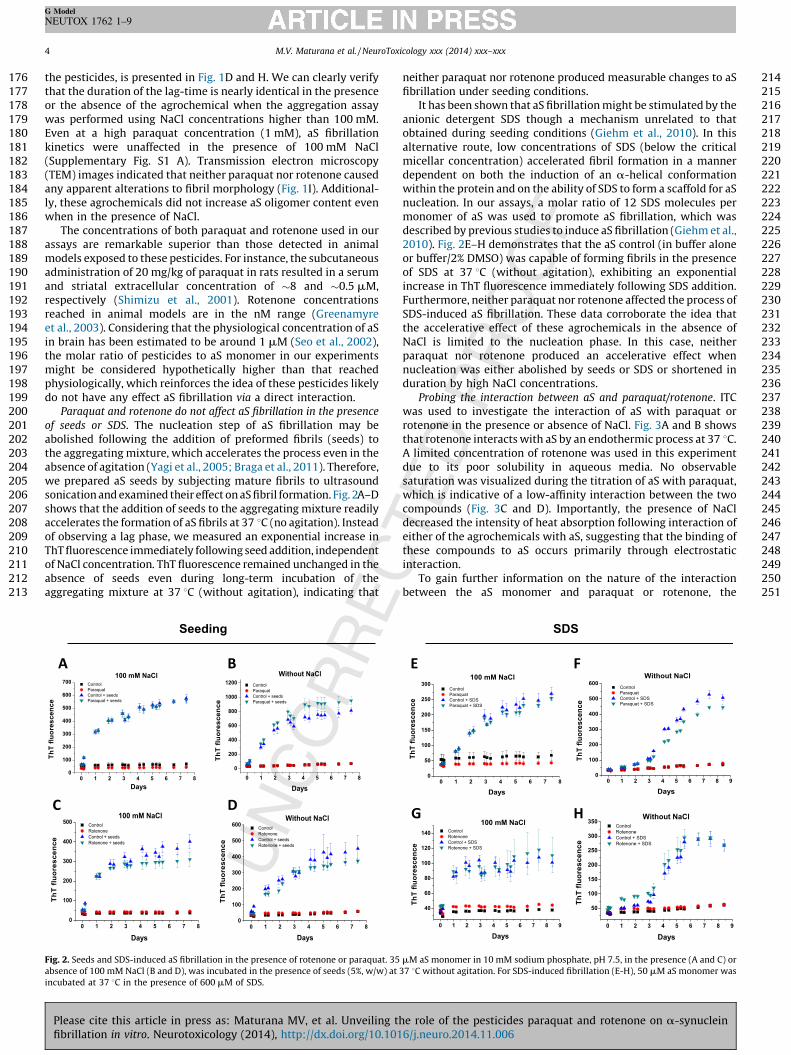
seeds or SDS. The nucleation step of aS fibrillation may beolished following the addition of preformed fibrils (seeds) toe aggregating mixture, which accelerates the process even in thesence of agitation (Yagi et al., 2005; Braga et al., 2011). Therefore,e prepared aS seeds by subjecting mature fibrils to ultrasoundnication and examined their effect on aS fibril formation. Fig. 2A–Dows that the addition of seeds to the aggregating mixture readilycelerates the formation of aS fibrils at 37 8C (no agitation). Instead
observing a lag phase, we measured an exponential increase inT fluorescence immediately following seed addition, independent
NaCl concentration. ThT fluorescence remained unchanged in thesence of seeds even during long-term incubation of thegregating mixture at 37 8C (without agitation), indicating that
0 1 2 3 4 5 6 7 80
100
200
300
400
500
600
700
Th
T f
luo
res
ce
nc
e
Days
100 mM NaCl
0 1 2 3 4 5 6 7 8
0
200
400
600
800
1000
1200
Th
T f
luo
res
ce
nc
e
Days
Wit hout NaCl
0 1 2 3 4 5 6 7 80
100
200
300
400
500
Th
T f
luo
res
ce
nc
e
Days
100 mM NaCl
0 1 2 3 4 5 6 7 80
100
200
300
400
500
600
Th
T f
luo
res
ce
nc
e
Days
Wit hout NaCl
A
See ding
B
D
. 2. Seeds and SDS-induced aS fibrillation in the presence of rotenone or paraquat. 3
sence of 100 mM NaCl (B and D), was incubated in the presence of seeds (5%, w/w) at
ubated at 37 8C in the presence of 600 mM of SDS.
Please cite this article in press as: Maturana MV, et al. Unveiling
fibrillation in vitro. Neurotoxicology (2014), http://dx.doi.org/10.10
neither paraquat nor rotenone produced measurable changes to aSfibrillation under seeding conditions.
It has been shown that aS fibrillation might be stimulated by theanionic detergent SDS though a mechanism unrelated to thatobtained during seeding conditions (Giehm et al., 2010). In thisalternative route, low concentrations of SDS (below the criticalmicellar concentration) accelerated fibril formation in a mannerdependent on both the induction of an a-helical conformationwithin the protein and on the ability of SDS to form a scaffold for aSnucleation. In our assays, a molar ratio of 12 SDS molecules permonomer of aS was used to promote aS fibrillation, which wasdescribed by previous studies to induce aS fibrillation (Giehm et al.,2010). Fig. 2E–H demonstrates that the aS control (in buffer aloneor buffer/2% DMSO) was capable of forming fibrils in the presenceof SDS at 37 8C (without agitation), exhibiting an exponentialincrease in ThT fluorescence immediately following SDS addition.Furthermore, neither paraquat nor rotenone affected the process ofSDS-induced aS fibrillation. These data corroborate the idea thatthe accelerative effect of these agrochemicals in the absence ofNaCl is limited to the nucleation phase. In this case, neitherparaquat nor rotenone produced an accelerative effect whennucleation was either abolished by seeds or SDS or shortened induration by high NaCl concentrations.
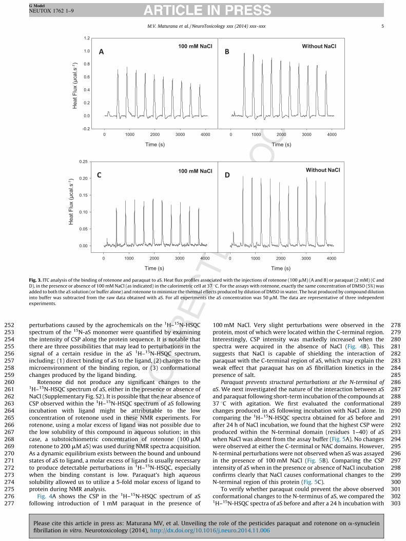
Probing the interaction between aS and paraquat/rotenone. ITCwas used to investigate the interaction of aS with paraquat orrotenone in the presence or absence of NaCl. Fig. 3A and B showsthat rotenone interacts with aS by an endothermic process at 37 8C.A limited concentration of rotenone was used in this experimentdue to its poor solubility in aqueous media. No observablesaturation was visualized during the titration of aS with paraquat,which is indicative of a low-affinity interaction between the twocompounds (Fig. 3C and D). Importantly, the presence of NaCldecreased the intensity of heat absorption following interaction ofeither of the agrochemicals with aS, suggesting that the binding ofthese compounds to aS occurs primarily through electrostaticinteraction.
To gain further information on the nature of the interactionbetween the aS monomer and paraquat or rotenone, the
SDS
0 1 2 3 4 5 6 7 8 90
100
200
300
400
500
600
Th
T f
luo
res
ce
nc
e
Days
Wit hout NaCl
0 1 2 3 4 5 6 7 8 9
50
100
150
200
250
300
350
Th
T f
luo
res
ce
nc
e
Days
Wit hout NaCl
0 1 2 3 4 5 6 7 80
50
100
150
200
250
300
Th
T f
luo
res
ce
nc
e
Days
100 mM NaCl
0 1 2 3 4 5 6 7 8 9
40
60
80
100
120
140
Th
T f
luo
res
ce
nc
e
Days
100 mM NaCl
E F
G H
5 mM aS monomer in 10 mM sodium phosphate, pH 7.5, in the presence (A and C) or
37 8C without agitation. For SDS-induced fibrillation (E-H), 50 mM aS monomer was
the role of the pesticides paraquat and rotenone on a-synuclein16/j.neuro.2014.11.006
252
253
254
255
256
257
258
259
260
261262
263
264
265
266
267
268
269
270
271
272
273
274
275
276
277
278279280281282283284285286287288289290291292293294295296297298299300301302303
Time (s)
0 100 0 200 0 300 0 400 0
Time (s)
0 100 0 200 0 300 0 400 0
-0.2
0.0
0.2
0.4
0.6
0.8
1.0
1.2
Time (s)
0 100 0 200 0 300 0 400 0
Time (s)
0 100 0 200 0 300 0 400 0
0.00
0.05
0.10
0.15
0.20
0.25
100 mM NaCl Without NaClA B
C D100 mM NaCl Without NaCl
Heat F
lux (
μcal.s
-1)
Heat F
lux (
μcal.s
-1)
Fig. 3. ITC analysis of the binding of rotenone and paraquat to aS. Heat flux profiles associated with the injections of rotenone (100 mM) (A and B) or paraquat (2 mM) (C and
D), in the presence or absence of 100 mM NaCl (as indicated) in the calorimetric cell at 37 8C. For the assays with rotenone, exactly the same concentration of DMSO (5%) was
added to both the aS solution (or buffer alone) and rotenone to minimize the thermal effects produced by dilution of DMSO in water. The heat produced by compound dilution
into buffer was subtracted from the raw data obtained with aS. For all experiments the aS concentration was 50 mM. The data are representative of three independent
experiments.
M.V. Maturana et al. / NeuroToxicology xxx (2014) xxx–xxx 5
G Model
NEUTOX 1762 1–9
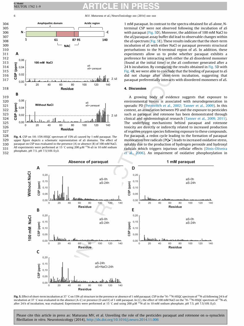
perturbations caused by the agrochemicals on the 1H–15N-HSQCspectrum of the 15N-aS monomer were quantified by examiningthe intensity of CSP along the protein sequence. It is notable thatthere are three possibilities that may lead to perturbations in thesignal of a certain residue in the aS 1H–15N-HSQC spectrum,including: (1) direct binding of aS to the ligand, (2) changes to themicroenvironment of the binding region, or (3) conformationalchanges produced by the ligand binding.
Rotenone did not produce any significant changes to the1H–15N-HSQC spectrum of aS, either in the presence or absence ofNaCl (Supplementary Fig. S2). It is possible that the near absence ofCSP observed within the 1H–15N-HSQC spectrum of aS followingincubation with ligand might be attributable to the lowconcentration of rotenone used in these NMR experiments. Forrotenone, using a molar excess of ligand was not possible due tothe low solubility of this compound in aqueous solution; in thiscase, a substoichiometric concentration of rotenone (100 mMrotenone to 200 mM aS) was used during NMR spectra acquisition.As a dynamic equilibrium exists between the bound and unboundstates of aS to ligand, a molar excess of ligand is usually necessaryto produce detectable perturbations in 1H–15N-HSQC, especiallywhen the binding constant is low. Paraquat’s high aqueoussolubility allowed us to utilize a 5-fold molar excess of ligand toprotein during NMR analysis.
Fig. 4A shows the CSP in the 1H–15N-HSQC spectrum of aSfollowing introduction of 1 mM paraquat in the presence of
Please cite this article in press as: Maturana MV, et al. Unveiling thfibrillation in vitro. Neurotoxicology (2014), http://dx.doi.org/10.101
100 mM NaCl. Very slight perturbations were observed in theprotein, most of which were located within the C-terminal region.Interestingly, CSP intensity was markedly increased when thespectra were acquired in the absence of NaCl (Fig. 4B). Thissuggests that NaCl is capable of shielding the interaction ofparaquat with the C-terminal region of aS, which may explain theweak effect that paraquat has on aS fibrillation kinetics in thepresence of salt.
Paraquat prevents structural perturbations at the N-terminal of
aS. We next investigated the nature of the interaction between aSand paraquat following short-term incubation of the compounds at37 8C with agitation. We first evaluated the conformationalchanges produced in aS following incubation with NaCl alone. Incomparing the 1H–15N-HSQC spectra obtained for aS before andafter 24 h of NaCl incubation, we found that the highest CSP wereproduced within the N-terminal domain (residues 1–40) of aSwhen NaCl was absent from the assay buffer (Fig. 5A). No changeswere observed at either the C-terminal or NAC domains. However,N-terminal perturbations were not observed when aS was assayedin the presence of 100 mM NaCl (Fig. 5B). Comparing the CSPintensity of aS when in the presence or absence of NaCl incubationconfirms clearly that NaCl causes conformational changes to theN-terminal region of this protein (Fig. 5C).
To verify whether paraquat could prevent the above observedconformational changes to the N-terminus of aS, we compared the1H–15N-HSQC spectra of aS before and after a 24 h incubation with
e role of the pesticides paraquat and rotenone on a-synuclein6/j.neuro.2014.11.006
304
305
306
307
308
309
310
311
312
313
314
315
316
317
318
319
320
321
322
323
324
325
326
327
328
329
330
331
332
NAC
Amph ipat hic domain Acidic regi on
N _ _C
1 61 95 14 087
Res idue
0 20 40 60 80 100 120 140
CS
P (
pp
m)
0,00
0,02
0,04
0,06
Without NaClaS
aS+ paraqua t
2 Δδ
Res idue
0 20 40 60 80 100 120 140
CS
P (
pp
m)
0,00
0,02
0,04
0,06100 mM NaCl
aS
aS+ paraqua t
2 Δδ
A
B
Fig. 4. CSP on 1H–15N-HSQC spectrum of 15N-aS caused by 1 mM paraquat. The
upper figure depicts a schematic representation of aS domains. The effect of
paraquat on CSP was evaluated in the presence (A) or absence (B) of 100 mM NaCl.
All experiments were performed at 15 8C using 200 mM 15N-aS in 10 mM sodium
phosphate, pH 7.5, pH 7.5/10% D2O.
Residue
0 20 40 60 80 10 0 12 0 14 0
CS
P (
pp
m)
0,00
0,05
0,10
0,15
0,20
Residue
0 20 40 60 80 10 0 12 0 14 0
CS
P (
pp
m)
0,00
0,05
0,10
0,15
0,20
Residue
0 20 40 60 80 10 0 12 0 14 0
CS
P (
pp
m)
0,00
0,05
0,10
0,15
0,20
Wit
ho
ut
NaC
l1
00
mM
Na
Cl
aS-0h
aS-24 h
aS-0h
aS-24 h
Abs enc e of para quat
aS-24 h
aS+NaCl-24 h
A
B
C
Fig. 5. Effect of short-term incubation at 37 8C on 15N-aS structure in the presence or abs
incubation at 37 8C was evaluated in the absence (A–C) or presence (D and E) of 1 mM
after 24 h of incubation, was evaluated. Experiments were performed at 15 8C and us
M.V. Maturana et al. / NeuroToxicology xxx (2014) xxx–xxx6
G Model
NEUTOX 1762 1–9
Please cite this article in press as: Maturana MV, et al. Unveiling
fibrillation in vitro. Neurotoxicology (2014), http://dx.doi.org/10.10
1 mM paraquat. In contrast to the spectra obtained for aS alone, N-terminal CSP were not observed following the incubation of aSwith paraquat (Fig. 5D). Moreover, the addition of 100 mM NaCl tothe aS/paraquat assay buffer did lead to observable changes withinthe aS spectrum (Fig. 5E). These results indicate that the short-termincubation of aS with either NaCl or paraquat prevents structuralperturbations to the N-terminal region of aS. In addition, theseexperiments allow us to probe whether paraquat exhibits apreference for interacting with either the aS disordered monomer(found at the initial time) or the aS conformer generated after a24 h incubation. By comparing the results obtained in Fig. 5D withFig. 4B, we were able to conclude that the binding of paraquat to aSdid not change after short-term incubation, suggesting thatparaquat preferentially interacts with disordered monomers of aS.
4. Discussion
A growing body of evidence suggests that exposure toenvironmental toxins is associated with neurodegeneration insporadic PD (Petrovitch et al., 2002; Tanner et al., 2009). In thiscontext, an association between PD and the exposure to pesticidessuch as paraquat and rotenone has been demonstrated throughclinical and epidemiological research (Tanner et al., 2009, 2011).The underlying mechanisms behind paraquat and rotenonetoxicity are directly or indirectly related to increased productionof reactive oxygen species following exposure to these compounds.For paraquat, a redox cycle leading to the formation of paraquatmonocation free radicals (PQ�+) leads to increased oxidative stress,notably due to the production of hydrogen peroxide and hydroxylradicals which triggers injurious cellular effects (Dinis-Oliveiraet al., 2006). An impairment of oxidative phosphorylation in
Residue
0 20 40 60 80 10 0 12 0 14 0
CS
P (
pp
m)
0,00
0,05
0,10
0,15
0,20
Residue
0 20 40 60 80 10 0 12 0 14 0
CS
P (
pp
m)
0,00
0,05
0,10
0,15
0,20
aS-0h
aS-24 h
aS-0h
aS-24 h
1 mM paraquat
D
E
ence of 1 mM paraquat. CSP in the 1H–15N-HSQC spectrum of 15N-aS following 24 h of
paraquat. In (C), the effect of 100 mM NaCl on the 1H–15N-HSQC spectrum of 15N-aS,
ing 200 mM 15N-aS in 10 mM sodium phosphate, pH 7.5, pH 7.5/10% D2O.
the role of the pesticides paraquat and rotenone on a-synuclein16/j.neuro.2014.11.006
333
334
335
336
337
338
339
340
341
342
343
344
345
346
347
348
349
350
351
352
353
354
355
356
357
358
359
360
361
362
363
364
365
366
367
368
369
370
371
372
373
374
375
376
377
378379380381382383384385386387388389390391392393394395396397398399400401402403404405406407408409410411412413414415416417418419420421422
M.V. Maturana et al. / NeuroToxicology xxx (2014) xxx–xxx 7
G Model
NEUTOX 1762 1–9
mitochondria by inhibiting complex I of the electron-transportchain is associated to the toxic effects of rotenone (Hartley et al.,1994; Silva et al., 2013a,b).
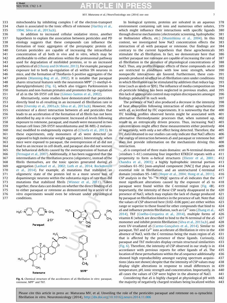
In addition to increased cellular oxidative stress, anotherhypothesis to explain the association between pesticides and PDetiology suggests that these compounds can influence theformation of toxic aggregates of the presynaptic protein aS.Certain pesticides are capable of increasing the intracellularconcentration of aS both in vivo and in vitro, which may beattributable to either alterations within the proteasomal pathwayused for degradation of misfolded proteins, or to an increasedsynthesis of aS (Ulusoy and Di Monte, 2013). For example, paraquatexposure led to both up-regulation of aS within SN neurons inmice, and the formation of Thioflavin-S positive aggregates of theprotein (Manning-Bog et al., 2002). It is notable that paraquatshares structural features with the neurotoxin MPP+ (1-methyl-4-phenylpyridinium) (Fig. 6), which also triggers Parkinsonism inhumans and non-human primates and promotes the up-regulationof aS in the SH-SY5Y cell line (Gomez-Santos et al., 2002).
Alternatively, some studies have demonstrated that pesticidesdirectly bind to aS resulting in an increased aS fibrillation rate in
vitro (Uversky et al., 2001a,b; Silva et al., 2013a,b). However, theexistence of a direct interaction between aS and pesticides thatleads to an acceleration of the formation of aS fibrils has not beenidentified by any in vivo experiment. Increased aS levels followingexposure to rotenone, paraquat, and maneb were measured in twohuman cell lines (SH-SY5Y neuroblastoma and SK-MEL-2 melano-ma) modified to endogenously express aS (Chorfa et al., 2013). Inthese experiments, only monomers of aS were detected (asopposed to high molecular weight aggregates). Interestingly, whenmice were exposed to paraquat, the overexpression of aS did notlead to an increase in cell death, and paraquat also did not worsenthe behavioral deficits caused by the overexpression of human aS(Fernagut et al., 2007). Additionally, it has been suggested that theintermediates of the fibrillation process (oligomers), instead of thefibrils themselves, are the toxic species generated during aSaggregation (Lashuel et al., 2002; Luth et al., 2014; Rockensteinet al., 2014). For example, aS mutations that stabilized anoligomeric state of the protein led to a more severe loss ofdopaminergic neurons within the substantia nigra of rats than didmutations that stabilized fibrils (Winner et al., 2011). Takentogether, these data cast doubts on whether the direct binding of aSto either paraquat or rotenone as demonstrated by a series of in
vitro experiments would even be relevant under physiologicalconditions.
423424425426427428429430431432433434435436437438439440441442443
Fig. 6. Chemical structure of the accelerators of aS fibrillation in vitro: paraquat,
rotenone, MPP+ and ThT.
Please cite this article in press as: Maturana MV, et al. Unveiling thfibrillation in vitro. Neurotoxicology (2014), http://dx.doi.org/10.101
In biological systems, proteins are solvated in an aqueousenvironment containing salt ions and numerous other solutes,which might influence their interactions with specific ligandsthrough diverse mechanisms (electrostatic screening, hydrophobicor Hofmeister effects, etc.) (Munishkina et al., 2004). In thiscontext, we investigated how NaCl concentration effects theinteraction of aS with paraquat or rotenone. Our findings arecontrary to the current hypothesis that these agrochemicalsstimulate the aS fibrillation. In fact, we demonstrate here thatneither paraquat nor rotenone are capable of increasing the rate ofaS fibrillation in the presence of physiological concentrations ofNaCl. Thus, any profibrillogenic effects of these compounds on aSare restricted to conditions with low ionic strength, wherenonspecific interactions are favored. Furthermore, these com-pounds produced no effect on aS fibrillation rates under conditionsin which fibrillation can be stimulated by additives that reduce lag-time (such as seeds or SDS). The influence of media composition onaS-pesticide binding has been neglected in previous studies, andthis lack of appropriate control may explain discrepancies amongstfindings produced in such work.
The presence of NaCl also produced a decrease in the intensityof heat absorption following interaction of either agrochemicalwith aS, as verified by ITC experiments. It is noteworthy that theITC binding profiles observed herein might be associated withalternative thermodynamic processes that, when summed up,result in an entropically driven process. Thus, increasing NaClconcentrations might affect these measurements either positivelyor negatively, with only a net effect being detected. Therefore, theITC data obtained in our studies can only indicate that NaCl affectsthe energy of interaction between aS and paraquat or rotenone butdoes not provide information on the mechanisms driving thisinteraction.
aS is comprised of three main domains: an N-terminal domain(residues 1–61) containing four imperfect KTKEGV repeats with apropensity to form a-helical structures (Eliezer et al., 2001;Chandra et al., 2003); a highly hydrophobic internal portion(residues 61–95) [non-amyloid component (NAC)] that plays anessential role in fibrillation; and an unstructured C-terminaldomain (residues 95–140) (Hoyer et al., 2004; Hong et al., 2011).CSP analysis in the 1H–15N HSQC spectra of aS indicates that themost significant alterations caused by the exposure of aS toparaquat were found within the C-terminal region (Fig. 4B).Importantly, the intensity of these CSP nearly disappeared in thepresence of NaCl, which may explain the lack of change producedby paraquat on fibrillation kinetics in the presence of salt. Note thatthe values of CSP observed here (0.02–0.06 ppm) are either withinrange or superior to those found for other compounds that bind toaS and influence protein fibrillation, such as Ca2+ ions (Zhang et al.,2014), ThT (Coelho-Cerqueira et al., 2014), multiple forms ofvitamin K (which are described to bind to the N-terminal of the aSmonomer and inhibit protein fibrillation (Silva et al., 2013a,b), andeven UV-irradiated aS (Carmo-Goncalves et al., 2014). Similar toparaquat, ThT and Ca2+ ions accelerate aS fibrillation in vitro in theabsence of NaCl, with the C-terminus being the main region of aSthat is affected by the presence of these ligands. Curiously,paraquat and ThT molecules display certain structural similarities(Fig. 6). Therefore, the intensity of CSP observed in our study is inaccordance with previous reports for other ligands of aS. Thelocation of these perturbations within the aS sequence additionallyshowed high reproducibility amongst varying spectrum acquisi-tions (data not shown) despite that the intensity of CSP values maydisplay slight alterations in response to small differences intemperature, pH, ionic strength and concentration. Importantly, inall cases the values of CSP were higher in the absence of NaCl.
Considering that aS is highly charged at physiological pH withthe majority of negatively charged residues being localized within
e role of the pesticides paraquat and rotenone on a-synuclein6/j.neuro.2014.11.006
444 th445 pa446 in447 te448 sio449 in450 ef451 ge452 20453 lo454 C-455 aS456 m457 et458 pa459 in460 do461 po462
463 el464 w465 co466 ag467 ei468 w469 hy470 ex471 re472 w473 (G474 io475 ac476 fib477 af478 fib479 ne480 m481 m482 ag483
484 ap485 in486 Na487 co488 pa489 fin490 ac491 pr492 w493 ve
494 pe
495 Co
496 Q3
497 Tr
498
499 fo
500 Ac
501 Q4
502 Es
503504505506
507
508509
510
511512513514515516517518519520521522523524525526527528529530531532533534535536537538539540541542543544545546547548549550551552553554555556557558559560561562563564565566567568569570571572573574575576577578579580
M.V. Maturana et al. / NeuroToxicology xxx (2014) xxx–xxx8
G Model
NEUTOX 1762 1–9
e C-terminal region of the protein, the positively chargedraquat could stimulate aS fibrillation by neutralizing thetermolecular repulsion caused by the negatively charged C-rminus. Neutralization of the Coulombic charge–charge repul-n between protein monomers, which favor fibrillation by
creasing local concentration, is also thought to be the dominantfect associated with the conformational changes and profibrillo-nic action promoted by NaCl, certain metal ions (Uversky et al.,01b) and polycations (Goers et al., 2003). On the other hand,
ng-range intra-molecular electrostatic interactions of theterminal region with either NAC or the N-terminal domain of stabilize the disordered monomer and any perturbation of themight stimulate or inhibit aS fibrillation (Hoyer et al., 2004; Hong
al., 2011). In this context, we can speculate that the binding ofraquat to the C-terminal of aS is potentially destabilizing to itsteraction with other regions of aS, such as the N-terminal or NACmain, thereby facilitating the conversion of the disorderedlypeptide into a b-sheet conformation.aS aggregation is associated with both hydrophobic and
ectrostatic interactions between protein residues, as well asith changes in protein hydration (Munishkina et al., 2004). In thisntext, the influence of salt on protein conformation andgregation might be attributable to ions binding directly tother protein residues or water molecules, potentially perturbingater–water and protein–water interactions and changing thedrophobic interactions of the protein. Far-UV circular dichroismperiments have indicated that NaCl induces a completelyversible increase in the content of ordered secondary structureithin aS, leading to a partially folded fibrillogenic aS intermediateoers et al., 2003). Similarly to NaCl, pesticides and certain metaln are also thought to favor this partially folded conformation andcelerate aS fibrillation (Silva et al., 2013a,b). Thus, when the aSrillation assay is performed in the presence of NaCl, the salt can
fect aS fibrillation by both stabilizing a partially foldedrillogenic monomer (via intramolecular interactions) and byutralizing the Coulombic repulsion between the proteinonomers (via intermolecular interactions) which together mightask the accelerative effect of paraquat or rotenone on aSgregation.In conclusion, our data indicate that paraquat and rotenone
pear to interact with aS via solvent effects rather than specificteractions, as their effects on aS fibrillation kinetics depend onCl concentration. For example, a slight increase in NaClncentration is sufficient to abolish any accelerative action ofraquat and rotenone on aS fibrillation in vitro. Based on thesedings, we propose a revision of the putative profibrillogeniction of rotenone and paraquat on aS, and suggest that theimary effect of these compounds on PD is more likely associatedith an increase in oxidative stress and/or the up-regulation of aSrsus the acceleration of aS fibrillation through direct protein–sticide interactions.
nflicts of interest
The authors declare that there are no conflicts of interest.
ansparency document
The Transparency document associated with this article can beund in the online version.
knowledgements
This work was supported by Fundacao de Amparo a Pesquisa dotado do Rio de Janeiro (FAPERJ), Conselho Nacional de
Please cite this article in press as: Maturana MV, et al. Unveiling
fibrillation in vitro. Neurotoxicology (2014), http://dx.doi.org/10.10
Desenvolvimento Cientıfico e Tecnologico (CNPq). The authorsare grateful to the Instituto Nacional de C,T&I em MateriaisComplexos Funcionais (INOMAT) – CNPq and Mr. Douglas Soaresda Silva (University of Campinas – SP) for TEM analysis.
Appendix A. Supplementary data
Supplementary data associated with this article can be found, inthe online version, at http://dx.doi.org/10.1016/j.neuro.2014.11.006.
References
Braga CA, Follmer C, Palhano FL, Khattar E, Freitas MS, Romao L, et al. The anti-Parkinsonian drug selegiline delays the nucleation phase of a-synuclein aggrega-tion leading to the formation of nontoxic species. J Mol Biol 2011;405:254–73.
Carmo-Goncalves P, Pinheiro AS, Romao L, Cortines J, Follmer C. UV-induced selectiveoxidation of Met5 to Met-sulfoxide leads to the formation of neurotoxic fibril-incompetent a-synuclein oligomers. Amyloid 2014;21:163–74.
Chandra S, Chen X, Rizo J, Jahn R, Sudhof TC. A broken alpha-helix in folded alpha-synuclein. J Biol Chem 2003;278:15313–8.
Chorfa A, Betemps D, Morignat E, Lazizzera C, Hogeveen K, Andrieu T, et al. Specificpesticide-dependent increases in a-synuclein levels in human neuroblastoma (SH-SY5Y) and melanoma (SK-MEL-2) cell lines. Toxicol Sci 2013;133:289–97.
Coelho-Cerqueira E, Carmo-Goncalves P, Pinheiro AS, Cortines J, Follmer C. a-Synucleinas an intrinsically disordered monomer – fact or artefact? FEBS J 2013;280:4915–27.
Coelho-Cerqueira E, Pinheiro AS, Follmer C. Pitfalls associated with the use of Thio-flavin-T to monitor anti-fibrillogenic activity. Bioorg Med Chem Lett 2014;24:3194–8.
Delaglio F, Grzesiek S, Vuister GW, Zhu G, Pfeifer J, Bax A. NMRPipe: a multidimensionalspectral processing system based on UNIX pipes. J Biomol NMR 1995;6:277–93.
Dinis-Oliveira RJ, Remiao F, Carmo H, Duarte JA, Navarro AS, Bastos ML, et al. CarvalhoF. Paraquat exposure as an etiological factor of Parkinson’s disease. Neurotoxicol-ogy 2006;27:1110–22.
Eliezer D, Kutluay E, Bussell R, Browne G. Conformational properties of alpha-synucleinin its free and lipid-associated states. J Mol Biol 2001;307:1061–73.
Fernagut PO, Hutson CB, Fleming SM, Tetreaut NA, Salcedo J, Masliah E, et al. ChesseletMF. Behavioral and histopathological consequences of paraquat intoxication inmice: effects of alpha-synuclein over-expression. Synapse 2007;61:991–1001.
Follmer C. Monoamine oxidase and a-synuclein as targets in Parkinson’s diseasetherapy. Expert Rev Neurother 2014;14:703–16.
Giehm L, Oliveira CL, Christiansen G, Pedersen JS, Otzen DE. SDS-induced fibrillation ofalpha-synuclein: an alternative fibrillation pathway. J Mol Biol 2010;401:115–33.
Goers J, Uversky VN, Fink AL. Polycation-induced oligomerization and acceleratedfibrillation of human alpha-synuclein in vitro. Protein Sci 2003;12:702–7.
Gomez-Santos C, Ferrer I, Reiriz J, Vinals F, Barrachina M, Ambrosio S. MPP+ increasesalpha-synuclein expression and ERK/MAP-kinase phosphorylation in human neu-roblastoma SH-SY5Y cells. Brain Res 2002;935:32–9.
Greenamyre JT, Betarbet R, Sherer TB. The rotenone model of Parkinson’s disease:genes, environment and mitochondria. Parkinsonism Relat Disord 2003;9:59–64.
Hartley A, Stone JM, Heron C, Cooper JM, Schapira AH. Complex I inhibitors inducedose-dependent apoptosis in PC12 cells: relevance to Parkinson’s disease. J Neu-rochem 1994;63:1987–90.
Hong DP, Xiong W, Chang JY, Jiang C. The role of the C-terminus of human a-synuclein:intra-disulfide bonds between the C-terminus and other regions stabilize non-fibrillar monomeric isomers. FEBS Lett 2011;585:561–6.
Hoyer W, Cherny D, Subramaniam V, Jovin TM. Impact of the acidic C-terminal regioncomprising amino acids 109–140 on alpha-synuclein aggregation in vitro. Bio-chemistry 2004;43:16233–42.
Johnson BA, Blevins RA. NMR view: a computer program for the visualization andanalysis of NMR data. J Biomol NMR 1994;4:603–14.
Kamel F, Tanner C, Umbach D, Hoppin J, Alavanja M, Blair A, et al. Pesticide exposureand self-reported Parkinson’s disease in the agricultural health study. Am JEpidemiol 2007;165:364–74.
Langston JW, Ballard P, Tetrud JW, Irwin I. Chronic Parkinsonism in humans due to aproduct of meperidine-analog synthesis. Science 1983;219:979–80.
Langston JW. The Parkinson’s complex: parkinsonism is just the tip of the iceberg. AnnNeurol 2006;59:591–6.
Lashuel HA, Hartley D, Petre BM, Walz T, Lansbury PT Jr. Neurodegenerative disease:amyloid pores from pathogenic mutations. Nature 2002;418:291.
Luth ES, Stavrovskaya IG, Bartels T, Kristal BS, Selkoe DJ. Soluble, prefibrillar a-synuclein oligomers promote complex I-dependent Ca2+-induced mitochondrialdysfunction. J Biol Chem 2014;289:21490–507.
Manning-Bog AB, McCormack AL, Li J, Uversky VN, Fink AL, Di Monte DA. The herbicideparaquat causes up-regulation and aggregation of alpha-synuclein in mice: para-quat and alpha-synuclein. J Biol Chem 2002;277:1641–4.
Munishkina LA, Henriques J, Uversky VN, Fink AL. Role of protein–water interactionsand electrostatics in alpha-synuclein fibril formation. Biochemistry 2004;43:3289–300.
Petrovitch H, Ross GW, Abbott RD, Sanderson WT, Sharp DS, Tanner CM, et al.Plantation work and risk of Parkinson disease in a population-based longitudinalstudy. Arch Neurol 2002;59:1787–92.
the role of the pesticides paraquat and rotenone on a-synuclein16/j.neuro.2014.11.006
581
582
583
584
585
586
587
588
589
590
591
592
593
594
595
596
597
598
599
600
601
602
603
604
605
606
607608609610611612613614615616617618619620621622623624625626627628629630631
M.V. Maturana et al. / NeuroToxicology xxx (2014) xxx–xxx 9
G Model
NEUTOX 1762 1–9
Priyadarshi A, Khuder SA, Schaub EA, Shrivastava S. A meta-analysis of Parkinson’sdisease and exposure to pesticides. Neurotoxicology 2000;21:435–40.
Rao JN, Kim YE, Park LS, Ulmer TS. Effect of pseudorepeat rearrangement on alpha-synuclein misfolding, vesicle binding, and micelle binding. J Mol Biol 2009;390:516–29.
Rockenstein E, Nuber S, Overk CR, Ubhi K, Mante M, Patrick C, et al. Accumulation ofoligomer-prone a-synuclein exacerbates synaptic and neuronal degeneration invivo. Brain 2014;137:1496–513.
Seo JH, Rah JC, Choi SH, Shin JK, Min K, Kim HS, et al. a-Synuclein regulates neuronalsurvival via Bcl-2 family expression and PI3/Akt kinase pathway. FASEB J2002;16:1826–8.
Shimizu K, Ohtaki K, Matsubara K, Aoyama K, Uezono T, Saito O, et al. Carrier-mediatedprocesses in blood–brain barrier penetration and neural uptake of paraquat. BrainRes 2001;906:135–42.
Silva BA, Breydo L, Fink AL, Uversky VN. Agrochemicals, a-synuclein, and Parkinson’sdisease. Mol Neurobiol 2013a;47:598–612.
Silva FL, Coelho Cerqueira E, de Freitas MS, Goncalves DL, Costa LT, Follmer C, et al.interact with N-terminus a-synuclein and modulate the protein fibrillization invitro. Exploring the interaction between quinones and a-synuclein. Neurochem Int2013b;62:103–12.
Spillantini MG, Crowther RA, Jakes R, Hasegawa M, Goedert M. Alpha-synuclein infilamentous inclusions of Lewy bodies from Parkinson’s disease and dementia withlewy bodies. Proc Natl Acad Sci USA 1998;95:6469–73.
Sulzer D. Multiple hit hypotheses for dopamine neuron loss in Parkinson’s disease.Trends Neurosci 2007;30:244–50.
Please cite this article in press as: Maturana MV, et al. Unveiling thfibrillation in vitro. Neurotoxicology (2014), http://dx.doi.org/10.101
Tanner CM, Kamel F, Ross GW, Hoppin JA, Goldman SM, Korell M, et al. Rotenone,paraquat, and Parkinson’s disease. Environ Health Perspect 2011;119:866–72.
Tanner CM, Ross GW, Jewell SA, Hauser RA, Jankovic J, Factor SA, et al. Occupation andrisk of parkinsonism: a multicenter case-control study. Arch Neurol 2009;66:1106–13.
Ulusoy A, Di Monte DA. a-Synuclein elevation in human neurodegenerative diseases:experimental, pathogenetic, and therapeutic implications. Mol Neurobiol 2013;47:484–94.
Uversky VN, Li J, Fink AL. Pesticides directly accelerate the rate of alpha-synucleinfibril formation: a possible factor in Parkinson’s disease. FEBS Lett 2001a;500:105–8.
Uversky VN, Li J, Fink AL. Metal-triggered structural transformations, aggregation,and fibrillation of human alpha-synucleins. A possible molecular link betweenParkinson’s disease and heavy metal exposure. J Biol Chem 2001b;276:44284–96.
Winner B, Jappelli R, Maji SK, Desplats PA, Boyer L, Aigner S, et al. In vivo demonstrationthat alpha-synuclein oligomers are toxic. Proc Natl Acad Sci USA 2011;108:4194–9.
Yagi H, Kusaka E, Hongo K, Mizobata T, Kawata Y. Amyloid fibril formation of alpha-synuclein is accelerated by preformed amyloid seeds of other proteins: implica-tions for the mechanism of transmissible conformational diseases. J Biol Chem2005;280:38609–16.
Zhang Z, Dai C, Bai J, Xu G, Liu M, Li C. Ca(2+) modulating a-synuclein membranetransient interactions revealed by solution NMR spectroscopy. Biochim BiophysActa 2014;1838:853–8.
e role of the pesticides paraquat and rotenone on a-synuclein6/j.neuro.2014.11.006


![Effects of Seeding on Lysozyme Amyloid Fibrillation in the ... · Alzheimer’s disease, type 2 diabetes, and several systemic amyloidoses [1-3]. These proteins, despite their unrelated](https://static.fdocument.org/doc/165x107/5f66a2f7828269373b7d097b/effects-of-seeding-on-lysozyme-amyloid-fibrillation-in-the-alzheimeras-disease.jpg)