Cytokine-mediated Downregulation of The Transcription Factor ...

Triptolide reverses hypoxia-induced epithelial–mesenchymaltransition and stem-like features in pancreatic cancer by NF-jBdownregulation
Li Liu1,2, Alexei V. Salnikov1,2,3, Nathalie Bauer1,2, Ewa Aleksandrowicz1,2, Sabrina Labsch1,2, Clifford Nwaeburu1,2,
J€urgen Mattern1,2, Jury Gladkich1,2, Peter Schemmer2, Jens Werner2 and Ingrid Herr1,2
1 Molecular OncoSurgery Group, Department of General and Transplantation Surgery, University of Heidelberg and German Cancer Research Center, Heidel-
berg, Germany2 Department of General and Transplantation Surgery, University of Heidelberg, Heidelberg, Germany3 Department of Translational Immunology, German Cancer Research Center and National Center for Tumor Diseases, Heidelberg, Germany
Pancreatic ductal adenocarcinoma (PDA) is one of the most lethal malignancies characterized by an intense tumor stroma with
hypoperfused regions, a significant inflammatory response and pronounced therapy resistance. New therapeutic agents are urgently
needed. The plant-derived agent triptolide also known as “thunder god vine” has a long history in traditional Chinese medicine for
treatment of rheumatoid arthritis and cancer and is now in a clinical phase II trial for establishing the efficacy against a placebo.
The authors mimicked the situation in patient tumors by induction of hypoxia in experimental models of pancreatic cancer stem
cells (CSCs) and evaluated the therapeutic effect of triptolide. Hypoxia led to induction of colony and spheroid formation, aldehyde
dehydrogenase 1 (ALDH1) and NF-jB activity, migratory potential and a switch in morphology to a fibroblastoid phenotype, as well
as stem cell- and epithelial–mesenchymal transition-associated protein expression. Triptolide efficiently inhibited hypoxia-induced
transcriptional signaling and downregulated epithelial–mesenchymal transition (EMT) and CSC features in established highly malig-
nant cell lines, whereas sensitive cancer cells or nonmalignant cells were less affected. In vivo triptolide inhibited tumor take and
tumor growth. In primary CSCs isolated from patient tumors, triptolide downregulated markers of CSCs, proliferation and mesenchy-
mal cells along with upregulation of markers for apoptosis and epithelial cells. This study is the first to show that triptolide
reverses EMT and CSC characteristics and therefore may be superior to current chemotherapeutics for treatment of PDA.
Pancreatic ductal adenocarcinoma (PDA) is one of the mostaggressive malignancies and is usually diagnosed in an advancedstate, with extensive local invasion, early systemic disseminationand marked resistance to chemotherapy and radiotherapy.1
PDA is characterized by a pronounced hypoxic tumor microen-vironment,2 resulting in high expression of the hypoxia markerHIF-1a in patient tissue, which is a predictor of poor clinicaloutcome.3 In experimental studies, hypoxia is a marker foraggressive growth and spontaneous metastasis formation in pan-creatic cancer xenografts,4 suggesting a role for hypoxia inenrichment of cancer cells with stem cell characteristics.
According to the theory, cancer stem cells (CSCs) survivehypoxia and conventional cytotoxic therapy due to defense andsurvival mechanisms.5 The small CSC population within thetumor mass is believed to possess self-renewal potential and theability to differentiate, thereby generating a heterogeneous cellpopulation of the originating tumor.6–8 In addition, CSCs areproposed to mediate uncontrolled growth, therapy resistance,invasion and metastasis.9 Markers for CSCs have been identifiedin PDA and involve typical surface marker expression ofCD441/CD241/ESA1, CD1331/CXCR41 and c-Met1.10–12
Recent reports have indicated that the emergence of CSCs occursin part because of epithelial–mesenchymal transition (EMT).13
EMT can be activated by hypoxia-induced HIF-1 signal-ing,14 followed by a cellular switch from epithelial to
Can
cer
The
rapy
Key words: pancreatic cancer, novel antitumor agents, hypoxia, epi-
thelial–mesenchymal transition, cancer stem cells
Additional Supporting Information may be found in the online
version of this article.
This is an open access article under the terms of the Creative Com-
mons Attribution Non-Commercial License, which permits use, dis-
tribution and reproduction in any medium, provided the original
work is properly cited and is not used for commercial purposes.
Grant sponsor: German Cancer Aid (Deutsche Krebshilfe); Grant
number: 109362; Grant sponsor: German Research Community;
Grant number: DFG HE 3186/11-1; Grant sponsor: German-Israeli
Foundation for Scientific Research and Development; Grant
number: GIF 1058-7.11/2008; Grant sponsor: Federal Ministry of
Education and Research; Grant number: BMBF 031A213; Grant
sponsors: Heidelberger Stiftung Chirurgie, Foundation Funds of the
University of Heidelberg
DOI: 10.1002/ijc.28583
History: Received 30 Apr 2013; Accepted 22 Oct 2013; Online 7
Nov 2013
Correspondence to: Ingrid Herr, Molecular OncoSurgery Group,
Department of General and Transplantation Surgery, University of
Heidelberg and German Cancer Research Center, Im Neuenheimer
Feld 365, 69120 Heidelberg, Germany, Tel.: 149-6221-56-5147, Fax:
149-6221-56-6119, E-mail: [email protected]
Can
cer
The
rapy
Int. J. Cancer: 134, 2489–2503 (2014) VC 2013 The Authors. Published by Wiley Periodicals, Inc. on behalf of UICC.
International Journal of Cancer
IJC

mesenchymal properties.15 This is accompanied by reducedintercellular adhesion and formation of migratory cells withinvasive properties.16 During EMT, the mesenchymal markerVimentin is upregulated, and the epithelial marker and trans-membrane adhesion molecule E-cadherin is downregulated. E-cadherin is transcriptionally repressed by Twist, Snail and Slugand causes adherens junction breakdown in concert with othersignaling events.13 The involvement of EMT in tumor metasta-sis in vivo has been controversial for a long time17 becausehuman carcinoma metastasis lacks a mesenchymal phenotypeand presents with an epithelial morphology.18 Therefore, it hasbeen proposed that invading tumor cells undergo mesenchy-mal–epithelial transition to form metastases with an epithelialphenotype.19 A recent article confirmed this hypothesis andshowed in vivo the requirement of “reversible EMT” in tumormetastasis.20 Recent data have demonstrated that EMT isinvolved in generating cells with stem cell properties.21
Furthermore, hypoxia leads to activation of the transcrip-tion factor NF-jB and its translocation to the nucleus, whereit binds to Ij-specific promoter regions of many genes.22,23
The functions of NF-jB are diverse and include regulation ofcell proliferation, resistance to apoptosis, EMT, metastasisand inflammation-induced cancer development and progres-sion.24–26 Recent studies have indicated a role for NF-jB acti-vation in providing signals that maintain mammary CSCs.27
Our data have demonstrated that constitutively enhancedNF-jB binding of the subunits c-Rel and Rel A confers CSCfeatures in highly aggressive PDA cells.28,29
Traditional Chinese medicine (TCM) provides a richsource of anti-inflammatory agents with NF-jB inhibitoryand anticarcinogenic activities. The herb Tripterygium wilfor-dii Hook f, known as the “thunder god vine” in China, has along history in the treatment of rheumatoid arthritis andcancer.30 The major active substance in this herb is triptolide,a diterpenoid triepoxide, which is currently being evaluatedin a clinical phase I trial for screening of safety (reviewed inRef. 31). Several experimental studies have explained theanti-inflammatory, proapoptotic and tumor-repressing effectsof triptolide by inhibition of NFAT, proteasome activity,topoisomerase, heat-shock response and NF-jB signaling(reviewed in Ref. 31). Whether triptolide might overcomehypoxia-induced NF-jB activity, EMT and CSC characteris-tics in PDA is unknown thus far, although these featuresmay be the prerequisite for therapeutic long-term responses.
In our study, we demonstrate that hypoxia induces CSCcharacteristics and NF-jB c-Rel-dependent EMT. Downregu-
lation of NF-jB by triptolide inhibited migration, self-renewal activity, stem cell-related signaling, tumor take andgrowth of established pancreatic cancer cells. Most notably,triptolide induced apoptosis and inhibited proliferation alongwith downregulation of CSC and EMT markers in spheroidalCSC-enriched cultures selected from patient tumors.
Material and MethodsTumor cell lines
BxPc-3, MIA-PaCa2 and AsPC-1 pancreatic cancer cell lineswere obtained from the American Type Culture Collection(Manassas, VA) and authenticated throughout the culture bythe typical morphology. To maintain authenticity of the celllines, frozen stocks were prepared from initial stocks, andevery 3 months, a new frozen stock was used for the experi-ments. Mycoplasma-negative cultures were ensured bymonthly testing. Cells were cultured in DMEM (PAA, Pasch-ing, Austria) supplemented with 10% heat-inactivated FCS(Sigma, Deisenhoffen, Germany) and 25 mmol/l HEPES(PAA).
Selection of CSC-enriched spheroidal cells from patient
tumors by subtransplantation
A surgical nondiagnostic specimen was mechanically minced,and 2 3 107 cells in matrigel were transplanted into theflanks of 6-week-old NMRI (nu/nu) female mice. After devel-opment of a tumor, the xenograft was resected, minced andsubtransplanted to new mice. Subtransplantation wasrepeated until a stably growing xenograft line after Passage 3was obtained. Pancreatic cancer spheres were generated asrecently described32 and used for experiments lasting between7 and 30 days in culture. Patient material was obtained underthe approval of the ethical committee of the University ofHeidelberg after written informed consent of patients. Thediagnoses were established by conventional clinical and histo-logical criteria according to the World Health Organization.All surgical resections were indicated by principles and prac-tice of oncological therapy.
Treatment of cells
For induction of hypoxia, 80% confluent cells were added toa hypoxia chamber (self-made), which was flushed using agas mixture of 1% O2, 5% CO2 and 94% N2 (Grandpair, Hei-delberg, Germany) for �4 min. Cells were incubated in thehypoxic environment for 24, 48 or 72 hr at 37�C. If the incu-bation period exceeded 24 hr, the chamber was refilled with
What’s new?
Current treatment for pancreatic cancer does not directly target tumor hypoxia, a major mediator of aggressive growth, early
metastasis, and therapy resistance. The plant-derived agent triptolide has a long history of use in rheumatoid arthritis and
cancer in traditional Chinese medicine and has been shown to have potent therapeutic properties in a variety of studies.
Here, the authors show for the first time that triptolide effectively inhibits hypoxia-induced signaling, leading to downregula-
tion of NF-jB activity, epithelial-mesenchymal transition, and stem cell-like features. Triptolide may therefore be superior to
current chemotherapeutics for treatment of pancreatic cancer.
Can
cer
The
rapy
2490 Triptolide Inhibits EMT and Stem-Like Features
Int. J. Cancer: 134, 2489–2503 (2014) VC 2013 The Authors. Published by Wiley Periodicals, Inc. on behalf of UICC.
the gas mixture every 24 hr to ensure constant oxygen con-centrations. Triptolide (PG-490) was obtained from Sigma-Aldrich (St. Louis, MO). The purity of triptolide was �98%(HPLC), and a 10 mM stock solution was prepared inDMSO.
Viability assay
Viability was measured using 3-(4,5-dimethylthiazol-2-yl)-2,5-diphenyltetrazolium bromide (MTT) as describedpreviously.29
Colony-forming assay
Cells cultured under normoxia or hypoxia for 24 hr wereseeded in complete medium in six-well tissue culture plates(TPP), and colony-forming assays were performed asdescribed previously.29
Spheroid assay
For formation of spheroids, cells were cultured in NeuroCultNS-A basal serum-free medium (human; StemCell Technolo-gies, Vancouver, Canada) supplemented with 2 lg/ml hepa-rin (StemCell Technologies), 20 ng/ml hEGF (R&D Systems,Wiesbaden-Nordenstadt, Germany), 10 ng/ml hFGF-b(PeproTech, Hamburg, Germany) and NeuroCult NS-A Pro-liferation Supplements (StemCell Technologies). Cells wereseeded at low densities (5 3 102 to 2 3 103 cells per millili-ter) in 12-well low-adhesion plates (1 milliliter per well). Forquantification of the percentage of spheroid-forming cells,cells were seeded at one cell per well in 96-well plates. Wellswith more than one cell were excluded from evaluation.
Aldehyde dehydrogenase 1 activity
ALDEFLUOR substrate (5 ml; Aldagen, Durham, NC) wasadded to 1 3 106 treated tumor cells in 500 ml of assaybuffer and incubated for 60 min at 37�C. Pretreatment withthe aldehyde dehydrogenase 1 (ALDH1) inhibitor diethylami-nobenzaldehyde was used as a negative control.
Scratch assay
Tumor cells (6 3 105) were seeded in six-well plates andgrown to confluence overnight. A line was then scrapedwithin confluent cells using the fine end of 10-ml pipette tips(Time 0). Cells were exposed to hypoxia for 24 hr. Images ofmigrating cells were sequentially acquired at 6, 12 and 24 hr.
Western blot analysis
Total cell lysates, whole-cell protein extracts were preparedby a standard protocol, and proteins were detected by West-ern blot analysis as described previously.29 Briefly, cells werewashed twice in ice-cold PBS and were lysed in three packedcell volumes of buffer containing Tris (30 mM, pH 7.4),NaCl (150 mM), 0.5% Triton X-100, 0.5% Na-Desoxycholate,1 ml/ml dithiothreitol (DTT) and protease inhibitor cocktails(Sigma-Aldrich). After incubation on ice for 5 min, lysedcells were centrifuged at 14,000 rpm for 20 min to pellet cel-
lular debris. Cellular proteins (40–60 lg) were separated on8%, 10% or 12% SDS-PAGE gel, and the proteins were trans-ferred to the ImmobilonVR -P Transfer membranes (Millipore,Billerica, MA). The following antibodies were used: mousemAbs against human Twist2, Vimentin, Notch4 (Abcam,Cambridge, UK), HIF-1a (R&D Systems, Abingdon, UK)and b-Actin (Sigma-Aldrich) and rabbit polyclonal Absagainst human Snail, Slug, Jagged1 (Abcam), E-cadherin,Sox2, Nanog (Cell Signaling, Danvers, MA), c-Rel and Rel-A(Santa Cruz Biotechnology, Santa Cruz, CA).
Gel retardation analysis of NF-jB binding
Nuclear protein extracts were prepared using NE-PERVR
Nuclear and Cytoplasmic Extraction Reagents, and the band-shift reaction was performed using the Light ShiftVR Chemilu-minescent EMSA Kit according to the manufacturer’sinstructions (Thermo Scientific). The following biotin 30 end-labeled oligonucleotides or unlabeled oligonucleotides (MWGBiotech) were used: NF-jB-antisense: 5-GCC TGG GAAAGT CCC CTC AA-30 and NF-jB-sense: 50-TTG AGG GGACTT TCC CAG GC-30.33
Downregulation of c-Rel by siRNA transfection
Nonsense or c-Rel siRNA oligonucleotides were obtainedfrom Santa Cruz Biotechnology (Heidelberg, Germany). Oneday before transfection, 2 3 105 cells per well were seeded insix-well plates. The medium was replaced with the siRNAtransfection reagent, and the transfection of cells was per-formed as recommended by the manufacturer. The cells wereanalyzed 72 hr after transfection.
Immunohistochemistry and immunofluorescence staining
Endogenous biotin was blocked using the Avidin/Biotinblocking kit (Vector, Burlingame, CA) according to theinstructions of the manufacturer. Endogenous peroxidase wasquenched by 0.3% in methanol. Primary antibodies were rab-bit polyclonal Abs against human CA IX (Santa Cruz Bio-technology), c-Met (Abcam), E-cadherin (Cell Signaling),Ki67 (Thermo Scientific, Rockford, IL) and cleaved fragmentof activated human caspase-3 (R&D Systems, Abingdon, UK)and mouse mAb against CD133 (Abcam). Biotinylated goatanti-rabbit or anti-mouse IgG (Vector) was used as a second-ary Ab. The signal was amplified using the ABC Elite kit(Vector). AEC (3-Amino-9-ethylcarbazole) was used as achromogen. Samples were counterstained with hematoxylin(Mayer) and mounted on Pro Tags Aqua mount (Quartett,Berlin, Germany). Omission of the primary Ab served as anegative control. The signal was detected at 4003 magnifica-tion using a Leica DMRB fluorescence microscope (Leica,Wetzlar, Germany). Immunofluorescence staining was per-formed in established cell lines growing in chambers of theNuncVR Lab-Tek Chamber SlideTM system (Sigma-Aldrich).According to a standard protocol, ice-cold acetone served asfixative. Primary antibodies were rabbit polyclonal Absagainst human Ki67 (Thermo Scientific, Rockford, IL) and
Can
cer
The
rapy
Liu et al. 2491
Int. J. Cancer: 134, 2489–2503 (2014) VC 2013 The Authors. Published by Wiley Periodicals, Inc. on behalf of UICC.

Slug (Abcam) and mouse Abs against cytokeratin 19(Abcam) and Vimentin (Abcam). Nuclei were stained withDAPI (4,6-diamidino-20-phenylindol, 1 mg/ml). Goat anti-rabbit Alexa Fluor 488 IgG and goat anti-mouse Alexa Fluor594 (Invitrogen, Camarillo, CA) were used as secondary Abs.
Images of representative fields were captured using aSPOTTM FLEX 15.2 64Mp shifting pixel digital color camera(Diagnostic Instruments) and analyzed with SPOT Basic/Advanced 4.6 software.
Human pluripotent stem cell antibody array
Nitrocellulose membranes to which capture antibodies havebeen spotted, and reagents for detection were obtained as akit from R&D SystemsVR (Wiesbaden, Germany). Accordingto the manufacturer’s instructions, protein extracts were pre-pared and incubated with the nitrocellulose membranes, fol-lowed by detection of specific protein binding withbiotinylated secondary antibodies using streptavidin-HRP andchemiluminescence detection reagents.
Transplantation of tumor cells to the chorioallantoic
membrane of fertilized chicken eggs
This assay was performed as described recently34 with modi-fications. Briefly, fertilized white Leghorn chicken eggs(Gefl€ugelzucht Hockenberger, Eppingen, Germany) wereincubated at a humidity of 45–55% at 37.8�C in digital motorbreeders Type 168/D (Siepmann GmbH, Herdecke, Ger-many). At Day 4 of embryonic development, 2–3 ml of albu-men was removed with a syringe, thus allowing detachmentof the embryo. Thereafter, a small window was cut into theeggshell, and the window was sealed with tape. At Day 9 ofembryonic development, small handmade rings from Ther-manoxTM cover discs (Thermo Scientific, Schwerte, Germany)were placed on the chorioallantoic membrane (CAM), and 53 105 of pretreated tumor cells in matrigel were depositedinto the rings. Alternatively, untreated tumor cells weretransplanted to eggs with viable embryos at Day 9 of embry-onic development followed by ovo treatment at Days 12 and15 of embryonic development. For treatment, a Whatman fil-ter paper was placed to the CAM directly adjacent to theplastic ring in which the tumor xenograft was growing anddropping 10 ml PBS to eggs of the CO and 10 ml triptolide(50 nM) to the triptolide group. Tumor take and tumorgrowth were evaluated at Day 17. All embryos that diedbefore Day 17 were excluded from further analyses. At thistime point, tumors of the control group were resected fol-lowed by evaluation of morphology by H&E staining anddetection of human cells by labeling Ki67-positive cells byimmunohistochemistry and double-immunofluorescenceagainst human Ki67 and cytokeratin 19. Tumor take was cal-culated by the following formula: N1 3 100/N2 (N1 5 num-ber of embryos with tumor; N2 5 number of live embryos).Tumor volumes were determined after resection of tumorxenografts by the following formula: Volume 5 4/3 3 p 3
r3 (r 5 1/2 3 square root of diameter 1 3 diameter 2).34
Tumor tissue was stored in dry ice and embedded in TissueTek O.C.T. Compound (Sakura, Zoeterwoude, NL) for fur-ther analyses.
Extraction of genomic DNA and human Alu PCR
amplification
Genomic DNA was isolated from fresh tissue derived fromthe CAM surrounding the plastic ring in which the tumorcells were xenografted to chicken eggs and from liver andlung tissue of the chicken embryo using the DNeasy Blood &Tissue Kit (Quiagen). Polymerase chain reaction was per-formed in a thermocycler (Biozym, Oldendorf, Germany)using 5 ng genomic DNA and FastStart PCR reagents fromRoche Applied Science (Mannheim, Germany). Specificprimer sequences for human Alu DNA, which is notexpressed in chicken, were as follows: Alu-sense: 50-GTAAGA GTT CCG TAA CAG GAC AGC T-30 and Alu-anti-sense: 50-CCC CAC CCT AGG AGA ACT TCT CTT T-30.The PCR conditions were 60 sec at 94�C, 120 sec at 58�Cand 120 sec at 72�C and 40 amplification cycles. About 20 mlof the PCR reaction was separated by electrophoresis on 4%agarose gels and visualized by ethidium bromide staining andUV transillumination.
Statistical analysis
Experiments with established cell lines were performed threetimes, and the quantitative data are presented as the mean 6
SD. An exception is the human pluripotent stem cell anti-body array, which was performed once in duplicate due tohigh costs of this experiment. However, the array data wereconfirmed by Western blot analysis. The experiments withprimary spheroidal cells isolated from patient tumor-derivedxenograft lines on mice were performed twice in triplicates.The significance of data was analyzed using Student’s t-test.P < 0.05 was considered to be statistically significant.
ResultsHypoxia increases self-renewal potential and migratory
activity
To evaluate the effect of hypoxia on progression of pancreaticcancer, we used three established human PDA cell lines.According to the degree of differentiation of the primarytumor, mutations in K-ras and/or p53, in vitro morphology,colony- and spheroid-forming capacity, ALDH1 activity,tumorigenicity in mice, histology of xenografts and expres-sion of E-cadherin and Vimentin, we classified MIA-PaCa2and AsPC-1 as CSChigh and BxPc-3 as CSClow (SupportingInformation Table S1). Hypoxia was induced by incubationof the cells in a gas mixture of 1% O2, 5% CO2 and 94% N2,which resulted in fast upregulation of HIF-1a and its targetgene VEGF within 2 hr as we showed recently,35 demonstrat-ing activation of hypoxia-induced signaling. To evaluate theeffect of hypoxia on CSC characteristics, we examined self-renewal potential by analyzing ALDH1 activity and colony-and spheroid-forming capacity. A high colony-formation
Can
cer
The
rapy
2492 Triptolide Inhibits EMT and Stem-Like Features
Int. J. Cancer: 134, 2489–2503 (2014) VC 2013 The Authors. Published by Wiley Periodicals, Inc. on behalf of UICC.

potential of the aggressive MIA-PaCa2 and AsPC-1 cells wasobserved under normoxia and was further increased byhypoxia. In contrast, the less aggressive BxPc-3 cells had onlyminimal colony-forming potential under normoxia, a processthat was induced by hypoxia (Fig. 1a). This finding was asso-ciated with significant upregulation of ALDH1 in BxPc-3cells from 5 to 15% by hypoxia, whereas the already constitu-tively enhanced ALDH1 levels in MIA-PaCa2 and AsPC-1cells under normoxia were not further increased significantlyby hypoxia (Fig. 1b). Likewise, culture of the cells in hypoxiafor 24 hr led to enhanced spheroid formation 7 days later,which was, however, not significant after this first round of
hypoxia (Fig. 1c). To increase the effect, viable cells from thefirst-generation spheroids were isolated and treated againwith hypoxia, followed by anchorage-independent growth forspheroid formation. This resulted in significant enhancedspheroid formation in the second-generation spheroids. Foranalysis of hypoxia-induced stem cell signaling, we examinedprotein expression in BxPc-3 cells before and after inductionof hypoxia using a stem cell antibody array. This array con-tained 15 protein markers for detection of pluripotency andtumorigenesis. Hypoxia led to upregulation of many of thesemarkers with the most pronounced effects in SOX2 (essentialfor self-renewal and pluripotency), FOXA2 (involved in
Figure 1. Hypoxia enhances stemness in pancreatic cancer cells. (a) BxPc-3, MIA-PaCa2 and AsPC-1 cells were seeded in six-well plates
and cultured under normoxic (N) or hypoxic (H) conditions. Twenty-four hours later, cells were trypsinized and replated in medium contain-
ing 10% FCS at different cell numbers (BxPc-3: 600, 1,000 or 2,000 cells per well; MIA-PaCa2: 200, 400 or 600 cells per well; AsPC-1: 200
or 400 cells per well). Cells were grown without a medium change for 2 weeks, followed by evaluation of fixed and Coomassie-stained colo-
nies consisting of at least 50 cells. The plating efficiency as a percent was calculated using the following formula: 100 3 number of colo-
nies/number of seeded cells. (b) The cells were cultured under normoxic or hypoxic conditions, and the ALDH1 activity was analyzed by
flow cytometry 24 hr later. The percentage of ALDH1-positive cells is presented. (c) The cells were cultured for 24 hr under normoxia or
hypoxia and then seeded at clonal density in low-adhesion plates. The spheroids were grown until Day 7 and photographed at 3100 mag-
nification or quantified as described in the “Material and Methods” section (first generation). Thereafter, the first-generation spheroids were
dissociated to single cells, and equal numbers of live cells were cultured under normoxia or hypoxia for 24 hr. Subsequently, the cells
were plated at clonal density, resulting in second-generation spheroids. The number of second-generation spheroids was quantified 7 days
after plating by counting viable spheroids per well. The number of surviving cells in the normoxia control was set to 100%. (d) BxPc-3 cells
were cultured under normoxia or hypoxia. Twenty-four hours later, protein extracts were prepared and incubated with the nitrocellulose
membranes of an antibody array kit for detection of human pluripotent stem cell markers. The binding of proteins to antibodies spotted to
the membrane was detected using biotinylated secondary antibodies, streptavidin-HRP and chemiluminescence. The pixel density was
quantified using ImageJ software. Left panel: BxPc-3 cells were cultured 24 hr under normoxia or hypoxia, and the protein expression of
Notch1, Jagged1, Notch4, SOX2 and Nanog was evaluated by Western blot analysis. *p < 0.05, **p < 0.01.
Can
cer
The
rapy
Liu et al. 2493
Int. J. Cancer: 134, 2489–2503 (2014) VC 2013 The Authors. Published by Wiley Periodicals, Inc. on behalf of UICC.

differentiation of pancreas and tumorigenesis) and Snail(involved in downregulation of E-cadherin and induction ofEMT). Because Notch antibodies were not included in theprotein array, we examined the expression of Notch signalingby Western blot analysis. Twenty-four hours after hypoxiawithout an additional reoxygenation phase, Notch1, its ligandJagged1 and Notch4 were upregulated compared to normoxia(Fig. 1d, left). Upregulation of SOX2 and Nanog, as demon-strated by Western blot analysis, confirmed the results of theprotein array. Although at 24 hr, hypoxia had no effect on E-cadherin protein levels in protein array analysis (Fig. 1d),almost complete downregulation of E-cadherin proteinexpression was observed by Western blot analysis 48 and 72hr after hypoxia without an additional reoxygenation phase
in BxPc-3 and AsPC-1 cells, whereas MIA-PaCa2 cells donot constitutively express E-cadherin (Fig. 2a). The result wehad already confirmed in our recent publications, that is, theabsence of E-cadherin expression in MIA-PaCa2 cells35,36
and also others found that E-cadherin expression is absent inMIA-PaCa2 cells.37 In parallel, the EMT markers Slug, Snail,Twist2 and Vimentin were upregulated, with the most pro-nounced effects in BxPc-3 cells. In contrast, the expression ofSnail and Twist2 was already constitutively enhanced undernormoxia in the two more aggressive cell lines MIA-PaCa2and AsPC-1. Hypoxia-induced signaling changed the mor-phology to a fibroblastoid, spindle-shaped phenotype asdetected by microscopy (Fig. 2b) and enhanced the migratoryactivity as measured by a scratch assay (Fig. 2c).
Figure 2. Hypoxia induces mesenchymal features. (a) Protein extracts were prepared from cells cultured under normoxia or after 24, 48 or
72 hr under hypoxia. The protein expression of E-cadherin, Vimentin, Slug, Snail and Twist2 was analyzed by Western blot analysis by the
use of 40–60 lg protein extract per well. The expression of b-actin was evaluated to ensure equal conditions. (b) Cells were cultured under
normoxic (N) or hypoxic (H) conditions, and 24 hr later, morphological changes to more spindle-shaped cells (arrows) were evaluated with
a Nikon Eclipse TS100 microscope at 3200 magnification. (c) Cells were cultured to 90% confluence, and the cell layer was scratched with
the tip of a yellow pipette, followed by culturing cells under normoxia or hypoxia. During this time, closure of the wounded region was eval-
uated 0, 12 and 24 hr after scratching by microscopy at 3100 magnification.
Can
cer
The
rapy
2494 Triptolide Inhibits EMT and Stem-Like Features
Int. J. Cancer: 134, 2489–2503 (2014) VC 2013 The Authors. Published by Wiley Periodicals, Inc. on behalf of UICC.

siRNA-mediated inhibition of c-Rel prevents
hypoxia-induced NF-jB and EMT
Hypoxia-induced EMT was associated with increased bindingof NF-jB to its consensus promoter site as shown by gelretardation assays (Fig. 3a). A control EMSA experimentusing the house-keeping gene AP-1 ensured functionality ofour nuclear extracts (data not shown). Because we knowfrom our previous studies that the highly transactivation-competent NF-jB subunit c-Rel is a key player in mediatingpancreatic CSC features,29 we transfected BxPc-3, MIA-PaCa2 and AsPC-1 cells with specific c-Rel siRNA using lipo-somes. This procedure led to strong downregulation of c-Relexpression in all cell lines compared to the nonsense controlsiRNA (Fig. 3b). Downregulation of c-Rel was associatedwith inhibition of hypoxia-induced migration (Fig. 3c) andinhibition of Slug and Vimentin expression, as measured bydouble immunofluorescence staining (Fig. 3d). In addition,Twist2 expression was inhibited in BxPc-3 and AsPC-1 cells,whereas the constitutively enhanced Twist2 expression inMIA-PaCa2 cells was not affected by the siRNA-mediateddownregulation of c-Rel. These results suggest that progres-sion of pancreatic cancer may be targeted by therapeuticdownregulation of NF-jB signaling.
Triptolide inhibits hypoxia-induced NF-jB activity, EMT and
CSC features
Because we found that hypoxia-induced EMT is associatedwith increased NF-jB activity in our system (compare Fig.3), which was also recently found by others,22,23 we evaluatedthe NF-jB inhibitor triptolide for therapeutic targeting. Trip-tolide is a diterpenoid triepoxide (Fig. 4a), with strong anti-inflammatory and NF-jB-inhibitory activity (reviewed in Ref.31). Treatment of BxPc-3, AsPC-1 and MIA-PaCa2 cells withtriptolide for 24 hr potently downregulated hypoxia-inducedNF-jB binding activity as examined by EMSA (Fig. 4b). Thiswas accompanied by inhibition of protein expression of HIF-1a, the NF-jB subunits c-Rel and Rel-A and the EMT-related protein Twist2 within 24 hr of treatment, as examinedby Western blot analysis (Fig. 4c). In line with the repressedNF-jB and EMT signaling, hypoxia-induced migratory activ-ity was inhibited by coincubation with triptolide for 24 hr(Fig. 4d). Importantly, within 24 hr of treatment with tripto-lide, cancer cells remained viable (Fig. 4e), suggesting thatthe inhibition of migration was not due to the death of cells.The viability was reduced within the first 48 hr after treat-ment of cells with triptolide (Fig. 4e). There were no signifi-cant differences in triptolide-reduced viability between cellscultured under normoxia or hypoxia; however, the effectswere most pronounced in the highly aggressive MIA-PaCa2and AsPC-1 cells. In contrast, the less malignant BxPc-3 cellsor nonmalignant pancreatic ductal cells or mesenchymalstem cells (MSCs) were less sensitive to triptolide (Fig. 4e),suggesting predominant targeting of highly malignant cellsand, thus, low side effects in patients. This assumption was
underlined by the inhibition of hypoxia-induced colony for-mation (Fig. 5a), ALDH1 activity (Fig. 5b) and spheroid-forming capacity, which was even more pronounced insecond-generation spheroids (Fig. 5c). These results indicatethe potent inhibition of self-renewal activity of CSC featuresby triptolide. Because BxPc-3 cells possess only weak self-renewal potential, we analyzed the effect of triptolide on stemcell signaling in BxPc-3 cells by a pluripotency andtumorigenesis-specific stem cell antibody array (compare Fig.1d). After treatment of BxPc-3 cells with hypoxia alone or inthe presence of triptolide, triptolide clearly reversed hypoxia-induced upregulation of many markers involved in pluripo-tency and tumorigenesis with the most pronounced effects inSOX2 and FOXA2 (Fig. 5d). Downregulation of SOX2 andNanog was confirmed by Western blot analysis, which alsorevealed downregulation of Notch1 and its ligand Jagged1(the latter two genes were not represented in the stem cellarray).
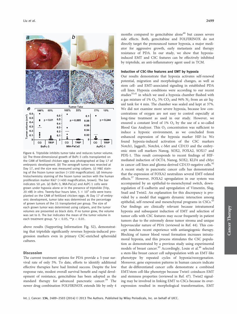
Furthermore, we evaluated the effect of triptolide ontumor engraftment (tumor take) and growth in vivo usingxenotransplantation to the CAM of fertilized chicken eggs.This model closely resembles the xenograft model in immu-nodeficient mice because the immune system of the chickenembryo is not fully developed,38 allowing the growth ofthree-dimensional tumors with a blood supply from thechicken embryo (Fig. 6a). Most importantly, the CAM con-tains no nerves, and transplantation is therefore not painfulto the embryo. The size of the resected tumors can be deter-mined using calipers (Fig. 6b), and human tumor cells growembedded in stroma formed by chicken cells (Fig. 6c). Stain-ing of a tumor section with the human-specific proliferationmarker Ki67 indicates that the large dark-brown labeled cellsare of human origin, and the smaller, nonlabeled stroma-embedded cells between the human cell islets are chickencells (Fig. 6d). BxPc-3, MIA-PaCa2 and AsPC-1 cells weretreated with hypoxia in the presence or absence of triptolidein vitro, followed by transplantation of viable cells 24 hr later(Fig. 6e). Although the percentage of tumor take of cellstreated with hypoxia alone was 100%, 77% and 100% inBxPc-3, AsPC-1 and MIA-Paca2, respectively, triptolidecotreatment strongly diminished tumor take to 50%, 15%and 33%, respectively. In addition, the tumors that started togrow on eggs demonstrated a strongly reduced tumor volumein the triptolide treatment groups. These results suggest thattriptolide targets CSCs, due to inhibition of tumor take,which is an essential feature of CSCs.
Finally, we examined the effect of triptolide on tumorgrowth under more clinically relevant conditions transplantedBxPc-3 cells first to the CAM followed by in ovo treatmentafter development of a tumor. Because of the rapid three-dimensional growth of xenograft tumors (Figs. 7a–7d), theblood supply by vessels was limited, as we detected by stain-ing of the tumor tissue with the human markers for cytoker-atin 19 and the Ki67, which detects a high proliferation rate(Fig. 7e). However, the human tissue was supplied by only
Can
cer
The
rapy
Liu et al. 2495
Int. J. Cancer: 134, 2489–2503 (2014) VC 2013 The Authors. Published by Wiley Periodicals, Inc. on behalf of UICC.

Figure 3. NF-jB is involved in hypoxia-induced progression. (a) Cells were incubated under normoxic (N) or hypoxic (H) conditions, followed
by isolation of nuclear protein extracts 24 hr later. DNA binding was analyzed by EMSA using a biotin-labeled oligonucleotide probe for the
NF-jB promoter consensus sequence. Specific (NF-jB) and unspecific shifts (UNSP) are marked. Competition with a tenfold excess of unla-
beled oligonucleotide (103) served as a control for specificity of binding. (b) Cells were transfected with siRNA oligonucleotides encoding a
nonsense (NS) sequence or a specific sequence for inhibition of c-Rel (sicRel). Three days later, proteins were prepared, and the expression
of c-Rel was evaluated by Western blot analysis. b-Actin served as a control for equal loading. The bar indicates 50 mm. (c) Three days after
transfection with nonsense or c-Rel siRNA, the confluent cell layers were scratched, and then, they were cultured under hypoxic conditions.
Closure of the wounded region was monitored 6 and 24 hr after scratching by microscopy and documented by photographs. The bar indi-
cates 100 mm. (d) Likewise, the expression of EMT-related proteins Slug and Vimentin was detected by double immunofluorescence stain-
ing of cells exposed to hypoxia for 48 hr. DAPI counterstaining was used for detection of nuclei. Mouse anti-CD44 mAb/Alexa Fluor 594 IgG
(red) and rabbit polyclonal anti-CA IX/Alexa Fluor 488 IgG (green) were used as secondary antibodies. Tissue sections were analyzed under
3400 magnification using a Leica DMRB fluorescence microscope. The bar indicates 25 mm. (e) Western blot analysis of Twist2 expression
was performed using protein extracts derived from siRNA-transfected cells cultured under normoxia or hypoxia for 3 days.
Can
cer
The
rapy

one chicken cell-derived blood vessel at the margin of thetumor, as detected by DAPI staining (Fig. 7f). As a conse-quence, hypoxia occurred in the tissue, as concluded from a
positive staining of control tumor tissue with the hypoxiamarker CA IX, which was accompanied by enhanced expres-sion of the CSC marker c-Met, the proliferation marker Ki67and the EMT marker Vimentin along with basal expressionof E-cadherin and the cleaved fragment of active caspase-3(Fig. 7i). In contrast, in ovo treatment with triptolide pre-vented the expression of CA IX, c-Met, Ki67 and Vimentinand enhanced the expression of E-cadherin and the cleavedfragment of active caspase-3, as shown by representativestainings and a diagram, which shows the number of averagepositive cells. These expression data were confirmed by themeasurement of tumor volume, which was 0.42 cm3 in thecontrol group but 0.079 cm3 in the triptolide group (Figs.7a–7d and 7g). In addition, we also examined invasion andmetastasis of human tumor cells by replication of human Alusequences from genomic DNA of chicken tissue by PCR.Although five of the five examined CAM pieces adjacent tothe tumor xenograft were positive in the control groups, sug-gesting invasion, triptolide totally prevented this (Fig. 7h). Incontrast, Alu sequences were not detectable in the liver andlung of the chicken embryo (Supporting Information Fig.S1), suggesting that metastasis of the less malignant BxPc3cells did not occur within the short time of growth in fertil-ized chicken eggs. Most importantly, side effects in chickenembryos were not observed, because the weight of chickenembryos was the same after treatment and necrosis in liversections was not detected (data not shown). These resultsdemonstrate that triptolide inhibits tumor engraftment,tumor growth, invasion, expression of markers for CSCs andEMT and induces E-cadherin expression and apoptosis invivo.
Triptolide inhibits hypoxia-induced CSC marker expression
in primary spheroidal cultures
To further highlight these findings in a patient-related model,we transplanted freshly resected PDA tissue subcutaneouslyin mice, followed by serial subtransplantation. With increas-ing passage number, the xenograft tumors started to growfaster, as evident from the enhanced doubling time andlatency (Fig. 8a). After each subtransplantation up to Passage7, the morphology of frozen tumor sections was examined,and the findings demonstrated that the typical duct-likestructures and pronounced stroma of PDA were preserved(Fig. 8b). In parallel, the CSC marker c-Met was enrichedfrom 4% in the primary patient tumor to 43% in xenograftsof Passage 7. Tumor cells were isolated from xenografts(T29) and cultured in vitro as anchorage-independent sphe-roidal colonies (Fig. 8c), which led to further enrichment ofc-Met1 cells to 64% (Fig. 8d). Culture of these spheres undernormoxia or hypoxia for 48 hr in the presence or absence oftriptolide resulted in marked reduction of viable spheroids inboth triptolide groups as evaluated by microscopy (Fig. 8c).Immunohistochemistry revealed that hypoxia enhanced thepercentage of hypoxia marker CA IX from 5.2 to 32%, of c-Met1 cells from 64 to 75%, of CD1331 cells from 74 to 85%,
Figure 4. Triptolide inhibits hypoxia-induced NF-jB and migration. (a)
The chemical structure of the diterpenoid triepoxide triptolide. (b)
Cells were incubated under hypoxic conditions alone (CO) or com-
bined with triptolide (Trip, 20 nM). Nuclear protein extracts were har-
vested 24 hr later, and DNA binding was analyzed by EMSA using a
biotin-labeled oligonucleotide probe for the NF-jB promoter consen-
sus sequence. Specific (NF-jB) and unspecific shifts (UNSP) are
marked. Competition with a tenfold excess of unlabeled oligonucleo-
tide (103) served as a control for specificity of binding. (c) Cells were
cultured under normoxia (N), hypoxia (H) or hypoxia plus triptolide
(H1Trip) for 24 hr. Protein extracts were isolated, and the expression
of HIF-1a, c-Rel, Rel-A and Twist2 was examined by Western blot
analysis. (d) Cells were grown to 90% confluence, followed by
scratching and incubation under hypoxia (H) or hypoxia plus triptolide
(H1Trip). Additionally, migration was examined as described in Figure
3c. The bar indicates 100 mm. (e) MTT assay of malignant (BxPc-3,
MIA-PaCa2 and AsPC-1) and nonmalignant (pancreatic ductal cells
and MSC) cells grown under normoxia or hypoxia in the presence or
absence of triptolide (20 nM) at the time points indicated.
Can
cer
The
rapy
Liu et al. 2497
Int. J. Cancer: 134, 2489–2503 (2014) VC 2013 The Authors. Published by Wiley Periodicals, Inc. on behalf of UICC.

of Ki671 cells from 23 to 36%, of caspase-31 cells from 9 to20% and of Vimentin1 cells from 85 to 91% along withdownregulation the percentage of E-cadherin1 cells from 16
to 10%. Similar experiments with two primary spheroidal cul-tures derived from two different patient tumors xenograftedto mice (T18 and T30) were performed and confirm the
Figure 5. Triptolide downregulates cancer stem cell features. (a) The colony-forming capacity of cells grown for 24 hr under hypoxia alone or in
the presence of triptolide (20 nM) was analyzed as described in Figure 1a. (b) The ALDH1 activity of cells treated as described above was meas-
ured as described in Figure 1b. (c) Spheroid formation was detected in MIA-PaCa2 and AsPC-1 cells treated as described above. The number of
second-generation spheroids was quantified 3 days after plating by counting viable spheroids per well. Photographs of second-generation sphe-
roids (left) and evaluation of the percentage of first- and second-generation spheroids (right) are shown. The bar indicates 100 mm. (d) Binding of
proteins to antibodies spotted on the membrane of a Human Pluripotent Stem Cell Array was detected in BxPc-3 cells treated as described above
and in Figure 1d. (e) Notch1, Jagged1, SOX2 and Nanog protein expression was detected by Western blot analysis. *p < 0.05, **p < 0.01.
Can
cer
The
rapy
2498 Triptolide Inhibits EMT and Stem-Like Features
Int. J. Cancer: 134, 2489–2503 (2014) VC 2013 The Authors. Published by Wiley Periodicals, Inc. on behalf of UICC.
above results (Supporting Information Fig. S2), demonstrat-ing that triptolide significantly reverses hypoxia-induced pro-gression markers even in primary CSC-enriched spheroidalcultures.
DiscussionThe current treatment options for PDA provide a 5-year sur-vival rate of only 5%. To date, efforts to identify additionaleffective therapies have had limited success. Despite the lowresponse rate, modest overall survival benefit and rapid devel-opment of resistance, gemcitabine has been adopted as thestandard therapy for advanced pancreatic cancer.39 Thenewer drug combination FOLFIRINOX extends life by only 4
months compared to gemcitabine alone40 but causes severeside effects. Both, gemcitabine and FOLFIRINOX do notdirectly target the pronounced tumor hypoxia, a major medi-ator for aggressive growth, early metastasis and therapyresistance of PDA. In our study, we show that hypoxia-induced EMT and CSC features can be effectively inhibitedby triptolide, an anti-inflammatory agent used in TCM.
Induction of CSC-like features and EMT by hypoxia
Our results demonstrate that hypoxia activates self-renewalpotential, migration and morphological changes, as well asstem cell- and EMT-associated signaling in established PDAcell lines. Hypoxia conditions were according to our recentstudies35,41 in which we used a hypoxia chamber flushed witha gas mixture of 1% O2, 5% CO2 and 94% N2 from an air liq-uid tank for 4 min. The chamber was sealed and kept at 37%.We did not examine more severe hypoxia, because low con-centrations of oxygen are not easy to control especially atlong-time treatment as used in our study. However, weensured a constant level of 1% O2 by the use of a so-calledBlood Gas Analyzer. This O2 concentration was sufficient toinduce a hypoxic environment, as we concluded fromenhanced expression of the hypoxia marker HIF-1a Wefound hypoxia-induced activation of the CSC markersNotch1, Jagged1, Notch4, c-Met and CD133 and the embry-onic stem cell markers Nanog, SOX2, FOXA2, SOX17 andPDX-1. This result corresponds to recent findings of HIF-mediated induction of OCT4, Nanog, SOX2, KLF4 and cMycin cancer cell lines and glioma-derived CD133-negative cells.42
A recent study in pancreatic cancer cells has demonstratedthat the expression of FOXA2 neutralizes several EMT-relatedeffects.37 However, FOXA2 upregulation in our system wasaccompanied by an epithelial-to-mesenchymal switch, down-regulation of E-cadherin and upregulation of Vimentin, Slug,Snail and Twist2. An explanation for this discrepancy is pro-vided by a model that suggests dynamic interaction amongepithelial, self-renewal and mesenchymal programs in CSCs.43
Our findings are clinically relevant because intratumoralhypoxia and subsequent induction of EMT and selection oftumor cells with CSC features may occur frequently in patienttumors due to the extremely dense tumor stroma and uniquehypovascular nature of PDA (reviewed in Ref. 44). This con-cept matches recent experience with antiangiogenic therapy.Blocking of tumor blood vessel formation increases intratu-moral hypoxia, and this process stimulates the CSC popula-tion as demonstrated by a previous study using experimentalmodels of breast cancer.45 Accordingly, Louie et al.46 selecteda stem-like breast cancer cell subpopulation with an EMT-likephenotype by repeated cycles of hypoxia/reoxygenation.Moreover, gene expression patterns in human cancers indicatethat de-differentiated cancer cells demonstrate a combinedEMT/stem cell-like phenotype because Twist1 coinduces EMTand stemness properties (reviewed in Ref. 47). Twist2 signal-ing may be involved in linking EMT to CSCs because its over-expression resulted in morphological transformation, EMT
Figure 6. Triptolide inhibits tumor take and reduces tumor volume.
(a) The three-dimensional growth of BxPc-3 cells transplanted on
the CAM of fertilized chicken eggs was photographed at Day 17 of
embryonic development. (b) The xenograft tumor was resected at
Day 17, and the size was measured using calipers. (c) H&E stain-
ing of the frozen tumor section (3100 magnification). (d) Immuno-
histochemistry staining of the frozen tumor section with the human
proliferation marker Ki67 (3400 magnification, brown). The bar
indicates 50 lm. (e) BxPc-3, MIA-PaCa2 and AsPC-1 cells were
grown under hypoxia alone or in the presence of triptolide (Trip,
20 nM) in vitro. Twenty-four hours later, 5 3 105 cells were trans-
planted on the CAM of fertilized chicken eggs. At Day 17 of embry-
onic development, tumor take was determined as the percentage
of grown tumors of the 15 transplanted per group. The size of
each grown tumor was determined using calipers, and the tumor
volumes are presented as black dots. If no tumor grew, the volume
was set to 0. The bar indicates the mean of the tumor volume in
each treatment group. *p < 0.05, **p < 0.01.
Can
cer
The
rapy
Liu et al. 2499
Int. J. Cancer: 134, 2489–2503 (2014) VC 2013 The Authors. Published by Wiley Periodicals, Inc. on behalf of UICC.

Figure 7. Triptolide inhibits tumor growth, invasion, CSC-like feature and EMT in vivo. Untreated BxPc-3 cells were transplanted on the CAM
of fertilized chicken eggs at Day 9 of embryonic development followed by in ovo treatment as described in the “Material and Methods” sec-
tion at Days 12 and 15 with triptolide (1Trip, 50 nM) or PBS (CO). At Day 17 of embryonic development, the tumors of (a and b) untreated
or (c and d) triptolide-treated eggs were photographed before and after resection. At the right lower edge of the eggs, the filter papers to
which PBS or triptolide were dropped are visible. The bar indicates 0.25 cm. (e) Double immunofluorescence staining of a frozen tumor sec-
tion (3400 magnification) with the human markers for cytokeratin 19 (red) and Ki67 for proliferation (green). The bar indicates 50 lm. (f)
Nuclei of the tumor section were stained with DAPI (blue). The arrows mark a blood vessel formed by chicken cells, which are not positive
for the human markers cytokeratin 19 and Ki67. (g) Diagram in which tumor volumes of all examined xenografts and the means are shown.
(h) Tissue from the CAM surrounding the plastic ring in which the tumor xenografts grow were excised from five eggs per group, followed
by isolation of genomic DNA. Human Alu sequences were amplified using 5 ng genomic DNA as template. Double-distilled water served as
negative control (Neg.), and genomic DNA isolated from tumor xenografts served as positive control (Tumor). The DNA marker is shown in
the first lane. (i) Frozen tumor sections were analyzed by immunohistochemistry for the expression of CA IX, c-Met, Ki67, cleaved fragment
of active caspase-3, E-cadherin and Vimentin. Representative photographs for each group under 3400 magnification are shown. The arrows
mark positive cells, which appear red to dark-red. The bar indicates 50 lm. The number of positive cells was quantified in ten vision fields
under 3400 magnification, and the means 6 SD are shown. *p < 0.05, **p < 0.01.
Can
cer
The
rapy

Figure 8. Triptolide prevents growth of patient-selected and CSC-enriched spheroids. (a) Tumor mush isolated from a surgical nondiagnostic
specimen (T29) from a patient with PDA was transplanted subcutaneously in the flanks of immunodeficient 6-week-old NMRI (nu/nu)
female mice (P1), followed by subtransplantation (P2 and P3) until a stably growing xenograft line developed after Passage 3. The time
until a first tumor was visible after transplantation (Latency) and the tumor doubling times are shown. (b) The morphology of the primary
patient tumor and tissue thereof transplanted to mice (xenograft tumors) in Passages 1, 3 and 7 (P1, P3 and P7) was detected by Tri-
chrome staining of frozen tissue, followed by microscopy. Likewise, the tissue was stained using a c-Met antibody, and the percentage of
positive cells (brown) was analyzed by counting ten vision fields under 3400 magnification. The bar indicates 50 mm. (c) Anchorage-
independent spheroidal cultures were established from xenografts and cultivated under normoxia (N) or hypoxia (H) alone or combined
with triptolide (Trip, 20 ng) for 48 hr. Photographs were then taken. The bar indicates 50 mm. (d) Alternatively, spheroidal cultures treated
as described above were cytospinned to glass slides, and the expression of hypoxia marker (CA IX), tumor stem cell markers (c-Met and
CD133), cell proliferation (Ki67), apoptosis marker (caspase-3) and EMT-related markers (E-cadherin and Vimentin) was examined by immu-
nohistochemistry. Representative pictures of positive-labeled cells (red, arrow) under normoxia are shown. The bar indicates 20 mm. The
number of positive cells was quantified in ten vision fields under 3400 magnification, and the means 6 SD are shown. **p < 0.01.
Can
cer
The
rapy
Can
cer
The
rapy
Liu et al. 2501
Int. J. Cancer: 134, 2489–2503 (2014) VC 2013 The Authors. Published by Wiley Periodicals, Inc. on behalf of UICC.

signaling, colony-forming abilities and an enhanced numberof stem-like cells in a breast cancer model.48
NF-jB as a mediator of hypoxia-induced CSC and EMT
features
Our data show hypoxia-mediated upregulation of Twist2 inBxPc-3 and AsPC-1 cells, whereas MIA-PaCa2 cells exhibitconstitutively enhanced Twist2 expression under normoxia.siRNA-mediated inhibition of NF-jB c-Rel downregulatedthe expression of hypoxia-induced Twist2, inhibited migra-tory activity and downregulated the expression of Slug andVimentin. Correspondingly, pancreatic CSC-enriched cellswere resensitized to cancer therapy by siRNA-mediateddownregulation of c-Rel as shown in one of our earlier stud-ies.29 Therefore, we assume that NF-jB may be a masterinducer of both EMT and CSC features. Our suggestion isunderscored in the study by Jiang et al.49 These authorsdescribed that during neoplastic transformation, human kera-tinocyte cells undergo EMT and then acquire a malignantCSC-like phenotype. Longer times for transformationincreased activation of IKKb, IjBa and NF-jB Rel-A andenhanced the expression of Snail; stem cell markers were alsodetected.49 Inhibition of Rel-A blocked EMT, acquisition of aCSC-like phenotype, and neoplastic transformation. Likewise,in the established gemcitabine-resistant PDA cell lineSW1990, siRNA-mediated knockdown of Rel-A inhibited theability of gemcitabine to increase the proportion of CSC andEMT markers.
Therapeutic potential of triptolide
The purity of triptolide that we used was �98% (HPLC).This high purity makes it unlikely that some of the observedeffects are due to impurities of the substance. Although trip-tolide is the main active ingredient in extracts from the Chi-nese herb Tripterygium wilfordii Hook f, 124 othersesquiterpene derivatives have been reported to be present inTripterygium, as extracts were often prepared in differentways (e.g., with different solvents), and they have differentconstituents and biological effects.31 However, this concerndoes not apply to our study as we used highly purified tripto-lide. We have identified triptolide as an effective inhibitor ofhypoxia-induced transcriptional signaling that blocks HIF-1a
expression and NF-jB binding activity, as well as downregu-lates c-Rel, Rel-A and Twist2 expression, consistent with thefinding that the twist gene promoters are under the controlof NF-jB and contain functional jB binding elements(reviewed in Ref. 50). Additionally, we found that triptolidecaused impairment of the migration, colony- and spheroid-forming ability and ALDH1 activity and repression ofhypoxia-induced stem cell signaling. We did not observecytotoxicity by triptolide 24 hr after incubation. Cytotoxicityoccurred during the first 48 hr after incubation with thehighest levels in CSC-enriched cell lines. In contrast, tripto-lide was less toxic in gemcitabine-sensitive BxPc-3 cells ornonmalignant MSCs and immortalized pancreatic ductal
cells. Indeed, recent data demonstrate that triptolide generallytakes �24 hr or more to induce apoptosis or cytotoxicity incancer cells, much longer than the time of several hours thatother cytotoxic agents require.31 Notably, although highlymalignant cells do not die within 24 hr, triptolide potentlyinhibited hypoxia-mediated EMT and CSC signaling 24 hrafter triptolide treatment. This assumption is underscored byour finding of the completely blocked in vivo tumor take ofthe highly malignant MIA-PaCa2 and AsPC-1 cells 24 hrafter treatment with triptolide. In contrast, the tumor take ofthe less aggressive BxPc-3 cells was only partially blocked.Therefore, it is tempting to speculate that triptolide predomi-nantly targets CSCs, while leaving more differentiated tumorcells or nonmalignant cells unaffected. Such a model wouldexplain why cytotoxic effects occurred rather late and whytriptolide can circumvent tumor drug resistance.31 The ideaof specific targeting of CSC by triptolide is emphasized bythe finding that no overt signs of toxicity occurred whenmice received the triptolide derivative minnelide for 385 daysat a concentration of 0.43 mg/kg.51 However, minnelide andtriptolide significantly decreased tumor weight, tumor volumeand tumor spread in orthotopic MIA-PaCa2 xenografts atthe same concentration, and tumors did not recur after ter-mination of treatment. In contrast, the first-line chemothera-peutic agent in pancreatic cancer, gemcitabine, at aconcentration of 100 mg/kg bodyweight, did not lead to asignificant decrease in tumor size versus the control.51 Thispromising in vivo effect of triptolide in mice was confirmedin a small clinical study where 45 leukemia patients received30–40 lg triptolide per kilogram of body weight daily inChina (reviewed in Ref. 31). Complete remission wasobserved in 18 patients, and partial remission was observedin six patients. The efficacy of triptolide was superior to thatof adriamycin and aclacinomycin. Toxicities were manifestedas phlebitis and gastrointestinal abnormalities. No heart, liverand kidney toxicities were observed (reviewed in Ref. 31).
Regarding inhibition of metastasis by triptolide in mousemodels, we believe that downregulated expression of Notch1and its ligand Jagged1 may be involved, as demonstrated inour study. This would further support Notch1’s ability toinhibit invasion by inactivation of NF-jB, VEGF and MMP-9in pancreatic cancer,52 implying a positive feedback loopbetween hypoxia-induced transcriptional signaling and targetgene expression that leads to EMT signaling and CSC features.
Undoubtedly, further studies will be needed to determinethe precise mechanisms by which triptolide potently elimi-nates highly malignant pancreatic cancer cells while sparingmore differentiated cells. However, our data provide strongevidence for effective inhibition of hypoxia-induced EMT andCSC signaling by triptolide in pancreatic cancer.
AcknowledgementsThe authors thank Dr. N. Giese for organization of the tissueservice at their clinic and Sonja Bauer for technicalassistance.
Can
cer
The
rapy
2502 Triptolide Inhibits EMT and Stem-Like Features
Int. J. Cancer: 134, 2489–2503 (2014) VC 2013 The Authors. Published by Wiley Periodicals, Inc. on behalf of UICC.

Author ContributionsI.H., L.L. and A.S.: concept and design; L.L., S.L., N.B.,E.A., C.N., J.M. and J.G.: development of methodology;L.L., S.L., N.B., E.A., C.N., J.M. and J.G.: acquisition of
data; L.L. and I.H.: analysis and interpretation of data; I.H.and L.L.: writing, review and/or revision of the manuscriptand J.W. and P.S.: administrative, technical or materialsupport.
References
1. Gukovskaya AS, Pandol SJ. Cell death pathwaysin pancreatitis and pancreatic cancer. Pancreatol-ogy 2004;4:567–86.
2. Brown JM, Giaccia AJ. The unique physiology ofsolid tumors: opportunities (and problems) forcancer therapy. Cancer Res 1998;58:1408–16.
3. Hoffmann AC, Mori R, Vallbohmer D, et al.High expression of HIF1a is a predictor of clini-cal outcome in patients with pancreatic ductaladenocarcinomas and correlated to PDGFA,VEGF, and bFGF. Neoplasia 2008;10:674–9.
4. Chang Q, Jurisica I, Do T, et al. Hypoxia predictsaggressive growth and spontaneous metastasisformation from orthotopically grown primaryxenografts of human pancreatic cancer. CancerRes 2011;71:3110–20.
5. Rasheed ZA, Kowalski J, Smith BD, et al. Concisereview: emerging concepts in clinical targeting ofcancer stem cells. Stem Cells 2011;29:883–7.
6. Lapidot T, Sirard C, Vormoor J, et al. A cell initiat-ing human acute myeloid leukaemia after transplan-tation into SCID mice. Nature 1994;367:645–8.
7. Al-Hajj M, Wicha MS, Benito-Hernandez A,et al. Prospective identification of tumorigenicbreast cancer cells. Proc Natl Acad Sci USA 2003;100:3983–8.
8. Singh SK, Hawkins C, Clarke ID, et al. Identifica-tion of human brain tumour initiating cells.Nature 2004;432:396–401.
9. Simeone DM. Pancreatic cancer stem cells: impli-cations for the treatment of pancreatic cancer.Clin Cancer Res 2008;14:5646–8.
10. Li C, Heidt DG, Dalerba P, et al. Identification ofpancreatic cancer stem cells. Cancer Res 2007;67:1030–7.
11. Hermann PC, Huber SL, Herrler T, et al. Distinctpopulations of cancer stem cells determine tumorgrowth and metastatic activity in human pancre-atic cancer. Cell Stem Cell 2007;1:313–23.
12. Li C, Wu JJ, Hynes M, et al. c-Met is a marker ofpancreatic cancer stem cells and therapeutic tar-get. Gastroenterology 2011;141:2218–27.
13. Singh A, Settleman J. EMT, cancer stem cells anddrug resistance: an emerging axis of evil in thewar on cancer. Oncogene 2010;29:4741–51.
14. Higgins DF, Kimura K, Bernhardt WM, et al.Hypoxia promotes fibrogenesis in vivo via HIF-1stimulation of epithelial-to-mesenchymal transi-tion. J Clin Invest 2007;117:3810–20.
15. Thiery JP. Epithelial–mesenchymal transitions indevelopment and pathologies. Curr Opin Cell Biol2003;15:740–6.
16. Huber MA, Beug H, Wirth T. Epithelial–mesen-chymal transition: NF-jB takes center stage. CellCycle 2004;3:1477–80.
17. Ledford H. Cancer theory faces doubts. Nature2011;472:273.
18. Peinado H, Olmeda D, Cano A. Snail, Zeb andbHLH factors in tumour progression: an allianceagainst the epithelial phenotype? Nat Rev Cancer2007;7:415–28.
19. Thiery JP. Epithelial–mesenchymal transitions intumour progression. Nat Rev Cancer 2002;2:442–54.
20. Tsai JH, Donaher JL, Murphy DA, et al. Spatio-temporal regulation of epithelial–mesenchymaltransition is essential for squamous cell carci-noma metastasis. Cancer Cell 2012;22:725–36.
21. Mani SA, Guo W, Liao MJ, et al. The epithelial–mesenchymal transition generates cells with prop-erties of stem cells. Cell 2008;133:704–15.
22. Koong AC, Chen EY, Mivechi NF, et al. Hypoxicactivation of nuclear factor-jB is mediated by aRas and Raf signaling pathway and does notinvolve MAP kinase (ERK1 or ERK2). Cancer Res1994;54:5273–9.
23. Baeuerle PA, Baltimore D. NF-jB: ten years after.Cell 1996;87:13–20.
24. Pikarsky E, Porat RM, Stein I, et al. NF-jB func-tions as a tumour promoter in inflammation-associated cancer. Nature 2004;431:461–6.
25. Karin M, Greten FR. NF-jB: linking inflamma-tion and immunity to cancer development andprogression. Nat Rev Immunol 2005;5:749–59.
26. Maier HJ, Schmidt-Strassburger U, Huber MA,et al. NF-jB promotes epithelial–mesenchymaltransition, migration and invasion of pancreaticcarcinoma cells. Cancer Lett 2010;295:214–28.
27. Cao YX, Luo JL, Karin M. IjB kinase a kinaseactivity is required for self-renewal of ErbB2/Her2-transformed mammary tumor-initiatingcells. Proc Natl Acad Sci USA 2007;104:15852–7.
28. Korkaya H, Liu S, Wicha MS. Breast cancer stemcells, cytokine networks, and the tumor microen-vironment. J Clin Invest 2011;121:3804–9.
29. Kallifatidis G, Rausch V, Baumann B, et al. Sul-foraphane targets pancreatic tumour-initiatingcells by NF-jB-induced antiapoptotic signalling.Gut 2009;58:949–63.
30. Brinker AM, Ma J, Lipsky PE, et al. Medicinalchemistry and pharmacology of genus Tripterygium(Celastraceae). Phytochemistry 2007;68:732–66.
31. Zhou ZL, Yang YX, Ding J, et al. Triptolide:structural modifications, structure–activity rela-tionships, bioactivities, clinical development andmechanisms. Nat Prod Rep 2012;29:457–75.
32. Lonardo E, Hermann PC, Mueller MT, et al.Nodal/Activin signaling drives self-renewal andtumorigenicity of pancreatic cancer stem cellsand provides a target for combined drug therapy.Cell Stem Cell 2011;9:433–46.
33. Klapproth K, Sander S, Marinkovic D, et al. TheIKK2/NF-{j}B pathway suppresses MYC-inducedlymphomagenesis. Blood 2009;114:2448–58.
34. Balke M, Neumann A, Szuhai K, et al. A short-term in vivo model for giant cell tumor of bone.BMC Cancer 2011;11:241.
35. Salnikov AV, Liu L, Platen M, et al. Hypoxiainduces EMT in low and highly aggressive pan-creatic tumor cells but only cells with cancerstem cell characteristics acquire pronouncedmigratory potential. PLoS One 2012;7:e46391.
36. Zhou W, Kallifatidis G, Baumann B, et al. Dietarypolyphenol quercetin targets pancreatic cancerstem cells. Int J Oncol 2010;37:551–61.
37. Song Y, Washington MK, Crawford HC. Loss ofFOXA1/2 is essential for the epithelial-to-
mesenchymal transition in pancreatic cancer.Cancer Res 2010;70:2115–25.
38. Janse EM, Jeurissen SH. Ontogeny and functionof two non-lymphoid cell populations in thechicken embryo. Immunobiology 1991;182:472–81.
39. Oberstein PE, Saif MW. First-line treatment foradvanced pancreatic cancer. Highlights from the2011 ASCO Gastrointestinal Cancers Symposium,San Francisco, CA, USA, January 20–22, 2011.JOP 2011;12:96–100.
40. Conroy T, Desseigne F, Ychou M, et al. FOLFIRI-NOX versus gemcitabine for metastatic pancreaticcancer. N Engl J Med 2011;364:1817–25.
41. Rausch V, Liu L, Apel A, et al. Autophagy medi-ates survival of pancreatic tumour-initiating cellsin a hypoxic microenvironment. J Pathol 2012;227:325–35.
42. Mathieu J, Zhang Z, Zhou W, et al. HIF induceshuman embryonic stem cell markers in cancercells. Cancer Res 2011;71:4640–52.
43. Celia-Terrassa T, Meca-Cortes O, Mateo F, et al.Epithelial–mesenchymal transition can suppressmajor attributes of human epithelial tumor-initiating cells. J Clin Invest 2012;122:1849–68.
44. Tuveson DA, Neoptolemos JP. Understandingmetastasis in pancreatic cancer: a call for newclinical approaches. Cell 2012;148:21–3.
45. Conley SJ, Gheordunescu E, Kakarala P, et al.Antiangiogenic agents increase breast cancer stemcells via the generation of tumor hypoxia. ProcNatl Acad Sci USA 2012;109:2784–9.
46. Louie E, Nik S, Chen JS, et al. Identification of astem-like cell population by exposing metastaticbreast cancer cell lines to repetitive cycles ofhypoxia and reoxygenation. Breast Cancer Res2010;12:R94.
47. Brabletz T. EMT and MET in metastasis: whereare the cancer stem cells? Cancer Cell 2012;22:699–701.
48. Fang X, Cai Y, Liu J, et al. Twist2 contributesto breast cancer progression by promoting anepithelial–mesenchymal transition and cancerstem-like cell self-renewal. Oncogene 2011;30:4707–20.
49. Jiang R, Li Y, Xu Y, et al. EMT and CSC-likeproperties mediated by the IKKb/IjBa/RelA sig-nal pathway via the transcriptional regulator,Snail, are involved in the arsenite-induced neo-plastic transformation of human keratinocytes.Arch Toxicol 2013;87:991–1000.
50. Sosic D, Olson EN. A new twist on twist—modu-lation of the NF-jB pathway. Cell Cycle 2003;2:76–8.
51. Chugh R, Sangwan V, Patil SP, et al. A preclinicalevaluation of Minnelide as a therapeutic agentagainst pancreatic cancer. Sci Transl Med 2012;4:156ra39.
52. Wang Z, Banerjee S, Li Y, et al. Down-regulationof notch-1 inhibits invasion by inactivation ofnuclear factor-jB, vascular endothelial growthfactor, and matrix metalloproteinase-9 in pancre-atic cancer cells. Cancer Res 2006;66:2778–84.
Can
cer
The
rapy
Liu et al. 2503
Int. J. Cancer: 134, 2489–2503 (2014) VC 2013 The Authors. Published by Wiley Periodicals, Inc. on behalf of UICC.