Downregulation of the DNA repair enzyme apurinic ... on this finding, we have investigated the effec...
30
Instructions for use Title Downregulation of the DNA repair enzyme apurinic/apyrimidinic endonuclease 1 stimulates transforming growth factor-β1 production and promotes actin rearrangement. Author(s) Sakai, Yuri; Yamamori, Tohru; Yasui, Hironobu; Inanami, Osamu Citation Biochemical and Biophysical Research Communications, 461(1): 35-41 Issue Date 2015-05-22 Doc URL http://hdl.handle.net/2115/61914 Rights © 2015. This manuscript version is made available under the CC-BY-NC-ND 4.0 license http://creativecommons.org/licenses/by-nc-nd/4.0/ Rights(URL) http://creativecommons.org/licenses/by-nc-nd/4.0/ Type article (author version) File Information manuscript.pdf Hokkaido University Collection of Scholarly and Academic Papers : HUSCAP
Transcript of Downregulation of the DNA repair enzyme apurinic ... on this finding, we have investigated the effec...
Instructions for use
Title Downregulation of the DNA repair enzyme apurinic/apyrimidinic endonuclease 1 stimulates transforming growthfactor-β1 production and promotes actin rearrangement.
Author(s) Sakai, Yuri; Yamamori, Tohru; Yasui, Hironobu; Inanami, Osamu
Citation Biochemical and Biophysical Research Communications, 461(1): 35-41
Issue Date 2015-05-22
Doc URL http://hdl.handle.net/2115/61914
Rights © 2015. This manuscript version is made available under the CC-BY-NC-ND 4.0 licensehttp://creativecommons.org/licenses/by-nc-nd/4.0/
Rights(URL) http://creativecommons.org/licenses/by-nc-nd/4.0/
Type article (author version)
File Information manuscript.pdf
Hokkaido University Collection of Scholarly and Academic Papers : HUSCAP
1
Downregulation of the DNA repair enzyme apurinic/apyrimidinic endonuclease 11
stimulates transforming growth factor-1 production and promotes actin 2
rearrangement3
4
Yuri Sakaia, Tohru Yamamori
a*, Hironobu Yasui
a, Osamu Inanami
a*5
6
aLaboratory of Radiation Biology, Department of Environmental Veterinary Sciences, 7
Graduate School of Veterinary Medicine, Hokkaido University, Sapporo, Japan8
9
E-mail addresses:10
15
*Corresponding authors: Dr. Tohru Yamamori and Prof. Osamu Inanami16
Address: Kita 18, Nishi 9, Kita-ku, Sapporo, Hokkaido 060-0818, Japan17
Tel: +81-11-706-5236 (TY), +81-11-706-5235 (OI) 18
Fax: +81-11-706-7373 19
E-mail: [email protected] (TY), [email protected] (OI) 20
21
Abbreviations:22
AP-1, activator protein 1; APE1, apurinic/apyrimidinic endonuclease 1; B2M,23
β-2-microglobulin; COL1A1, collagen type I α1; COL3A1, collagen type III α1; COL4A1, 24
collagen type IV α1; DMEM, Dulbecco’s modified Eagle’s medium; ECM, extracellular 25

2
matrix; EMT, epithelial-mesenchymal transition; FBS, fetal bovine serum; FN1, fibronectin 1
1; HIF-1α, hypoxia-inducible factor-1α; LAMC1, laminin γ1; NF-κB, nuclear factor-κB; PBS,2
phosphate buffered saline; PMS, phenazine methosulfate; qRT-PCR, quantitative reverse 3
transcription polymerase chain reaction; ROS, reactive oxygen species; TGF, transforming 4
growth factor; TGFB1, TGF-β1; TRITC, tetramethylrhodamine isothiocyanate; WST-1, 5
water-soluble tetrazolium salts6
7
3
ABSTRACT1
The DNA repair enzyme apurinic/apyrimidinic endonuclease 1 (APE1) plays a central role in 2
base excision repair and functions as a reductive activator of various transcription factors. 3
Multiple other functionalities have been ascribed to APE1 in addition to these major 4
functions. A recent study showed that APE1 knockdown upregulated the expression of a set 5
of genes related to extracellular matrix (ECM) production, indicating an additional novel 6
biological role for this enzyme. Based on this finding, we have investigated the effect of 7
APE1 downregulation on ECM-related gene expression and its biological consequences. 8
Endogenous APE1 expression was downregulated in human cervical carcinoma HeLa cells9
and human lung carcinoma A549 cells using siRNA. When the expression of six10
ECM-related genes (TGFB1, LAMC1, FN1, COL1A1, COL3A1, and COL4A1) was evaluated, 11
we found that APE1 knockdown upregulated the expression of TGFB1 in both cell lines.12
APE1 downregulation promoted actin rearrangement, inducing F-actin accumulation in HeLa 13
cells and the dissipation of stress fibers in A549 cells. We also discovered that APE1 14
knockdown enhanced cellular motility in A549 cells, which was suppressed by the inhibition 15
of transforming growth factor (TGF)-1 signaling. These results suggested that APE1 16
controls the organization of actin cytoskeleton through the regulation of TGF-β1 expression,17
providing novel insights into the biological significance of APE1. 18
19

4
Keywords: APE1, TGF-β1, extracellular matrix, actin cytoskeleton, cellular motility1
5
INTRODUCTION1
Apurinic/apyrimidinic endonuclease 1 (APE1) is an homolog of Escherichia coli exonuclease 2
III and primarily functions as a DNA repair enzyme [1]. APE1 has an apurinic/apyrimidinic3
endonuclease domain at its C-terminal region that is highly conserved in various organisms. 4
In addition, mammalian APE1 has a nuclear localization signal and a redox domain in its 5
N-terminal domain. APE1 is indispensable for cellular and organismal survival, as 6
demonstrated by early embryonic lethality in APE1 knockout mice; attempts to isolate stable 7
APE1-knockout cells have also been unsuccessful [2, 3]. Mammalian APE1 is also involved 8
in the activation of transcription factors. The DNA binding affinities of various transcription9
factors, such as activator protein 1 (AP-1), nuclear factor-κB (NF-κB), and hypoxia-inducible 10
factor-1α (HIF-1α), are modulated by the redox status of the cysteine residues in their DNA 11
binding domains [4-6]. The redox activity of APE1 causes the reduction of redox-sensitive 12
cysteine residues, leading to their activation. In addition to these major functions, various 13
studies have reported the other functions of APE1, such as redox-independent transcriptional 14
activation [7-9], RNA quality control [10, 11], and telomere maintenance [12]. Therefore, 15
APE1 has a wide range of functions that potentially influence many biological processes. 16
However, the multifunctional properties of APE1 complicate the elucidation of17
specific functions regulating cellular physiology and increase the difficulty of determining 18
the corresponding mechanism. In addition, the inability to establish an APE1-deficient cell 19
6
line or APE1 knockout mouse has hampered investigations into this protein. Vascotto et al.1
recently performed a study analyzing the effect of APE1 downregulation on global gene 2
expression by microarrays using a conditional knockout system [13]. The authors reported3
significant changes in the expression of genes related to multiple cellular functions including4
lipid metabolism, cell cycle regulation, protein synthesis, and DNA repair. Notably, a set of 5
genes related to the production of the extracellular matrix (ECM) was upregulated as a result 6
of APE1 downregulation. The aforementioned APE1 functions, including cell cycle 7
regulation and DNA repair, are well characterized and closely associated with the enzymatic 8
activity of the protein. However, the involvement of APE1 in ECM production was 9
unexpected. We were intrigued by this finding; therefore, we sought to decipher the 10
mechanism by which APE1 regulates ECM-related gene expression, and determine the 11
biological consequences of this novel aspect of APE1 function.12
13
MATERIALS AND METHODS14
Reagents15
Lipofectamine®
2000, Stealth RNAiTM
siRNA Negative Control LO GC (Control siRNA), 16
and Stealth RNAiTM
siRNA targeting APE1 (ID: HSS100557 and HSS100556, hereafter17
referred to as siAPE1 #1 and siAPE1 #2, respectively) were purchased from Life 18
Technologies (Carlsbad, CA, USA). Anti-APE1 antibody, anti-actin antibody, and 19
7
horseradish peroxidase-conjugated anti-mouse and anti-goat antibodies were purchased from 1
Santa Cruz Biotechnology (Santa Cruz, CA, USA). Anti-transforming growth factor 2
(TGF)-β1 antibody (clone 9016) was obtained from R&D Systems (Minneapolis, MN, USA).3
TGF-β type I receptor kinase inhibitor LY364947 was obtained from Cayman Chemical (Ann 4
Arbor, MI, USA). Tetramethylrhodamine isothiocyanate (TRITC)-conjugated phalloidin was5
obtained from Sigma-Aldrich (St. Louis, MO, USA). Water-soluble tetrazolium salts 6
(WST-1) and 1-methoxy phenazine methosulfate (PMS) were purchased from Dojindo 7
Molecular Technologies, Inc. (Kumamoto, Japan).8
9
Cell culture and siRNA transfection10
Human cervical carcinoma HeLa cells were maintained in Dulbecco’s modified Eagle’s 11
medium (DMEM; Life Technologies) supplemented with 10% fetal bovine serum (FBS) at 12
37°C in 5% CO2. Human lung carcinoma A549 cells were maintained in RPMI 1640 medium 13
(Life Technologies) supplemented with 10% FBS at 37°C in 5% CO2. The cells were 14
transfected with APE1-targeting siRNA or control siRNA using Lipofectamine 2000. 15
Following siRNA transfection, the medium was replaced with fresh medium and the 16
transfected cells were used for subsequent analyses.17
18
Western blot analysis19
8
Cells were lysed with lysis buffer (50 mM Tris-HCl [pH 7.5], 1% (v/v) Triton X-100, 5%1
(v/v) glycerol, 5 mM EDTA, 150 mM NaCl) and centrifuged at 20,000 × g for 15 min at 4°C. 2
The supernatant was collected, and 3× Laemmli’s sample buffer (0.1875 M Tris-HCl [pH 6.8], 3
15% (v/v) -mercaptoethanol, 6% (w/v) SDS, 30% (v/v) glycerol and 0.006% (w/v) 4
bromophenol blue) was added to each supernatant. Equal amounts of protein were separated 5
by SDS-PAGE and transferred onto nitrocellulose membranes (Advantec TOYO, Tokyo, 6
Japan). The membranes were blocked with 10 mM Tris-HCl (pH 7.4), 0.1 M NaCl, and 0.1%7
(v/v) Tween-20 (TBST) supplemented with 5% (w/v) nonfat skim milk for 1 h. Subsequently, 8
the membranes were probed with specific primary antibodies diluted with TBST 9
supplemented with 5% (w/v) nonfat skim milk overnight at 4°C. The membranes were then 10
probed with horseradish peroxidase-conjugated secondary antibodies and the blots developed 11
using a Western LightningTM
Chemiluminescence Reagent Plus kit (Perkin Elmer Life 12
Sciences, Boston, MA, USA).13
14
Quantitative reverse transcription polymerase chain reaction (qRT-PCR)15
Total RNA was extracted using the Relia PrepTM
RNA Cell Miniprep System (Promega 16
Corporation, Madison, WI, USA), as per the manufacturer protocols. The RNA sample (2 g)17
was reverse-transcribed using the ReverTra Ace®
qPCR RT Master Mix (TOYOBO, Osaka, 18
Japan). The obtained cDNA was subjected to real-time PCR analysis using a LightCycler 19
9
Nano System (Roche Applied Science, Mannheim, Germany), with the samples being 1
prepared using FastStart Essential DNA Green Master mix (Roche Applied Science). The 2
sequences of the PCR primers used in this study are as follows: TGF-β1 (TGFB1), 5′-TGC 3
TGA GGC TCA AGT TAA AA-3′ and 5′-TCC GGT GAC ATC AAA AGA TA-3′; laminin γ14
(LAMC1), 5′-CTG TTG TTC CCA AGA CAA AG-3′ and 5′-TCT CCA AAG TAG CCA TCA 5
TC-3′; fibronectin 1 (FN1), 5′-ATT TCA TGT CAT CCT GTT GG-3′ and 5′-CTT TCA GTG 6
CCT CCA CTA TG-3′; collagen type I α1 (COL1A1), 5′-ATG TTC AGC TTT GTG GAC 7
CT-3′ and 5′-GTG ATT GGT GGG ATG TCT T-3′; collagen type III α1 (COL3A1), 5′-CTA 8
CTT CTC GCT CTG CTT CA-3′ and 5′-ACA TAT TTG GCA TGG TTC TG-3′; collagen 9
type IV α1 (COL4A1), 5′-ATG GAA TTG TGG AAT GTC AG-3′ and 5′-AGG CAA CTC 10
TCT CCT TTT TG-3′; andβ-2-microglobulin (B2M), 5′-TTC TGG CCT GGA GGC TAT C-3′11
and 5′-TCA GGA AAT TTG ACT TTC CAT TC-3′. The following cycle conditions were12
applied for PCR analysis: initial denaturation at 95°C for 10 min, followed by 40 cycles at 13
95°C for 20 s, 60°C for 20 s, and 72°C for 20 s. The relative mRNA level of each 14
ECM-related gene was normalized to that of B2M, which was used as the internal control. 15
16
WST-1 assay17
Cells (1,000 per well) were seeded into each well of 96-well plates containing growth 18
medium 24 h after siRNA transfection. The medium was replaced with medium 19
10
supplemented with 1% FBS at 48 h post-transfection; subsequently, 10 μL WST-1 solution 1
(3.6 μg/μL WST-1, 70 ng/μL 1-methoxy PMS in 20 mM HEPES-KOH [pH 7.4]) was added 2
to each well 48, 72, and 96 h post-transfection. The cells were incubated for 2 h at 37°C, and 3
the absorbance of each well was recorded at 450 nm using a Model 680 Microplate Reader 4
(Bio-Rad Laboratories, Hercules, CA, USA).5
6
F-actin staining7
Cells were seeded to collagen-coated glass coverslips 24 h after siRNA transfection. 8
Following incubation for 24 h or 48 h, the cells were fixed with 3.7% (w/v)9
paraformaldehyde/phosphate buffered saline (PBS) for 15 min at room temperature, and 10
subsequently permeabilized with 0.1% (v/v) Triton X-100/PBS for 5 min at room temperature.11
The cells were then incubated with 1% (v/v) TRITC-conjugated phalloidin/PBS for 30 min at 12
room temperature in the dark. The coverslips were mounted with Prolong Gold antifade 13
reagent (Life Technologies). Fluorescent microscopic analysis and image acquisition were 14
performed using an Olympus BX61 microscope (Olympus, Tokyo, Japan) equipped with a 15
CCD camera. F-actin focal accumulation was evaluated by measuring the fluorescent 16
intensities and area sizes of F-actin foci in over 200 randomly chosen cells per condition 17
using the Image J software (National Institutes of Health, Bethesda, MD, USA). F-actin focal 18
accumulation was quantified by multiplying the focal fluorescence intensity with its size. The 19
11
average F-actin focal accumulation for each condition was determined by dividing the sum of 1
F-actin focal accumulation by the cell number. Relative F-actin focal accumulation was 2
calculated by normalizing the average F-actin accumulation for each condition to that of the 3
control.4
5
Wound healing assay6
Cells (2.8 × 10⁴
cells) were seeded onto each side of an Ibidi 2-well culture-insert (Ibidi, 7
Planegg, Germany) 24 h after siRNA transfection, and incubated for 24 h.The culture-insert 8
was removed to create an area containing no cells, and the medium replaced with RPMI 9
containing 1% FBS. The gap area was photographed at 400× magnification using an 10
Olympus IX71 light microscope (Olympus, Tokyo, Japan) at 0 and 48 h after the medium 11
change. Cell migration was evaluated by counting the number of cells that migrated to the 12
gap area.13
14
Statistical analysis15
All results are displayed as mean ± standard deviation (S.D.) of at least three independent 16
experiments. Statistical analyses were performed using the Student’s t-test. p-values < 0.05 17
were considered to be statistically significant.18
19
12
RESULTS1
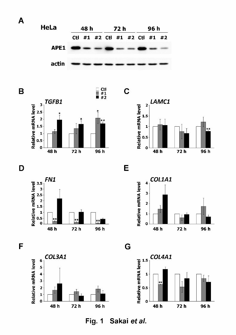
Effect of APE1 downregulation on the expression of ECM-related genes2
The effect of siRNA on APE1 protein expression was verified in human cervical carcinoma 3
HeLa cells and human lung carcinoma A549 cells at 48, 72, and 96 h after the transfection of 4
control siRNA or the two types of APE1-targeting siRNA (siAPE1 #1 and #2). There was a 5
decrease in APE1 protein expression 48 h after siRNA transfection, as seen in Figs. 1A and 6
2A; this effect was sustained for up to 96 h in both cell lines. The microarray analysis 7
performed in a previous study revealed that the conditional knockdown of APE1 modified the 8
expression of several ECM-related genes [13]. This finding was re-evaluated in our 9
experimental settings by qRT-PCR analysis in HeLa and A549 cells. Six ECM-related genes,10
TGFB1, LAMC1, FN1, COL1A1, COL3A1, and COL4A1, reported to be upregulated as a 11
result of APE1 downregulation, were selected as analysis targets [13]. TGFB1 expression in 12
HeLa cells was increased by the introduction of both siAPE1 #1 and #2 (Fig. 1B). Expression 13
of LAMC1 was downregulated 96 h after siAPE1 #2 transfection (Fig. 1C). The introduction 14
of siAPE1 #1 (and not siAPE1 #2) effected a significant decrease in FN1 expression (Fig. 15
1D), whereas siAPE1 did not cause any significant changes in the expression of either 16
COL1A1 or COL3A1 (Figs. 1E and F). COL4A1 expression was downregulated 48 h after the 17
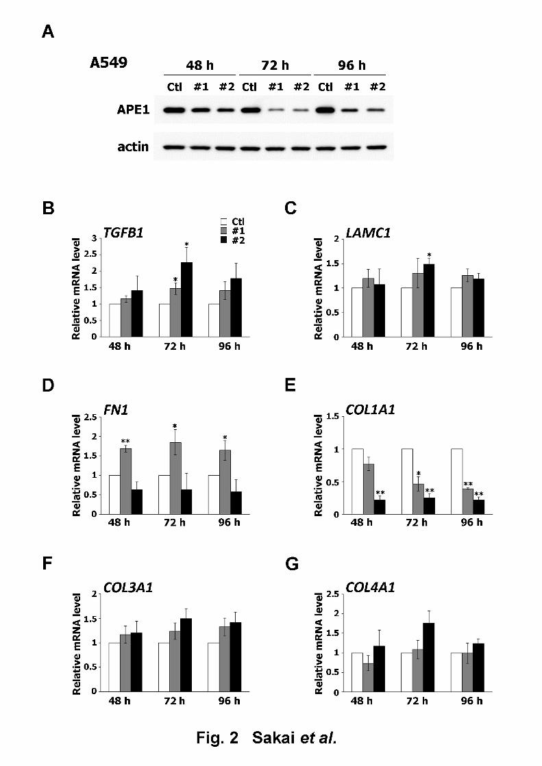
introduction of siAPE1 #1 (Fig. 1G). The same analyses conducted with A549 cells revealed 18
that the introduction of siAPE1 #1 and #2 led to an elevation in TGFB1 expression, with a 19
13
peak observed at 72 h post-transfection (Fig. 2B). On the other hand, the expression of other 1
ECM-related genes did not increase significantly, with the exception of FN1, following the 2
introduction of siAPE1 #1 (Figs. 2C–G). The level of TGFB1 expression was similarly 3
upregulated by APE1 siRNA in both cell lines, which suggested that APE1 regulates the gene 4
expression of TGF-β1.5
6
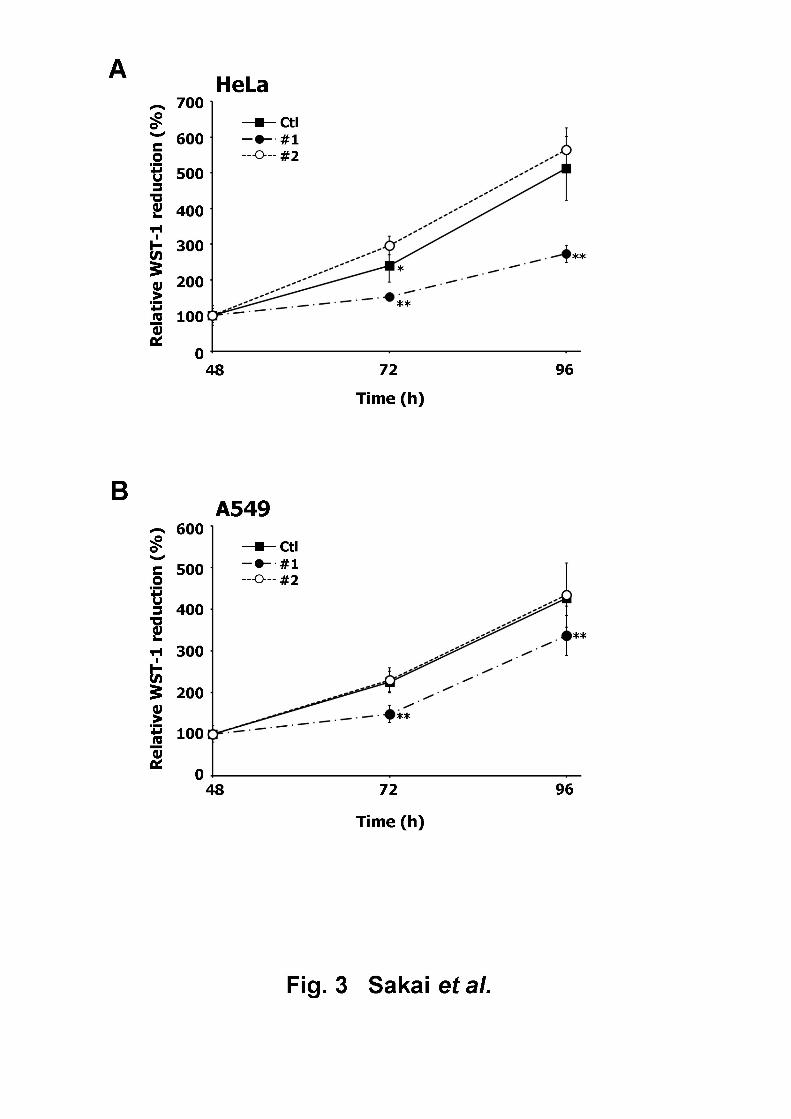
Effect of APE1 downregulation on cellular proliferation7
The effect of siRNA oligonucleotide introduction on cellular viability or proliferation was 8
examined by subjecting the transfected HeLa and A549 cells to a WST-1 assay. While neither 9
of the siRNA constructs caused cytotoxicity in HeLa cells, the cells transfected with siAPE1 10
#1 demonstrated reduced proliferation, compared to those transfected with control siRNA and 11
siAPE1 #2 (Fig. 3A). This suppressive effect of siAPE1 #1 on cell proliferation was observed 12
to increase in a time-dependent manner. Similarly, siAPE #1 (and not siAPE #2 or the control 13
siRNA) was observed to attenuate the proliferation of A549 cells (Fig. 3B). These results14
implied that despite the near-equivalent efficacy of siAPE1 #1 and #2 on APE1 knockdown, 15
the resulting biological effects were not identical. As the induction of TGF-β1 was more 16
potent in cells (of both cell lines) transfected with siAPE1 #2 than those transfected with 17
siAPE1 #1 (Figs. 1B and 2B), this disparity in cell proliferation is unlikely to be related to 18
TGF-β1-dependent inhibition of cell growth [14], but rather more likely to be due to a 19
14
non-specific side effect of siAPE1 #1 [15]. Therefore, we used siAPE1 #2 but not #1 to avoid 1
unwanted effects in the subsequent experiments.2
3
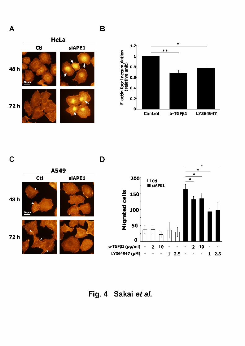
Effect of APE1 downregulation on actin cytoskeleton organization4
TGF-β has been associated with cytoskeletal rearrangement [16]; therefore, we attempted to 5
determine if APE1 downregulation affected actin cytoskeleton organization via TGF-β1. 6
Following the suppression of APE1, its impact on F-actin organization was monitored in 7
HeLa cells. APE1-downregulated cells displayed a strong accumulation of F-actin around the 8
central region of the cell (Fig. 4A). When APE1-downregulated cells were treated with an 9
anti-TGF-β1 antibody or TGF-β receptor kinase inhibitor, LY364947, both treatments 10
resulted in a significant reduction in F-actin focal accumulation (Fig. 4B), suggesting the 11
involvement of TGF-β1 in APE1 downregulation-induced actin rearrangement. The same 12
analysis performed in A549 cells resulted in the dissipation of actin stress fibers, which were13
evident in control cells, as a result of APE1 downregulation (Fig. 4C), unlike the HeLa cells. 14
Because of the difficulty involved in the quantification of the dissipation of stress fibers, an 15
assay was required to examine the involvement of TGF-β1 in the APE1 siRNA-induced actin 16
rearrangement in A549 cells. TGF-β is reported to stimulate cellular migration, which was 17
accompanied by the reduction of stress fiber formation [17]. Therefore, the effect of APE1 18
downregulation on A549 cellular motility was tested using a wound healing assay. APE1 19
15
downregulation led to a significant increase in the number of migrated cells (Fig. 4D); the 1
cell number was diminished by treatment with anti-TGF-β1 antibody or LY364947. These 2
results indicated that APE1 controls actin cytoskeleton organization, through the regulation of 3
TGF-β1 expression.4
5
DISCUSSION6
The aim of this study was to elucidate the effect of APE1 downregulation on ECM-related 7
gene expression and cellular function. APE1 expression was transiently inhibited using8
APE1-targeting siRNA in two cell lines, A549 cells and HeLa cells. In this study, qRT-PCR 9
analysis showed that APE1 downregulation clearly increased TGFB1 gene expression in both 10
cell lines (Figs. 1B and 2B). This result suggested that APE1 regulates the expression of 11
TGF-β1. TGF-β is a multifunctional cytokine that modulates a variety of cellular functions,12
including cellular proliferation, and differentiation [18]. Although the precise transcriptional 13
regulatory mechanism controlling the expression of TGFB1 remains unclear, it has been14
suggested that reactive oxygen species (ROS) are involved in the upregulation of TGFB115
gene expression [19]. APE1 has been shown to influence intracellular ROS levels [20, 21]; 16
therefore, siRNA-induced downregulation of APE1 could have increased TGF-β1 expression 17
by enhancing ROS production. Regulation of microRNA expression by APE1 is another18
possible transcriptional regulatory mechanism of TGF-β1. Dai et al. reported that APE1 19
16
downregulation affected the expression of various microRNAs, including miR-29b [22]. 1
Because miR-29b is believed to repress TGF-β expression [23], it could be hypothesized that 2
APE1 downregulation increases TGF-β gene expression via miR-29b regulation.3
The expression of several ECM-related genes, such as LAMC1, FN1, COL1A1, 4
COL3A1, and COL4A1, was also upregulated by APE1 knockdown, in parallel with the5
upregulated expression of TGFB1 in a previous study [13]. The effect of APE1 siRNA on the 6
expression of these five genes analyzed in this study revealed no significant changes (Figs. 7
1C–G and 2C–G). This inconsistency might be a result of the different methodologies used in 8
the two studies (microarray vs. qPCR). Furthermore, analysis of the effect of APE1 siRNA on 9
ECM-related gene expression in HeLa and A549 cells revealed the increase in TGF-β1 to be 10
the only consistent change in both cell lines; the transcriptional response of the other genes 11
differed, following APE1 knockdown (Figs. 1B–G and 2B–G). These results suggested that 12
APE1 downregulation causes different effects, depending on the cell type. The cause of this 13
phenomenon remains unclear. However, since APE1 downregulation stimulated TGF-β1 14
expression under all experimental conditions, TGF-β1 gene expression is highly likely to be15
regulated by APE1.16
In this study, APE1 downregulation caused rearrangements in the actin cytoskeleton, 17
which was diminished in HeLa cells by inhibiting TGF- signaling. This was consistent with 18
a previous study, which demonstrated TGF-1-dependent actin cytoskeleton rearrangement 19
17
through the activation of Rho GTPases, such as cdc42 and RhoA [24]. However, the effect of 1
APE1 downregulation on actin cytoskeleton was observed to be different in HeLa and A549 2
cells. This suggests that, although APE1 downregulation commonly influences actin 3
cytoskeleton organization, its consequence differs according to cell type. Our results where4
APE1 knockdown enhanced cellular motility in A549 cells (Fig. 4D), but not in HeLa cells 5
(unpublished results), partly supports this assumption. The mechanism behind this 6
inconsistency remains unknown, and must be investigated further. Furthermore, APE1 7
downregulation enhanced the motility of A549 cells (reduced by inhibited TGF-β signaling;8
Fig. 4D), which suggested that APE1 influences cellular motility via TGF-β1 gene regulation. 9
TGF-β is a major epithelial-mesenchymal transition (EMT)-inducing factor [25]. EMT10
induced by TGF-β is associated with the downregulation of E-cadherin [26] and actin 11
cytoskeletal rearrangement [16], thereby increasing cellular motility. Therefore, promotion of 12
cellular motility by APE1 downregulation could be associated with the induction of EMT 13
through the production of TGF-β1.14
In conclusion, this study demonstrated that APE1 downregulation promoted the 15
expression of TGF-β1, causing the rearrangement of actin cytoskeleton. We also discovered16
that APE1 knockdown enhanced cellular motility in A549 cells, which was suppressed by the 17
inhibition of TGF-1 signaling. We believe our study provides novel insights into elucidating18
the biological significance of APE1 function.19
18
1
CONFLICT OF INTEREST2
The authors declare no conflict of interest.3
4
ACKNOWLEDGEMENTS5
The authors would like to thank Prof. Kaikobad Irani (University of Iowa) for his careful 6
review of the manuscript. This work was supported, in part, by the JSPS KAKENHI (Grant 7
Numbers 23780286, 2646187504 [TY], 25861045 [HY], 24659551 [OI]), and the Takeda 8
Science Foundation [TY].9
10
11
REFERENCES12
[1] P.W. Doetsch, R.P. Cunningham, The enzymology of apurinic/apyrimidinic 13
endonucleases, Mutat. Res. 236 (1990) 173–201.14
[2] S. Xanthoudakis, R.J. Smeyne, J.D. Wallace, et al., The redox/DNA repair protein, Ref-1, 15
is essential for early embryonic development in mice, Proc. Natl. Acad. Sci. U.S.A. 16
93 (1996) 8919–8923.17
[3] T. Izumi, D.B. Brown, C.V. Naidu, et al., Two essential but distinct functions of the 18
mammalian abasic endonuclease, Proc. Natl. Acad. Sci. U.S.A. 102 (2005) 5739–19
19
5743.1
[4] S. Xanthoudakis, T. Curran, Identification and characterization of Ref-1, a nuclear protein 2
that facilitates AP-1 DNA-binding activity, EMBO J. 11 (1992) 653–665.3
[5] T. Nishi, N. Shimizu, M. Hiramoto, et al., Spatial redox regulation of a critical cysteine 4
residue of NF-kappa B in vivo, J. Biol. Chem. 277 (2002) 44548–44556.5
[6] L.E. Huang, Z. Arany, D.M. Livingston, et al., Activation of hypoxia-inducible 6
transcription factor depends primarily upon redox-sensitive stabilization of its alpha 7
subunit, J. Biol. Chem. 271 (1996) 32253–32259.8
[7] T. Okazaki, U. Chung, T. Nishishita, et al., A redox factor protein, ref1, is involved in 9
negative gene regulation by extracellular calcium, J. Biol. Chem. 269 (1994) 27855–10
27862.11
[8] M.J. Gray, J. Zhang, L.M. Ellis, et al., HIF-1alpha, STAT3, CBP/p300 and Ref-1/APE are 12
components of a transcriptional complex that regulates Src-dependent 13
hypoxia-induced expression of VEGF in pancreatic and prostate carcinomas, 14
Oncogene 24 (2005) 3110–3120.15
[9] L. Jayaraman, K.G. Murthy, C. Zhu, et al., Identification of redox/repair protein Ref-1 as 16
a potent activator of p53, Genes Dev. 11 (1997) 558–570.17
[10] D.T. Kuninger, T. Izumi, J. Papaconstantinou, et al., Human AP-endonuclease 1 and 18
hnRNP-L interact with a nCaRE-like repressor element in the AP-endonuclease 1 19
20
promoter, Nucleic Acids Res. 30 (2002) 823–829.1
[11] W.C. Kim, B.R. Berquist, M. Chohan, et al., Characterization of the endoribonuclease 2
active site of human apurinic/apyrimidinic endonuclease 1, J. Mol. Biol. 411 (2011) 3
960–971.4
[12] S. Madlener, T. Ströbel, S. Vose, et al., Essential role for mammalian 5
apurinic/apyrimidinic (AP) endonuclease Ape1/Ref-1 in telomere maintenance, Proc.6
Natl. Acad. Sci. U.S.A. 110 (2013) 17844–17849.7
[13] C. Vascotto, L. Cesaratto, L.A. Zeef, et al., Genome-wide analysis and proteomic studies 8
reveal APE1/Ref-1 multifunctional role in mammalian cells, Proteomics 9 (2009) 9
1058–1074.10
[14] M. Laiho, J.A. DeCaprio, J.W. Ludlow, et al., Growth inhibition by TGF-beta linked to 11
suppression of retinoblastoma protein phosphorylation, Cell 62 (1990) 175–185.12
[15] D. Tschaharganeh, V. Ehemann, T. Nussbaum, et al., Non-specific effects of siRNAs on 13
tumor cells with implications on therapeutic applicability using RNA interference, 14
Pathol. Oncol. Res. 13 (2007) 84-90.15
[16] N.A. Bhowmick, M. Ghiassi, A. Bakin, et al., Transforming growth factor-beta1 16
mediates epithelial to mesenchymal transdifferentiation through a RhoA-dependent 17
mechanism, Mol. Biol. Cell. 12 (2001) 27–36.18
[17] N. Fils-Aimé, M. Dai, J. Guo, et al., MicroRNA-584 and the protein phosphatase and 19
21
actin regulator 1 (PHACTR1), a new signaling route through which transforming 1
growth factor-β Mediates the migration and actin dynamics of breast cancer cells, J.2
Biol. Chem. 288 (2013) 11807-11823.3
[18] P.M. Siegel, J. Massagué, Cytostatic and apoptotic actions of TGF-beta in homeostasis 4
and cancer, Nat. Rev. Cancer 3 (2003) 807–821.5
[19] M.G. Morales, Y. Vazquez, M.J. Acuña, et al., Angiotensin II-induced pro-fibrotic effects 6
require p38MAPK activity and transforming growth factor beta 1 expression in 7
skeletal muscle cells, Int. J. Biochem. Cell Biol. 44 (2012) 1993–2002.8
[20] G.M. Zou, A. Maitra, Small-molecule inhibitor of the AP endonuclease 1/REF-1 E3330 9
inhibits pancreatic cancer cell growth and migration, Mol. Cancer Ther. 7 (2008) 10
2012–2021.11
[21] P. Angkeow, S.S. Deshpande, B. Qi, et al., Redox factor-1: an extra-nuclear role in the 12
regulation of endothelial oxidative stress and apoptosis, Cell Death Differ. 9 (2002) 13
717–725.14
[22] N. Dai, Z.Y. Zhong, Y.P. Cun, et al., Alteration of the microRNA expression profile in 15
human osteosarcoma cells transfected with APE1 siRNA, Neoplasma 60 (2013) 384–16
394.17
[23] C. Luna, G. Li, J. Qiu, et al., Cross-talk between miR-29 and transforming growth 18
factor-betas in trabecular meshwork cells, Invest. Ophthalmol. Vis. Sci. 52 (2011) 19
22
3567–3572.1
[24] S. Lamouille, J. Xu, R. Derynck, Molecular mechanisms of epithelial-mesenchymal 2
transition, Nat. Rev. Mol. Cell Biol. 15 (2014) 178–196.3
[25] J. Xu, S. Lamouille, R. Derynck, TGF-beta-induced epithelial to mesenchymal transition, 4
Cell Res. 19 (2009) 156–172.5
[26] E. Fransvea, U. Angelotti, S. Antonaci, et al., Blocking transforming growth factor-beta 6
up-regulates E-cadherin and reduces migration and invasion of hepatocellular 7
carcinoma cells, Hepatology 47 (2008) 1557–1566.8
9
10
11
23
Figure legends1
Fig. 12
Effect of APE1 downregulation on the expression of ECM-related genes in HeLa cells. (A) 3
APE1 protein levels in HeLa cells transfected with control siRNA (Ctl), siAPE1 #1 (#1), or 4
siAPE1 #2 (#2) and collected 48, 72, and 96 h after transfection were determined by western 5
blot. Loading control: Actin; representative blots are shown. (B–G) Effect of APE1 6
downregulation on the expression of ECM-related genes. The mRNA levels in HeLa cells 7
transfected with Ctl, #1, or #2 and collected 48, 72, and 96 h after transfection were analyzed8
by qRT-PCR. The mRNA levels were expressed as the ratio relative to the control group. (B) 9
TGFB1 (C) LAMC1 (D) FN1 (E) COL1A1 (F) COL3A1 (G) COL4A1. All data are presented 10
as mean ± S.D. of three independent experiments. *p < 0.05; **p < 0.01 vs. Ctl (Student’s 11
t-test).12
13
Fig. 214
Effect of APE1 downregulation on the expression of ECM-related genes in A549 cells. (A) 15
APE1 protein levels in A549 cells transfected with control siRNA (Ctl), siAPE1 #1 (#1), or 16
siAPE1 #2 (#2) and collected 48, 72, and 96 h after transfection were determined by western 17
blot. Loading control: Actin; representative blots are shown. (B–G) Effect of APE1 18
downregulation on the expression of ECM-related genes. The mRNA levels of A549 cells 19
24
transfected with Ctl, #1, or #2 and collected 48, 72, and 96 h after transfection were analyzed1
by qRT-PCR. The mRNA levels were expressed as the ratio relative to the control group. (B) 2
TGFB1 (C) LAMC1 (D) FN1 (E) COL1A1 (F) COL3A1 (G) COL4A1. All data are presented3
as mean ± S.D. of three independent experiments. *p < 0.05; **p < 0.01 vs. Ctl (Student’s 4
t-test).5
6
Fig. 37
Effect of APE1 downregulation on cellular viability/proliferation. WST-1 assay was 8
performed 48, 72, and 96 h post-transfection in (A) HeLa cells and (B) A549 cells. The 9
reduced WST-1 levels were expressed as percentages relative to the WST-1 levels at 48 h. All 10
data are expressed as mean ± S.D. of six independent experiments. *p < 0.05; **p < 0.01 vs.11
Ctl (Student’s t-test).12
13
Fig. 414
Effect of APE1 downregulation on actin cytoskeleton organization. (A, C) F-actin was 15
visualized by TRITC-conjugated phalloidin staining at 48 h and 72 h after transfection in (A) 16
HeLa cells and (C) A549 cells. Arrows and arrowheads indicate F-actin focal accumulation 17
and actin stress fibers, respectively. (B) Effect of TGF-β signaling inhibition on F-actin focal 18
accumulation after APE1 downregulation. F-actin localization in HeLa cells, treated with the 19
25
anti-TGF-β1 antibody or LY364947 after transfection with siAPE1 #2, was visualized.1
F-actin focal accumulation was expressed as the ratio relative to the control. All data are 2
expressed as mean ± S.D. of three independent experiments. *p < 0.05; **p < 0.01 (Student’s 3
t-test). (D) Effect of TGF-β signaling inhibition on cellular motility promoted by APE1 4
downregulation. The number of cells that migrated to the gap area was counted 48 h after the 5
removal of inserts. All data are presented as mean ± S.D. of three independent experiments. 6
*p < 0.05 (Student’s t-test)7
8