The small molecule ISRIB reverses the effects of eIF2α ...
16
elifesciences.org RESEARCH ADVANCE The small molecule ISRIB reverses the effects of eIF2α phosphorylation on translation and stress granule assembly Carmela Sidrauski 1 *, Anna M McGeachy 2 , Nicholas T Ingolia 2 , Peter Walter 1 * 1 Department of Biochemistry and Biophysics, Howard Hughes Medical Institute, University of California, San Francis, San Francisco, United States; 2 Department of Molecular and Cell Biology, University of California, Berkeley, Berkeley, United States Abstract Previously, we identified ISRIB as a potent inhibitor of the integrated stress response (ISR) and showed that ISRIB makes cells resistant to the effects of eIF2α phosphorylation and enhances long-term memory in rodents (Sidrauski et al., 2013). Here, we show by genome-wide in vivo ribosome profiling that translation of a restricted subset of mRNAs is induced upon ISR activation. ISRIB substantially reversed the translational effects elicited by phosphorylation of eIF2α and induced no major changes in translation or mRNA levels in unstressed cells. eIF2α phosphorylation-induced stress granule (SG) formation was blocked by ISRIB. Strikingly, ISRIB addition to stressed cells with pre-formed SGs induced their rapid disassembly, liberating mRNAs into the actively translating pool. Restoration of mRNA translation and modulation of SG dynamics may be an effective treatment of neurodegenerative diseases characterized by eIF2α phosphorylation, SG formation, and cognitive loss. DOI: 10.7554/eLife.05033.001 Introduction Diverse cellular conditions activate an integrated stress response (ISR) that rapidly reduces overall protein synthesis while sustaining or enhancing translation of specific transcripts whose products support adaptive stress responses. The ISR is mediated by diverse stress-sensing kinases that converge on a common target, serine 51 in eukaryotic translation initiation factor alpha (eIF2α) eliciting both global and gene-specific translational effects (Harding et al., 2003; Wek et al., 2006). Mammalian genomes encode four eIF2α kinases that drive this response: PKR-like endoplasmic reticulum (ER) kinase (PERK) is activated by the accumulation of unfolded polypeptides in the lumen of the ER, general control non-derepressible 2 (GCN2) kinase by amino acid starvation and UV light, protein kinase RNA-activated (PKR) by viral infection, and heme-regulated eIF2α kinase (HRI) by heme deficiency and redox stress. The eIF2α kinase PERK is also part of the unfolded protein response (UPR). This intracellular stress signaling network is comprised of three ER-localized transmembrane sensors, IRE1, ATF6, and PERK, which initiate unique signaling cascades upon sensing an increase in unfolded proteins in the ER lumen (Walter and Ron, 2011; Pavitt and Ron, 2012). The common mediator of the ISR, eIF2α, is a subunit of an essential translation initiation factor conserved throughout eukaryotes and archaea. The heterotrimeric eIF2 complex (composed of subunits α, β and γ) brings initiator methionyl tRNA (Met-tRNA i ) to translation initiation complexes and mediates start codon recognition. It binds GTP along with Met-tRNA i to form a ternary complex (eIF2- GTP-Met-tRNA i ) that assembles, along with the 40S ribosomal subunit and several other initiation factors, into the 43S pre-initiation complex (PIC). The 43S PIC is recruited to the 5′ methylguanine cap of an mRNA and scans the 5′UTR for an AUG initiation codon (Hinnebusch and Lorsch, 2012). Start site codon recognition triggers GTP hydrolysis and phosphate release, which is followed by release of *For correspondence: [email protected] (CS); Peter. [email protected] (PW) Competing interests: The authors declare that no competing interests exist. Funding: See page 12 Received: 07 October 2014 Accepted: 04 February 2015 Published: 26 February 2015 Reviewing editor: David Ron, University of Cambridge, United Kingdom Copyright Sidrauski et al. This article is distributed under the terms of the Creative Commons Attribution License, which permits unrestricted use and redistribution provided that the original author and source are credited. Sidrauski et al. eLife 2015;4:e05033. DOI: 10.7554/eLife.05033 1 of 16
Transcript of The small molecule ISRIB reverses the effects of eIF2α ...
elifesciences.org
RESEARCH ADVANCE
The small molecule ISRIB reverses theeffects of eIF2α phosphorylation ontranslation and stress granule assemblyCarmela Sidrauski1*, Anna M McGeachy2, Nicholas T Ingolia2, Peter Walter1*
1Department of Biochemistry and Biophysics, Howard Hughes Medical Institute,University of California, San Francis, San Francisco, United States; 2Department ofMolecular and Cell Biology, University of California, Berkeley, Berkeley, United States
Abstract Previously, we identified ISRIB as a potent inhibitor of the integrated stress response
(ISR) and showed that ISRIB makes cells resistant to the effects of eIF2α phosphorylation and
enhances long-term memory in rodents (Sidrauski et al., 2013). Here, we show by genome-wide in
vivo ribosome profiling that translation of a restricted subset of mRNAs is induced upon ISR
activation. ISRIB substantially reversed the translational effects elicited by phosphorylation of eIF2αand induced no major changes in translation or mRNA levels in unstressed cells. eIF2αphosphorylation-induced stress granule (SG) formation was blocked by ISRIB. Strikingly, ISRIB
addition to stressed cells with pre-formed SGs induced their rapid disassembly, liberating mRNAs
into the actively translating pool. Restoration of mRNA translation and modulation of SG dynamics
may be an effective treatment of neurodegenerative diseases characterized by eIF2αphosphorylation, SG formation, and cognitive loss.
DOI: 10.7554/eLife.05033.001
IntroductionDiverse cellular conditions activate an integrated stress response (ISR) that rapidly reduces overall
protein synthesis while sustaining or enhancing translation of specific transcripts whose products
support adaptive stress responses. The ISR is mediated by diverse stress-sensing kinases that
converge on a common target, serine 51 in eukaryotic translation initiation factor alpha (eIF2α)eliciting both global and gene-specific translational effects (Harding et al., 2003; Wek et al., 2006).
Mammalian genomes encode four eIF2α kinases that drive this response: PKR-like endoplasmic
reticulum (ER) kinase (PERK) is activated by the accumulation of unfolded polypeptides in the lumen of
the ER, general control non-derepressible 2 (GCN2) kinase by amino acid starvation and UV light,
protein kinase RNA-activated (PKR) by viral infection, and heme-regulated eIF2α kinase (HRI) by heme
deficiency and redox stress. The eIF2α kinase PERK is also part of the unfolded protein response
(UPR). This intracellular stress signaling network is comprised of three ER-localized transmembrane
sensors, IRE1, ATF6, and PERK, which initiate unique signaling cascades upon sensing an increase in
unfolded proteins in the ER lumen (Walter and Ron, 2011; Pavitt and Ron, 2012).
The common mediator of the ISR, eIF2α, is a subunit of an essential translation initiation factor
conserved throughout eukaryotes and archaea. The heterotrimeric eIF2 complex (composed of
subunits α, β and γ) brings initiator methionyl tRNA (Met-tRNAi) to translation initiation complexes and
mediates start codon recognition. It binds GTP along with Met-tRNAi to form a ternary complex (eIF2-
GTP-Met-tRNAi) that assembles, along with the 40S ribosomal subunit and several other initiation
factors, into the 43S pre-initiation complex (PIC). The 43S PIC is recruited to the 5′ methylguanine cap
of an mRNA and scans the 5′UTR for an AUG initiation codon (Hinnebusch and Lorsch, 2012). Start
site codon recognition triggers GTP hydrolysis and phosphate release, which is followed by release of
*For correspondence:
[email protected] (CS); Peter.
[email protected] (PW)
Competing interests: The
authors declare that no
competing interests exist.
Funding: See page 12
Received: 07 October 2014
Accepted: 04 February 2015
Published: 26 February 2015
Reviewing editor: David Ron,
University of Cambridge, United
Kingdom
Copyright Sidrauski et al. This
article is distributed under the
terms of the Creative Commons
Attribution License, which
permits unrestricted use and
redistribution provided that the
original author and source are
credited.
Sidrauski et al. eLife 2015;4:e05033. DOI: 10.7554/eLife.05033 1 of 16
eIF2 from the 40S subunit, allowing binding of the 60S ribosomal subunit to join. After these events,
the elongation phase of protein synthesis ensues. To engage in a new round of initiation, the newly
released eIF2 complex has to be re-loaded with GTP, a reaction catalyzed by its dedicated guanine
nucleotide exchange factor (GEF), the heteropentameric eukaryotic initiation factor 2B (eIF2B).
Phosphorylation of eIF2α does not directly affect its function in the PIC, but rather inhibits eIF2B,
thereby depleting ternary complex and reducing translation initiation (Krishnamoorthy et al., 2001).
eIF2B complex is limiting in cells, present in lower abundance than eIF2; a small amount of phospho-
eIF2α therefore acts as a competitive inhibitor with dramatic effects on eIF2B activity. When eIF2B is
inhibited and ternary complex is unavailable, the rate of translation initiation decreases.
Unimpaired elongation in the face of reduced initiation allows translating ribosomes to run off of
their mRNAs, generating naked mRNAs that can then bind to RNA-binding proteins (RBPs) and form
messenger ribonucleoproteins, which can further assemble into stress granules (SGs). These
cytoplasmic, non-membrane bounded organelles contain translationally stalled and silent mRNAs,
40S ribosomal subunits and their associated pre-initiation factors and RBPs; these RBPs facilitate the
nucleation and reversible aggregation of SGs through reversible, low-affinity protein–protein
interactions mediated by their low complexity domains (Buchan and Parker, 2009; Kedersha and
Anderson, 2009; Kato et al., 2012).
Paradoxically, under conditions of reduced ternary complex formation and protein synthesis,
a group of mRNAs is translationally up-regulated. These mRNAs contain short upstream open reading
frames (uORFs) in their 5′ UTRs, which are required for their ISR-responsive translational control
(Hinnebusch, 2005; Jackson et al., 2010). These target transcripts include mammalian ATF4 (a cAMP
response element binding transcription factor) and CHOP (a pro-apoptotic transcription factor)
(Harding et al., 2000; Vattem and Wek, 2004; Palam et al., 2011). ATF4 regulates the transcription
of many genes involved in metabolism and nutrient uptake and thus is a major regulator of the
transcriptional changes that ensue upon eIF2α phosphorylation and ISR induction (Harding et al.,
2003). Although activation of this cellular program can initially mitigate the stress and confer
cytoprotection, persistent and severe stress and its associated reduction in protein synthesis and
CHOP activation lead to apoptosis (Tabas and Ron, 2011; Lu et al., 2014).
In animals, the ISR has been implicated in diverse processes ranging from the regulation of insulin
production to learning and memory. These effects were studied first using genetics by generating knock-
out mice lacking individual eIF2α kinases as well as a knock-in of the non-phosphorylatable allele
eIF2αS51A (Eif2s1S51A). Homozygous loss of eIF2α phosphorylation leads to perinatal death but
heterozygous eIF2α+/S51A animals, which have reduced levels of eIF2α phosphorylation, grow into
healthy adults showing phenotypes that demonstrate the importance of translation initiation in
establishment of long-term memories (Scheuner et al., 2001). Behavioral tests demonstrated that PKR−/−, GCN2−/− and eIF2α+/S51A animals display enhanced memory consolidation in learning paradigms of
light training (Costa-Mattioli et al., 2005, 2007; Zhu et al., 2011). Pharmacological modulation of eIF2αphosphorylation represented an important advance, allowing easier discrimination between de-
velopmental and acute effects of ISR reduction and circumventing the lethal phenotype of homozygous
eIF2αS51A/S51A. Recent work identified small molecules that modulate the ISR pathway at distinct steps: (1)
kinase inhibitors that target PERK or PKR (Jammi et al., 2003; Atkins et al., 2013); (2) an activator of HRI
(Chen et al., 2011); (3) salubrinal, an inhibitor of eIF2α phosphatases (Boyce et al., 2005); and (4) ISRIB
(Sidrauski et al., 2013). By a yet unknown mechanism, ISRIB blunts the effects of eIF2α phosphorylation
in cells and thus represents the first bona fide ISR inhibitor acting downstream of all eIF2α kinases.
Here, we show that ISRIB reverses comprehensively and specifically the effects of eIF2αphosphorylation. By profiling the genome-wide translational program downstream of the ISR, we
present the application of ribosome profiling to the ISR in mammalian cells, which allowed us to
identify and quantify the translational changes that take place upon its induction and ISRIB treatment.
Moreover, live cell imaging revealed that ISRIB addition can trigger a remarkably fast dissolution of
phospho-eIF2α-dependent SGs in stressed cells, restoring translation.
Results
Ribosome profiling of ER stress in mammalian cellsWe used ribosome profiling to characterize translational changes induced by ER stress. Deep
sequencing of ribosome-protected mRNA fragments provides global, quantitative measurements of
Sidrauski et al. eLife 2015;4:e05033. DOI: 10.7554/eLife.05033 2 of 16
Research advance Cell biology | Neuroscience
translation and reveals the precise location of ribosomes on each mRNA (Ingolia et al., 2009, 2011).
We triggered the UPR in HEK293T cells by treating them with tunicamycin (Tm), a toxin that blocks N-
linked glycosylation of ER-resident proteins. We chose to analyze an early time point (1 hr) in order to
focus on translational changes preceding the extensive transcriptional induction that takes place upon
activation of the three branches of the UPR (for a time course of UPR induction, see Figure 3—figure
supplement 1 in 10.7554/eLife.00498). After 1 hr of Tm or mock treatment, we added cycloheximide
(CHX) to arrest translating ribosomes, lysed the cells, and digested the extract with nuclease to
degrade mRNAs not protected by ribosomes. In parallel, we isolated total mRNAs to monitor any
changes in mRNA levels. Ribosome profiling data revealed a discrete subset of mRNAs that were
translationally up- or down-regulated more than twofold after UPR induction (Figure 1A, above or
below box) as seen by changes in abundance of ribosome-protected fragments (RPF) (‘Ribo-Seq’,
y-axis) without corresponding changes in mRNA levels (‘mRNA-Seq’, x-axis). Data points representing
statistically significant changes in expression between Tm-treated and untreated (‘UT’) samples are
highlighted in black.
Consistent with the well-established presence of regulatory uORFs in their 5′-UTRs, this genome-
wide analysis identified four previously extensively studied mRNAs that displayed significant
translational upregulation: ATF4, ATF5, CHOP, and GADD34, (Figure 1A, colored pink). The mRNAs
encoding the closely paralogous transcription factors ATF4 and ATF5 are known translational targets
of the ISR (Lu et al., 2004; Vattem and Wek, 2004; Zhou et al., 2008). They contain two uORFs (the
second one overlapping with the coding sequence [CDS]) that govern their enhanced translational
efficiency. The mRNAs encoding the pro-apoptotic transcription factor CHOP and the regulatory
subunit of the eIF2α phosphatase GADD34 were also significantly upregulated at the translational
level. Although both CHOP and GADD34 are also known transcriptional targets of ATF4, we did not
detect significant induction of their mRNAs at this early time point (Figure 1A, lack of displacement
along x-axis) indicating that at the time point chosen our analysis exclusively reports on translational
effects. CHOP and GADD34 mRNAs also contain uORFs that allow for translational regulation upon
eIF2α phosphorylation (Lee et al., 2009; Palam et al., 2011).
We identified a total of 78 mRNAs whose translation changed significantly and substantially (more than
twofold) upon ER stress in HEK293T cells (listed in Figure 1—source data 2A). GO term analysis revealed
the involvement of these genes in diverse functions and several encode for proteins with entirely unknown
functions. Besides the four known ISR translational targets described above, six mRNAs in the list contain
previously mapped uORFs as validated by ribosome profiling in the presence of a translation initiation
inhibitor to mark initiation sites (Figure 1A, colored green and Figure 1—source data 2B) (Lee et al.,
2012). Whereas 5% of the non-significantly changed genes in the Tm sample contain previously identified
AUG-initiated uORFs, 14% of genes in the list of ISR-translational targets contain uORFs, indicating
a significant enrichment (p < 0.003, chi-squared test with Yates correction).
A seventh and novel uORF-containing translational target of the ISR encodes SLC35A4, a putative
nucleotide-sugar transporter (Song, 2013). It was recently shown that the longest uORF of SLC35A4 is
indeed translated because peptides corresponding to the encoded polypeptide were found in a whole
proteome mass spectrometry study (Kim et al., 2014). Analysis of RPFs in the uORFs of the SLC35A4 and
ATF4 mRNAs revealed significant ribosome density, further confirming that these regulatory uORFs are
normally translated (Figure 1—figure supplement 1). Due to the reduced mRNA expression levels of
ATF5, CHOP, and GADD34 in the absence of stress or at early time-points of UPR activation, we did not
analyze the RPFs or mRNA reads at specific locations along these genes, as the read numbers were low.
Interestingly, there was a slight reduction in translation of mRNAs encoding ribosomal proteins and
translation elongation factors (Figure 1—figure supplement 2, panel A). The translation of this
functionally related class of ∼100 abundant mRNAs, which have a 5′ terminal oligopyrimidine (5′ TOP)
motif, is controlled by the activity of the mTOR kinase (Meyuhas, 2000; Tang et al., 2001; Hsieh et al.,
2012; Thoreen et al., 2012). The concerted changes that we observed in their translation upon UPR
activation suggest that ER stress and eIF2α phosphorylation affects 5′ TOP translation in HEK293T cells.
ISRIB substantially reduced the translational effects elicited by stressand eIF2α phosphorylationTo study the translational effects of the small molecule ISRIB at a genome-wide level, we analyzed
changes in RPFs and mRNA levels after addition of the drug to both ER-stressed and unstressed cells.
Sidrauski et al. eLife 2015;4:e05033. DOI: 10.7554/eLife.05033 3 of 16
Research advance Cell biology | Neuroscience
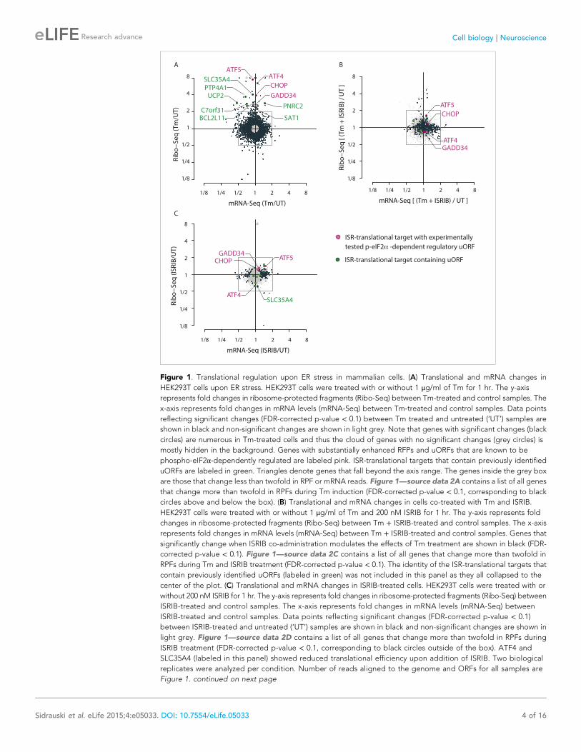
Figure 1. Translational regulation upon ER stress in mammalian cells. (A) Translational and mRNA changes in
HEK293T cells upon ER stress. HEK293T cells were treated with or without 1 μg/ml of Tm for 1 hr. The y-axis
represents fold changes in ribosome-protected fragments (Ribo-Seq) between Tm-treated and control samples. The
x-axis represents fold changes in mRNA levels (mRNA-Seq) between Tm-treated and control samples. Data points
reflecting significant changes (FDR-corrected p-value < 0.1) between Tm treated and untreated (‘UT’) samples are
shown in black and non-significant changes are shown in light grey. Note that genes with significant changes (black
circles) are numerous in Tm-treated cells and thus the cloud of genes with no significant changes (grey circles) is
mostly hidden in the background. Genes with substantially enhanced RFPs and uORFs that are known to be
phospho-eIF2α-dependently regulated are labeled pink. ISR-translational targets that contain previously identified
uORFs are labeled in green. Triangles denote genes that fall beyond the axis range. The genes inside the grey box
are those that change less than twofold in RPF or mRNA reads. Figure 1—source data 2A contains a list of all genes
that change more than twofold in RPFs during Tm induction (FDR-corrected p-value < 0.1, corresponding to black
circles above and below the box). (B) Translational and mRNA changes in cells co-treated with Tm and ISRIB.
HEK293T cells were treated with or without 1 μg/ml of Tm and 200 nM ISRIB for 1 hr. The y-axis represents fold
changes in ribosome-protected fragments (Ribo-Seq) between Tm + ISRIB-treated and control samples. The x-axis
represents fold changes in mRNA levels (mRNA-Seq) between Tm + ISRIB-treated and control samples. Genes that
significantly change when ISRIB co-administration modulates the effects of Tm treatment are shown in black (FDR-
corrected p-value < 0.1). Figure 1—source data 2C contains a list of all genes that change more than twofold in
RPFs during Tm and ISRIB treatment (FDR-corrected p-value < 0.1). The identity of the ISR-translational targets that
contain previously identified uORFs (labeled in green) was not included in this panel as they all collapsed to the
center of the plot. (C) Translational and mRNA changes in ISRIB-treated cells. HEK293T cells were treated with or
without 200 nM ISRIB for 1 hr. The y-axis represents fold changes in ribosome-protected fragments (Ribo-Seq) between
ISRIB-treated and control samples. The x-axis represents fold changes in mRNA levels (mRNA-Seq) between
ISRIB-treated and control samples. Data points reflecting significant changes (FDR-corrected p-value < 0.1)
between ISRIB-treated and untreated (‘UT’) samples are shown in black and non-significant changes are shown in
light grey. Figure 1—source data 2D contains a list of all genes that change more than twofold in RPFs during
ISRIB treatment (FDR-corrected p-value < 0.1, corresponding to black circles outside of the box). ATF4 and
SLC35A4 (labeled in this panel) showed reduced translational efficiency upon addition of ISRIB. Two biological
replicates were analyzed per condition. Number of reads aligned to the genome and ORFs for all samples are
Figure 1. continued on next page
Sidrauski et al. eLife 2015;4:e05033. DOI: 10.7554/eLife.05033 4 of 16
Research advance Cell biology | Neuroscience
As seen in Figure 1B, ISRIB comprehensively blocked the translational changes that take place upon
ER-stress. A large number of genes with a significant change in expression upon stress collapsed to
the center of the plot with ISRIB and Tm co-treatment (Figure 1B, highlighted in black). Importantly,
ISRIB abolished the induction of the known phospho-eIF2α-dependent translational targets
(Figure 1B, colored pink) and the seven ISR-translational targets with previously identified uORFs
(Figure 1B, colored green). The mRNAs that remained translationally induced in the presence of ISRIB
are listed in Figure 1—source data 2C. In addition, ISRIB reversed the reduction in translation of
mTOR target mRNAs upon ER stress (Figure 1—figure supplement 2, panel B).
Importantly, ISRIB treatment alone did not have general effects on translation in non-stressed cells,
as revealed by the lack of substantial changes in RPFs in most cellular mRNAs, nor did it cause any
significant changes in mRNA levels (Figure 1C) and mTOR target expression (Figure 1—figure
supplement 2, panel C). In the absence of ER stress, ISRIB-treated cells behaved like untreated cells
with the exception of a reduction in the basal level of translation of ATF4 and SLC35A4 mRNAs and
a few additional mRNAs (Figure 1C and Figure 1—source data 2C). Taken together, these data
strongly support the notion that ISRIB does not have global effects on translation, transcription, or
mRNA stability in non-stressed cells and underscores its remarkable ability to counteract selectively
the translational changes elicited by eIF2α phosphorylation in stressed cells.
ISRIB prevents formation of stress granules exclusively triggered byeIF2α phosphorylationPhosphorylation of eIF2α and reduction of ternary complex formation are tightly linked to the
formation of stress granules (SGs) (Kedersha et al., 2002). ISRIB renders cells insensitive to the effects
of eIF2α phosphorylation, thus leading to the prediction that it prevents SG formation as well. We
tested this hypothesis by inducing SG formation using thapsigargin (Tg), a potent ER stressor that
inhibits the ER calcium pump and was recently shown by ribosome profiling to yield analogous
translational effects to tunicamycin (Reid et al., 2014). Microscopic detection of SGs required
a stronger induction of ER stress than commonly achieved with Tm, making Tg the preferred inducer.
We monitored SGs by performing immunofluorescence on eIF3a, a translation initiation factor that is
recruited into SGs. As expected, we found that ISRIB significantly reduced their assembly upon co-
treatment with Tg (Figure 2A,B). In addition, ISRIB prevented SG formation induced by arsenite (Ars),
another widely used inducer of eIF2α phosphorylation via activation of HRI. As expected, both
treatments induced eIF2α phosphorylation but only Tg induced the ER-resident kinase, PERK, as
seen by its shift in mobility that is due to its extensive auto-phosphorylation (Figure 2C). Both the
Figure 1. Continued
found in Figure 1—source data 2E. Correlation plots for the replicates for each condition are found in Figure
1—figure supplement 3. mRNA abundance for all ORFs mapped are found in Figure 1—figure supplement 4.
Read counts for all conditions and each individual transcript are found in Figure 1—source data 1. The Ribo-seq
and mRNA-seq data have been deposited in NCBI’s Gene Expression Omnibus and are accessible through GEO
series accession number GSE65778.
DOI: 10.7554/eLife.05033.002
The following source data and figure supplements are available for figure 1:
Source data 1. Read counts for all conditions and each individual transcript.
DOI: 10.7554/eLife.05033.003
Source data 2. Source data for Figure 1.
DOI: 10.7554/eLife.05033.004
Figure supplement 1. Ribosome and mRNA densities in the 5’UTR of ATF4 and SLC35A4.
DOI: 10.7554/eLife.05033.005
Figure supplement 2. Translational regulation of mTOR targets upon ER-stress.
DOI: 10.7554/eLife.05033.006
Figure supplement 3. Correlation plots for duplicate ribosome profiling experiments.
DOI: 10.7554/eLife.05033.007
Figure supplement 4. Mean mRNA abundance of all genes mapped.
DOI: 10.7554/eLife.05033.008
Sidrauski et al. eLife 2015;4:e05033. DOI: 10.7554/eLife.05033 5 of 16
Research advance Cell biology | Neuroscience
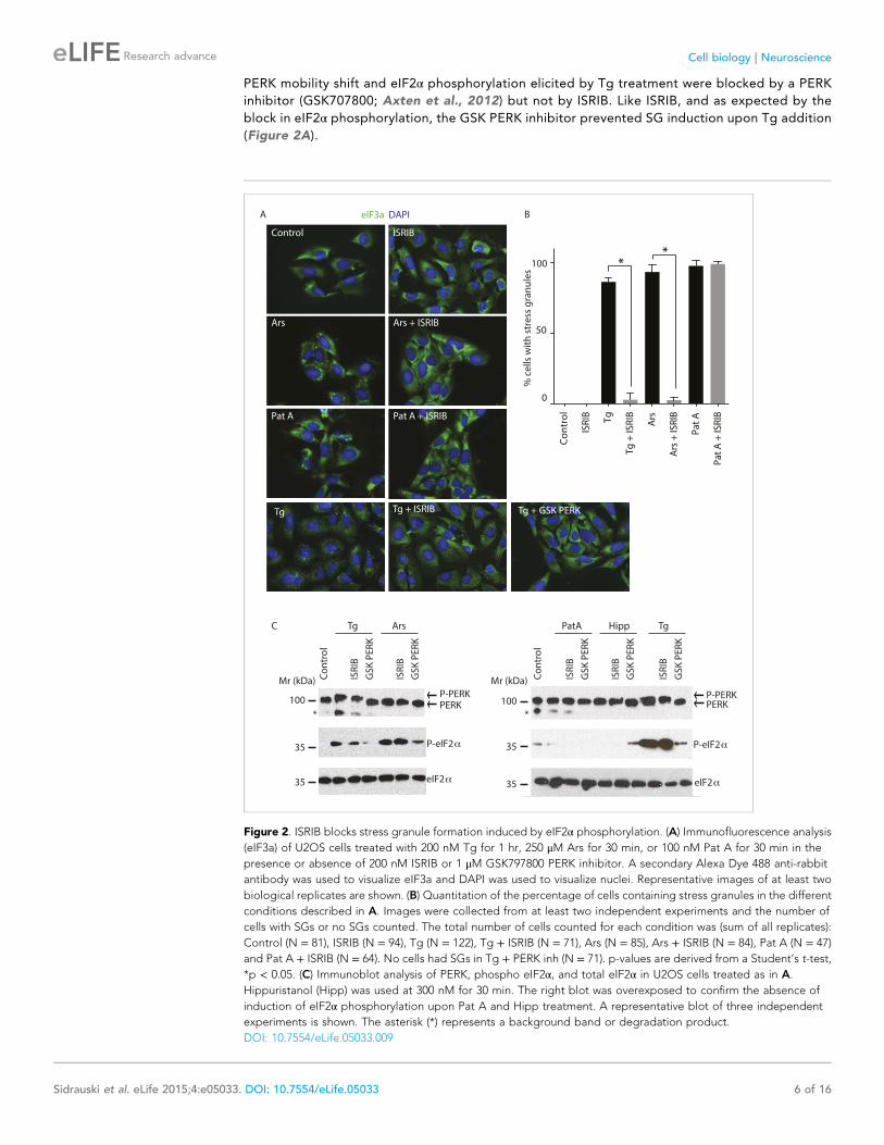
PERK mobility shift and eIF2α phosphorylation elicited by Tg treatment were blocked by a PERK
inhibitor (GSK707800; Axten et al., 2012) but not by ISRIB. Like ISRIB, and as expected by the
block in eIF2α phosphorylation, the GSK PERK inhibitor prevented SG induction upon Tg addition
(Figure 2A).
Figure 2. ISRIB blocks stress granule formation induced by eIF2α phosphorylation. (A) Immunofluorescence analysis
(eIF3a) of U2OS cells treated with 200 nM Tg for 1 hr, 250 μM Ars for 30 min, or 100 nM Pat A for 30 min in the
presence or absence of 200 nM ISRIB or 1 μM GSK797800 PERK inhibitor. A secondary Alexa Dye 488 anti-rabbit
antibody was used to visualize eIF3a and DAPI was used to visualize nuclei. Representative images of at least two
biological replicates are shown. (B) Quantitation of the percentage of cells containing stress granules in the different
conditions described in A. Images were collected from at least two independent experiments and the number of
cells with SGs or no SGs counted. The total number of cells counted for each condition was (sum of all replicates):
Control (N = 81), ISRIB (N = 94), Tg (N = 122), Tg + ISRIB (N = 71), Ars (N = 85), Ars + ISRIB (N = 84), Pat A (N = 47)
and Pat A + ISRIB (N = 64). No cells had SGs in Tg + PERK inh (N = 71). p-values are derived from a Student’s t-test,
*p < 0.05. (C) Immunoblot analysis of PERK, phospho eIF2α, and total eIF2α in U2OS cells treated as in A.
Hippuristanol (Hipp) was used at 300 nM for 30 min. The right blot was overexposed to confirm the absence of
induction of eIF2α phosphorylation upon Pat A and Hipp treatment. A representative blot of three independent
experiments is shown. The asterisk (*) represents a background band or degradation product.
DOI: 10.7554/eLife.05033.009
Sidrauski et al. eLife 2015;4:e05033. DOI: 10.7554/eLife.05033 6 of 16
Research advance Cell biology | Neuroscience
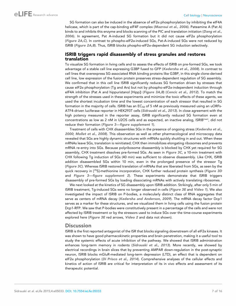
SG formation can also be induced in the absence of eIF2α phosphorylation by inhibiting the eIF4A
helicase, which is part of the cap-binding eIF4F complex (Mazroui et al., 2006). Pateamine A (Pat A)
binds to and inhibits this enzyme and blocks scanning of the PIC and translation initiation (Dang et al.,
2006). In agreement, Pat A-induced SG formation but it did not cause eIF2α phosphorylation
(Figure 2A,C). In contrast to phospho-eIF2α-induced SGs, Pat-A-induced SGs were not reduced by
ISRIB (Figure 2A,B). Thus, ISRIB blocks phospho-eIF2α-dependent SG induction selectively.
ISRIB triggers rapid disassembly of stress granules and restorestranslationTo visualize SG formation in living cells and to assess the effects of ISRIB on pre-formed SGs, we took
advantage of a stable cell line expressing G3BP fused to GFP (Kedersha et al., 2008). In contrast to
cell lines that overexpress SG-associated RNA binding proteins like G3BP, in this single clone-derived
cell line, low expression of the fusion protein preserves stress-dependent regulation of SG assembly.
We confirmed that in this cell line ISRIB significantly reduces SG formation driven by stresses that
cause eIF2α phosphorylation (Tg and Ars) but not by phospho-eIF2α-independent induction through
eIF4A inhibition (Pat A and hippuristanol [Hipp]) (Figure 3A,B) (Cencic et al., 2012). To match the
strength of the stresses used in these experiments and minimize the toxic effects of these agents, we
used the shortest incubation time and the lowest concentration of each stressor that resulted in SG
formation in the majority of cells. ISRIB has an EC50 of 5 nM as previously measured using an uORFs-
ATF4-driven luciferase reporter in HEK293T cells (Sidrauski et al., 2013). In close agreement with the
high potency measured in the reporter assay, ISRIB significantly reduced SG formation even at
concentrations as low as 2 nM in U2OS cells and as expected, an inactive analog, ISRIBinact, did not
reduce their formation (Figure 3—figure supplement 1).
Treatment of cells with CHX disassembles SGs in the presence of ongoing stress (Kedersha et al.,
2000; Mollet et al., 2008). This observation as well as other pharmacological and microscopy data
revealed that SGs are highly dynamic structures with mRNAs quickly shuttling in and out. When these
mRNAs leave SGs, translation is reinitiated; CHX then immobilizes elongating ribosomes and prevents
mRNA re-entry into SGs. Because polyribosome disassembly is blocked by CHX yet required for SG
assembly, CHX treatment dissolves pre-formed SGs. As seen in Figure 3C, a 10-min treatment with
CHX following Tg induction of SGs (40 min) was sufficient to observe disassembly. Like CHX, ISRIB
addition disassembled SGs within 10 min, even in the prolonged presence of the stressor Tg
(Figure 3C). Whereas ISRIB restored translation of mRNAs that are liberated from SGs, as seen by the
quick recovery in [35S]-methionine incorporation, CHX further reduced protein synthesis (Figure 3D
and Figure 3—figure supplement 2). These experiments demonstrate that ISRIB triggers
disassembly of pre-formed SGs by loading dissociating mRNAs with actively translating ribosomes.
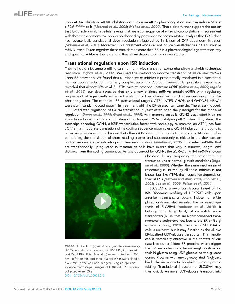
We next looked at the kinetics of SG disassembly upon ISRIB addition. Strikingly, after only 5 min of
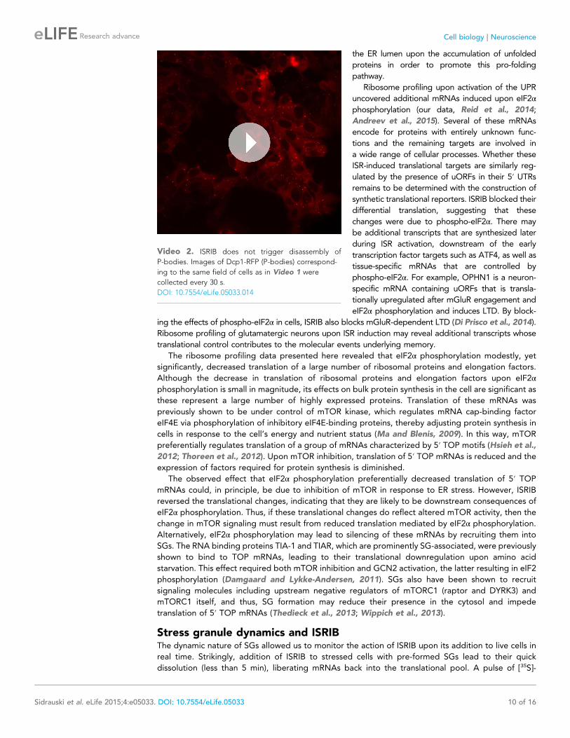
ISRIB treatment, Tg-induced SGs were no longer observed in cells (Figure 3E and Video 1). We also
investigated the impact of ISRIB on P-bodies, a molecularly distinct class of RNA aggregates that
serve as centers of mRNA decay (Kedersha and Anderson, 2009). The mRNA decay factor Dcp1
serves as a marker for these structures, and we visualized them in living cells using the fusion protein
Dcp1-RFP. We saw that P-bodies were constitutively present in a percentage of the cells and were not
affected by ISRIB treatment or by the stressors used to induce SGs over the time-course experiments
explored here (Figure 3E red arrows, Video 2 and data not shown).
DiscussionISRIB is the first reported antagonist of the ISR that blocks signaling downstream of all eIF2α kinases. It
was shown to have good pharmacokinetic properties and brain penetration, making it a useful tool to
study the systemic effects of acute inhibition of the pathway. We showed that ISRIB administration
enhances long-term memory in rodents (Sidrauski et al., 2013). More recently, we showed by
electrical recordings in brain slices that by preventing AMPAR down-regulation in the post-synaptic
neuron, ISRIB blocks mGluR-mediated long-term depression (LTD), an effect that is dependent on
eIF2α phosphorylation (Di Prisco et al., 2014). Comprehensive analyses of the cellular effects and
kinetics of action of ISRIB are critical for interpretation of its in vivo effects and assessment of its
therapeutic potential.
Sidrauski et al. eLife 2015;4:e05033. DOI: 10.7554/eLife.05033 7 of 16
Research advance Cell biology | Neuroscience
Our translational and transcriptional profiling confirmed that ISRIB treatment of ER-stressed cells
substantially and comprehensively blocks the translational effects of eIF2α phosphorylation. ISRIB
blocked SG formation that was triggered by eIF2α phosphorylation but did not abolish their assembly
Figure 3. ISRIB addition rapidly dissolves pre-formed stress granules in live cells restoring translation. (A) Live cell imaging of stress granules in U2OS cells
stably expressing G3BP-GFP (SG marker). Cells were treated with 200 nM Tg for 40 min, 250 μM Ars for 30 min, 100 nM Pat A for 30 min, or 300 nM Hipp in
the presence or absence of 200 nM ISRIB. Cells were imaged using an epifluorescence microscope. Representative images of at least two biological
replicates are shown. (B) Quantitation of the percentage of cells containing stress granules in the different conditions described in A. Images were
collected from at least two independent experiments and the number of cells with SGs or no SGs counted. The number of cells analyzed for each
condition was (sum of replicates): Control (N = 98), ISRIB (N = 81), Tg (N = 101), Tg + ISRIB (N = 84), Ars (N = 80), Ars + ISRIB (N = 55), Pat A (N = 58), Pat A
+ ISRIB (N = 50), Hipp (N = 41) and Hipp + ISRIB (N = 52). p-values are derived from a Student’s t-test, *p < 0.05. (C) Stress granules were pre-formed with
Tg for 40 min (as in Figure 3A) and then CHX (50 μg/ml) or ISRIB (200 nM) was added to the well, incubated for 10 min and images were collected.
Representative images of at least two biological replicates are shown. (D) ISRIB quickly restores mRNA translation upon disassembly of stress granules.
Cells were treated as in C with 200 nM Tg for 40 min and then DMSO, CHX (50 μg/ml), or ISRIB (200 nM) was added at the same time as [35S]-methionine.
Cells were lysed after 15 min, protein was run in an SDS-PAGE gel and radioactivity was measured in each lane (N = 2, mean ± SD). (E) ISRIB quickly
dissolves stress granules but does not affect P-bodies. Live cell imaging of U2OS cells stably expressing G3BP-GFP (SG marker) and Dcp1-RFP (P-body
marker). Cells were treated with 200 nM Tg for 45 min followed by addition of 200 nM ISRIB at t = 0 min to the well and then imaged using spinning disk
confocal microscopy. Images were collected every 30 s. The red arrows point to two representative P-bodies. Representative images of at least three
biological replicates are shown.
DOI: 10.7554/eLife.05033.010
The following figure supplements are available for figure 3:
Figure supplement 1. ISRIB dose response and inactive analog in stress granule assay.
DOI: 10.7554/eLife.05033.011
Figure supplement 2. Representative SDS-PAGE gel of [35S]-methionine pulse as described in Figure 3D.
DOI: 10.7554/eLife.05033.012
Sidrauski et al. eLife 2015;4:e05033. DOI: 10.7554/eLife.05033 8 of 16
Research advance Cell biology | Neuroscience
upon eIF4A inhibition; eIF4A inhibitors do not cause eIF2α phosphorylation and can induce SGs in
eIF2αS51A/S51A cells (Mazroui et al., 2006; Mokas et al., 2009). These data further support the notion
that ISRIB solely inhibits cellular events that are a consequence of eIF2α phosphorylation. In agreement
with these observations, we previously showed by polyribosome sedimentation analysis that ISRIB does
not reverse bulk translational down-regulation triggered by inhibition of CAP-dependent initiation
(Sidrauski et al., 2013). Moreover, ISRIB treatment alone did not induce overall changes in translation or
mRNA levels. Taken together these data demonstrate that ISRIB is a pharmacological agent that acutely
and specifically blocks the ISR and is thus an invaluable tool for in vivo studies.
Translational regulation upon ISR inductionThe method of ribosome profiling can monitor in vivo translation comprehensively and with nucleotide
resolution (Ingolia et al., 2009). We used this method to monitor translation of all cellular mRNAs
upon ISR activation. We found that a limited set of mRNAs is preferentially translated in a substantial
manner upon a reduction in ternary complex assembly. Although previous large-scale analyses have
revealed that almost 45% of all 5′ UTRs have at least one upstream uORF (Calvo et al., 2009; Ingolia
et al., 2011), our data revealed that only a few of these mRNAs contain uORFs with regulatory
properties that significantly enhance translation of their downstream coding sequences upon eIF2αphosphorylation. The canonical ISR translational targets, ATF4, ATF5, CHOP, and GADD34 mRNAs
were significantly induced upon 1 hr treatment with the ER-stressor tunicamycin. The stress-induced,
uORF-mediated regulation of GCN4 translation in yeast established the paradigm for this mode of
regulation (Dever et al., 1995; Grant et al., 1995). As in mammalian cells, GCN2 is activated in amino
acid-starved yeast by the accumulation of uncharged tRNAs, catalyzing eIF2α phosphorylation. The
transcript encoding GCN4, a bZIP transcription factor with homology to mammalian ATF4, has four
uORFs that modulate translation of its coding sequence upon stress. GCN4 induction is thought to
occur via a re-scanning mechanism that allows 40S ribosomal subunits to remain mRNA-bound after
completing the translation of short reading frames and subsequently reinitiate in the downstream
coding sequence after reloading with ternary complex (Hinnebusch, 2005). The select mRNAs that
are translationally upregulated in mammalian cells have uORFs that vary in number, length, and
distance from the coding sequences. As was observed for GCN4, the uORF2 of ATF4 mRNA showed
ribosome density, supporting the notion that it is
translated under normal growth conditions (Ingo-
lia et al., 2009). Whether the same mechanism of
rescanning is utilized by all these mRNAs is not
known but, like ATF4, their regulation depends on
their uORFs (Vattem andWek, 2004; Zhou et al.,
2008; Lee et al., 2009; Palam et al., 2011).
SLC35A4 is a novel translational target of the
ISR. Ribosome profiling of HEK293T cells upon
arsenite treatment, a potent inducer of eIF2αphosphorylation, also revealed the increased syn-
thesis of SLC35A4 (Andreev et al., 2015). It
belongs to a large family of nucleotide sugar
transporters (NSTs) that are highly conserved trans-
membrane antiporters localized to the ER or Golgi
apparatus (Song, 2013). The role of SLC35A4 in
cells is unknown but it may function as the elusive
ER-localized UDP-glucose transporter. This hypoth-
esis is particularly attractive in the context of our
data because unfolded ER proteins, which trigger
the ISR, are continuously de- and re-glucosylated on
their N-glycans using UDP-glucose as the glucose
donor. Proteins with monoglucosylated N-glycans
bind calnexin or calreticulin which promote protein
folding. Translational induction of SLC35A4 may
thus quickly enhance UDP-glucose transport into
Video 1. ISRIB triggers stress granule disassembly.
U2OS cells stably expressing G3BP-GFP (SG marker)
and Dcp1-RFP (P-body marker) were treated with 200
nM Tg for 40 min and then 200 nM ISRIB was added at
t = 0 min to the well and imaged using an epifluor-
escence microscope. Images of G3BP-GFP (SGs) were
collected every 30 s.
DOI: 10.7554/eLife.05033.013
Sidrauski et al. eLife 2015;4:e05033. DOI: 10.7554/eLife.05033 9 of 16
Research advance Cell biology | Neuroscience
the ER lumen upon the accumulation of unfolded
proteins in order to promote this pro-folding
pathway.
Ribosome profiling upon activation of the UPR
uncovered additional mRNAs induced upon eIF2αphosphorylation (our data, Reid et al., 2014;
Andreev et al., 2015). Several of these mRNAs
encode for proteins with entirely unknown func-
tions and the remaining targets are involved in
a wide range of cellular processes. Whether these
ISR-induced translational targets are similarly reg-
ulated by the presence of uORFs in their 5′ UTRsremains to be determined with the construction of
synthetic translational reporters. ISRIB blocked their
differential translation, suggesting that these
changes were due to phospho-eIF2α. There may
be additional transcripts that are synthesized later
during ISR activation, downstream of the early
transcription factor targets such as ATF4, as well as
tissue-specific mRNAs that are controlled by
phospho-eIF2α. For example, OPHN1 is a neuron-
specific mRNA containing uORFs that is transla-
tionally upregulated after mGluR engagement and
eIF2α phosphorylation and induces LTD. By block-
ing the effects of phospho-eIF2α in cells, ISRIB also blocks mGluR-dependent LTD (Di Prisco et al., 2014).
Ribosome profiling of glutamatergic neurons upon ISR induction may reveal additional transcripts whose
translational control contributes to the molecular events underlying memory.
The ribosome profiling data presented here revealed that eIF2α phosphorylation modestly, yet
significantly, decreased translation of a large number of ribosomal proteins and elongation factors.
Although the decrease in translation of ribosomal proteins and elongation factors upon eIF2αphosphorylation is small in magnitude, its effects on bulk protein synthesis in the cell are significant as
these represent a large number of highly expressed proteins. Translation of these mRNAs was
previously shown to be under control of mTOR kinase, which regulates mRNA cap-binding factor
eIF4E via phosphorylation of inhibitory eIF4E-binding proteins, thereby adjusting protein synthesis in
cells in response to the cell’s energy and nutrient status (Ma and Blenis, 2009). In this way, mTOR
preferentially regulates translation of a group of mRNAs characterized by 5′ TOP motifs (Hsieh et al.,
2012; Thoreen et al., 2012). Upon mTOR inhibition, translation of 5′ TOP mRNAs is reduced and the
expression of factors required for protein synthesis is diminished.
The observed effect that eIF2α phosphorylation preferentially decreased translation of 5′ TOP
mRNAs could, in principle, be due to inhibition of mTOR in response to ER stress. However, ISRIB
reversed the translational changes, indicating that they are likely to be downstream consequences of
eIF2α phosphorylation. Thus, if these translational changes do reflect altered mTOR activity, then the
change in mTOR signaling must result from reduced translation mediated by eIF2α phosphorylation.
Alternatively, eIF2α phosphorylation may lead to silencing of these mRNAs by recruiting them into
SGs. The RNA binding proteins TIA-1 and TIAR, which are prominently SG-associated, were previously
shown to bind to TOP mRNAs, leading to their translational downregulation upon amino acid
starvation. This effect required both mTOR inhibition and GCN2 activation, the latter resulting in eIF2
phosphorylation (Damgaard and Lykke-Andersen, 2011). SGs also have been shown to recruit
signaling molecules including upstream negative regulators of mTORC1 (raptor and DYRK3) and
mTORC1 itself, and thus, SG formation may reduce their presence in the cytosol and impede
translation of 5′ TOP mRNAs (Thedieck et al., 2013; Wippich et al., 2013).
Stress granule dynamics and ISRIBThe dynamic nature of SGs allowed us to monitor the action of ISRIB upon its addition to live cells in
real time. Strikingly, addition of ISRIB to stressed cells with pre-formed SGs lead to their quick
dissolution (less than 5 min), liberating mRNAs back into the translational pool. A pulse of [35S]-
Video 2. ISRIB does not trigger disassembly of
P-bodies. Images of Dcp1-RFP (P-bodies) correspond-
ing to the same field of cells as in Video 1 were
collected every 30 s.
DOI: 10.7554/eLife.05033.014
Sidrauski et al. eLife 2015;4:e05033. DOI: 10.7554/eLife.05033 10 of 16
Research advance Cell biology | Neuroscience
methionine confirmed the fast recovery in protein synthesis even in the presence of stress. Although
the molecular target of ISRIB remains unknown, its quick action suggests a direct effect on translation
initiation. Phospho-eIF2α resistance has been observed both in yeast and in mammalian cells. In yeast,
mutations in eIF2B (the GEF for eIF2) and eIF5 (the 48S PIC-associated GTPase-activating protein for
eIF2) have been reported to make cells insensitive to this phosphorylation event (Vazquez de Aldana
and Hinnebusch, 1994; Pavitt et al., 1997, 1998). In mammalian cells, TLR4 engagement in
macrophages leads to increased eIF2B activity by removal of an inhibitory phosphorylation and
insensitivity to ISR activation (Woo et al., 2012). Thus, ISRIB may directly or indirectly enhance the
activity of eIF2B, eIF5, or other initiation factors, thus quickly reversing the cellular effects of
phosphorylated eIF2α.SGs contain a large number of RBPs that harbor low complexity sequence domains that nucleate
through transient, low affinity interactions (Kato et al., 2012). These RBPs usually contain several
RNA-binding domains and can associate with more than one mRNA; this multi-valency further favors
the coalescence of RNA-protein granules. A conspicuous feature of some degenerative diseases is the
cytoplasmic or nuclear aggregation of RBPs, driven in some cases by pathogenic mutations. TDP-43
and FUS mutations are found in amyetropic lateral sclerosis (ALS) and fronto temporal lobar
degeneration (FTLD) (Li et al., 2013), and mutations in hnRNPA1 and hnRPNPA2/B1 have also been
found in ALS (Kim et al., 2013). Recent reports have also described the presence of RNA and RBPs in
aggregates that form in prion disease, taupathies, and Alzheimer’s (Vanderweyde et al., 2012; Ash
et al., 2014). The impact of these cytosolic aggregates on SG dynamics is not known, though they
may hamper the ability of SGs to properly dissolve, thereby contributing to sustained translational
attenuation and neurodegeneration. By quickly disassembling SGs even in the presence of stress,
ISRIB may provide a useful therapeutic intervention in these diseases by antagonizing the cellular
effects of pathogenic RNA-protein assemblies.
Materials and methods
ChemicalsTunicamycin was obtained from Calbiochem EMB Bioscience. Thapsigargin, cycloheximide and
sodium arsenite were obtained from Sigma–Aldrich. Hippuristanol and pateamine A were a kind gift
from Jerry Pelletier. GSK797800 (PERK inhibitor) was obtained from TRC Inc. ISRIB (Sidrauski et al.,
2013) and an inactive analog (754125) (Di Prisco et al., 2014) were synthesized in-house.
Cell cultureHEK293T, U2OS, and U2OSGFP-G3BP/Dcp1-RFP cells were maintained at 37˚C, 5% CO2 in DMEMmedia
supplemented with 10% FBS, L-glutamine and antibiotics (penicillin and streptomycin). U2OS cells stably
expressing G3BP-GFP/Dcp1-RFP cells were a kind gift from Nancy Kedersha (Kedersha et al., 2008).
Isolation of ribosome footprints and RNAHEK293T cells were treated with or without 1 μg/ml of tunicamycin, tunicamycin and ISRIB (200 nM), or
ISRIB for 1 hr. Cycloheximide (CHX) (100 μg/ml) was added for 2 min, cells were washed with ice cold
PBS (with 100 μg/ml of CHX) and lysed in 20 mM Tris pH = 7.4 (RT), 200 mM NaCl, 15 mM MgCl, 1 mM
DTT, 8% glycerol, 100 μg/ml CHX, 1% Triton and protease inhibitors (Roche complete EDTA-free). A
syringe (25G5/8) was used to triturate cells, the lysate was clarified at 12,000 rpm for 10 min and half of
the lysate was used for RNA extraction (Trizol, Invitrogen, Carlsbad, CA) and the other half was digested
with RNase I (Ambion). The amount of RNase I and time of incubation was optimized for each sample
based on the collapse of polyribosomes to the monosome peak as analyzed by analytical polyribosome
gradients. The reaction was quenched with SUPERaseIn (Ambion, Life Technologies) and the digested
lysate was then loaded on an 800 μl sucrose cushion (1.7 g of sucrose was dissolved in 3.9 ml of lysis
buffer without Triton) and centrifuged in a TLA100.2 rotor at 70,000 rpm for 4 hr. The pellet was
resuspended in 10 mM Tris pH = 7 (RT), and RNA was extracted (phenol/chloroform).
Generation of sequencing libraries and data analysisSequencing libraries were generated as described in Ingolia et al., 2012. For data analysis, we used
DESeq as described by Anders and Huber (2010). P-adj values (p-values) were calculated using the R
command ‘p.adjust’ for multiple comparisons and the BH method (Benjamini and Hochberg, 1995) to
Sidrauski et al. eLife 2015;4:e05033. DOI: 10.7554/eLife.05033 11 of 16
Research advance Cell biology | Neuroscience
correct for false discovery rate. The data in this publication have been deposited in NCBI’s Gene
Expression Omnibus and are accessible through GEO series accession number GSE65778.
ImmunofluorescenceU2OS cells were seeded on 4-well chamber slides (Lab-Tek) 18 hr prior to processing for
immunofluorescence. Cells (80% confluent) were fixed with ice-cold methanol. The cells were then
rinsed with PBS (Sigma) and blocked for 1 hr at room temperature in 0.5% BSA in PBS. The cells were
then incubated overnight at 4˚C with an anti-eIF3A rabbit antibody (#3411; Cell Signaling Technology)
at a 1:1000 dilution in blocking buffer. The next morning the slides were washed three times (5 min
each time) with PBS and then incubated for 1 hr at room temperature in a 1:1000 dilution (in 0.5% BSA
in PBS) of secondary anti-rabbit antibody labeled with Alexa Dye 488 (Molecular Probes). The slides
were washed three additional times with PBS. The slides were then mounted with antifade reagent
with DAPI (Life Technologies P-36931). Lastly, the slides were imaged using a Zeiss Axiovert 200M
epifluorescence microscope.
Live cell microscopyU2OS G3BP-GFP/Dcp1-RFP cells were plated in 8-well Lab-Tek chamber slides and switched to
imaging media (lacking phenol red) upon addition of different stress inducers. Cells were
either imaged using a Zeiss Axiovert 200M epifluorescence microscope or in a heated chamber
using a spinning confocal epifluorescence microscope (Eclipse Ti-Nikon) and an Andor iXon3
camera.
Protein analysisCells were washed with PBS and lysed in SDS-PAGE loading buffer (1% SDS, 62.5 mM Tris–HCl pH
6.8, 10% glycerol). Lysates were sonicated and loaded on Any-kD SDS-PAGE gels (BioRad). Proteins
were transferred onto nitrocellulose and probed with primary antibodies diluted in Tris-buffered
saline supplemented with 0.1% Tween 20 and 5% BSA. The following antibodies were used: PERK
(D11A8) (1:1000), eIF2α (#9722; Cell Signaling technology) (1:1000), phospho-eIF2α (Ser51)
(44728G; Invitrogen). An HRP-conjugated secondary antibody (Amersham) was employed to
detect immune-reactive bands using enhanced chemiluminescence (SuperSignal, Thermo
Scientific).
[35S]-methionine incorporationU2OS GFP-G3BP/mRFP-DCP1a cells were seeded on 12-well plates, allowed to recover overnight and
treated with 100 nM Tg for 40 min. ISRIB (200 nM) or CHX (50 μg/ml) was added at the same time as
50 μCi of [35S]-methionine (Perkin Elmer) and incubated for 15 min. Cells were lysed by addition of
SDS-PAGE loading buffer. Lysates were sonicated and equal amounts were loaded on SDS-PAGE gels
(BioRad). The gel was dried and radioactive methionine incorporation was detected by exposure to
a phosphor-screen and visualized with a Typhoon 9400 Variable Mode Imager (GE Healthcare).
AcknowledgementsWe thank Margaret Elvekrog, Voytek Okreglak, Shelley Starck, and Jirka Peschek for editing the
manuscript and the members of the Walter lab for helpful discussions.
Additional information
Funding
Funder Grant reference number Author
Howard Hughes MedicalInstitute (HHMI)
Carmela Sidrauski,Peter Walter
Kinship Foundation Searle Scholars Program11-SSP-229
Nicholas T Ingolia
National Institutes ofHealth (NIH)
T32GM007231 Anna M McGeachy
Sidrauski et al. eLife 2015;4:e05033. DOI: 10.7554/eLife.05033 12 of 16
Research advance Cell biology | Neuroscience
Funder Grant reference number Author
Carnegie Institution ofWashington
Nicholas T Ingolia
The funders had no role in study design, data collection and interpretation, or thedecision to submit the work for publication.
Author contributions
CS, NTI, Conception and design, Acquisition of data, Analysis and interpretation of data, Drafting or
revising the article; AMMG, Acquisition of data, Analysis and interpretation of data, Drafting or
revising the article; PW, Analysis and interpretation of data, Drafting or revising the article
ReferencesAnders S, Huber W. 2010. Differential expression analysis for sequence count data. Genome Biology 11:R106.doi: 10.1186/gb-2010-11-10-r106.
Andreev DE, O’Connor PB, Fahey C, Kenny EM, Terenin IM, Dimitriev SE, Cormican P, Morris DW, Shatsky IN,Baranov PV. 2015. Translation of 5’ leaders is pervasive in genes resistant to eIF2 repression. eLife 4:e03971.doi: 10.7554/eLife.03971.
Ash PE, Vanderweyde TE, Youmans KL, Apicco DJ, Wolozin B. 2014. Pathological stress granules in Alzheimer’sdisease. Brain Research 1584:52–58. doi: 10.1016/j.brainres.2014.05.052.
Atkins C, Liu Q, Minthorn E, Zhang S, Figueroa DJ, Moss KG, Stanley TB, Sanders B, Goetz A, Gaul N, ChoudhryAE, Alsaid H, Jucker BM, Axten JM, Kumar R. 2013. Characterization of a novel PERK kinase inhibitor with anti-tumor and anti-angiogenic activity. Cancer Research 73:1993–2002. doi: 10.1158/0008-5472.CAN-12-3109.
Axten JM, Medina JR, Feng Y, Shu A, Romeril SP, Grant SW, Li WH, Heerding DA, Minthorn E, Mencken T, AtkinsC, Liu Q, Rabindran S, Kumar R, Hong X, Goetz A, Stanley T, Taylor JD, Sigethy SD, Tomberlin GH, Hassell AM,Kahler KM, Shewchuk LM, Gampe RT. 2012. Discovery of 7-methyl-5-(1-{[3-(trifluoromethyl)phenyl]acetyl}-2,3-dihydro-1 H-indol-5-yl)-7 H-pyrrolo[2,3- d]pyrimidin-4-amine (GSK2606414), a potent and selective first-in-classInhibitor of protein kinase R (PKR)-like endoplasmic reticulum kinase (PERK). Journal of Medicinal Chemistry 55:7193–7207. doi: 10.1021/jm300713s.
Benjamini Y, Hochberg Y. 1995. Controlling the False Discovery Rate: A Practical and Powerful Approach toMultiple Testing. Journal of the Royal Statistical Society Series B 57:289–300.
Boyce M, Bryant KF, Jousse C, Long K, Harding HP, Scheuner D, Kaufman RJ, Ma D, Coen DM, Ron D, Yuan J.2005. A selective inhibitor of eIF2alpha dephosphorylation protects cells from ER stress. Science 307:935–939.doi: 10.1126/science.1101902.
Buchan JR, Parker R. 2009. Eukaryotic stress granules: the ins and outs of translation. Molecular Cell 36:932–941.doi: 10.1016/j.molcel.2009.11.020.
Calvo SE, Pagliarini DJ, Mootha VK. 2009. Upstream open reading frames cause widespread reduction of proteinexpression and are polymorphic among humans. Proceedings of the National Academy of Sciences of USA 106:7507–7512. doi: 10.1073/pnas.0810916106.
Cencic R, Galicia-Vazquez G, Pelletier J. 2012. Inhibitors of translation targeting eukaryotic translation initiationfactor 4A. Methods in Enzymology 511:437–461. doi: 10.1016/B978-0-12-396546-2.00020-6.
Chen T, Ozel D, Qiao Y, Harbinski F, Chen L, Denoyelle S, He X, Zvereva N, Supko JG, Chorev M, Halperin JA,Aktas BH. 2011. Chemical genetics identify eIF2α kinase heme-regulated inhibitor as an anticancer target. NatureChemical Biology 7:608–614. doi: 10.1038/nchembio.613.
Costa-Mattioli M, Gobert D, Harding H, Herdy B, Azzi M, Bruno M, Bidinosti M, Ben Mamou C, Marcinkiewicz E,Yoshida M, Imataka H, Cuello AC, Seidah N, Sossin W, Lacaille JC, Ron D, Nader K, Sonenberg N. 2005.Translational control of hippocampal synaptic plasticity and memory by the eIF2alpha kinase GCN2. Nature 436:1166–1173. doi: 10.1038/nature03897.
Costa-Mattioli M, Gobert D, Stern E, Gamache K, Colina R, Cuello C, Sossin W, Kaufman R, Pelletier J, RosenblumK, Krnjevic K, Lacaille JC, Nader K, Sonenberg N. 2007. eIF2alpha phosphorylation bidirectionally regulates theswitch from short- to long-term synaptic plasticity and memory. Cell 129:195–206. doi: 10.1016/j.cell.2007.01.050.
Damgaard CK, Lykke-Andersen J. 2011. Translational coregulation of 5’TOP mRNAs by TIA-1 and TIAR. Genes &Development 25:2057–2068. doi: 10.1101/gad.17355911.
Dang Y, Kedersha N, Low WK, Romo D, Gorospe M, Kaufman R, Anderson P, Liu JO. 2006. Eukaryotic initiationfactor 2alpha-independent pathway of stress granule induction by the natural product pateamine A. The Journalof Biological Chemistry 281:32870–32878. doi: 10.1074/jbc.M606149200.
Dever TE, Yang W, Astrom S, Bystrom AS, Hinnebusch AG. 1995. Modulation of tRNA(iMet), eIF-2, and eIF-2Bexpression shows that GCN4 translation is inversely coupled to the level of eIF-2.GTP.Met-tRNA(iMet) ternarycomplexes. Molecular and Cellular Biology 15:6351–6363.
Di Prisco GV, Huang W, Buffington SA, Hsu CC, Bonnen PE, Placzek AN, Sidrauski C, Krnjevic K, Kaufman RJ,Walter P, Costa-Mattioli M. 2014. Translational control of mGluR-dependent long-term depression and object-place learning by eIF2α. Nature Neuroscience 17:1073–1082. doi: 10.1038/nn.3754.
Sidrauski et al. eLife 2015;4:e05033. DOI: 10.7554/eLife.05033 13 of 16
Research advance Cell biology | Neuroscience
Grant CM, Miller PF, Hinnebusch AG. 1995. Sequences 5’ of the first upstream open reading frame in GCN4mRNA are required for efficient translational reinitiation. Nucleic Acids Research 23:3980–3988. doi: 10.1093/nar/23.19.3980.
Harding HP, Novoa I, Zhang Y, Zeng H, Wek R, Schapira M, Ron D. 2000. Regulated translation initiation controlsstress-induced gene expression in mammalian cells. Molecular Cell 6:1099–1108. doi: 10.1016/S1097-2765(00)00108-8.
Harding HP, Zhang Y, Zeng H, Novoa I, Lu PD, Calfon M, Sadri N, Yun C, Popko B, Paules R, Stojdl DF, Bell JC,Hettmann T, Leiden JM, Ron D. 2003. An integrated stress response regulates amino acid metabolism andresistance to oxidative stress. Molecular Cell 11:619–633. doi: 10.1016/S1097-2765(03)00105-9.
Hinnebusch AG. 2005. Translational regulation of GCN4 and the general amino acid control of yeast. AnnualReview of Microbiology 59:407–450. doi: 10.1146/annurev.micro.59.031805.133833.
Hinnebusch AG, Lorsch JR. 2012. The mechanism of eukaryotic translation initiation: new insights and challenges.Cold Spring Harbor Perspectives in Biology 4:. doi: 10.1101/cshperspect.a011544.
Hsieh AC, Liu Y, Edlind MP, Ingolia NT, Janes MR, Sher A, Shi EY, Stumpf CR, Christensen C, Bonham MJ, Wang S,Ren P, Martin M, Jessen K, Feldman ME, Weissman JS, Shokat KM, Rommel C, Ruggero D. 2012. Thetranslational landscape of mTOR signalling steers cancer initiation and metastasis. Nature 485:55–61. doi: 10.1038/nature10912.
Hurtaud C, Gelly C, Bouillaud F, Levi-Meyrueis C. 2006. Translation control of UCP2 synthesis by the upstreamopen reading frame. Cellular and Molecular Life Sciences 63:1780–1789. doi: 10.1007/s00018-006-6129-0.
Ingolia NT, Brar GA, Rouskin S, McGeachy AM, Weissman JS. 2012. The ribosome profiling strategy for monitoringtranslation in vivo by deep sequencing of ribosome-protected mRNA fragments. Nature Protocols 7:1534–1550.doi: 10.1038/nprot.2012.086.
Ingolia NT, Ghaemmaghami S, Newman JR, Weissman JS. 2009. Genome-wide analysis in vivo of translation withnucleotide resolution using ribosome profiling. Science 324:218–223. doi: 10.1126/science.1168978.
Ingolia NT, Lareau LF, Weissman JS. 2011. Ribosome profiling of mouse embryonic stem cells reveals thecomplexity and dynamics of mammalian proteomes. Cell 147:789–802. doi: 10.1016/j.cell.2011.10.002.
Jackson RJ, Hellen CUT, Pestova TV. 2010. The mechanism of eukaryotic translation initiation and principles of itsregulation. Nature Reviews Molecular Cell Biology 11:113–127. doi: 10.1038/nrm2838.
Jammi NV, Whitby LR, Beal PA. 2003. Small molecule inhibitors of the RNA-dependent protein kinase. Biochemicaland Biophysical Research Communications 308:50–57. doi: 10.1016/S0006-291X(03)01318-4.
Kato M, Han TW, Xie S, Shi K, Du X, Wu LC, Mirzaei H, Goldsmith EJ, Longgood J, Pei J, Grishin NV,Frantz DE,Schneider JW, Chen S, Li L, Sawaya MR, Eisenberg D, Tycko R, McKnight SL. 2012. Cell-free formation of RNAgranules: low complexity sequence domains form dynamic fibers within hydrogels. Cell 149:753–767. doi: 10.1016/j.cell.2012.04.017.
Kedersha N, Cho MR, Li W, Yacono PW, Chen S, Gilks N, Golan DE, Anderson P. 2000. Dynamic shuttling of TIA-1accompanies the recruitment of mRNA to mammalian stress granules. The Journal of Cell Biology 151:1257–1268. doi: 10.1083/jcb.151.6.1257.
Kedersha N, Anderson P. 2009. Chapter 4-Regulation of translation by stress granules and processing bodies.Elsevier inc.
Kedersha N, Chen S, Gilks N, Li W, Miller IJ, Stahl J, Anderson P. 2002. Evidence that ternary complex (eIF2-GTP-tRNA(i)(Met))-deficient preinitiation complexes are core constituents of mammalian stress granules. MolecularBiology of the Cell 13:195–210. doi: 10.1091/mbc.01-05-0221.
Kedersha N, Tisdale S, Hickman T, Anderson P. 2008. Real-time and quantitative imaging of mammalian stressgranules and processing bodies. Methods in Enzymology 448:521–552. doi: 10.1016/S0076-6879(08)02626-8.
Kim HJ, Kim NC, Wang YD, Scarborough EA, Moore J, Diaz Z, MacLea KS, Freibaum B, Li S, Molliex A, KanagarajAP, Carter R, Boylan KB, Wojtas AM, Rademakers R, Pinkus JL, Greenberg SA, Trojanowski JQ, Traynor BJ, SmithBN, Topp S, Gkazi AS, Miller J, Shaw CE, Kottlors M, Kirschner J, Pestronk A, Li YR, Ford AF, Gitler AD, BenatarM, King OD, Kimonis VE, Ross ED, Weihl CC, Shorter J, Taylor JP. 2013. Mutations in prion-like domains inhnRNPA2B1 and hnRNPA1 cause multisystem proteinopathy and ALS. Nature 495:467–473. doi: 10.1038/nature11922.
Kim MS, Pinto SM, Getnet D, Nirujogi RS, Manda SS, Chaerkady R, Madugundu AK, Kelkar DS, Isserlin R, Jain S,Jain S, Thomas JK, Muthusamy B, Leal-Rojas P, Kumar P, Sahasrabuddhe NA, Balakrishnan L, Advani J, George B,Renuse S, Selvan LD, Patil AH, Nanjappa V, Radhakrishnan A, Prasad S, Subbannayya T, Raju R, Kumar M,Sreenivasamurthy SK, Marimuthu A, Sathe GJ, Chavan S, Datta KK, Subbannayya Y, Sahu A, Yelamanchi SD,Jayaram S, Rajagopalan P, Sharma J, Murthy KR, Syed N, Goel R, Khan AA, Ahmad S, Dey G, Mudgal K,Chatterjee A, Huang TC, Zhong J, Wu X, Shaw PG, Freed D, Zahari MS, Mukherjee KK, Shankar S, Mahadevan A,Lam H, Mitchell CJ, Shankar SK, Satishchandra P, Schroeder JT, Sirdeshmukh R, Maitra A, Leach SD, Drake CG,Halushka MK, Prasad TS, Hruban RH, Kerr CL, Bader GD, Iacobuzio-Donahue CA, Gowda H, Pandey A. 2014. Adraft map of the human proteome. Nature 509:575–581. doi: 10.1038/nature13302.
Krishnamoorthy T, Pavitt GD, Zhang F, Dever TE, Hinnebusch AG. 2001. Tight binding of the phosphorylatedalpha subunit of initiation factor 2 (eIF2alpha) to the regulatory subunits of guanine nucleotide exchange factoreIF2B is required for inhibition of translation initiation. Molecular and Cellular Biology 21:5018–5030. doi: 10.1128/MCB.21.15.5018-5030.2001.
Lee S, Liu B, Lee S, Huang SX, Shen B, Qian SB. 2012. Global mapping of translation initiation sites in mammaliancells at single-nucleotide resolution. Proceedings of the National Academy of Sciences of USA 109:E2424–E2432.doi: 10.1073/pnas.1207846109.
Sidrauski et al. eLife 2015;4:e05033. DOI: 10.7554/eLife.05033 14 of 16
Research advance Cell biology | Neuroscience
Lee YY, Cevallos RC, Jan E. 2009. An upstream open reading frame regulates translation of GADD34 duringcellular stresses that induce eIF2alpha phosphorylation. The Journal of Biological Chemistry 284:6661–6673.doi: 10.1074/jbc.M806735200.
Li YR, King OD, Shorter J, Gitler AD. 2013. Stress granules as crucibles of ALS pathogenesis. The Journal of CellBiology 201:361–372. doi: 10.1083/jcb.201302044.
Lu M, Lawrence DA, Marsters S, Acosta-Alvear D, Kimmig P, Mendez AS, Paton AW, Paton JC, Walter P, AshkenaziA. 2014. Cell death. Opposing unfolded-protein-response signals converge on death receptor 5 to controlapoptosis. Science 345:98–101. doi: 10.1126/science.1254312.
Lu PD, Harding HP, Ron D. 2004. Translation reinitiation at alternative open reading frames regulates geneexpression in an integrated stress response. The Journal of Cell Biology 167:27–33. doi: 10.1083/jcb.200408003.
Ma XM, Blenis J. 2009. Molecular mechanisms of mTOR-mediated translational control. Nature Reviews MolecularCell Biology 10:307–318. doi: 10.1038/nrm2672.
Mandal S, Mandal A, Johansson HE, Orjalo AV, Park MH. 2013. Depletion of cellular polyamines, spermidine andspermine, causes a total arrest in translation and growth in mammalian cells. Proceedings of the NationalAcademy of Sciences of USA 110:2169–2174. doi: 10.1073/pnas.1219002110.
Mazroui R, Sukarieh R, Bordeleau ME, Kaufman RJ, Northcote P, Tanaka J, Gallouzi I, Pelletier J. 2006. Inhibition ofribosome recruitment induces stress granule formation independently of eukaryotic initiation factor 2alphaphosphorylation. Molecular Biology of the Cell 17:4212–4219. doi: 10.1091/mbc.E06-04-0318.
Meyuhas O. 2000. Synthesis of the translational apparatus is regulated at the translational level. European Journalof Biochemistry 267:6321–6330. doi: 10.1046/j.1432-1327.2000.01719.x.
Mokas S, Mills JR, Garreau C, Fournier MJ, Robert F, Arya P, Kaufman RJ, Pelletier J, Mazroui R. 2009. Uncouplingstress granule assembly and translation initiation inhibition. Molecular Biology of the Cell 20:2673–2683. doi: 10.1091/mbc.E08-10-1061.
Mollet S, Cougot N, Wilczynska A, Dautry F, Kress M, Bertrand E, Weil D. 2008. Translationally repressed mRNAtransiently cycles through stress granules during stress. Molecular Biology of the Cell 19:4469–4479. doi: 10.1091/mbc.E08-05-0499.
Palam LR, Baird TD, Wek RC. 2011. Phosphorylation of eIF2 facilitates ribosomal bypass of an inhibitory upstreamORF to enhance CHOP translation. Journal of Biological Chemistry 286:10939–10949. doi: 10.1074/jbc.M110.216093.
Pavitt GD, Ramaiah KV, Kimball SR, Hinnebusch AG. 1998. eIF2 independently binds two distinct eIF2Bsubcomplexes that catalyze and regulate guanine-nucleotide exchange. Genes & Development 12:514–526.doi: 10.1101/gad.12.4.514.
Pavitt GD, Yang W, Hinnebusch AG. 1997. Homologous segments in three subunits of the guanine nucleotideexchange factor eIF2B mediate translational regulation by phosphorylation of eIF2. Molecular and CellularBiology 17:1298–1313.
Pavitt GD, Ron D. 2012. New insights into translational regulation in the endoplasmic reticulum unfolded proteinresponse. Cold Spring Harbor Perspectives in Biology 4:. doi: 10.1101/cshperspect.a012278.
Reid DW, Chen Q, Tay AS, Shenolikar S, Nicchitta CV. 2014. The unfolded protein response triggers selectivemRNA release from the endoplasmic reticulum. Cell 158:1362–1374. doi: 10.1016/j.cell.2014.08.012.
Scheuner D, Song B, McEwen E, Liu C, Laybutt R, Gillespie P, Saunders T, Bonner-Weir S, Kaufman RJ. 2001.Translational control is required for the unfolded protein response and in vivo glucose homeostasis. MolecularCell 7:1165–1176. doi: 10.1016/S1097-2765(01)00265-9.
Sidrauski C, Acosta-Alvear D, Khoutorsky A, Vedantham P, Hearn BR, Li H, Gamache K, Gallagher CM, Ang KK,Wilson C, Okreglak V, Ashkenazi A, Hann B, Nader K, Arkin MR, Renslo AR, Sonenberg N, Walter P. 2013.Pharmacological brake-release of mRNA translation enhances cognitive memory. eLife 2:e00498. doi: 10.7554/eLife.00498.
Song Z. 2013. Roles of the nucleotide sugar transporters (SLC35 family) in health and disease.Molecular Aspects ofMedicine 34:590–600. doi: 10.1016/j.mam.2012.12.004.
Tabas I, Ron D. 2011. Integrating the mechanisms of apoptosis induced by endoplasmic reticulum stress. NatureCell Biology 13:184–190. doi: 10.1038/ncb0311-184.
Tang H, Hornstein E, Stolovich M, Levy G, Livingstone M, Templeton D, Avruch J, Meyuhas O. 2001. Amino acid-induced translation of TOP mRNAs is fully dependent on phosphatidylinositol 3-kinase-mediated signaling, ispartially inhibited by rapamycin, and is independent of S6K1 and rpS6 phosphorylation. Molecular and CellularBiology 21:8671–8683. doi: 10.1128/MCB.21.24.8671-8683.2001.
Thedieck K, Holzwarth B, Prentzell MT, Boehlke C, Klasener K, Ruf S, Sonntag AG, Maerz L, Grellscheid S-N,Kremmer E, Nitschke R, Kuehn EW, Jonker JW, Groen AK, Reth M, Hall MN, Baumeister R. 2013. Inhibition ofmTORC1 by astrin and stress granules prevents apoptosis in cancer cells. Cell 154:859–874. doi: 10.1016/j.cell.2013.07.031.
Thoreen CC, Chantranupong L, Keys HR, Wang T, Gray NS, Sabatini DM. 2012. A unifying model for mTORC1-mediated regulation of mRNA translation. Nature 485:109–113. doi: 10.1038/nature11083.
Vanderweyde T, Yu H, Varnum M, Liu-Yesucevitz L, Citro A, Ikezu T, Duff K, Wolozin B. 2012. Contrastingpathology of the stress granule proteins TIA-1 and G3BP in tauopathies. Journal of Neuroscience 32:8270–8283.doi: 10.1523/JNEUROSCI.1592-12.2012.
Vattem KM, Wek RC. 2004. Reinitiation involving upstream ORFs regulates ATF4 mRNA translation in mammaliancells. Proceedings of the National Academy of Sciences of USA 101:11269–11274. doi: 10.1073/pnas.0400541101.
Sidrauski et al. eLife 2015;4:e05033. DOI: 10.7554/eLife.05033 15 of 16
Research advance Cell biology | Neuroscience
Vazquez de Aldana CR, Hinnebusch AG. 1994. Mutations in the GCD7 subunit of yeast guanine nucleotideexchange factor eIF-2B overcome the inhibitory effects of phosphorylated eIF-2 on translation initiation.Molecular and Cellular Biology 14:3208–3222.
Walter P, Ron D. 2011. The unfolded protein response: from stress pathway to homeostatic regulation. Science334:1081–1086. doi: 10.1126/science.1209038.
Wek RC, Jiang HY, Anthony TG. 2006. Coping with stress: eIF2 kinases and translational control. BiochemicalSociety Transactions 34:7–11.
Wippich F, Bodenmiller B, Trajkovska MG, Wanka S, Aebersold R, Pelkmans L. 2013. Dual specificity kinase DYRK3Couples stress granule condensation/dissolution to mTORC1 signaling. Cell 152:791–805. doi: 10.1016/j.cell.2013.01.033.
Woo CW, Kutzler L, Kimball SR, Tabas I. 2012. Toll-like receptor activation suppresses ER stress factor CHOP andtranslation inhibition through activation of eIF2B. Nature Cell Biology 14:192–200. doi: 10.1038/ncb2408.
Yu L, Kelly U, Ebright JN, Malek G, Saloupis P, Rickman DW, McKay BS, Arshavsky VY, Bowes Rickman C. 2007.Oxidative stress-induced expression and modulation of Phosphatase of Regenerating Liver-1 (PRL-1) inmammalian retina. Biochimica Et Biophysica Acta 1773:1473–1482. doi: 10.1016/j.bbamcr.2007.06.005.
Zhou D, Palam LR, Jiang L, Narasimhan J, Staschke KA, Wek RC. 2008. Phosphorylation of eIF2 directs ATF5translational control in response to diverse stress conditions. The Journal of Biological Chemistry 283:7064–7073.doi: 10.1074/jbc.M708530200.
Zhu PJ, Huang W, Kalikulov D, Yoo JW, Placzek AN, Stoica L, Zhou H, Bell JC, Friedlander MJ, Krnjevic K, NoebelsJL, Costa-Mattioli M. 2011. Suppression of PKR promotes network excitability and enhanced Cognition byInterferon-γ-mediated disinhibition. Cell 147:1384–1396. doi: 10.1016/j.cell.2011.11.029.
Sidrauski et al. eLife 2015;4:e05033. DOI: 10.7554/eLife.05033 16 of 16
Research advance Cell biology | Neuroscience