Tiffany Horng NIH Public Access Author Manuscript School ...
19
Calcuim signaling and mitochondrial destabilization in the triggering of the NLRP3 inflammasome Tiffany Horng Department of Genetics & Complex Diseases and Immunology & Infectious Diseases, Harvard School of Public Health, Boston, Massachusetts Abstract The NLRP3 inflammasome is a cytosolic complex that activates Caspase-1, leading to maturation of IL-1β and IL-18 and induction of a pro-inflammatory cell death in sentinel cells of the innate immune system. Diverse stimuli have been shown to activate the NLRP3 inflammasome during infection and metabolic diseases, thus implicating the pathway in triggering both adaptive and maladaptive inflammation in a variety of clinically important settings. Here I discuss the emerging model that signals associated with mitochondrial destabilization may critically activate the NLRP3 inflammasome. Together with studies indicating an important role for Ca 2+ signaling, these findings suggest that many stimuli engage Ca 2+ signaling as an intermediate step to trigger mitochondrial destabilization, thus generating the mitochondria-associated ligands that activate the NLRP3 inflammasome. Keywords Mitochondrial destabilization; mitochondrial damage; calcium signaling; NLRP3 inflammasome Activating the NLRP3 inflammasome The NLRP3 inflammasome was first identified about a decade ago[1]. Informed by recent characterization of other inflammasome complexes (including NLRP1), NLRP3 was shown to assemble a cytoplasmic complex in which interaction with the adaptor protein Asc enables recruitment and activation of Caspase-1. Soon afterwards, the generation of NLRP3−/− mice provided genetic confirmation for a role of this complex in Caspase-1 activation and consequent IL-1β and IL-18 maturation. These studies also uncovered several stimuli that can trigger inducible activation of this complex, including extracellular ATP, nigericin, RNA, and uric acid crystals[2–4]. Since then, many additional stimuli have been shown to activate the NLRP3 inflammasome, thus implicating the pathway in a variety of clinically important settings, including host defense where its activity is beneficial as well as © 2014 Elsevier Ltd. All rights reserved. Corresponding author: Horng, T. ([email protected]), Ph: (617) 432-7526, Fax: (617) 432-5236. Publisher's Disclaimer: This is a PDF file of an unedited manuscript that has been accepted for publication. As a service to our customers we are providing this early version of the manuscript. The manuscript will undergo copyediting, typesetting, and review of the resulting proof before it is published in its final citable form. Please note that during the production process errors may be discovered which could affect the content, and all legal disclaimers that apply to the journal pertain. NIH Public Access Author Manuscript Trends Immunol. Author manuscript; available in PMC 2015 June 01. Published in final edited form as: Trends Immunol. 2014 June ; 35(6): 253–261. doi:10.1016/j.it.2014.02.007. NIH-PA Author Manuscript NIH-PA Author Manuscript NIH-PA Author Manuscript
Transcript of Tiffany Horng NIH Public Access Author Manuscript School ...
Calcuim signaling and mitochondrial destabilization in thetriggering of the NLRP3 inflammasome
Tiffany HorngDepartment of Genetics & Complex Diseases and Immunology & Infectious Diseases, HarvardSchool of Public Health, Boston, Massachusetts
Abstract
The NLRP3 inflammasome is a cytosolic complex that activates Caspase-1, leading to maturation
of IL-1β and IL-18 and induction of a pro-inflammatory cell death in sentinel cells of the innate
immune system. Diverse stimuli have been shown to activate the NLRP3 inflammasome during
infection and metabolic diseases, thus implicating the pathway in triggering both adaptive and
maladaptive inflammation in a variety of clinically important settings. Here I discuss the emerging
model that signals associated with mitochondrial destabilization may critically activate the NLRP3
inflammasome. Together with studies indicating an important role for Ca2+ signaling, these
findings suggest that many stimuli engage Ca2+ signaling as an intermediate step to trigger
mitochondrial destabilization, thus generating the mitochondria-associated ligands that activate the
NLRP3 inflammasome.
Keywords
Mitochondrial destabilization; mitochondrial damage; calcium signaling; NLRP3 inflammasome
Activating the NLRP3 inflammasome
The NLRP3 inflammasome was first identified about a decade ago[1]. Informed by recent
characterization of other inflammasome complexes (including NLRP1), NLRP3 was shown
to assemble a cytoplasmic complex in which interaction with the adaptor protein Asc
enables recruitment and activation of Caspase-1. Soon afterwards, the generation of
NLRP3−/− mice provided genetic confirmation for a role of this complex in Caspase-1
activation and consequent IL-1β and IL-18 maturation. These studies also uncovered several
stimuli that can trigger inducible activation of this complex, including extracellular ATP,
nigericin, RNA, and uric acid crystals[2–4]. Since then, many additional stimuli have been
shown to activate the NLRP3 inflammasome, thus implicating the pathway in a variety of
clinically important settings, including host defense where its activity is beneficial as well as
© 2014 Elsevier Ltd. All rights reserved.
Corresponding author: Horng, T. ([email protected]), Ph: (617) 432-7526, Fax: (617) 432-5236.
Publisher's Disclaimer: This is a PDF file of an unedited manuscript that has been accepted for publication. As a service to ourcustomers we are providing this early version of the manuscript. The manuscript will undergo copyediting, typesetting, and review ofthe resulting proof before it is published in its final citable form. Please note that during the production process errors may bediscovered which could affect the content, and all legal disclaimers that apply to the journal pertain.
NIH Public AccessAuthor ManuscriptTrends Immunol. Author manuscript; available in PMC 2015 June 01.
Published in final edited form as:Trends Immunol. 2014 June ; 35(6): 253–261. doi:10.1016/j.it.2014.02.007.
NIH
-PA
Author M
anuscriptN
IH-P
A A
uthor Manuscript
NIH
-PA
Author M
anuscript
metabolic diseases where it may play a pathophysiological role[5–9](Table 1). In addition,
mutations in NLRP3 lead to aberrantly increased NLRP3 inflammasome activity and
inflammation-mediated impairment of multiple tissues in Cryopyrin-Associated Periodic
Syndrome (CAPS), a group of rare, inherited autoinflammatory diseases[10].
Intriguingly, many NLRP3 inflammasome activators are chemically and structurally distinct
from each other and/or known to engage the cell in disparate ways (Table 1). For example,
extracellular ATP signals through the cell surface receptor P2X7R; nigericin is a bacterial
toxin with potassium ionophore activity; and crystal formation is necessary for several
stimuli to activate the NLRP3 inflammasome (e.g. uric acid, silica). Therefore, it is
generally believed that these stimuli engage the NLRP3 inflammasome indirectly, by
triggering some signal(s) associated with cellular stress that can be recognized by the
complex (see below)[6, 7, 11]. Activation of the NLRP3 inflammasome also requires a
priming step, typically provided by LPS pretreatment, which is thought to be needed for
transcriptional upregulation of the NLRP3 subunit. Common assays for inflammasome
activation include analysis of Caspase-1 processing, which indicates Caspase-1 activation in
most settings; Caspase-1 activity as reflected in processing and secretion of IL-1β and IL-18;
and complex formation (including formation of puncta that are visible by
immunofluorescence)[6, 7, 11].
Understanding how the NLRP3 inflammasome pathway is activated by diverse stimuli has
been an outstanding question in the field and the focus of considerable attention in the last
several years. At least a few models have been advanced[6, 7, 11]. One model proposes a
key role for ROS production, but later studies suggest that this process is likely important
for the priming step rather than regulation of complex assembly. Other models implicate
phagolysosomal damage (in the case of crystals) and K+ efflux. However, how these
processes lead to cytosolic assembly of the NLRP3 inflammasome has remained unclear[6,
7, 11]. Here I discuss recent studies that implicate mitochondria-associated signal(s),
generated by mitochondrial destabilization or dysfunction, in many settings of NLRP3
inflammasome activation. In particular, there is compelling evidence to indicate that mtDNA
released into the cytosol and externalized cardiolipin can interact with and activate the
NLRP3 inflammasome, potentially serving as ligands of the complex. I also discuss a
potential role for Ca2+ signaling in triggering such mitochondrial destabilization, in response
to diverse upstream stimuli.
Ca2+ signaling and NLRP3 inflammasome activation
Mobilizing Ca2+
Early studies linked Ca2+ mobilization to NLRP3 inflammasome activation largely based on
the ability of BAPTA-AM to inhibit IL-1β secretion[12–14]. These studies did not address
the critical Ca2+-regulated step, and Ca2+ signaling was speculated to control IL-1β secretion
but not NLRP3 inflammasome activation[12]. Ca2+ mobilization was first implicated in the
upstream steps of this pathway in the context of Mycobacterium abscessus infection[15].
Subsequently, two studies revealed a general requirement for Ca2+ signaling in the proximal
steps upstream of NLRP3 inflammasome activation[16, 17]. Multiple NLRP3
inflammasome activators mobilize Ca2+, including ATP, nigericin, and high levels of
Horng Page 2
Trends Immunol. Author manuscript; available in PMC 2015 June 01.
NIH
-PA
Author M
anuscriptN
IH-P
A A
uthor Manuscript
NIH
-PA
Author M
anuscript
extracellular Ca2+. This is also true of crystals and lysosomotropic peptides, indicating that
lysosomal destabilization or rupture is sufficient to trigger Ca2+ mobilization. Importantly,
disrupting Ca2+ signaling inhibits complex assembly, Caspase-1 processing, and IL-1β
processing during NLRP3 inflammasome activation, but does not have similar effects on the
AIM2 inflammasome pathway[16, 17]. Several follow-up studies identified additional
stimuli that activate the NLRP3 inflammasome in a manner dependent on Ca2+ signaling
[18–22].
These studies raised the question of how Ca2+ is mobilized during NLRP3 inflammasome
activation. As in many other settings of Ca2+ signaling, Ca2+ appears to be mobilized from
the extracellular space as well as intracellular stores. At least for some stimuli, blocking
either attenuates inflammasome activation, suggesting contributions of both processes[16,
18]. The ER is a major reservoir of intracellular Ca2+ and indeed ER Ca2+ release is critical,
since pharmacological inhibition or shRNA knockdown of IP3R, a ER-resident Ca2+ release
channel, blocks NLRP3 inflammasome activation by many stimuli, including extracellular
ATP, high extracellular Ca2+, crystals, lysosomotropic peptides, and nigericin[16, 17]. The
IP3R is gated by IP3, a product of PLC-mediated PIP2 cleavage, and consistently
pharmacological inhibition of PLC proteins also reduces inflammasome activation[16, 17].
Other Ca2+ release channels, such as ryanodine receptors (RyRs), may also liberate ER Ca2+
during inflammasome activation[19]. Therefore, many NLRP3 inflammasome activators
may critically promote ER Ca2+ release through IP3R and other Ca2+ channels.
The mechanisms of Ca2+ mobilization upstream of ER Ca2+ release are less clear. Lee et al
proposed a critical role for the Calcium Sensing Receptor (CaSR), a G-protein coupled
receptor (GPCR) that is known to be activated by high levels of extracellular Ca2+. In
support, CaSR knockdown reduces Ca2+ mobilization as well as inflammasome activation
by many stimuli, including extracellular ATP, nigericin, crystals, and high extracellular
Ca2+ levels[17]. However, a subsequent study implicated CaSR and the related family
member GPRC6A in extracellular Ca2+- but not ATP-mediated inflammasome
activation[22], in line with a dominant role for the P2X7R in extracellular Ca2+ influx
during ATP stimulation[18, 23]. In general, how the CaSR may be engaged by stimuli such
as ATP, crystals, and nigericin would be an important issue to resolve.
In other studies, the Ca2+-permeant plasma membrane channel TRPM2 was important for
Ca2+ mobilization and NLRP3 inflammasome activation by crystals and charged
liposomes[18], while additional members of the same family (TRPM7 and TRPV2) were
linked to inflammasome activation during cell swelling[24]. Finally, store-operated Ca2+
entry (SOCE), which is often coupled to ER Ca2+ release, was also implicated with
pharmacological inhibitors[16], warranting the use of genetic models to confirm a role for
this process in NLRP3 inflammasome activation. Thus it appears likely that Ca2+ is
mobilized by multiple pathways distal to NLRP3 inflammasome activation (Table 1).
Taken together, these findings support a model whereby many stimuli may engage Ca2+
signaling as a common denominator in activation of the NLRP3 inflammasome pathway
(Fig. 1 and Table 1). Distal to the NLRP3 inflammasome complex, the mechanisms of Ca2+
mobilization are likely to be varied, reflecting the diversity of activating stimuli; examples
Horng Page 3
Trends Immunol. Author manuscript; available in PMC 2015 June 01.
NIH
-PA
Author M
anuscriptN
IH-P
A A
uthor Manuscript
NIH
-PA
Author M
anuscript
include Ca2+-permeant channels (e.g. P2X7R and TRPM2 in the case of ATP and crystals
respectively), G-protein coupled receptors (e.g. CaSR and GPRC6A in the case of
extracellular Ca2+) and membrane and/or lysosomal damage leading to increased Ca2+
permeability (e.g. crystals). In contrast, Ca2+ liberation from the ER may be common to
NLRP3 inflammasome activation in many settings, given the ability of IP3R and/or RyR
inhibitors to inhibit inflammasome activation by most stimuli. How might NLRP3
inflammasome activators converge on ER Ca2+ release? Upstream of IP3R, PLC can be
activated by GPCRs or Ca2+ influx across the plasma membrane, and Ca2+ itself can
regulate opening of the RyR [25, 26].
It is worth noting here that many studies have relied on pharmacological inhibitors to link
Ca2+ signaling to NLRP3 inflammasome activation. This is acceptable given the redundancy
in the mechanisms of Ca2+ mobilization (e.g. multiple PLC proteins) and the lethality of
mice lacking critical regulators (e.g. IP3R1), but such findings are greatly strengthened if
Ca2+ signaling inhibitors can be shown to block Ca2+ mobilization in the given experimental
system.
The role of Ca2+ signaling
Although Ca2+ mobilization is necessary to activate the NLRP3 inflammasome, it can not be
sufficient given that Ca2+ signaling is triggered in many settings without coincident
inflammasome activation. Different models have been proposed to explain such a role for
Ca2+ signaling. Lee et al suggested that Ca2+ binds to the NLRP3 inflammasome complex to
regulate assembly, since addition of Ca2+ to lysates of LPS-stimulated macrophages induced
co-immunoprecipitation of Asc and NLRP3. This study also described an inhibitory role for
cAMP, levels of which fell during NLRP3 inflammasome activation. Increasing cAMP
production inhibited activation of the NLRP3 inflammasome, likely via direct regulation of
complex assembly and/or activity since cAMP was shown to interact with NLRP3. Many
biological processes are controlled by coordinate activation of Ca2+ and cAMP
signaling[27], and Lee et al proposed that a balance between the levels of Ca2+ and cAMP,
and their respective positive and negative effects, determine the outcome of NLRP3
inflammasome activation[17] (Fig. 2). Additional studies investigating the effects of Ca2+
and cAMP binding on conformational changes and/or complex assembly would further
support this model.
An alternative model is that Ca2+ mobilization induces mitochondrial damage as an
intermediate step in activation of the NLRP3 inflammasome[16] (Fig. 2). At sites of ER-
mitochondria contact, ER Ca2+ release is closely coordinated with mitochondrial Ca2+
uptake to regulate multiple aspects of mitochondria biology and function[28]. While
physiological Ca2+ uptake increases mitochondrial metabolism, excessive and/or sustained
Ca2+ influx can trigger mitochondrial Ca2+ overload, a pathophysiological condition
characterized by multiple parameters of mitochondrial damage such as high levels of mROS
production and induction of MPT[29–31] (see Glossary Box). Therefore, Ca2+ signaling per
se is insufficient and only Ca2+ mobilization that leads to Ca2+ overload and/or
mitochondrial damage can trigger NLRP3 inflammasome activation. Supporting this model,
ER-mitochondrial juxtaposition, which is critical for Ca2+ transfer from the ER to the
Horng Page 4
Trends Immunol. Author manuscript; available in PMC 2015 June 01.
NIH
-PA
Author M
anuscriptN
IH-P
A A
uthor Manuscript
NIH
-PA
Author M
anuscript
mitochondria, is necessary for NLRP3 inflammasome activation[32]. Mitochondrial
dysfunction is observed in many settings of NLRP3 inflammasome activation (see below),
and inhibiting Ca2+ mobilization reduces mROS production and depolarization in addition
to inflammasome activation[16]. Finally, mitochondria take up Ca2+ during NLRP3
inflammasome activation by the membrane attack complex, and shRNA knockdown of the
mitochondrial calcium uniporter (MCU)[33, 34], which reduces mitochondrial Ca2+ uptake,
diminished IL-1β production[19]. This supports an intra-mitochondrial rather than a
cytoplasmic role for Ca2+ in inflammasome activation, because defects in mitochondrial
Ca2+ buffering (i.e. uptake of cytoplasmic Ca2+) should increase cytoplasmic Ca2+. The
hypothesis that some stimuli may engage Ca2+-dependent mitochondrial destabilization
and/or damage to activate the inflammasome could be tested further by examining
mitochondrial Ca2+ uptake during various settings of NLRP3 inflammasome activation, and
determining consequences of perturbing this process for mitochondrial destabilization and
generation of the putative NLRP3 inflammasome ligands, cytosolic mtDNA and
externalized cardiolipin (see below).
Finally, because Ca2+ controls many aspects of mitochondrial biology, Ca2+ mobilization
may modulate inflammasome activation by impacting other aspects of mitochondrial
function. For example, Ca2+ regulates mitochondrial motility[32, 35], which is of interest
given that microtubule-driven apposition of mitochondria to the ER is necessary for
assembly of the NLRP3 inflammasome complex[32]. Ca2+ is also known to control a related
process of mitochondrial fusion and fission[35]. Although fusion-fission dynamics has not
been directly linked to NLRP3 inflammasome activation, Mitofusin 2, a critical regulator of
mitochondrial fusion, has been implicated[36]. Therefore, in addition to stimulus-dependent
mitochondrial destabilization, it is plausible that Ca2+ signaling regulates basal
mitochondrial activities such as motility and fusion and fission to govern NLRP3
inflammasome activation.
Cryopyrin Associated Periodic syndrome (CAPS)
Interestingly, Ca2+ signaling contributes to dysregulated NLRP3 inflammasome activation
in CAPS, a spectrum of autoinflammatory diseases caused by mutations in NLRP3[10]. In
monocytes from CAPS patients, the NLRP3 inflammasome is aberrantly active such that
LPS stimulation or cold exposure is sufficient to trigger Caspase-1 activation and IL-1β
maturation, presumably because of a reduced threshold for activation. Importantly, Lee et al
showed that Ca2+ signaling inhibitors, including an inhibitor of the IP3R, blocks LPS-
mediated NLRP3 inflammasome activation in CAPS PBMCs, and that CAPS mutations
decrease cAMP binding[17]. It would also be informative to determine if CAPS mutations
reduce the threshold for Ca2+ binding and/or recognition of the putative NLRP3
inflammasome ligands, cytosolic mtDNA and externalized cardiolipin.
Ca2+ mobilization and K+ efflux
It is worth discussing the role of Ca2+ mobilization vis-à-vis K+ efflux, since the latter has
been linked to NLRP3 inflammasome activation[37, 38]. Many stimuli, including ATP,
nigericin and crystals, can trigger K+ efflux. Buffers containing high, supraphysiological K+
levels block NLRP3 inflammasome activation by most/all stimuli, while lowering
Horng Page 5
Trends Immunol. Author manuscript; available in PMC 2015 June 01.
NIH
-PA
Author M
anuscriptN
IH-P
A A
uthor Manuscript
NIH
-PA
Author M
anuscript
cytoplasmic K+ levels is sufficient for activation[38]. Such decreases in cytoplasmic K+ may
allow complex assembly since physiological levels of K+ inhibit complex formation in
vitro[37]. Alternatively, since manipulating K+ levels will perturb Ca2+ mobilization, it is
possible that K+ fluxes impinge on Ca2+ signaling to control inflammasome activation, at
least in some settings; plerhaps consistently, Ca2+ signaling inhibitors[17] but not high K+
buffers[38] attenuate activation of NLRP3 inflammasomes harboring CAPS mutations.
Finally, the mitochondrial potassium cycle and various mitochondrial K+ channels regulate
multiple aspects of mitochondrial physiology, including mitochondrial volume, Ca2+ uptake,
and mROS generation[39, 40]. Thus it may be useful to examine the effects of
experimentally manipulating extracellular K+ levels on Ca2+ signaling and generation of
putative NLRP3 inflammasome ligands such as cardiolipin (see below).
The role of mitochondria in NLRP3 inflammasome activation
An increasing number of studies implicate mitochondrial damage, including mROS
production and MPT induction, in activation of the NLRP3 inflammasome in a variety of
settings. Because the precise mitochondrial processes involved remain unclear and may vary
depending on the context (see below), I use the terms destabilization, damage, and
dysfunction interchangeably in this review to refer to some perturbation of normal
mitochondrial function.
Mitochondrial destabilization or dysfunction
Zhou et al were the first to link mitochondrial damage to NLRP3 inflammasome activation.
Blocking mitophagy, which leads to the accumulation of damaged mitochondria, increased
activation of the NLRP3 inflammasome. Elevated mitochondrial ROS (mROS) production
was suggested to be the relevant signal of mitochondrial damage, since disruption of the
electron transport chain was sufficient to activate the NLRP3 inflammasome[41]. Moreover,
stimulation with nigericin, uric acid crystals, and ATP increased mROS levels, and mROS
scavenging by treatment with MitoTempo attenuated inflammasome activation[41–43].
Although the precise role of mROS is not clear, these findings are provocative because
increased mROS production is a feature of mitochondrial dysfunction in many settings[29].
Other studies indicated a role for mitochondrial permeability transition (MPT), another
common feature of damaged mitochondria, in activating the NLRP3 inflammasome[42, 44].
MPT is the increase in mitochondrial permeability caused by opening of the permeability
transition pore (PTP), and can be triggered by high/sustained Ca2+ influx and/or mROS
production[30, 45]. MPT was implicated based on the ability of Cyclosporin A to block
inflammasome activation by several stimuli. Given that Cyclosporin A has other cellular
targets in addition to the PTP and that induction of MPT was not demonstrated in these
studies, the role of MPT could be examined further. These experiments could employ the
use of macrophages deficient in Cyclophilin D, a subunit of the PTP, with the caveat that
MPT is triggered independently of Cyclophilin D in some settings[46].
Horng Page 6
Trends Immunol. Author manuscript; available in PMC 2015 June 01.
NIH
-PA
Author M
anuscriptN
IH-P
A A
uthor Manuscript
NIH
-PA
Author M
anuscript
Mitochondria-derived ligands
If the NLRP3 inflammasome is activated by mitochondrial destabilization and damage, what
signals associated with this process are being recognized? Recent studies have revealed two
putative ligands, mtDNA and cardiolipin. mtDNA is released into the cytosol during NLRP3
inflammasome activation by ATP, while depleting mitochondria of mtDNA (by ethidium
bromide treatment) impairs inflammasome activation. Furthermore, NLRP3 can co-
immunoprecipitate mtDNA that is either exogenously introduced (via transfection) or
released from the mitochondria during ATP stimulation[42, 47]. Interestingly, two models
have been proposed with respect to the role of mtDNA. Shimada et al place mtDNA release
upstream of NLRP3 inflammasome activation and argue that it initiates activation of this
pathway[47]. In contrast, Nakahira et al show that ATP-mediated mtDNA release is
dependent on the NLRP3 inflammasome, thus mtDNA amplifies inflammasome activation
downstream of the initial trigger (proposed to be mROS production). How might mtDNA be
released from the mitochondria to enable NLRP3 inflammasome activation? The study
suggests that mtDNA release is mediated by MPT based on sensitivity to Cyclosporin
A[42], which is plausible if MPT is followed by osmotic swelling and mitochondrial rupture
(since the PTP allows permeability only to low molecular weight solutes).
Additionally, a recent study suggests that cardiolipin[48, 49], a phospholipid abundant in the
mitochondria, may be a ligand for the NLRP3 inflammasome[44]. In vitro studies indicate
direct interactions between cardiolipin and recombinant NLRP3, mediated by the latter’s
leucine rich repeat (LRR) domain. In a broken cell system, addition of cardiolipin containing
liposomes is sufficient for NLRP3 inflammasome activation. Moreover, inhibiting
cardiolipin synthesis, either through palmitate treatment or knockdown of cardiolipin
synthase, reduces NLRP3 inflammasome activation by ATP and silica. Although not
addressed in this study, cardiolipin externalization is presumably prerequisite for
inflammasome activation since the phospholipid is normally sequestered in the inner
membrane of the mitochondria. Future studies to demonstrate such externalization and to
understand how it occurs during inflammasome activation are warranted. Interestingly, a
recent study showed that mitochondrial damage induced by rotenone and CCCP treatment
(which induce mROS production and membrane depolarization respectively) are sufficient
to induce cardiolipin externalization[50]. Another regulated aspect of cardiolipin biology
that may be worth exploring in the context of NLRP3 inflammasome activation is
peroxidation, since the peroxidized forms of the phospholipid are thought to be important in
triggering mitochondrial damage and cell death in other settings. Although not examined by
Iyer et al[44], the high unsaturated content of cardiolipin renders it very susceptible to
oxidation by mROS, which is elevated in many settings of NLRP3 inflammasome
activation. Perhaps not surprisingly, mitochondrial Ca2+ influx has been linked to
cardiolipin peroxidation[48, 49].
Therefore, mitochondrial dysfunction may be critical for NLRP3 inflammasome activation
in some contexts[16, 19, 41–44, 47, 51], and two putative ligands associated with this
process have been identified[42, 44, 47]. In contrast, other studies indicate no role for
mROS production and/or MPT induction in NLRP3 inflammasome activation. In response
to some stimuli, the NLRP3 inflammasome can be activated independent of detectable
Horng Page 7
Trends Immunol. Author manuscript; available in PMC 2015 June 01.
NIH
-PA
Author M
anuscriptN
IH-P
A A
uthor Manuscript
NIH
-PA
Author M
anuscript
increases in mROS generation, and mROS or total ROS scavengers are not able to block
NLRP3 inflammasome activation in some settings[36, 38, 44]. Induction of MPT is also
unlikely to be a universal requirement for activating the NLRP3 inflammasome, given that
dissipating the membrane potential can inhibit inflammasome activation, at least in some
settings[36]. These differences could be reconciled if the proximal signals for NLRP3
inflammasome activation are triggered by multiple mechanisms, including but likely not
limited to mROS production and MPT induction. Such mechanisms could be differentially
engaged depending on the context, and/or act redundantly to regulate the availability of the
NLRP3 inflammasome ligand(s), consistent with the intimate connection between multiple
parameters of mitochondrial damage. Altogether, it is likely that in many settings of NLRP3
inflammasome activation, some perturbation of normal mitochondrial function may be
needed to generate the proximal signals that activate this pathway (Fig. 2). This model can
be readily tested with the recent identification of cytosolic mtDNA and externalized
cardiolipin, by examining their role in NLRP3 inflammasome activation by various stimuli,
as well as their generation by Ca2+ signaling and mitochondrial destabilization.
Alternative models?
An alternative (but not mutually exclusive) role for mitochondria in NLRP3 inflammasome
activation may be to provide a platform for complex assembly. Recently, the mitochondrial-
resident protein MAVS was shown to facilitate NLRP3 oligomerization and inflammasome
activation during infection by Sendai virus[52]. Regulation of inflammasome activation by
mitofusin-2[36] could also be related to such a scaffolding function for the mitochondria,
given the changes in mitochondrial morphology and dynamics when fusion is impaired[35].
Finally, a recent study showed that DHX33, a RNA helicase of the DExD/H-box family,
bridges RNA ligands with NLRP3 to enable inflammasome activation[53], reminiscent of
NAIP proteins which confer specificity to NLRC4 inflammasome activation by bacterial
products[54, 55]. Therefore, engagement of accessory proteins may increase the repertoire
of NLRP3 inflammasome activators, perhaps to include those that do not trigger
mitochondrial destabilization or otherwise engage the mitochondria.
Mitochondrial surveillance by the inflammasome and apoptosome
Interestingly, the emerging view that some mitochondria associated signal (e.g. mtDNA or
externalized cardiolipin) is necessary for NLRP3 inflammasome activation bears striking
parallels to activation of the apoptosome during apoptosis (Fig 3). Activation of Caspase-9
in this complex is mediated by binding of cytochrome c to the regulatory subunit Apaf, and
thus is dependent on cytochrome c release from the mitochondrial intermembrane space.
Such cyt c release is a consequence of mitochondrial outer membrane permeabilization
(MOMP), which is regulated by the Bcl-2 family of proteins[56]. Indeed, modulation of
NLRP3 inflammasome activation by Bcl-2 proteins[41, 47] represents another parallel with
the apoptosome and presumably reflects a related role of these proteins in mitochondrial
stabilization.
Thus, both the apoptosome and the NLRP3 inflammasome can be considered sensors of
mitochondrial integrity. However, the two complexes are activated by distinct signals
(cytochrome c versus cardiolipin and mtDNA) that are likely associated with different
Horng Page 8
Trends Immunol. Author manuscript; available in PMC 2015 June 01.
NIH
-PA
Author M
anuscriptN
IH-P
A A
uthor Manuscript
NIH
-PA
Author M
anuscript
modes of mitochondrial destabilization. Although cardiolipin has been linked to activation
of the apoptosome in addition to the inflammasome, presumably its differential modification
(e.g. peroxidation) or other context dependent signals would enable specific activation of
one or the other pathway. Additionally, the different modes of mitochondrial dysfunction
may have consequences for the inflammatory nature of the ensuing cell death, which is anti-
inflammatory in the case of apoptosome-mediated apoptosis and pro-inflammatory in the
case of inflammasome-mediated pyroptosis. Indeed the mitochondria is a source of Danger
Associated Molecular Patterns (DAMPs) that can be externalized and/or released
extracellularly, including not only mtDNA but also N-formylated peptides, cardiolipin, and
mitochondrial transcription factor A[57]. Thus the apoptosome and the NLRP3
inflammasome may survey mitochondrial integrity in a context dependent manner to
regulate anti-inflammatory versus pro-inflammatory cell death.
Concluding Remarks
In conclusion, mitochondrial destabilization or dysfunction is likely critical for activation of
the NLRP3 inflammasome by many stimuli. Diverse forms of cellular stress, including
plasma membrane damage, lysosomal damage and ionic imbalance, are likely to impinge on
the mitochondria to activate the NLRP3 inflammasome (Table 1). Consistently, the NLRP3
inflammasome is localized to the mitochondria, facilitating its function as a mitochondrial
surveillance system analogous to the apoptosome. In some settings, mROS production and
MPT induction play key roles in inflammasome activation, likely by generating proximal
signals of inflammasome activation (e.g. mtDNA and cardiolipin). Increased mROS and
MPT induction are not observed and/or dispensable in other conditions, perhaps because of
redundancy in the mechanisms for triggering mitochondrial destabilization, and suggesting
that additional mechanisms exist. Recent studies implicate Ca2+ signaling in activation of
the NLRP3 inflammasome, perhaps by facilitating mitochondrial destabilization. While
diverse stimuli may mobilize Ca2+ through distinct mechanisms distal to inflammasome
activation, Ca2+ transfer at ER-mitochondria contact sites is likely to be of particular
significance. This process is closely associated with induction of mitochondrial damage,
including mROS production and MPT induction. Additional support for such a role for Ca2+
signaling could come from more careful analysis of cells deficient in mitochondrial Ca2+
uptake. Moreover, it would be informative to revisit the role of Ca2+ vis-à-vis mtDNA
release and cardiolipin externalization, and conversely, determine if introducing the ligands
can bypass the requirement for Ca2+ mobilization. Ca2+ signaling could also regulate other
aspects of mitochondrial biology (e.g. ER-mitochondrial apposition, fusion-fission, motility)
or complex assembly to control NLRP3 inflammasome activation. There is compelling
evidence to indicate that mtDNA and cardiolipin can serve as proximal signals for NLRP3
inflammasome activation, downstream of mitochondrial destabilization (although as noted
above mtDNA may not be the initiating trigger in some contexts). In addition to functional
data, biochemical studies indicate direct interactions with the inflammasome complex,
consistent with their role as bona fide ligands. Such identification of the proximal signals
warrants an examination of their role in various settings of NLRP3 inflammasome
activation, and additionally will enable clarification of other related questions, including the
role played by Ca2+ signaling and mitochondrial destabilization. These recent findings also
Horng Page 9
Trends Immunol. Author manuscript; available in PMC 2015 June 01.
NIH
-PA
Author M
anuscriptN
IH-P
A A
uthor Manuscript
NIH
-PA
Author M
anuscript
raise important new questions. For example, of the two putative ligands that have been
identified, which one(s) is relevant in various settings of NLRP3 inflammasome activation?
Are there other ligands yet to be uncovered? Are there additional accessory proteins that can
interact with NLRP3 to diversify the repertoire of ligands? Second, can mitochondrial Ca2+
uptake, mitochondrial destabilization, and cardiolipin externalization and mtDNA release be
manipulated therapeutically in CAPS and other diseases associated with aberrant activation
of the NLRP3 inflammasome? Could these processes be targeted by pathogens to evade
NLRP3 inflammasome recognition? Finally, one consequence of apoptosome activation by
MOMP is feedback amplification of mitochondrial damage. Is this also true of NLRP3
inflammasome activation, and if so, what are the implications for cell death and the
inflammatory nature of such cell death? Can the NLRP3 inflammasome and the apoptosome
be activated coordinately, and if so, what determines if the ensuing cell death is
inflammatory or not?
Acknowledgments
The author is supported by NIH grant R01AI102964.
Glossay Box
MitochondrialCa2+ uptake andoverload
In addition to shaping spatiotemporal patterns of Ca2+ in the
cytosol, mitochondrial uptake and release of Ca2+ during Ca2+
signaling has important consequences for mitochondrial function.
Mitochondrial Ca2+ uptake boosts oxidative phosphorylation and
mROS production, but excessive and/or sustained Ca2+ influx,
especially along with increased mROS production, leads to
mitochondrial Ca2+ overload, a dysfunctional state that can promote
further increases in mROS and MPT induction[30, 31]. The ER is
also a key player in these processes, since it is an important source
of Ca2+ release and is a feedback target of mROS in modulating
such Ca2+ release[28, 29]
Mitochondrialpermeabilitytransition (MPT)
MPT is defined as an increase in permeability of the mitochondria
to small solutes and ions (<1.5kD), and is mediated by opening of
the permeability transition pore (PTP) in the mitochondrial inner
membrane. The best-characterized triggers of MPT are
mitochondrial Ca2+ uptake and mROS generation, which are
thought to act synergistically[30, 45]. Although the molecular basis
of the PTP remains poorly defined, one important subunit is
cyclophilin D, which is critical for pore opening in response to
mitochondrial Ca2+ accumulation and oxidative stress and is a
target of Cyclosporin A[46]. Irreversible MPT dissipates the
membrane potential; disrupts oxidative phosphorylation; promotes
mROS production; and may lead to osmotic swelling and
mitochondrial rupture
Horng Page 10
Trends Immunol. Author manuscript; available in PMC 2015 June 01.
NIH
-PA
Author M
anuscriptN
IH-P
A A
uthor Manuscript
NIH
-PA
Author M
anuscript
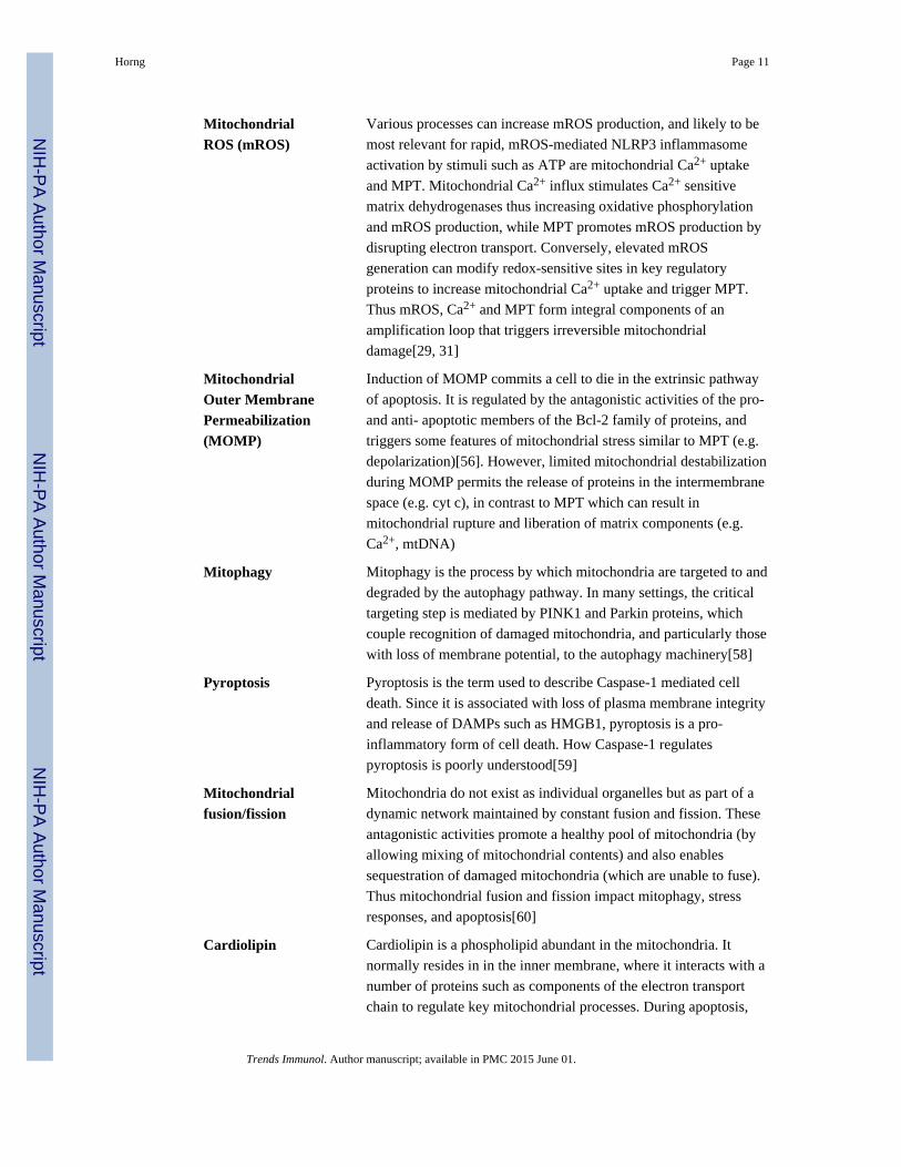
MitochondrialROS (mROS)
Various processes can increase mROS production, and likely to be
most relevant for rapid, mROS-mediated NLRP3 inflammasome
activation by stimuli such as ATP are mitochondrial Ca2+ uptake
and MPT. Mitochondrial Ca2+ influx stimulates Ca2+ sensitive
matrix dehydrogenases thus increasing oxidative phosphorylation
and mROS production, while MPT promotes mROS production by
disrupting electron transport. Conversely, elevated mROS
generation can modify redox-sensitive sites in key regulatory
proteins to increase mitochondrial Ca2+ uptake and trigger MPT.
Thus mROS, Ca2+ and MPT form integral components of an
amplification loop that triggers irreversible mitochondrial
damage[29, 31]
MitochondrialOuter MembranePermeabilization(MOMP)
Induction of MOMP commits a cell to die in the extrinsic pathway
of apoptosis. It is regulated by the antagonistic activities of the pro-
and anti- apoptotic members of the Bcl-2 family of proteins, and
triggers some features of mitochondrial stress similar to MPT (e.g.
depolarization)[56]. However, limited mitochondrial destabilization
during MOMP permits the release of proteins in the intermembrane
space (e.g. cyt c), in contrast to MPT which can result in
mitochondrial rupture and liberation of matrix components (e.g.
Ca2+, mtDNA)
Mitophagy Mitophagy is the process by which mitochondria are targeted to and
degraded by the autophagy pathway. In many settings, the critical
targeting step is mediated by PINK1 and Parkin proteins, which
couple recognition of damaged mitochondria, and particularly those
with loss of membrane potential, to the autophagy machinery[58]
Pyroptosis Pyroptosis is the term used to describe Caspase-1 mediated cell
death. Since it is associated with loss of plasma membrane integrity
and release of DAMPs such as HMGB1, pyroptosis is a pro-
inflammatory form of cell death. How Caspase-1 regulates
pyroptosis is poorly understood[59]
Mitochondrialfusion/fission
Mitochondria do not exist as individual organelles but as part of a
dynamic network maintained by constant fusion and fission. These
antagonistic activities promote a healthy pool of mitochondria (by
allowing mixing of mitochondrial contents) and also enables
sequestration of damaged mitochondria (which are unable to fuse).
Thus mitochondrial fusion and fission impact mitophagy, stress
responses, and apoptosis[60]
Cardiolipin Cardiolipin is a phospholipid abundant in the mitochondria. It
normally resides in in the inner membrane, where it interacts with a
number of proteins such as components of the electron transport
chain to regulate key mitochondrial processes. During apoptosis,
Horng Page 11
Trends Immunol. Author manuscript; available in PMC 2015 June 01.
NIH
-PA
Author M
anuscriptN
IH-P
A A
uthor Manuscript
NIH
-PA
Author M
anuscript
cardiolipin oxidation and redistribution to the outer mitochondrial
membrane not only disrupts mitochondrial function, but appears to
actively trigger cell death through multiple mechanisms, including
regulation of MOMP induction, cytochrome c release, Caspase-8
activation, and as discussed here, NLRP3 inflammasome
activation[48, 49]
References
1. Agostini L, et al. NALP3 forms an IL-1beta-processing inflammasome with increased activity inMuckle-Wells autoinflammatory disorder. Immunity. 2004; 20(3):319–25. [PubMed: 15030775]
2. Mariathasan S, et al. Cryopyrin activates the inflammasome in response to toxins and ATP. Nature.2006; 440(7081):228–32. [PubMed: 16407890]
3. Martinon F, et al. Gout-associated uric acid crystals activate the NALP3 inflammasome. Nature.2006; 440(7081):237–41. [PubMed: 16407889]
4. Kanneganti TD, et al. Bacterial RNA and small antiviral compounds activate caspase-1 throughcryopyrin/Nalp3. Nature. 2006; 440(7081):233–6. [PubMed: 16407888]
5. Franchi L, et al. The inflammasome: a caspase-1-activation platform that regulates immuneresponses and disease pathogenesis. Nature immunology. 2009; 10(3):241–7. [PubMed: 19221555]
6. Lamkanfi M V, Dixit M. Inflammasomes and their roles in health and disease. Annual review of celland developmental biology. 2012; 28:137–61.
7. Wen H, Miao EA, Ting JP. Mechanisms of NOD-like receptor-associated inflammasome activation.Immunity. 2013; 39(3):432–41. [PubMed: 24054327]
8. Strowig T, et al. Inflammasomes in health and disease. Nature. 2012; 481(7381):278–86. [PubMed:22258606]
9. Rathinam VA, Vanaja SK, Fitzgerald KA. Regulation of inflammasome signaling. Natureimmunology. 2012; 13(4):333–2. [PubMed: 22430786]
10. Hoffman HM, Brydges SD. Genetic and molecular basis of inflammasome-mediated disease. TheJournal of biological chemistry. 2011; 286(13):10889–96. [PubMed: 21296874]
11. Jin C, Flavell RA. Molecular mechanism of NLRP3 inflammasome activation. J Clin Immunol.2010; 30(5):628–31. [PubMed: 20589420]
12. Brough D, et al. Ca2+ stores and Ca2+ entry differentially contribute to the release of IL-1 betaand IL-1 alpha from murine macrophages. J Immunol. 2003; 170(6):3029–36. [PubMed:12626557]
13. Feldmeyer L, et al. The inflammasome mediates UVB-induced activation and secretion ofinterleukin-1beta by keratinocytes. Curr Biol. 2007; 17(13):1140–5. [PubMed: 17600714]
14. Chu J, et al. Cholesterol-dependent cytolysins induce rapid release of mature IL-1beta from murinemacrophages in a NLRP3 inflammasome and cathepsin B-dependent manner. J Leukoc Biol.2009; 86(5):1227–38. [PubMed: 19675207]
15. Lee HM, et al. Mycobacterium abscessus activates the NLRP3 inflammasome via Dectin-1-Sykand p62/SQSTM1. Immunology and cell biology. 2012; 90(6):601–10. [PubMed: 21876553]
16. Murakami T, et al. Critical role for calcium mobilization in activation of the NLRP3inflammasome. Proceedings of the National Academy of Sciences of the United States ofAmerica. 2012; 109(28):11282–7. [PubMed: 22733741]
17. Lee GS, et al. The calcium-sensing receptor regulates the NLRP3 inflammasome through Ca2+and cAMP. Nature. 2012; 492(7427):123–7. [PubMed: 23143333]
18. Zhong Z, et al. TRPM2 links oxidative stress to NLRP3 inflammasome activation. Naturecommunications. 2013; 4:1611.
19. Triantafilou K, et al. The complement membrane attack complex triggers intracellular Ca2+ fluxesleading to NLRP3 inflammasome activation. Journal of cell science. 2013; 126(Pt 13):2903–13.[PubMed: 23613465]
Horng Page 12
Trends Immunol. Author manuscript; available in PMC 2015 June 01.
NIH
-PA
Author M
anuscriptN
IH-P
A A
uthor Manuscript
NIH
-PA
Author M
anuscript
20. Abdul-Sater AA, et al. Cyclic-di-GMP and cyclic-di-AMP activate the NLRP3 inflammasome.EMBO reports. 2013; 14(10):900–6. [PubMed: 24008845]
21. Ito M, Yanagi Y, Ichinohe T. Encephalomyocarditis virus viroporin 2B activates NLRP3inflammasome. PLoS pathogens. 2012; 8(8):e1002857. [PubMed: 22916014]
22. Rossol M, et al. Extracellular Ca2+ is a danger signal activating the NLRP3 inflammasomethrough G protein-coupled calcium sensing receptors. Nature communications. 2012; 3:1329.
23. Lopez-Castejon G, et al. P2X(7) receptor-mediated release of cathepsins from macrophages is acytokine-independent mechanism potentially involved in joint diseases. J Immunol. 2010; 185(4):2611–9. [PubMed: 20639492]
24. Compan V, et al. Cell volume regulation modulates NLRP3 inflammasome activation. Immunity.2012; 37(3):487–500. [PubMed: 22981536]
25. Lau BW, et al. Deoxycholic acid activates protein kinase C and phospholipase C via increasedCa2+ entry at plasma membrane. Gastroenterology. 2005; 128(3):695–707. [PubMed: 15765405]
26. Witting A, et al. P2X7 receptors control 2-arachidonoylglycerol production by microglial cells.Proc Natl Acad Sci U S A. 2004; 101(9):3214–9. [PubMed: 14976257]
27. Borodinsky LN, Spitzer NC. Second messenger pas de deux: the coordinated dance betweencalcium and cAMP. Science’s STKE: signal transduction knowledge environment. 2006;2006(336):pe22.
28. Patergnani S, et al. Calcium signaling around Mitochondria Associated Membranes (MAMs). CellCommun Signal. 2011; 9:19. [PubMed: 21939514]
29. Csordas G, Hajnoczky G. SR/ER-mitochondrial local communication: calcium and ROS. BiochimBiophys Acta. 2009; 1787(11):1352–62. [PubMed: 19527680]
30. Lemasters JJ, et al. Mitochondrial calcium and the permeability transition in cell death. BiochimBiophys Acta. 2009; 1787(11):1395–401. [PubMed: 19576166]
31. Camello-Almaraz C, et al. Mitochondrial reactive oxygen species and Ca2+ signaling. Americanjournal of physiology Cell physiology. 2006; 291(5):C1082–8. [PubMed: 16760264]
32. Misawa T, et al. Microtubule-driven spatial arrangement of mitochondria promotes activation ofthe NLRP3 inflammasome. Nature immunology. 2013; 14(5):454–60. [PubMed: 23502856]
33. Baughman JM, et al. Integrative genomics identifies MCU as an essential component of themitochondrial calcium uniporter. Nature. 2011; 476(7360):341–5. [PubMed: 21685886]
34. De Stefani D, et al. A forty-kilodalton protein of the inner membrane is the mitochondrial calciumuniporter. Nature. 2011; 476(7360):336–40. [PubMed: 21685888]
35. Liu X, Hajnoczky G. Ca2+-dependent regulation of mitochondrial dynamics by the Miro-Miltoncomplex. The international journal of biochemistry & cell biology. 2009; 41(10):1972–6.[PubMed: 19481172]
36. Ichinohe T, et al. Mitochondrial protein mitofusin 2 is required for NLRP3 inflammasomeactivation after RNA virus infection. Proceedings of the National Academy of Sciences of theUnited States of America. 2013; 110(44):17963–8. [PubMed: 24127597]
37. Petrilli V, et al. Activation of the NALP3 inflammasome is triggered by low intracellularpotassium concentration. Cell Death Differ. 2007; 14(9):1583–9. [PubMed: 17599094]
38. Munoz-Planillo R, et al. K(+) efflux is the common trigger of NLRP3 inflammasome activation bybacterial toxins and particulate matter. Immunity. 2013; 38(6):1142–53. [PubMed: 23809161]
39. Szabo I, et al. Physiology of potassium channels in the inner membrane of mitochondria. PflugersArchiv: European journal of physiology. 2012; 463(2):231–46. [PubMed: 22089812]
40. Nowikovsky K, Schweyen RJ, Bernardi P. Pathophysiology of mitochondrial volume homeostasis:potassium transport and permeability transition. Biochimica et biophysica acta. 2009; 1787(5):345–50. [PubMed: 19007745]
41. Zhou R, et al. A role for mitochondria in NLRP3 inflammasome activation. Nature. 2011;469(7329):221–5. [PubMed: 21124315]
42. Nakahira K, et al. Autophagy proteins regulate innate immune responses by inhibiting the releaseof mitochondrial DNA mediated by the NALP3 inflammasome. Nat Immunol. 2011; 12(3):222–30. [PubMed: 21151103]
Horng Page 13
Trends Immunol. Author manuscript; available in PMC 2015 June 01.
NIH
-PA
Author M
anuscriptN
IH-P
A A
uthor Manuscript
NIH
-PA
Author M
anuscript
43. Lupfer C, et al. Receptor interacting protein kinase 2-mediated mitophagy regulates inflammasomeactivation during virus infection. Nature immunology. 2013; 14(5):480–8. [PubMed: 23525089]
44. Iyer SS, et al. Mitochondrial cardiolipin is required for Nlrp3 inflammasome activation. Immunity.2013; 39(2):311–23. [PubMed: 23954133]
45. Bernardi P, et al. The mitochondrial permeability transition. Biofactors. 1998; 8(3–4):273–81.[PubMed: 9914829]
46. Elrod JW, Molkentin JD. Physiologic functions of cyclophilin D and the mitochondrialpermeability transition pore. Circulation journal: official journal of the Japanese CirculationSociety. 2013; 77(5):1111–22. [PubMed: 23538482]
47. Shimada K, et al. Oxidized Mitochondrial DNA Activates the NLRP3 Inflammasome duringApoptosis. Immunity. 2012
48. Paradies G, et al. Role of cardiolipin peroxidation and Ca2+ in mitochondrial dysfunction anddisease. Cell Calcium. 2009; 45(6):643–50. [PubMed: 19368971]
49. Chicco AJ, Sparagna GC. Role of cardiolipin alterations in mitochondrial dysfunction and disease.American journal of physiology Cell physiology. 2007; 292(1):C33–44. [PubMed: 16899548]
50. Chu CT, et al. Cardiolipin externalization to the outer mitochondrial membrane acts as anelimination signal for mitophagy in neuronal cells. Nature cell biology. 2013; 15(10):1197–205.
51. Wen H, et al. Fatty acid-induced NLRP3-ASC inflammasome activation interferes with insulinsignaling. Nature immunology. 2011; 12(5):408–15. [PubMed: 21478880]
52. Park S, et al. The mitochondrial antiviral protein MAVS associates with NLRP3 and regulates itsinflammasome activity. Journal of immunology. 2013; 191(8):4358–66.
53. Mitoma H, et al. The DHX33 RNA helicase senses cytosolic RNA and activates the NLRP3inflammasome. Immunity. 2013; 39(1):123–35. [PubMed: 23871209]
54. Zhao Y, et al. The NLRC4 inflammasome receptors for bacterial flagellin and type III secretionapparatus. Nature. 2011; 477(7366):596–600. [PubMed: 21918512]
55. Kofoed EM, Vance RE. Innate immune recognition of bacterial ligands by NAIPs determinesinflammasome specificity. Nature. 2011; 477(7366):592–5. [PubMed: 21874021]
56. Green DR, Kroemer G. The pathophysiology of mitochondrial cell death. Science. 2004;305(5684):626–9. [PubMed: 15286356]
57. Galluzzi L, Kepp O, Kroemer G. Mitochondria: master regulators of danger signalling. Naturereviews Molecular cell biology. 2012; 13(12):780–8.
58. Youle RJ, Narendra DP. Mechanisms of mitophagy. Nature reviews Molecular cell biology. 2011;12(1):9–14.
59. Lamkanfi M V, Dixit M. Manipulation of host cell death pathways during microbial infections.Cell host & microbe. 2010; 8(1):44–54. [PubMed: 20638641]
60. Chan DC. Fusion and fission: interlinked processes critical for mitochondrial health. Annualreview of genetics. 2012; 46:265–87.
Horng Page 14
Trends Immunol. Author manuscript; available in PMC 2015 June 01.
NIH
-PA
Author M
anuscriptN
IH-P
A A
uthor Manuscript
NIH
-PA
Author M
anuscript
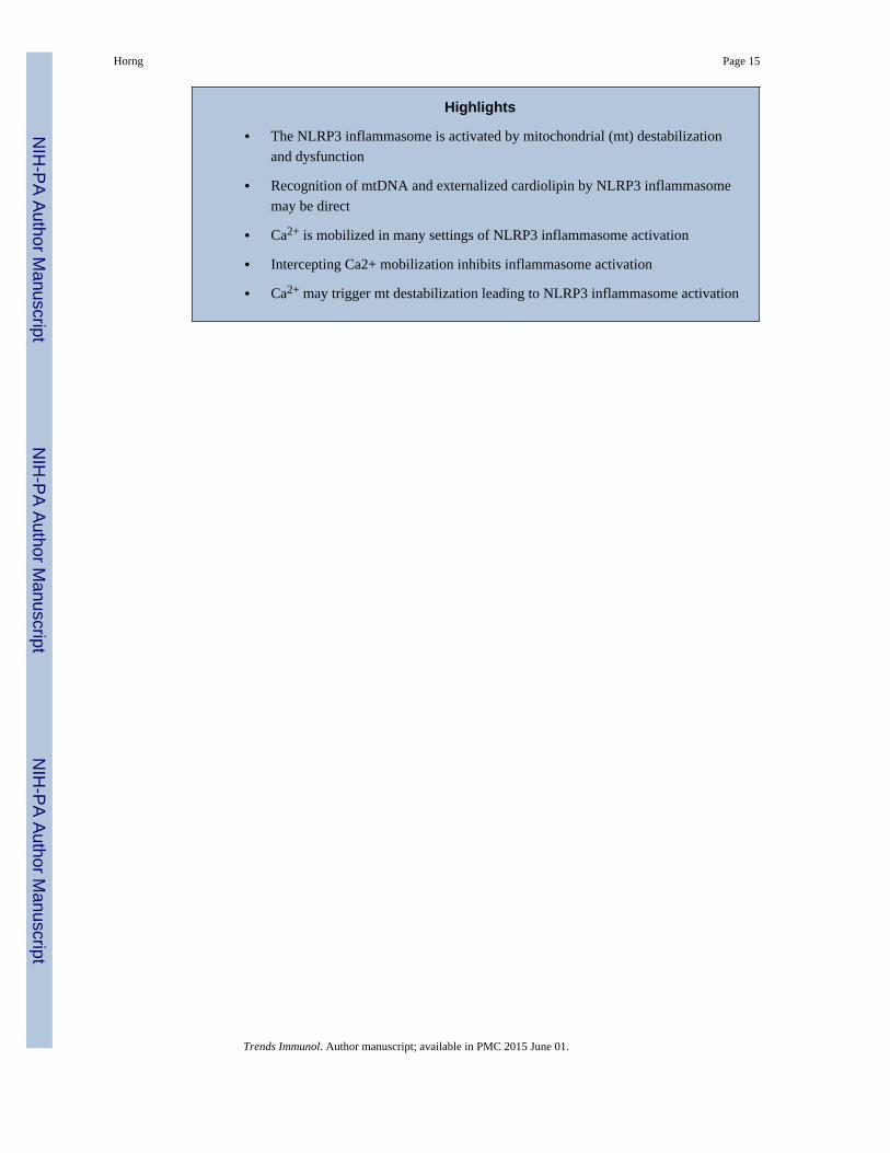
Highlights
• The NLRP3 inflammasome is activated by mitochondrial (mt) destabilization
and dysfunction
• Recognition of mtDNA and externalized cardiolipin by NLRP3 inflammasome
may be direct
• Ca2+ is mobilized in many settings of NLRP3 inflammasome activation
• Intercepting Ca2+ mobilization inhibits inflammasome activation
• Ca2+ may trigger mt destabilization leading to NLRP3 inflammasome activation
Horng Page 15
Trends Immunol. Author manuscript; available in PMC 2015 June 01.
NIH
-PA
Author M
anuscriptN
IH-P
A A
uthor Manuscript
NIH
-PA
Author M
anuscript
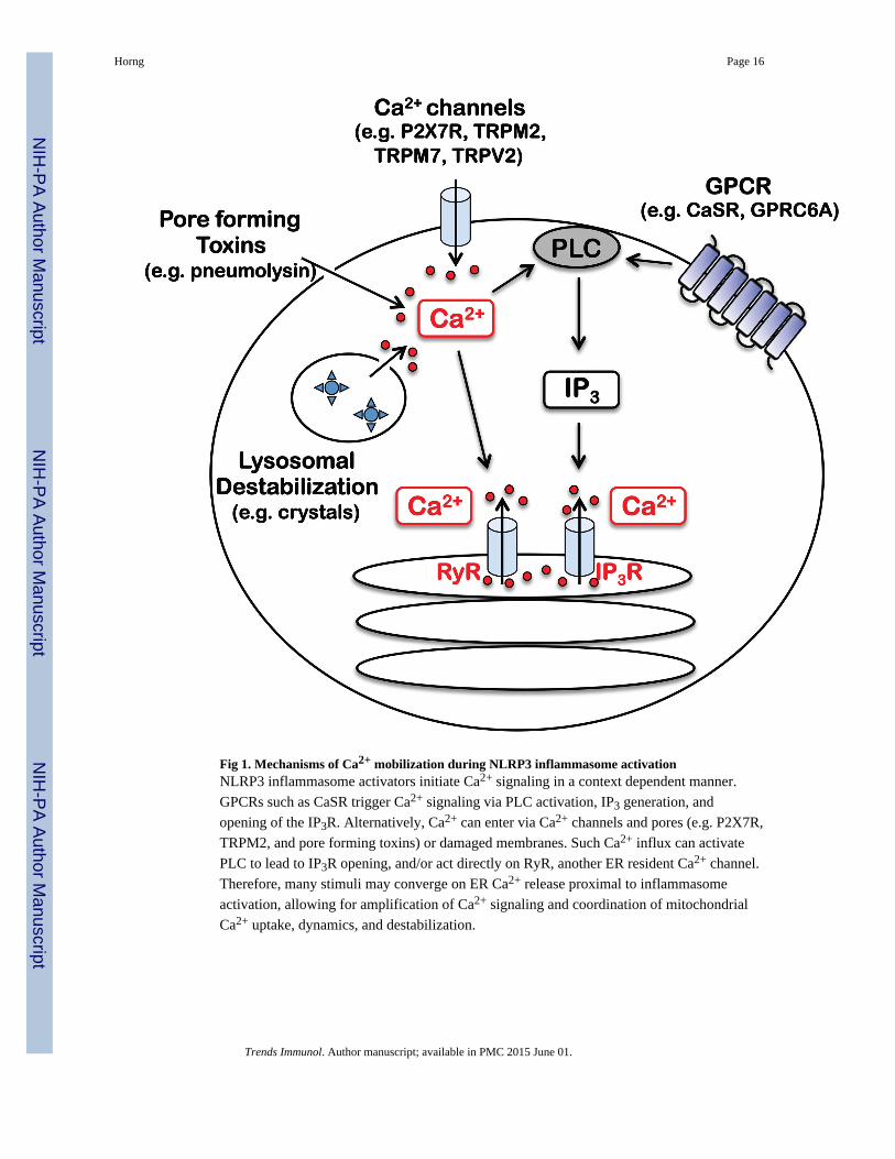
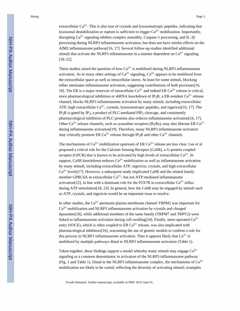
Fig 1. Mechanisms of Ca2+ mobilization during NLRP3 inflammasome activationNLRP3 inflammasome activators initiate Ca2+ signaling in a context dependent manner.
GPCRs such as CaSR trigger Ca2+ signaling via PLC activation, IP3 generation, and
opening of the IP3R. Alternatively, Ca2+ can enter via Ca2+ channels and pores (e.g. P2X7R,
TRPM2, and pore forming toxins) or damaged membranes. Such Ca2+ influx can activate
PLC to lead to IP3R opening, and/or act directly on RyR, another ER resident Ca2+ channel.
Therefore, many stimuli may converge on ER Ca2+ release proximal to inflammasome
activation, allowing for amplification of Ca2+ signaling and coordination of mitochondrial
Ca2+ uptake, dynamics, and destabilization.
Horng Page 16
Trends Immunol. Author manuscript; available in PMC 2015 June 01.
NIH
-PA
Author M
anuscriptN
IH-P
A A
uthor Manuscript
NIH
-PA
Author M
anuscript
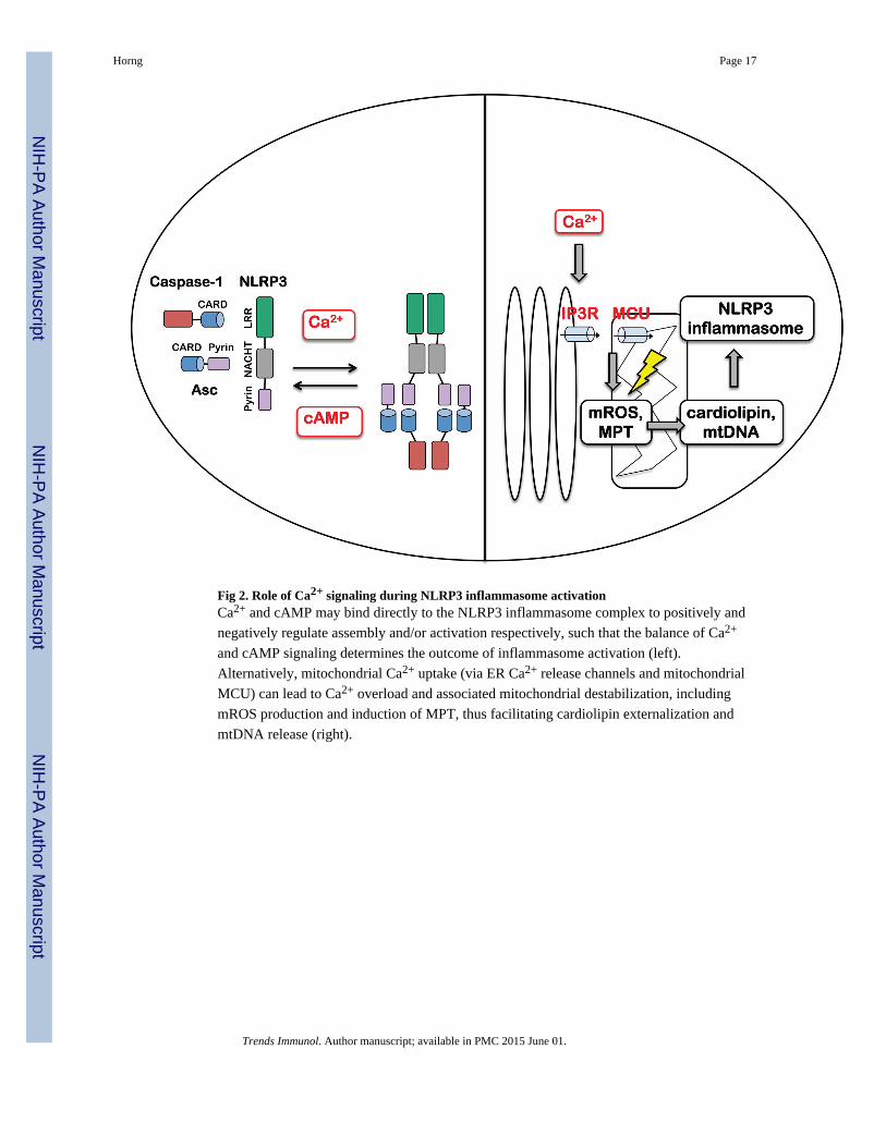
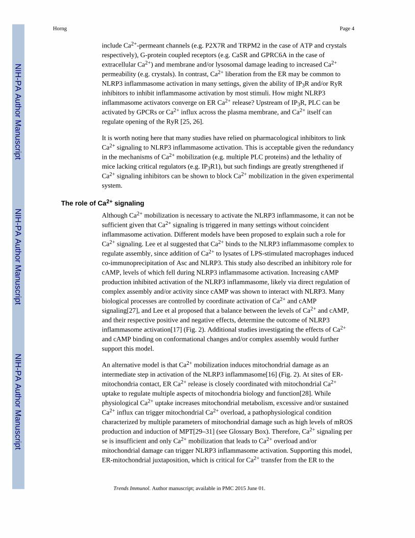
Fig 2. Role of Ca2+ signaling during NLRP3 inflammasome activationCa2+ and cAMP may bind directly to the NLRP3 inflammasome complex to positively and
negatively regulate assembly and/or activation respectively, such that the balance of Ca2+
and cAMP signaling determines the outcome of inflammasome activation (left).
Alternatively, mitochondrial Ca2+ uptake (via ER Ca2+ release channels and mitochondrial
MCU) can lead to Ca2+ overload and associated mitochondrial destabilization, including
mROS production and induction of MPT, thus facilitating cardiolipin externalization and
mtDNA release (right).
Horng Page 17
Trends Immunol. Author manuscript; available in PMC 2015 June 01.
NIH
-PA
Author M
anuscriptN
IH-P
A A
uthor Manuscript
NIH
-PA
Author M
anuscript
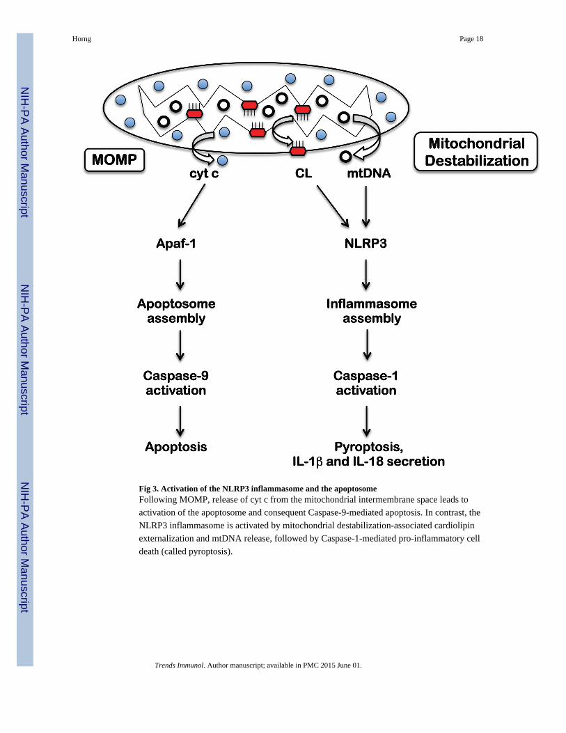
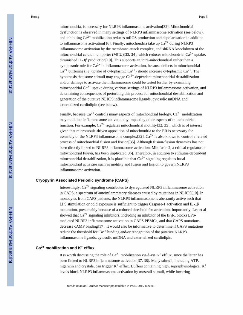
Fig 3. Activation of the NLRP3 inflammasome and the apoptosomeFollowing MOMP, release of cyt c from the mitochondrial intermembrane space leads to
activation of the apoptosome and consequent Caspase-9-mediated apoptosis. In contrast, the
NLRP3 inflammasome is activated by mitochondrial destabilization-associated cardiolipin
externalization and mtDNA release, followed by Caspase-1-mediated pro-inflammatory cell
death (called pyroptosis).
Horng Page 18
Trends Immunol. Author manuscript; available in PMC 2015 June 01.
NIH
-PA
Author M
anuscriptN
IH-P
A A
uthor Manuscript
NIH
-PA
Author M
anuscript
NIH
-PA
Author M
anuscriptN
IH-P
A A
uthor Manuscript
NIH
-PA
Author M
anuscript
Horng Page 19
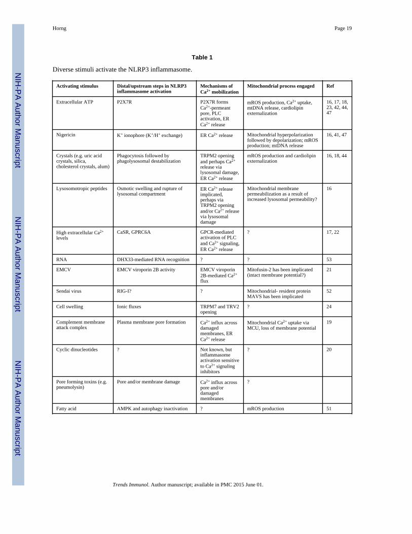
Table 1
Diverse stimuli activate the NLRP3 inflammasome.
Activating stimulus Distal/upstream steps in NLRP3inflammasome activation
Mechanisms ofCa2+ mobilization
Mitochondrial process engaged Ref
Extracellular ATP P2X7R P2X7R formsCa2+-permeantpore, PLCactivation, ERCa2+ release
mROS production, Ca2+ uptake,mtDNA release, cardiolipinexternalization
16, 17, 18,23, 42, 44,47
Nigericin K+ ionophore (K+/H+ exchange) ER Ca2+ release Mitochondrial hyperpolarizationfollowed by depolarization; mROSproduction; mtDNA release
16, 41, 47
Crystals (e.g. uric acidcrystals, silica,cholesterol crystals, alum)
Phagocytosis followed byphagolysosomal destabilization
TRPM2 openingand perhaps Ca2+
release vialysosomal damage,ER Ca2+ release
mROS production and cardiolipinexternalization
16, 18, 44
Lysosomotropic peptides Osmotic swelling and rupture oflysosomal compartment
ER Ca2+ releaseimplicated,perhaps viaTRPM2 openingand/or Ca2+ releasevia lysosomaldamage
Mitochondrial membranepermeabilization as a result ofincreased lysosomal permeability?
16
High extracellular Ca2+
levelsCaSR, GPRC6A GPCR-mediated
activation of PLCand Ca2+ signaling,ER Ca2+ release
? 17, 22
RNA DHX33-mediated RNA recognition ? ? 53
EMCV EMCV viroporin 2B activity EMCV viroporin2B-mediated Ca2+
flux
Mitofusin-2 has been implicated(intact membrane potential?)
21
Sendai virus RIG-I? ? Mitochondrial- resident proteinMAVS has been implicated
52
Cell swelling Ionic fluxes TRPM7 and TRV2opening
? 24
Complement membraneattack complex
Plasma membrane pore formation Ca2+ influx acrossdamagedmembranes, ERCa2+ release
Mitochondrial Ca2+ uptake viaMCU, loss of membrane potential
19
Cyclic dinucleotides ? Not known, butinflammasomeactivation sensitiveto Ca2+ signalinginhibitors
? 20
Pore forming toxins (e.g.pneumolysin)
Pore and/or membrane damage Ca2+ influx acrosspore and/ordamagedmembranes
?
Fatty acid AMPK and autophagy inactivation ? mROS production 51
Trends Immunol. Author manuscript; available in PMC 2015 June 01.