NIH Public Access films J Memb Sci Effect of
17
Effect of β-sheet crystalline content on mass transfer in silk films Kiran A. Karve a , Eun Seok Gil b , Stephen P. McCarthy c , and David L. Kaplan b,* a Biomedical Engineering and Biotechnology Program, University of Massachusetts at Lowell, Lowell MA 01854 USA b Departments of Biomedical Engineering, Chemical and Biological Engineering, Tufts University, 4 Colby Street, Medford MA 02155 USA c Department of Plastics Engineering University of Massachusetts at Lowell, 1 University Avenue, Lowell MA 01854 USA Abstract The material properties of silk are favorable for drug delivery due to the ability to control material structure and morphology under ambient, aqueous processing conditions. Mass transport of compounds with varying physical-chemical characteristics was studied in silk fibroin films with control of β-sheet crystalline content. Two compounds, vitamin B12 and fluorescein isothiocynate (FITC) labeled lysozyme were studied in a diffusion apparatus to determine transport through silk films. The films exhibited size exclusion phenomenon with permeability coefficients with contrasting trends with increases in β-sheet crystallinity. The size exclusion phenomenon observed with the two model compounds was characterized by contrasting trends in permeability coefficients of the films as a function of β-sheet crystallinity. The diffusivity of the compounds was examined in the context of free volume theory. Apart from the β-sheet crystallinity, size of the compound and its interactions with silk influenced mass transfer. Diffusivity of vitamin B12 was modeled to define a power law relationship with β-sheet crystallinity. The results of the study demonstrate that diffusion of therapeutic agents though silk fibroin films can be directed to match a desired rate by modulating secondary structure of the silk proteins. Keywords Silk; β-sheet; Diffusion; Permeability; Controlled drug delivery 1. Introduction Controlled release is desirable for therapeutics as it enables sustained release of drugs within effective therapeutic limits over extended periods of time [1,2]. This control enhances the efficacy of therapeutic agents [1,2]. Synthetic biodegradable polymers such as polyesters [3] and polyanhydrides [4] have been crafted into matrices with well-defined release kinetics. However, limits to the compatibility between the degradation products of these polymers © 2011 Elsevier B.V. All rights reserved. * Corresponding author: Tel.: +1 617 627 3251; fax +1 617 627 3231. [email protected] (D.L. Kaplan). Publisher's Disclaimer: This is a PDF file of an unedited manuscript that has been accepted for publication. As a service to our customers we are providing this early version of the manuscript. The manuscript will undergo copyediting, typesetting, and review of the resulting proof before it is published in its final citable form. Please note that during the production process errors may be discovered which could affect the content, and all legal disclaimers that apply to the journal pertain. NIH Public Access Author Manuscript J Memb Sci. Author manuscript; available in PMC 2012 November 1. Published in final edited form as: J Memb Sci. 2011 November 1; 383(1-2): 44–49. doi:10.1016/j.memsci.2011.08.032. NIH-PA Author Manuscript NIH-PA Author Manuscript NIH-PA Author Manuscript
Transcript of NIH Public Access films J Memb Sci Effect of

Effect of β-sheet crystalline content on mass transfer in silkfilms
Kiran A. Karvea, Eun Seok Gilb, Stephen P. McCarthyc, and David L. Kaplanb,*
aBiomedical Engineering and Biotechnology Program, University of Massachusetts at Lowell,Lowell MA 01854 USAbDepartments of Biomedical Engineering, Chemical and Biological Engineering, Tufts University,4 Colby Street, Medford MA 02155 USAcDepartment of Plastics Engineering University of Massachusetts at Lowell, 1 University Avenue,Lowell MA 01854 USA
AbstractThe material properties of silk are favorable for drug delivery due to the ability to control materialstructure and morphology under ambient, aqueous processing conditions. Mass transport ofcompounds with varying physical-chemical characteristics was studied in silk fibroin films withcontrol of β-sheet crystalline content. Two compounds, vitamin B12 and fluorescein isothiocynate(FITC) labeled lysozyme were studied in a diffusion apparatus to determine transport through silkfilms. The films exhibited size exclusion phenomenon with permeability coefficients withcontrasting trends with increases in β-sheet crystallinity. The size exclusion phenomenon observedwith the two model compounds was characterized by contrasting trends in permeabilitycoefficients of the films as a function of β-sheet crystallinity. The diffusivity of the compoundswas examined in the context of free volume theory. Apart from the β-sheet crystallinity, size of thecompound and its interactions with silk influenced mass transfer. Diffusivity of vitamin B12 wasmodeled to define a power law relationship with β-sheet crystallinity. The results of the studydemonstrate that diffusion of therapeutic agents though silk fibroin films can be directed to matcha desired rate by modulating secondary structure of the silk proteins.
KeywordsSilk; β-sheet; Diffusion; Permeability; Controlled drug delivery
1. IntroductionControlled release is desirable for therapeutics as it enables sustained release of drugs withineffective therapeutic limits over extended periods of time [1,2]. This control enhances theefficacy of therapeutic agents [1,2]. Synthetic biodegradable polymers such as polyesters [3]and polyanhydrides [4] have been crafted into matrices with well-defined release kinetics.However, limits to the compatibility between the degradation products of these polymers
© 2011 Elsevier B.V. All rights reserved.*Corresponding author: Tel.: +1 617 627 3251; fax +1 617 627 3231. [email protected] (D.L. Kaplan).Publisher's Disclaimer: This is a PDF file of an unedited manuscript that has been accepted for publication. As a service to ourcustomers we are providing this early version of the manuscript. The manuscript will undergo copyediting, typesetting, and review ofthe resulting proof before it is published in its final citable form. Please note that during the production process errors may bediscovered which could affect the content, and all legal disclaimers that apply to the journal pertain.
NIH Public AccessAuthor ManuscriptJ Memb Sci. Author manuscript; available in PMC 2012 November 1.
Published in final edited form as:J Memb Sci. 2011 November 1; 383(1-2): 44–49. doi:10.1016/j.memsci.2011.08.032.
NIH
-PA Author Manuscript
NIH
-PA Author Manuscript
NIH
-PA Author Manuscript
with the encapsulated therapeutic [2,5] and the surrounding tissue [6,7], limit applications.Natural polymers such as chitosan [8] and collagen [9] have enabled the design of releasematrices with biocompatibility and biodegradability, however often modifications [10] orcrosslinking [9] are required. Such changes can be problematic in terms of compatibilitywith the entrapped drug and control of degradation lifetime. Moreover, these polymers areunstable under many processing conditions, such as temperature, pH and other variables [9].Therefore, there remains a need for biomaterials which (i) excel in biocompatibility andcontrolled biodegradability, (ii) permit ambient processing conditions and (iii) facilitateefficacious and predictable delivery of a multitude of therapeutic agents.
Over the last decade, silk fibroin obtained from cocoons of Bombxy mori (B. mori)silkworms has been studied for drug delivery applications. Known typically for highmechanical strength [11], silk is biocompatible [12,13] and biodegradable with control oflifetime from hours to years depending on processing conditions and treatment [14,15]. Thistunable characteristic is particularly suited for applications of silk biomaterials for controlledrelease devices. Furthermore, the properties of silk biomaterials can be manipulated withoutincorporating harmful chemical agents, thereby positively influencing the utility of silk-based devices for medically-related applications.. Since beta sheet content reflectsdegradation lifetime in vivo, options for long term implants that retain degradability are alsoa plus with silk-based devices. The protein can be processed into multiple material formatsfor encapsulating therapeutic agents. The mild conditions during processing favor thestability of both the therapeutic and the matrix [16–18]. Hence, a number of strategies basedon silk coating thickness and β-sheet crystalline content have been used to facilitatecontrolled release [19–22]. Efforts to understand mass transport in silk biomaterials arelimited, and studies into this process can help provide improved fundamental insight as wellas more predictable control of drug delivery from silk.
In the present study, the mass transport of model two compounds vitamin B12 and lysozymewas studied as a function of β-sheet crystallinity of silk. Thin films were cast to mimicsingle layer silk coatings to control drug availability. The β-sheet crystallinity was varied bywater annealing (W.A.) and/or methanol annealing (M.A.) during film processing. Masstransfer was analyzed using free volume theory.
2. Materials and Methods2.1. Materials
Bombyx mori (silkworm) cocoons were supplied by Tajima Shoji Co (Yokohama, Japan).All chemicals, analytical or pharmaceutical grade, were purchased from Sigma-Aldrich (St.Louis, MO, USA) and used as received.
2.2. Preparation of silk filmsAqueous silk fibroin solution was prepared as described previously [23]. Briefly, B. moricocoons were boiled for 30 min in 0.02 M Na2CO3 solution to extract the fibroin from thesericin glue-like proteins. The fibers were then dissolved in a 9.3 M solution of lithiumbromide at 60°C and dialyzed in Slide-a-Lyser cassettes (MWCO 3500 g/mol) (Pierce,Woburn, MA, USA) against Milli-Q water. The resulting solution was centrifuged toremove any suspended impurities. The concentration of the stock was estimated by weighingthe residual solid content of a known volume of fibroin solution. The concentration of thesilk fibroin solution was adjusted to 1% (w/v) and a 386 μL aliquot, estimated from therelation between fibroin concentration and film thickness [24], was cast on circularpoly(dimethoxysiloxane) molds (28 mm diameter) to obtain films with 2 μm thickness. Thecast films were dried overnight in a clean chemical hood. β-sheet crystallinity was induced
Karve et al. Page 2
J Memb Sci. Author manuscript; available in PMC 2012 November 1.
NIH
-PA Author Manuscript
NIH
-PA Author Manuscript
NIH
-PA Author Manuscript
in the films by four different treatments (i) 6 h W.A., (ii) 15 h W.A., (iii) 15 h W.A.followed by 2 h M.A. or (iv) 2 h M.A. treatments. Films were water annealed by subjectingthem to a water vapor environment inside an air evacuated desiccator, partially filled withwater for a duration defined by the treatment [24]. To methanol anneal, the films weresoaked in a solution of 80% methanol and subsequently rinsed with Milli-Q water to removetraces of solvent.
2.3 Characterization2.3.1 Thickness of dry films—The thickness of the cast films was measured by imagingtheir cross-sections under a scanning electron microscope. The 2 h M.A. films were freezefractured in liquid nitrogen and sputter coated with platinum-palladium alloy. The cross-sections were imaged using a Zeiss Ultra 55 Field Emission Scanning Electron Microscope(FESEM) (Carl Zeiss SMT Inc., Germany). Film thicknesses were evaluated using Image Janalysis software at three random locations along the cross section. The thickness of the dryfilm was reported as the average of three films.
2.3.2 Hydration of silk films—The hydration of silk films was studied gravimetricallywith films 100 μm thick and 15 mm in diameter. A linear relationship independent of theannealing process was assumed between the thickness of the film and its hydration. Briefly,an annealed film was soaked in Milli-Q water. At regular time intervals, the film wasremoved from the water, and gently blotted to remove excess water on the surface. The filmwas weighed and then allowed to soak further in water. The process was repeated until noincrease in the weight of the wet films (Ww) (mg) was observed. The films were then driedin an oven and weighed (Wd) (mg). The percent hydration of the film was calculated by (Eq.1) [25]
(1)
The percent hydration was reported as an average of five samples for each annealingprocess.
2.3.3 Fourier transform infrared spectroscopy (FTIR)—The changes in thesecondary structure of silk resulting from the different annealing treatments were analyzedwith a JASCO FT/IR 6200 spectrometer (JASCO, Tokyo, Japan) [26]. The films werescanned 32 times over a wave-number range of 400 – 4000 cm−1 with a resolution of 4cm−1. The spectra were deconvoluted using Opus 5.0 software [27]. The secondarystructures of the films were evaluated in the amide I region (1595 – 1705 cm−1) [27]. The β-sheet crystalline content of the films was quantified by integrating the area under the silk IIpeaks (1614 – 1635 cm−1 and 1695 – 1705 cm−1) [27] and normalizing to the total areaunder the curve in the amide I region.
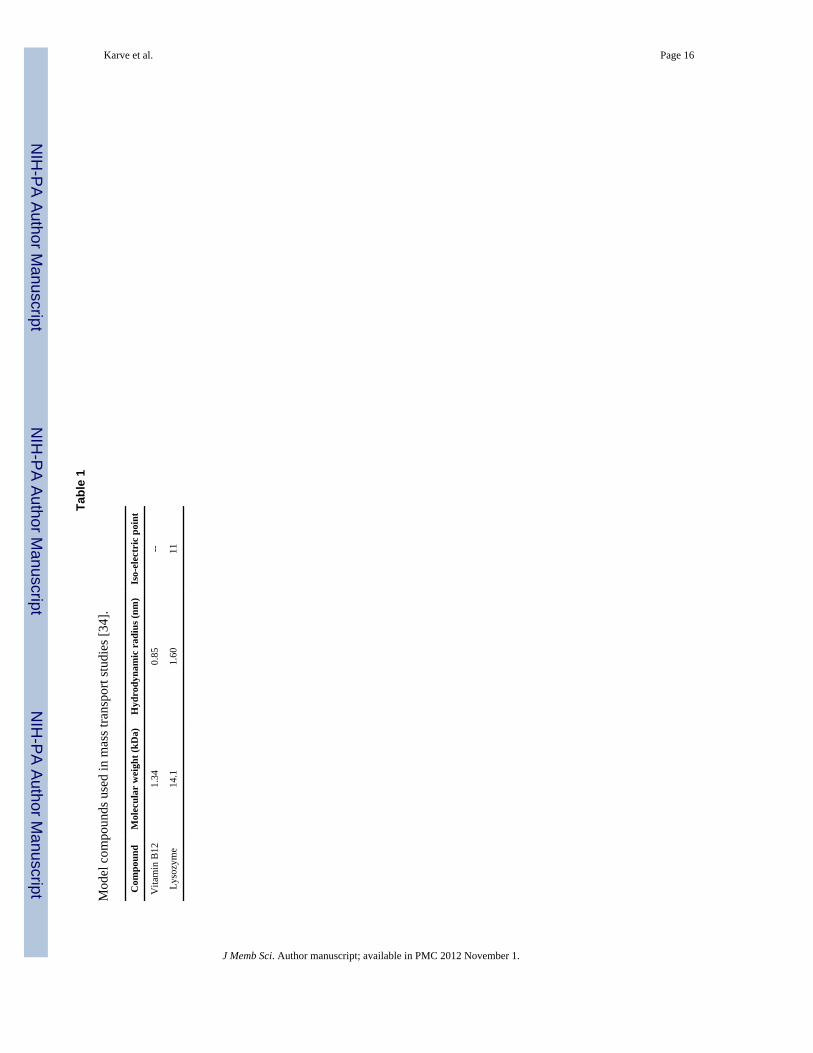
2.4 Model compoundsTwo compounds varying in molecular weight, hydrodynamic radius and ionic character(Table 1) were used to elucidate mass transport across the silk films. Stock solutions of thetwo compounds vitamin B12 (1mg mL−1) and FITC-lysozyme (0.5 mg mL−1) wereprepared in Dulbecco’s phosphate buffered saline (DPBS, pH 7.4) and stored at 8°C.Compound standards in DPBS were analyzed with a UV spectrophotometer (MolecularDevices, CA) by measuring the absorbance (λ = 361 nm) for vitamin B12 and florescence(λexcitation = 488 nm and λemission = 520 nm) for FITC-lysozyme. Standard curves were
Karve et al. Page 3
J Memb Sci. Author manuscript; available in PMC 2012 November 1.
NIH
-PA Author Manuscript
NIH
-PA Author Manuscript
NIH
-PA Author Manuscript
plotted for both compounds. Stability of the compounds for the experiments was ascertainedby spectrophotometric analysis of the compound standards incubated at 37°C (data notincluded).
2.4.1 Conjugation of fluorescein isothiocynate (FITC) to lysozyme—FITClabeled lysozyme was synthesized by adding 500 μL of 1 mg.mL−1 FITC/dimethyl sulfoxideto a 2 mg.mL−1 solution of the protein in 0.1 M carbonate-bicarbonate buffer at pH 9. Thereaction was stirred in dark for 12 h and dialyzed against Milli-Q water to remove the salts.The labeled protein was then lyophilized and stored for further use. The fluorescein toprotein molar ratio was calculated to be 0.55 as per the manufacturer’s instructions. Sincethe calculated molecular weight of 14.3 kDa of the FITC labeled lysozyme did not varysignificantly from that of the unlabeled protein (Table 1), it was assumed to have anegligible effect on the hydrodynamic radius of lysozyme. From the following section,lysozyme and FITC lysozyme have been used inter-changeably in the text.
2.5 Permeation studiesTransport of the model compounds across the silk films with varying crystallinity wasstudied using a standard horizontal twin cell side-by-side diffusion apparatus (PermegearInc. Hellertown, PA). The total capacity of each cell in the apparatus was 7 mL. Filmsequilibrated in DPBS were mounted between the two cells of the apparatus providing a totalarea of 1.54 cm2 for diffusion of compounds. Due to the potential for fracture during storageand handling, the silk films were evaluated immediately after the annealing treatments. Thecompound stock was loaded in the donor cell. An equal volume of DPBS was loaded in thereceptor cell. To prevent concentration polarization, solutions in both the cells were stirredwith magnetic stir bars at 600 rpm. The temperature was maintained at 37°C with a constanttemperature circulating water bath. One hundred μL samples were withdrawn from thereceptor cell at defined time points and replaced with equal amounts of DPBS. The sampleswere analyzed by UV spectrophotometry and concentrations evaluated from standardcurves. Background absorbance and auto-fluorescence associated with silk, were accountedfor by conducting the experiment without any compounds and subtracting the blank readingsfrom the sample readings at the respective time points.
2.5.1 Data Analysis—To calculate the transport parameters, the cumulative mass of acompound permeating through the film was calculated and normalized by the area availablefor diffusion. These data were then plotted as a function of time. The net flux J, (mg cm−2
s−1) of the compound passing through the film was given by the slope of the linear portionthis curve (R2 > 0.97). Thus, the permeability coefficients of the silk films were calculatedunder steady state conditions by Fick’s first law of diffusion expressed as (Eq. 2) [28]
(2)
Here, P (cm s−1) is the permeability coefficient of the film and C0 (mg mL−1) is theconcentration of the donor chamber maintained at sink conditions. The diffusion coefficientsof the model compounds under steady state conditions were calculated from thepermeability coefficient using (Eq. 3)
(3)
Karve et al. Page 4
J Memb Sci. Author manuscript; available in PMC 2012 November 1.
NIH
-PA Author Manuscript
NIH
-PA Author Manuscript
NIH
-PA Author Manuscript
Here, D (cm2 s−1) is the diffusion coefficient, Kd (dimensionless) is the partition coefficientof the model compound between silk and the buffer and h (cm) is the hydrated filmthickness. The hydrated film thickness was estimated indirectly, by calculating the swellingratio of solvent equilibrated 100 μm films.
2.6 Determination of partition coefficient (Kd)The partition coefficient is a measure of solubility and defines the distribution of thecompound in the film relative to that in the surrounding solvent. Higher solubility of thecompound in the films is attributed to greater partitioning resulting from strong interactionsbetween the two. For Kd ≤ 1 the compound is practically insoluble in the film. Partitioncoefficients of the model compounds between the film and the solvent were estimated usingthe solvent depletion method. Silk films, 2 μm in thickness and 15 mm in diameter, weresoaked in stock solutions of the compounds and allowed to equilibrate. The equilibriumconcentrations of the solutions were measured by UV-spectroscopy and partition coefficientwas given by (Eq. 4)
(4)
Here, Ci (mg mL−1) is the initial concentration of compound solution, Cs (mg mL−1) is theequilibrium drug concentration of the solution, Vs (mL) is the volume of the compoundsolution, and Vf (cm3) is the volume of the solvent equilibrated silk films. Vf was calculatedusing the equivalent hydrated film thicknesses estimated from the swelling ratios of films100 μm thick. The partition coefficient is reported as an average of three samples for eachannealing condition.
2.7 Statistical AnalysisThe data were analyzed by single factor analysis of variance (ANOVA). Data wereconsidered statistically significant for p < 0.05.
3 Results and DiscussionSilk matrices have been investigated for delivery of a variety of therapeutics such asadenosine, theophylline emodin, bone morphogenic protein (BMP-2) and plasmid DNA[29]. To characterize the transport of this wide range of compounds we have selectedvitamin B12 to model small molecule therapeutics and lysozyme for peptide therapeutics.
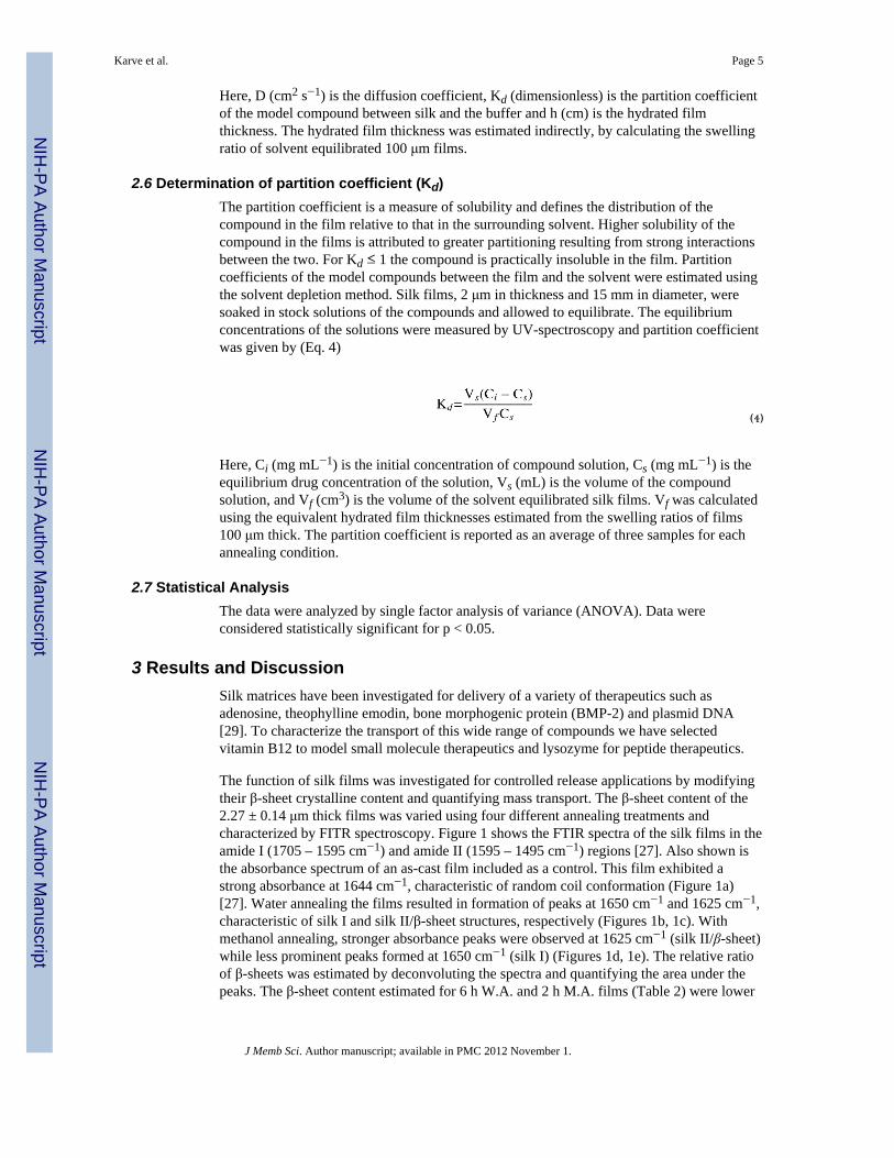
The function of silk films was investigated for controlled release applications by modifyingtheir β-sheet crystalline content and quantifying mass transport. The β-sheet content of the2.27 ± 0.14 μm thick films was varied using four different annealing treatments andcharacterized by FITR spectroscopy. Figure 1 shows the FTIR spectra of the silk films in theamide I (1705 – 1595 cm−1) and amide II (1595 – 1495 cm−1) regions [27]. Also shown isthe absorbance spectrum of an as-cast film included as a control. This film exhibited astrong absorbance at 1644 cm−1, characteristic of random coil conformation (Figure 1a)[27]. Water annealing the films resulted in formation of peaks at 1650 cm−1 and 1625 cm−1,characteristic of silk I and silk II/β-sheet structures, respectively (Figures 1b, 1c). Withmethanol annealing, stronger absorbance peaks were observed at 1625 cm−1 (silk II/β-sheet)while less prominent peaks formed at 1650 cm−1 (silk I) (Figures 1d, 1e). The relative ratioof β-sheets was estimated by deconvoluting the spectra and quantifying the area under thepeaks. The β-sheet content estimated for 6 h W.A. and 2 h M.A. films (Table 2) were lower
Karve et al. Page 5
J Memb Sci. Author manuscript; available in PMC 2012 November 1.
NIH
-PA Author Manuscript
NIH
-PA Author Manuscript
NIH
-PA Author Manuscript
than those observed by Lu et. al (6 h W.A. 30.3 ± 0.7%, 1 h M.A. 40.8 ± 2.1%), butfollowed the same trend with respect to the two treatments [30]. Increasing the duration ofwater annealing process and treating these films with methanol caused only a minor increasein β-sheets. Further, the increase in β-sheet of the 15 h W.A + 2 h M.A. films may beassociated with the inability of silk I formed after water annealing to convert to silk II (β-sheets) on further exposure to methanol [31]. Overall, methanol annealing yielded higher β-sheet content when compared to the water annealing treatments.
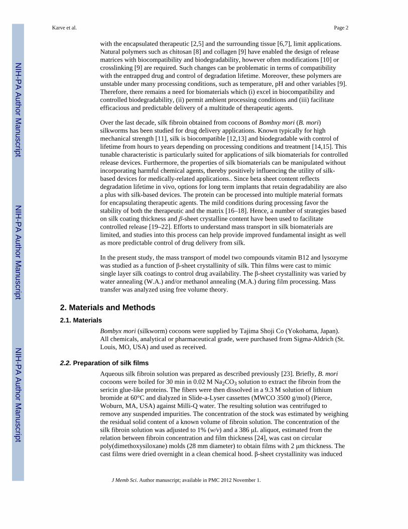
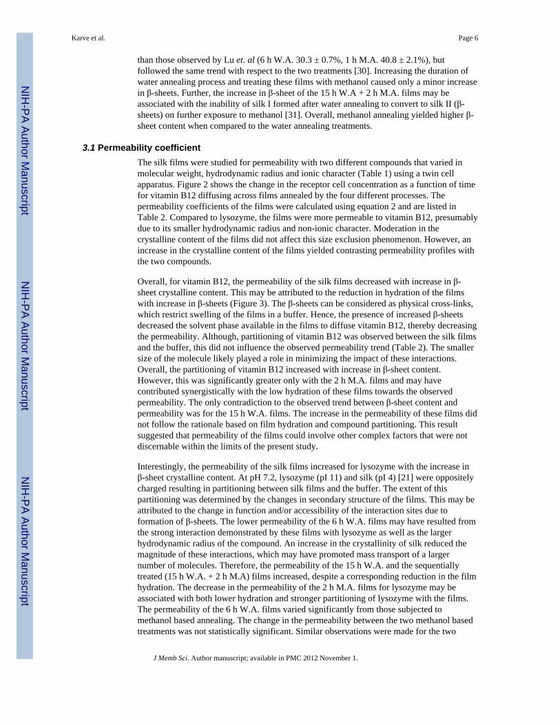
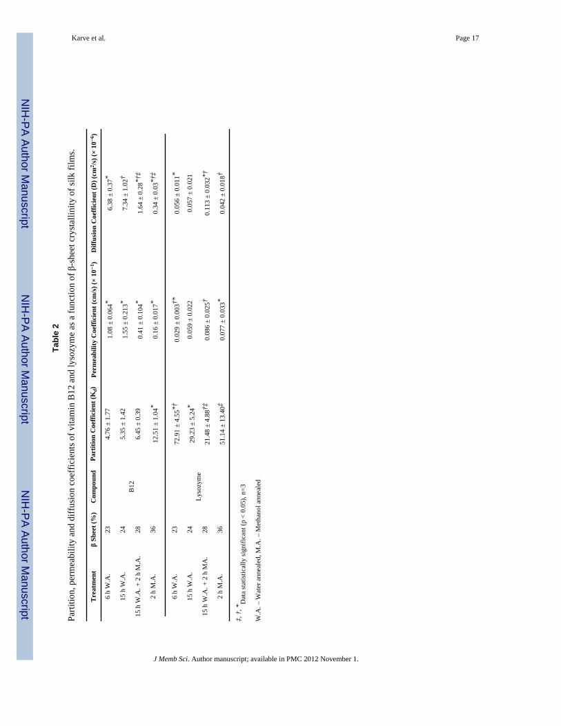
3.1 Permeability coefficientThe silk films were studied for permeability with two different compounds that varied inmolecular weight, hydrodynamic radius and ionic character (Table 1) using a twin cellapparatus. Figure 2 shows the change in the receptor cell concentration as a function of timefor vitamin B12 diffusing across films annealed by the four different processes. Thepermeability coefficients of the films were calculated using equation 2 and are listed inTable 2. Compared to lysozyme, the films were more permeable to vitamin B12, presumablydue to its smaller hydrodynamic radius and non-ionic character. Moderation in thecrystalline content of the films did not affect this size exclusion phenomenon. However, anincrease in the crystalline content of the films yielded contrasting permeability profiles withthe two compounds.
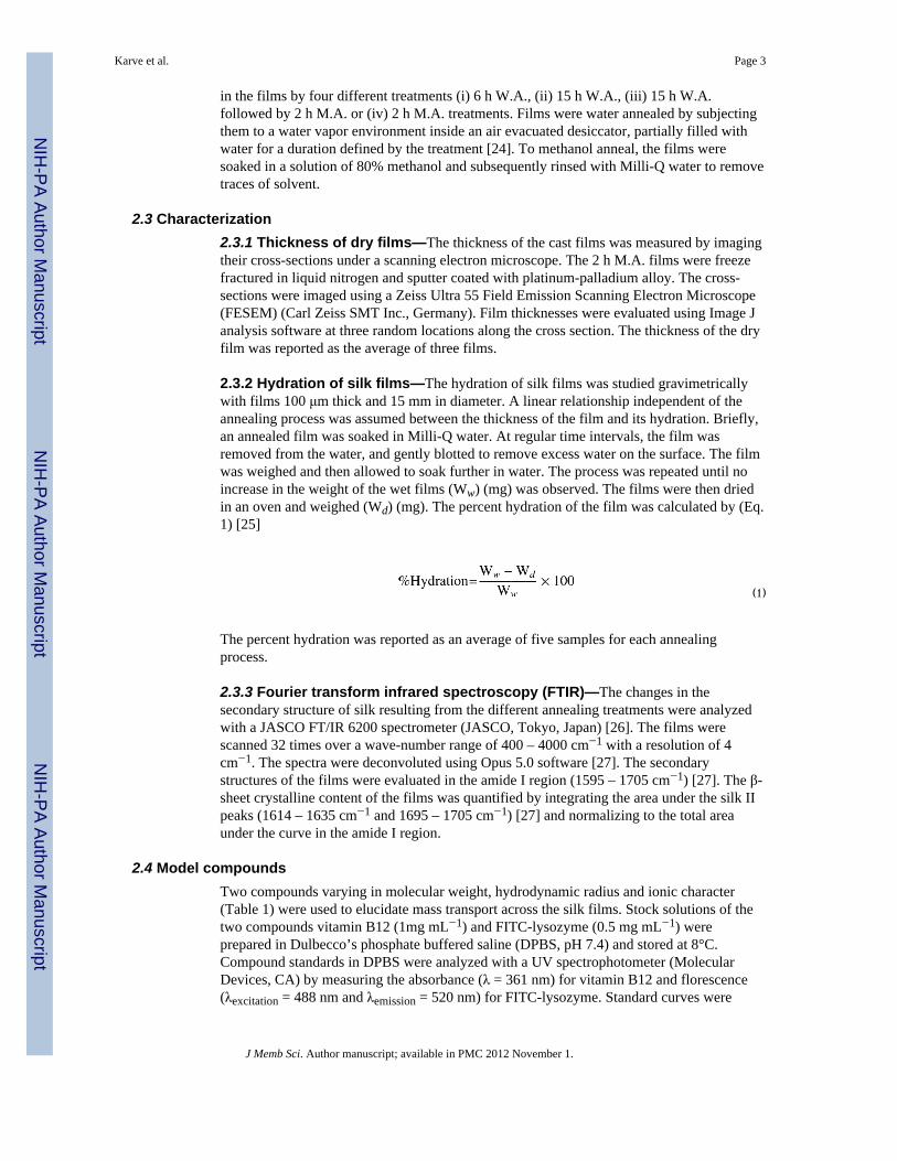
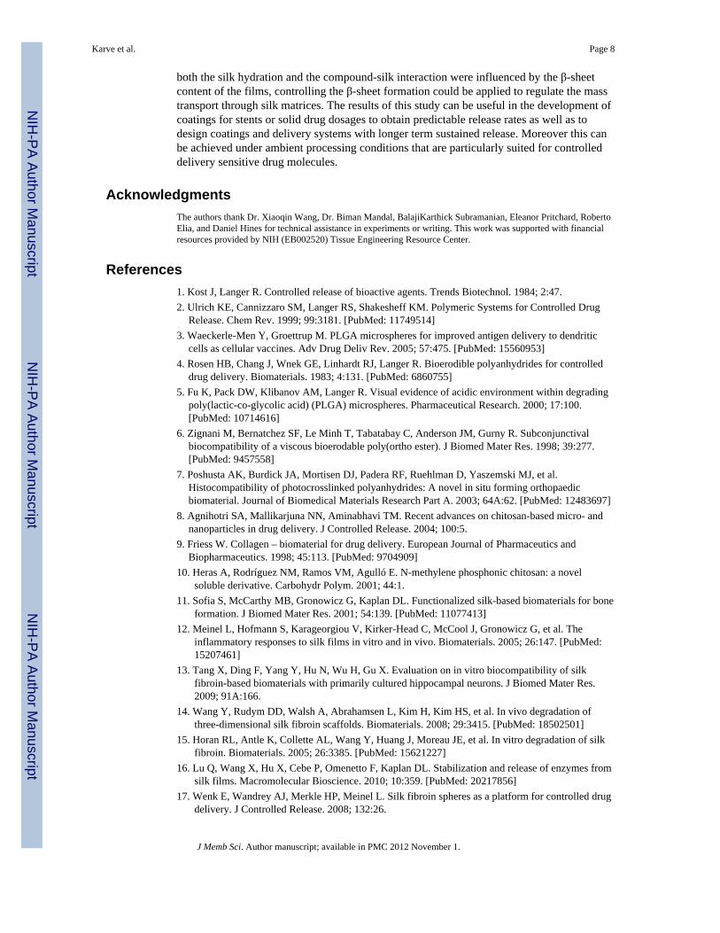
Overall, for vitamin B12, the permeability of the silk films decreased with increase in β-sheet crystalline content. This may be attributed to the reduction in hydration of the filmswith increase in β-sheets (Figure 3). The β-sheets can be considered as physical cross-links,which restrict swelling of the films in a buffer. Hence, the presence of increased β-sheetsdecreased the solvent phase available in the films to diffuse vitamin B12, thereby decreasingthe permeability. Although, partitioning of vitamin B12 was observed between the silk filmsand the buffer, this did not influence the observed permeability trend (Table 2). The smallersize of the molecule likely played a role in minimizing the impact of these interactions.Overall, the partitioning of vitamin B12 increased with increase in β-sheet content.However, this was significantly greater only with the 2 h M.A. films and may havecontributed synergistically with the low hydration of these films towards the observedpermeability. The only contradiction to the observed trend between β-sheet content andpermeability was for the 15 h W.A. films. The increase in the permeability of these films didnot follow the rationale based on film hydration and compound partitioning. This resultsuggested that permeability of the films could involve other complex factors that were notdiscernable within the limits of the present study.
Interestingly, the permeability of the silk films increased for lysozyme with the increase inβ-sheet crystalline content. At pH 7.2, lysozyme (pI 11) and silk (pI 4) [21] were oppositelycharged resulting in partitioning between silk films and the buffer. The extent of thispartitioning was determined by the changes in secondary structure of the films. This may beattributed to the change in function and/or accessibility of the interaction sites due toformation of β-sheets. The lower permeability of the 6 h W.A. films may have resulted fromthe strong interaction demonstrated by these films with lysozyme as well as the largerhydrodynamic radius of the compound. An increase in the crystallinity of silk reduced themagnitude of these interactions, which may have promoted mass transport of a largernumber of molecules. Therefore, the permeability of the 15 h W.A. and the sequentiallytreated (15 h W.A. + 2 h M.A) films increased, despite a corresponding reduction in the filmhydration. The decrease in the permeability of the 2 h M.A. films for lysozyme may beassociated with both lower hydration and stronger partitioning of lysozyme with the films.The permeability of the 6 h W.A. films varied significantly from those subjected tomethanol based annealing. The change in the permeability between the two methanol basedtreatments was not statistically significant. Similar observations were made for the two
Karve et al. Page 6
J Memb Sci. Author manuscript; available in PMC 2012 November 1.
NIH
-PA Author Manuscript
NIH
-PA Author Manuscript
NIH
-PA Author Manuscript

treatments based exclusively on water annealing. A lack of observed statistical significancemay be a result of the intrinsic variability associated with silk from different preparations.
3.2 Diffusion coefficientThe mass transport characteristics were further studied by calculating the diffusioncoefficients of the two model compounds using equation 3. With moderation in the β-sheetcontent of the films, the diffusion coefficients of vitamin B12 varied from 6.38 ± 0.37 ×10⁻⁶ to 0.34 ± 0.03 × 10⁻⁶ cm2 s−1 while those of lysozyme varied from 0.113 ± 0.032 ×10⁻⁶ to 0.042 ± 0.018 × 10⁻⁶ cm2 s−1. With the exception of 2 h M.A. films, diffusivity ofvitamin B12 was at least an order of magnitude greater than that of lysozyme at equivalentβ-sheet content (Table 1).
The diffusivity of the compounds was examined in light of the free volume theory describedby Yasuda [32]. According to this theory, diffusion of a solute through a polymer matrix isassumed to occur through the free volume present in the matrix. The free volume isdescribed as the fluctuating space within the matrix, not occupied by the polymer chains.This theory relates the diffusivity of the solute to its size and hydration of the film. It can beexpressed mathematically by (Eq 5) [25]
(5)
Here, D (cm2 s−1) is the diffusion coefficient of the solute through the polymer, D0 (cm2
s−1) is the diffusion coefficient of the solute in water, Y (dimensionless) is a characteristicconstant of a solute-solvent combination and H (dimensionless) is the hydration of the film.Compounds obeying the free volume theory are expected to have a linear relationshipbetween ln D and 1/H.
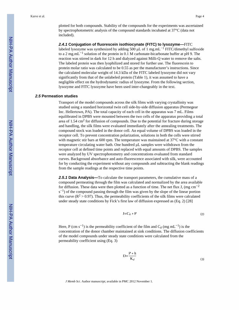
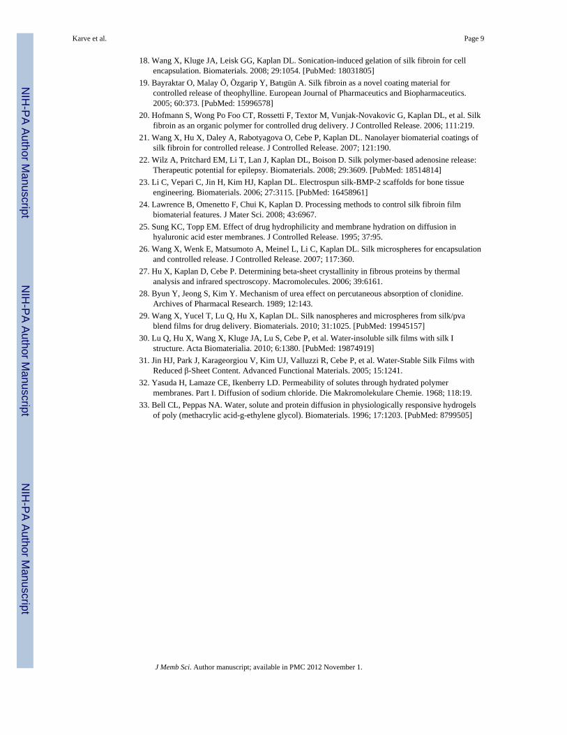
The diffusion of vitamin B12 was consistent with the free volume theory as demonstrated bythe high linearity (R2 > 0.96) between the two parameters shown in Figure 4. Thisconfirmed that diffusion of B12 was independent of the compound-matrix interactions andoccurred through the solvent filled channels within the film. The diffusivity of lysozymedeviated significantly from the free volume theory. This may be a result of the interactionbetween the lysozyme and silk that was not accounted for by this theory.
Further mathematical models were investigated to define the relationship betweendiffusivity of the model compounds and β-sheet crystallinity of the silk films. Only thediffusivity of vitamin B12 could be fitted adequately to a mathematical model. Therelationship between diffusivity of vitamin B12 and β-sheet crystallinity was modeled by apower law curve defined in Figure 5. This indicated that the diffusivity of vitamin B12 wasmore sensitive to small changes in the crystallinity at low β-sheet content. A limiting valueof diffusivity may also be postulated at high β-sheet content of the film. However at high β-sheet content, silk exhibited brittle characteristics that may impede measurement of thetransport parameters.
4 ConclusionsControlled mass transfer of two compounds varying in molecular size and ionic characteracross silk films was achieved by sequentially varying β-sheet crystalline content. Therelationship between β-sheet content and diffusivity of vitamin B12 was defined by powerlaw kinetics. The diffusion of vitamin B12 appeared to rely predominantly on the hydrationof the films, while that of lysozyme was likely influenced by interactions with silk. Since,
Karve et al. Page 7
J Memb Sci. Author manuscript; available in PMC 2012 November 1.
NIH
-PA Author Manuscript
NIH
-PA Author Manuscript
NIH
-PA Author Manuscript
both the silk hydration and the compound-silk interaction were influenced by the β-sheetcontent of the films, controlling the β-sheet formation could be applied to regulate the masstransport through silk matrices. The results of this study can be useful in the development ofcoatings for stents or solid drug dosages to obtain predictable release rates as well as todesign coatings and delivery systems with longer term sustained release. Moreover this canbe achieved under ambient processing conditions that are particularly suited for controlleddelivery sensitive drug molecules.
AcknowledgmentsThe authors thank Dr. Xiaoqin Wang, Dr. Biman Mandal, BalajiKarthick Subramanian, Eleanor Pritchard, RobertoElia, and Daniel Hines for technical assistance in experiments or writing. This work was supported with financialresources provided by NIH (EB002520) Tissue Engineering Resource Center.
References1. Kost J, Langer R. Controlled release of bioactive agents. Trends Biotechnol. 1984; 2:47.2. Ulrich KE, Cannizzaro SM, Langer RS, Shakesheff KM. Polymeric Systems for Controlled Drug
Release. Chem Rev. 1999; 99:3181. [PubMed: 11749514]3. Waeckerle-Men Y, Groettrup M. PLGA microspheres for improved antigen delivery to dendritic
cells as cellular vaccines. Adv Drug Deliv Rev. 2005; 57:475. [PubMed: 15560953]4. Rosen HB, Chang J, Wnek GE, Linhardt RJ, Langer R. Bioerodible polyanhydrides for controlled
drug delivery. Biomaterials. 1983; 4:131. [PubMed: 6860755]5. Fu K, Pack DW, Klibanov AM, Langer R. Visual evidence of acidic environment within degrading
poly(lactic-co-glycolic acid) (PLGA) microspheres. Pharmaceutical Research. 2000; 17:100.[PubMed: 10714616]
6. Zignani M, Bernatchez SF, Le Minh T, Tabatabay C, Anderson JM, Gurny R. Subconjunctivalbiocompatibility of a viscous bioerodable poly(ortho ester). J Biomed Mater Res. 1998; 39:277.[PubMed: 9457558]
7. Poshusta AK, Burdick JA, Mortisen DJ, Padera RF, Ruehlman D, Yaszemski MJ, et al.Histocompatibility of photocrosslinked polyanhydrides: A novel in situ forming orthopaedicbiomaterial. Journal of Biomedical Materials Research Part A. 2003; 64A:62. [PubMed: 12483697]
8. Agnihotri SA, Mallikarjuna NN, Aminabhavi TM. Recent advances on chitosan-based micro- andnanoparticles in drug delivery. J Controlled Release. 2004; 100:5.
9. Friess W. Collagen – biomaterial for drug delivery. European Journal of Pharmaceutics andBiopharmaceutics. 1998; 45:113. [PubMed: 9704909]
10. Heras A, Rodríguez NM, Ramos VM, Agulló E. N-methylene phosphonic chitosan: a novelsoluble derivative. Carbohydr Polym. 2001; 44:1.
11. Sofia S, McCarthy MB, Gronowicz G, Kaplan DL. Functionalized silk-based biomaterials for boneformation. J Biomed Mater Res. 2001; 54:139. [PubMed: 11077413]
12. Meinel L, Hofmann S, Karageorgiou V, Kirker-Head C, McCool J, Gronowicz G, et al. Theinflammatory responses to silk films in vitro and in vivo. Biomaterials. 2005; 26:147. [PubMed:15207461]
13. Tang X, Ding F, Yang Y, Hu N, Wu H, Gu X. Evaluation on in vitro biocompatibility of silkfibroin-based biomaterials with primarily cultured hippocampal neurons. J Biomed Mater Res.2009; 91A:166.
14. Wang Y, Rudym DD, Walsh A, Abrahamsen L, Kim H, Kim HS, et al. In vivo degradation ofthree-dimensional silk fibroin scaffolds. Biomaterials. 2008; 29:3415. [PubMed: 18502501]
15. Horan RL, Antle K, Collette AL, Wang Y, Huang J, Moreau JE, et al. In vitro degradation of silkfibroin. Biomaterials. 2005; 26:3385. [PubMed: 15621227]
16. Lu Q, Wang X, Hu X, Cebe P, Omenetto F, Kaplan DL. Stabilization and release of enzymes fromsilk films. Macromolecular Bioscience. 2010; 10:359. [PubMed: 20217856]
17. Wenk E, Wandrey AJ, Merkle HP, Meinel L. Silk fibroin spheres as a platform for controlled drugdelivery. J Controlled Release. 2008; 132:26.
Karve et al. Page 8
J Memb Sci. Author manuscript; available in PMC 2012 November 1.
NIH
-PA Author Manuscript
NIH
-PA Author Manuscript
NIH
-PA Author Manuscript
18. Wang X, Kluge JA, Leisk GG, Kaplan DL. Sonication-induced gelation of silk fibroin for cellencapsulation. Biomaterials. 2008; 29:1054. [PubMed: 18031805]
19. Bayraktar O, Malay Ö, Özgarip Y, Batıgün A. Silk fibroin as a novel coating material forcontrolled release of theophylline. European Journal of Pharmaceutics and Biopharmaceutics.2005; 60:373. [PubMed: 15996578]
20. Hofmann S, Wong Po Foo CT, Rossetti F, Textor M, Vunjak-Novakovic G, Kaplan DL, et al. Silkfibroin as an organic polymer for controlled drug delivery. J Controlled Release. 2006; 111:219.
21. Wang X, Hu X, Daley A, Rabotyagova O, Cebe P, Kaplan DL. Nanolayer biomaterial coatings ofsilk fibroin for controlled release. J Controlled Release. 2007; 121:190.
22. Wilz A, Pritchard EM, Li T, Lan J, Kaplan DL, Boison D. Silk polymer-based adenosine release:Therapeutic potential for epilepsy. Biomaterials. 2008; 29:3609. [PubMed: 18514814]
23. Li C, Vepari C, Jin H, Kim HJ, Kaplan DL. Electrospun silk-BMP-2 scaffolds for bone tissueengineering. Biomaterials. 2006; 27:3115. [PubMed: 16458961]
24. Lawrence B, Omenetto F, Chui K, Kaplan D. Processing methods to control silk fibroin filmbiomaterial features. J Mater Sci. 2008; 43:6967.
25. Sung KC, Topp EM. Effect of drug hydrophilicity and membrane hydration on diffusion inhyaluronic acid ester membranes. J Controlled Release. 1995; 37:95.
26. Wang X, Wenk E, Matsumoto A, Meinel L, Li C, Kaplan DL. Silk microspheres for encapsulationand controlled release. J Controlled Release. 2007; 117:360.
27. Hu X, Kaplan D, Cebe P. Determining beta-sheet crystallinity in fibrous proteins by thermalanalysis and infrared spectroscopy. Macromolecules. 2006; 39:6161.
28. Byun Y, Jeong S, Kim Y. Mechanism of urea effect on percutaneous absorption of clonidine.Archives of Pharmacal Research. 1989; 12:143.
29. Wang X, Yucel T, Lu Q, Hu X, Kaplan DL. Silk nanospheres and microspheres from silk/pvablend films for drug delivery. Biomaterials. 2010; 31:1025. [PubMed: 19945157]
30. Lu Q, Hu X, Wang X, Kluge JA, Lu S, Cebe P, et al. Water-insoluble silk films with silk Istructure. Acta Biomaterialia. 2010; 6:1380. [PubMed: 19874919]
31. Jin HJ, Park J, Karageorgiou V, Kim UJ, Valluzzi R, Cebe P, et al. Water-Stable Silk Films withReduced β-Sheet Content. Advanced Functional Materials. 2005; 15:1241.
32. Yasuda H, Lamaze CE, Ikenberry LD. Permeability of solutes through hydrated polymermembranes. Part I. Diffusion of sodium chloride. Die Makromolekulare Chemie. 1968; 118:19.
33. Bell CL, Peppas NA. Water, solute and protein diffusion in physiologically responsive hydrogelsof poly (methacrylic acid-g-ethylene glycol). Biomaterials. 1996; 17:1203. [PubMed: 8799505]
Karve et al. Page 9
J Memb Sci. Author manuscript; available in PMC 2012 November 1.
NIH
-PA Author Manuscript
NIH
-PA Author Manuscript
NIH
-PA Author Manuscript
Highlights
• Effect of silk β-sheet crystallinity on transport of model compounds is studied.
• Crystalline content relates to vitamin B12 diffusivity through a power lawmodel.
• Lysozyme-silk interactions result in non-compliance with free volume theory.
• We underscore potential of silk for controlled delivery of therapeutic agents.
Karve et al. Page 10
J Memb Sci. Author manuscript; available in PMC 2012 November 1.
NIH
-PA Author Manuscript
NIH
-PA Author Manuscript
NIH
-PA Author Manuscript
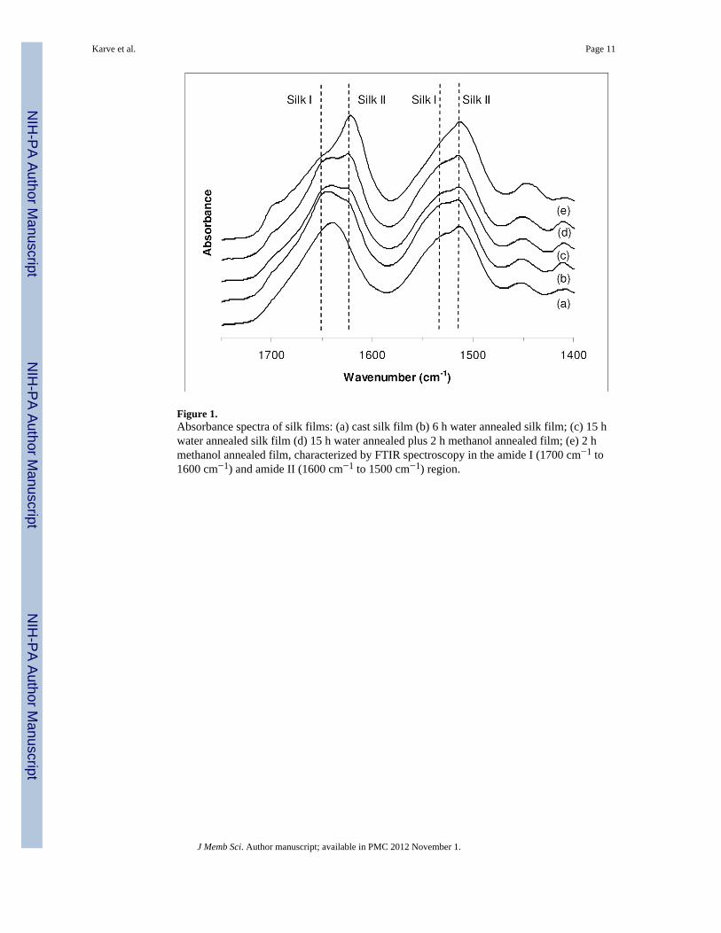
Figure 1.Absorbance spectra of silk films: (a) cast silk film (b) 6 h water annealed silk film; (c) 15 hwater annealed silk film (d) 15 h water annealed plus 2 h methanol annealed film; (e) 2 hmethanol annealed film, characterized by FTIR spectroscopy in the amide I (1700 cm−1 to1600 cm−1) and amide II (1600 cm−1 to 1500 cm−1) region.
Karve et al. Page 11
J Memb Sci. Author manuscript; available in PMC 2012 November 1.
NIH
-PA Author Manuscript
NIH
-PA Author Manuscript
NIH
-PA Author Manuscript
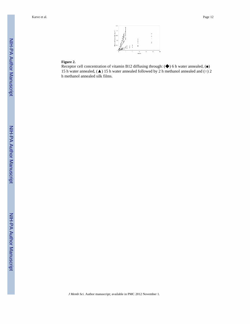
Figure 2.Receptor cell concentration of vitamin B12 diffusing through: (◆) 6 h water annealed, (■)15 h water annealed, (▲) 15 h water annealed followed by 2 h methanol annealed and (○) 2h methanol annealed silk films.
Karve et al. Page 12
J Memb Sci. Author manuscript; available in PMC 2012 November 1.
NIH
-PA Author Manuscript
NIH
-PA Author Manuscript
NIH
-PA Author Manuscript
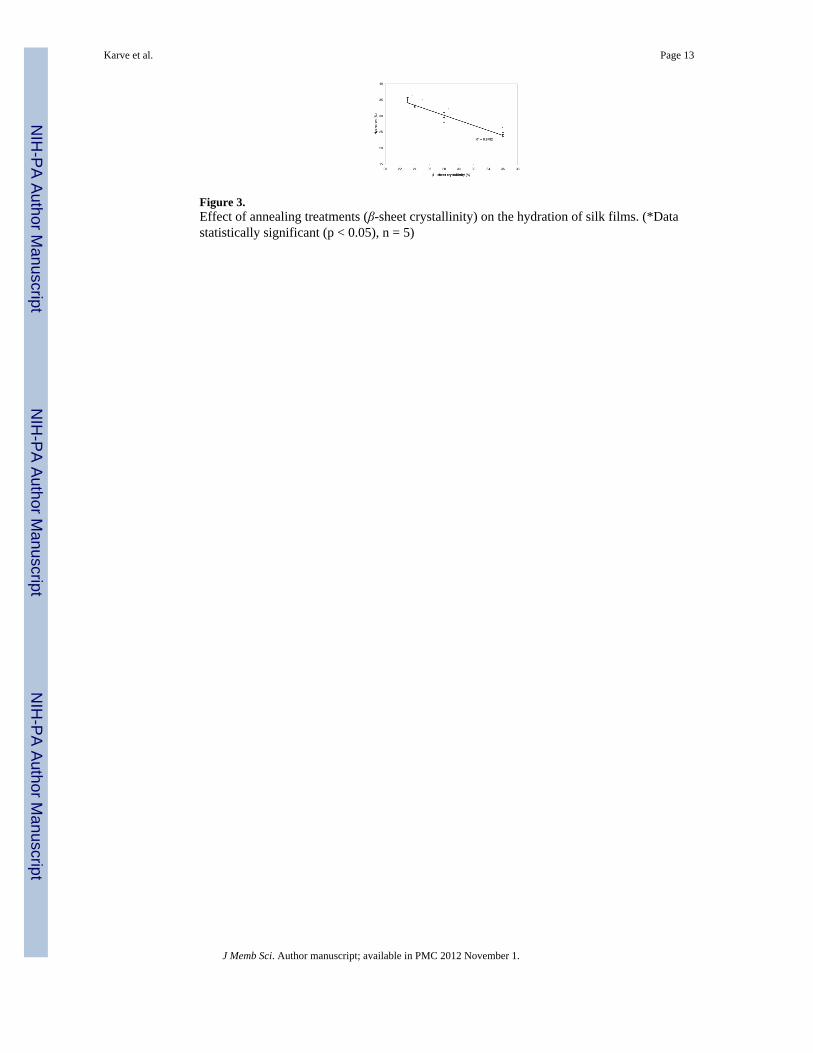
Figure 3.Effect of annealing treatments (β-sheet crystallinity) on the hydration of silk films. (*Datastatistically significant (p < 0.05), n = 5)
Karve et al. Page 13
J Memb Sci. Author manuscript; available in PMC 2012 November 1.
NIH
-PA Author Manuscript
NIH
-PA Author Manuscript
NIH
-PA Author Manuscript
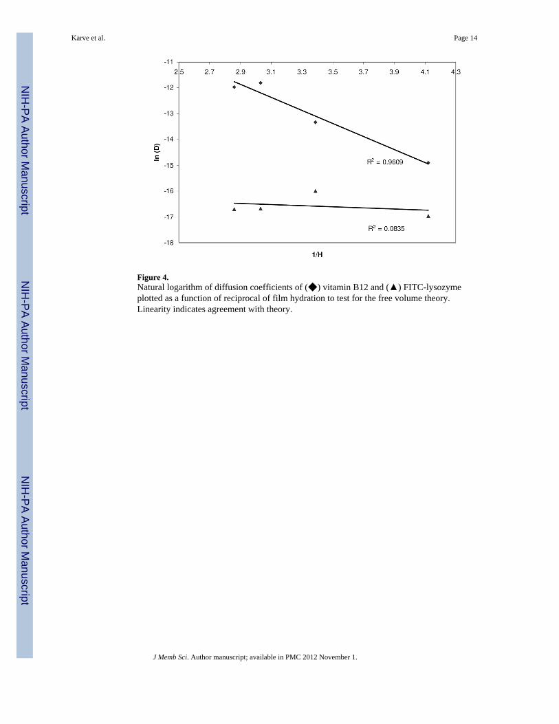
Figure 4.Natural logarithm of diffusion coefficients of (◆) vitamin B12 and (▲) FITC-lysozymeplotted as a function of reciprocal of film hydration to test for the free volume theory.Linearity indicates agreement with theory.
Karve et al. Page 14
J Memb Sci. Author manuscript; available in PMC 2012 November 1.
NIH
-PA Author Manuscript
NIH
-PA Author Manuscript
NIH
-PA Author Manuscript
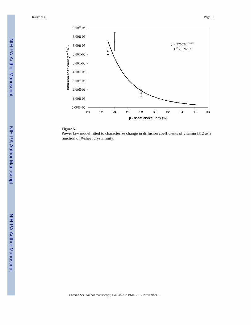
Figure 5.Power law model fitted to characterize change in diffusion coefficients of vitamin B12 as afunction of β-sheet crystallinity.
Karve et al. Page 15
J Memb Sci. Author manuscript; available in PMC 2012 November 1.
NIH
-PA Author Manuscript
NIH
-PA Author Manuscript
NIH
-PA Author Manuscript
NIH
-PA Author Manuscript
NIH
-PA Author Manuscript
NIH
-PA Author Manuscript
Karve et al. Page 16
Tabl
e 1
Mod
el c
ompo
unds
use
d in
mas
s tra
nspo
rt st
udie
s [34
].
Com
poun
dM
olec
ular
wei
ght (
kDa)
Hyd
rody
nam
ic r
adiu
s (nm
)Is
o-el
ectr
ic p
oint
Vita
min
B12
1.34
0.85
--
Lyso
zym
e14
.11.
6011
J Memb Sci. Author manuscript; available in PMC 2012 November 1.
NIH
-PA Author Manuscript
NIH
-PA Author Manuscript
NIH
-PA Author Manuscript
Karve et al. Page 17
Tabl
e 2
Parti
tion,
per
mea
bilit
y an
d di
ffus
ion
coef
ficie
nts o
f vita
min
B12
and
lyso
zym
e as
a fu
nctio
n of
β-s
heet
cry
stal
linity
of s
ilk fi
lms.
Tre
atm
ent
β Sh
eet (
%)
Com
poun
dPa
rtiti
on C
oeffi
cien
t (K
d)Pe
rmea
bilit
y C
oeffi
cien
t (cm
/s) (
× 10
−1 )
Diff
usio
n C
oeffi
cien
t (D
) (cm
2 /s) (
× 10
−6 )
6 h
W.A
.23
B12
4.76
± 1
.77
1.08
± 0
.064
*6.
38 ±
0.3
7*
15 h
W.A
.24
5.35
± 1
.42
1.55
± 0
.213
*7.
34 ±
1.0
2†
15 h
W.A
. + 2
h M
.A.
286.
45 ±
0.3
90.
41 ±
0.1
04*
1.64
± 0
.28*
†‡
2 h
M.A
.36
12.5
1 ±
1.04
*0.
16 ±
0.0
17*
0.34
± 0
.03*
†‡
6 h
W.A
.23
Lyso
zym
e
72.9
1 ±
4.55
*†0.
029
± 0.
003†
*0.
056
± 0.
011*
15 h
W.A
.24
29.2
3 ±
5.24
*0.
059
± 0.
022
0.05
7 ±
0.02
1
15 h
W.A
. + 2
h M
A.
2821
.48
± 4.
88†‡
0.08
6 ±
0.02
5†0.
113
± 0.
032*
†
2 h
M.A
.36
51.1
4 ±
13.4
0‡0.
077
± 0.
033*
0.04
2 ±
0.01
8†
‡, †
, *D
ata
stat
istic
ally
sign
ifica
nt (p
< 0
.05)
, n=3
W.A
. – W
ater
ann
eale
d, M
.A. –
Met
hano
l ann
eale
d
J Memb Sci. Author manuscript; available in PMC 2012 November 1.


![a, arXiv:1906.06378v3 [cond-mat.mtrl-sci] 4 Aug 2019](https://static.fdocument.org/doc/165x107/61c0d52e1c1cea23c461e775/a-arxiv190606378v3-cond-matmtrl-sci-4-aug-2019.jpg)
