Tiangang Li and John Y.L. Chiang Department of...
38
DMD#7575 Rifampicin Induction of CYP3A4 Requires PXR crosstalk with HNF4α and co-activators, and suppression of SHP gene Expression Tiangang Li and John Y.L. Chiang Department of Biochemistry and Molecular Pathology, Northeastern Ohio Universities College of Medicine, Rootstown, Ohio 44272,USA DMD Fast Forward. Published on February 2, 2006 as doi:10.1124/dmd.105.007575 Copyright 2006 by the American Society for Pharmacology and Experimental Therapeutics. This article has not been copyedited and formatted. The final version may differ from this version. DMD Fast Forward. Published on February 2, 2006 as DOI: 10.1124/dmd.105.007575 at ASPET Journals on March 30, 2020 dmd.aspetjournals.org Downloaded from
Transcript of Tiangang Li and John Y.L. Chiang Department of...
DMD#7575
Rifampicin Induction of CYP3A4 Requires PXR crosstalk with HNF4α and co-activators,
and suppression of SHP gene Expression
Tiangang Li and John Y.L. Chiang
Department of Biochemistry and Molecular Pathology, Northeastern Ohio Universities College
of Medicine, Rootstown, Ohio 44272,USA
DMD Fast Forward. Published on February 2, 2006 as doi:10.1124/dmd.105.007575
Copyright 2006 by the American Society for Pharmacology and Experimental Therapeutics.
This article has not been copyedited and formatted. The final version may differ from this version.DMD Fast Forward. Published on February 2, 2006 as DOI: 10.1124/dmd.105.007575
at ASPE
T Journals on M
arch 30, 2020dm
d.aspetjournals.orgD
ownloaded from
DMD#7575
2
Running Title: PXR regulation of CYP3A4 Correspondence address: John Y. L. Chiang, Ph.D. Department of Biochemistry and Molecular Pathology Northeastern Ohio Univ’s College of Medicine 4209 State Route 44, P. O. Box 95 Rootstown, OH 44272 Tel: 330-325-6694 Fax: 330-325-5910 E-mail: [email protected]
Number of text pages: 28 Number of figures: 9 Number of references: 32 Number of words in Abstract: 238 Number of words in Introduction: 626 Number of words in Discussion: 697
Abbreviations: ChIP, chromatin immunoprecipitation assay; CYP3A4, cytochrome P4503A4, CYP7A1, cholesterol 7α-hydroxylase; FXR, farnesoid X receptor; GST, glutathione S-transferase; HNF4α, hepatocyte nuclear factor 4α; LCA, lithocholic acid; PXR, pregnane X receptor; PGC-1α, peroxisome proliferators-activated receptor γ co-activator 1α; SHP, small heterodimer partner; SRC-1, steroid receptor coactivator-1; XREM, xenobiotic responsive element module.
This article has not been copyedited and formatted. The final version may differ from this version.DMD Fast Forward. Published on February 2, 2006 as DOI: 10.1124/dmd.105.007575
at ASPE
T Journals on M
arch 30, 2020dm
d.aspetjournals.orgD
ownloaded from
DMD#7575
3
ABSTRACT
Bile acids and drugs activate PXR to induce CYP3A4, which is the predominant cytochrome
P450 enzymes expressed in the liver and intestine and plays a critical role in detoxifying bile
acids and drugs, and protecting against cholestasis. The aim of this study is to investigate the
molecular mechanism of PXR cross talk with other nuclear receptors and co-activators in
regulating human CYP3A4 gene transcription. Rifampicin dose-dependently induced the
CYP3A4 but inhibited small heterodimer partner (SHP) mRNA expression levels in primary
human hepatocytes. Rifampicin strongly stimulated PXR and HNF4α interaction, and CYP3A4
reporter activity, which was further stimulated by PGC-1α and SRC-1 but inhibited by SHP.
Mutation of the putative HNF4α binding site in the distal xenobiotic responsive element module
(dXREM) did not affect CYP3A4 basal promoter activity and synergistic stimulation by PXR
and HNF4α. Chromatin immunoprecipitation assays revealed that rifampicin-activated PXR
recruited HNF4α and SRC-1 to the CYP3A4 chromatin. On the other hand, SHP reduced PXR
recruitment of HNF4α and SRC-1 to the CYP3A4 chromatin. The human SHP promoter was
stimulated by HNF4α and PGC-1α. Upon activation by rifampicin, PXR inhibited SHP promoter
activity. Results suggest that PXR strongly induces CYP3A4 gene transcription by interacting
with HNF4α, SRC-1 and PGC-1α. PXR concomitantly inhibits SHP gene transcription and
maximizes the PXR induction of the CYP3A4 gene in human livers. Drugs targeted to PXR may
be developed for treating cholestatic liver diseases induced by bile acids and drugs.
This article has not been copyedited and formatted. The final version may differ from this version.DMD Fast Forward. Published on February 2, 2006 as DOI: 10.1124/dmd.105.007575
at ASPE
T Journals on M
arch 30, 2020dm
d.aspetjournals.orgD
ownloaded from
DMD#7575
4
INTRODUCTION
CYP3A4 is the most abundant cytochrome P450 enzymes expressed in the liver and
small intestine and plays a crucial role in metabolism and detoxification of xenobiotics and
endobiotics (Guengerich, 1999). CYP3A4 has very broad substrate specificity and metabolizes
over 50% of clinical drugs. The expression levels of CYP3A4 vary widely among individuals
(Lehmann et al., 1998). Compounds that activate human PXR (NR1I2) also induce CYP3A4 and
drug metabolism activity (Bertilsson et al., 1998; Kliewer et al., 1998; Lehmann et al., 1998;
Goodwin et al., 2002). An antibiotic rifampicin is a highly efficacious human PXR agonist
(Kliewer et al., 2002), and has been used to treat pruritus of primary biliary cirrhosis patients for
many years without knowing its molecular mechanism of drug action (Bachs et al., 1989;
Hofmann, 2002). The ligand-activated PXR forms a heterodimer with RXR and binds to the
AGGTCA-like direct repeats spaced by 3, 4 or 5 bases (DR3, DR4, and DR5), and the everted
repeats separated by 6 or 8 bases (ER6 and ER8) located in the PXR target genes (Kliewer et al.,
1998). In the human CYP3A4 promoter, PXR binds to an ER6 located in the proximal promoter
and a DR3 and ER6 in the distal xenobiotic responsive enhancer module (XREM) located about
8 kb upstream of the transcription start site (Bertilsson et al., 1998; Lehmann et al., 1998;
Goodwin et al., 1999). The maximal induction of the CYP3A4 gene by PXR requires both the
proximal ER6 and the distal XREM (Goodwin et al., 1999).
Lithocholic acid (LCA) is the most efficacious bile acid that activates PXR and induces
CYP3A4 to catalyze 6-hydroxylation of LCA to hyodeoxycholic acid (Staudinger et al., 2001;
Xie et al., 2001). Feeding LCA causes liver damage in Pxr null mice, but transgenic mice
expressing a human PXR are protected from bile acid toxicity and cholestasis (Staudinger et al.,
2001). PXR inhibits cholesterol 7α-hydroxylase (CYP7A1), the regulatory gene in bile acid
synthesis, but induces organic anion transport protein 2, a sinusoidal Na+-independent bile acid
This article has not been copyedited and formatted. The final version may differ from this version.DMD Fast Forward. Published on February 2, 2006 as DOI: 10.1124/dmd.105.007575
at ASPE
T Journals on M
arch 30, 2020dm
d.aspetjournals.orgD
ownloaded from
DMD#7575
5
transporter (Staudinger et al., 2001). Nuclear receptors interact with their co-activators such as
steroid receptor co-activator-1 (SRC-1) or co-repressors and activate or inhibit their target genes.
We reported previously that rifampicin-activated PXR interacts with hepatocyte nuclear factor
4α (HNF4α) and blocks HNF4α recruitment of peroxisome proliferators-activated receptor γ co-
activator-1α (PGC-1α), and results in inhibition of the human CYP7A1 gene (Li and Chiang,
2004). HNF4α plays a vital role in liver development and regulation of bile acid synthesis, lipid
homeostasis, and xenobiotic response (Stroup and Chiang, 2000; Hayhurst et al., 2001; Kamiya
et al., 2003). Both PXR and HNF4α interact with PGC-1α, which is a nuclear receptor co-
activator induced by glucagon and glucocorticoid to regulate gluconeogenesis and bile acid
synthesis (De Fabiani et al., 2003; Puigserver et al., 2003; Shin et al., 2003). HNF4α induces
PXR gene transcription in fetal liver and enhances PXR induction of CYP3A4 (Kamiya et al.,
2003; Tirona et al., 2003). It has been reported recently that SHP interacts with rifampicin-
activated PXR and inhibits the CYP3A4 gene expression in vitro (Ourlin et al., 2003). SHP is a
negative nuclear receptor that is induced by bile acid-activated farnesoid X receptor (FXR) and
inhibits the CYP7A1 gene (Goodwin et al., 2000; Lu et al., 2000). Therefore, the bile acid-
induced FXR/SHP pathway could counteract the bile acid activation of the CYP3A4 gene
transcription by PXR. The physiological role of SHP in regulation of CYP3A4 needs to be
further studied.
In this study, we investigated the roles of human PXR, HNF4α and SHP on the
regulation of human CYP3A4 gene transcription. Our results suggest that CYP3A4 is subjected
to a complex regulation by nuclear receptors and co-activators, and that maximal induction of
CYP3A4 gene expression requires concomitant inhibition of SHP gene transcription by PXR.
This article has not been copyedited and formatted. The final version may differ from this version.DMD Fast Forward. Published on February 2, 2006 as DOI: 10.1124/dmd.105.007575
at ASPE
T Journals on M
arch 30, 2020dm
d.aspetjournals.orgD
ownloaded from
DMD#7575
6
MATERIALS AND METHODS
Cell culture. Human hepatoblastoma HepG2 cells were obtained from the American Type
Culture Collection (Manassas, VA). Primary human hepatocytes were isolated from human
donors (HH1115, 22 yr male; HH1117, 68 yr female; HH1119, 29 yr female) and were obtained
through the Liver Tissue Procurement and Distribution System (LTPADS) of NIH (S. Strom,
University of Pittsburgh, Pittsburgh, PA). Cells were cultured and maintained as described
previously (Li and Chiang, 2004)
Plasmids. Human CYP3A4/luciferase construct (p3A4-5’dDR3/dER6/pER6-LUC) was
obtained from Marie-Jose Vilarem (Institut Federatif de Recherche 24, France). This reporter
contains an HNF4α binding site (DR1) and PXR binding sites (DR3 and ER6) in the distal
XREM (dXREM, -7800/-7600), which was linked to a 5’flanking fragment containing a
proximal ER6 (pER6) of the human CYP3A4 gene (Drocourt et al., 2002). A human
CYP7A1/Luc reporter (ph-298/Luc) was constructed as described previously (Li and Chiang,
2004). Human SHP/luciferase reporter construct (SHP-luc) containing 2 kb promoter sequence
and expression plasmid (pCDM8-mSHP) were provided by Yoonkwang Lee (Bayor College of
Medicine, Houston, TX). Expression plasmids for SHP (pcDNA3-HA-SHP), SRC-1 (pCR3.1-
hSRC-1A), PGC-1α (pcDNA3/HA-PGC-1α), human RXR (pcDNA3-hRXR), rat HNF4α
(pCMX-rHNF4α), and human PXR (pSG5-hPXR) were obtained from H.S. Choi (Chungnam
National University, Korea), M. J. Tsai (Bayor College of Medicine, Houston, TX), A. Kralli
(The Scripp Research Institute, La Jolla, CA), R. Evans (Scripps Research Institute, La Jolla,
CA), W. Chin (Eli Lily Research Laboratories, Indianapolis, ID), and S. Kliewer (University of
Texas Southwestern Medical Center, Dallas, TX), respectively. For mammalian hybrid assays,
the reporter 5XUAS-TK-Luc contains 5 copies of the upstream activating sequence (UAS) fused
upstream of a thymidine kinase minimum promoter (TK) and the luciferase reporter gene. The
This article has not been copyedited and formatted. The final version may differ from this version.DMD Fast Forward. Published on February 2, 2006 as DOI: 10.1124/dmd.105.007575
at ASPE
T Journals on M
arch 30, 2020dm
d.aspetjournals.orgD
ownloaded from
DMD#7575
7
GAL4 and VP16 fusion plasmids contained the ligand-binding domain (LBD) of nuclear
receptors or nuclear receptor-interacting domain (RID) of co-activators fused to Gal4-DBD
(DNA binding domain) or VP16-AD (activation domain) vector. Mammalian two-hybrid fusion
plasmids used were: pCMX-VP16-rHNF4 (a.a 125 to 364) from D. Moore (Baylor College of
Medicine, Houston, TX); Gal4-hPXR-LBD (a.a. 107-434, VP16-SRC-1-RID (a.a. 595-780) and
VP16-hPXR-LBD (a.a. 107-437) from A. Takeshita (Toranomon Hospital, Tokyo, Japan), and
Gal4-PGC-1α (full length) from A. Kralli (The Scripp Research Institute, La Jolla, CA). Gal4-
HNF4α fusion plasmids pBx-HNF4α-FL (full-length) was obtained from I. Talianidis (Institute
of Molecular Biology and Biotechnology Foundation for Research and Technology, Hellas,
Herakleion Crete, Greece). For GST fusion protein expressions, pGEX-4T-1 plasmid encoding
the Glutathione S- transferase (GST) protein was obtained from Amersham Pharmacia Biotech.
The pET23a-GST-HNF4α plasmid encoding a fusion protein of GST with full length human
HNF4 was a kind gift from Todd Leff (Wayne State University, Detroit, MI). GST-hPXR-LBD
was cloned by inserting PCR-amplified human PXR ligand binding domain (a.a. 241-434) into
pGEX-4T-1 BamH1/XhoI sites. GST-mSHP containing full length SHP was obtained from D.
Moore (Bayor College of Medicine, Houston, TX).
Quantitative real-time PCR assay. Primary human hepatocytes were maintained in 6-well
plates. Cells were treated with increasing amount of rifampicin (1-10 µM) (Sigma, St. Louis,
MO) for 24 h. Total RNA was isolated from the cells using Tri-Reagent (Sigma) according to the
manufacturer’s protocol. Reverse transcription reactions were performed using RETROscript kit
according to manufacturer’s instructions (Ambion Inc., Austin, TX). For real-time PCR, samples
were prepared according to the PCR Taqman Universal Master Mix 2X protocol (Applied
Biosystems, Foster City, CA). Amplification of human ubiquitin C (UBC) was used as an
internal reference gene. Quantitative PCR analysis was conducted on the ABI 7500 System
This article has not been copyedited and formatted. The final version may differ from this version.DMD Fast Forward. Published on February 2, 2006 as DOI: 10.1124/dmd.105.007575
at ASPE
T Journals on M
arch 30, 2020dm
d.aspetjournals.orgD
ownloaded from
DMD#7575
8
software. Relative mRNA expression was quantified using the comparative CT (Ct) method
according to the ABI manual. Assay-on-Demand PCR primers and Taqman MGB probe mix
used were: human CYP3A4 (Cat. #: Hs00604506_m1); human CYP7A1 (Cat. #
Hs00167982_ml); human SHP (Cat. # Hs00222627_ml); human UBC (Cat. # Hs00824723_m1).
Transient transfection assay. HepG2 cells were grown to ~80% confluence in 24-well
tissue culture plates. Reporter plasmid (0.2 µg) was transfected with expression plasmid (0.1 µg)
using lipofectamine 2000 reagent (Invitrogen, Carlsbad, CA) according to manufacturer’s
protocol. The pCMV-galactosidase plasmid (0.05 µg) was transfected as an internal standard for
normalizing the transfection efficiency in each assay. In some samples empty expression vectors
were added to equalize the total amounts of plasmid DNA transfected in each assay. Cells were
treated with vehicle (DMSO) or 10 µM rifampicin in serum free media for 40 h, and assayed for
luciferase reporter activity using Luciferase Assay System (Promega). Luciferase activity was
normalized by dividing the relative light units (RLU) by β-galactosidase activity. Each assay was
done in triplicate and individual experiments were repeated at least three times.
Site-directed mutagenesis. Mutations (shown in Fig. 3B) were introduced into the HNF4α
binding site in dXREM of CYP3A4/luciferase construct (p3A4-5’dDR3/dER6/ pER6-LUC)
using PCR-based QuikChange Site-directed Mutagenesis Kit (Stratagene, La Jolla, CA). Two
complementary oligonucleotides containing the mutations were used as PCR primers. PCR
reactions were set up according to the manufacturer's instruction using 50 ng of template DNA
and 125 ng of primers. PCR cycling parameters were set as following: denaturing at 95 ° for 2
min, followed by 18 cycles at 95 ° for 30 s, 52 ° for 1 min, and 68 ° for 18 min. The reaction
mixture was digested by Dpn I for 2 h to remove the template DNA and transformed into XL1-
Blue super competent cells (Stratagene) for selection of mutant clones. Mutations were
confirmed by DNA sequencing.
This article has not been copyedited and formatted. The final version may differ from this version.DMD Fast Forward. Published on February 2, 2006 as DOI: 10.1124/dmd.105.007575
at ASPE
T Journals on M
arch 30, 2020dm
d.aspetjournals.orgD
ownloaded from

DMD#7575
9
Electrophoretic Mobility Shift Assay (EMSA). Double-stranded synthetic probes
containing the HNF4α binding site were labeled with [35S] dATP by filling-in reaction using the
Klenow fragment of DNA polymerase I. Nuclear receptor HNF4α was synthesized in vitro using
Quick-coupled Transcription/Translation Systems (Promega) programmed with receptor
expression plasmids according to the manufacturer's instruction. EMSA assay was performed as
described in detail previously (Li and Chiang, 2004).
Immunoblotting analysis. HepG2 cells were cultured in 100 mm dishes to 80% confluence.
Cells were transfected with pcDNA3-HA-SHP plasmid (5 or 10 µg) and incubated in serum free
media for 40 h. Cell lysates were analyzed by SDS-polyacrylamide gel electrophoresis. Antibody
against HA-tag (Santa Cruz Biotechnology, CA) were used for western blotting and detected by
ECL Western blotting detection kit (Amersham Biosciences, UK).
Chromatin immunoprecipitation (ChIP) assay. HepG2 cells were grown in 100 mm
culture dishes to 80% confluence. HA-PGC-1α (10 µg) or HA-SHP plasmid was transfected into
HepG2 cells. Cells were treated with 10 µM rifampicin or vehicle (DMSO) in serum free media
for 40 h. ChIP assays were performed using ChIP Assay kit (Upstate Biotec, NY) according to
manufacturer’s protocol and described previously (Li and Chiang, 2004). Ten percent of the cell
lysates before immunoprecipitation was saved and used as “input”. The cross-linked DNA-
protein complexes were precipitated by incubating the cell lysates with 10 µg of antibodies or
non-immuno IgG (Santa Cruz Biotec. Inc, Santa Cruz, CA) followed by protein A-agarose
beads. The antibodies used were: goat anti-HNF4α (C-19), Cat#: sc-6556, lot#: I1203; rabbit
anti-HNF4α (H171), Cat#: sc-8987, lot#: K0204; goat anti-PXR (A-20), Cat#: sc-7737, lot#:
J0704; rabbit anti-SRC-1 (M341), Cat#: sc-8995, lot#: C3004; goat anti-PGC-1α (P-19), Cat#:
sc-5815, lot#: I141; goat Normal IgG, Cat#: sc-2027, lot#: E1204; rabbit Normal IgG, Cat#: sc-
2028, lot#: F0304.The beads were washed and samples eluted with 250 µl of ChIP elution
This article has not been copyedited and formatted. The final version may differ from this version.DMD Fast Forward. Published on February 2, 2006 as DOI: 10.1124/dmd.105.007575
at ASPE
T Journals on M
arch 30, 2020dm
d.aspetjournals.orgD
ownloaded from
DMD#7575
10
buffer. DNA fragments containing either dXREM or pER6 were PCR amplified for 35 cycles
and analyzed on a 2% agarose gel. The PCR primers used were: dXREM region (dDR3/ER6, -
7939 to -7576), forward-GCAGGTCCCCTGGAAAGTCAC, reverse-
CACCTGGGGTCAACACAGGAC; proximal ER6 region (-277 to -62), forward-
ATGCCAATGGCTCCACTTGAG, and reverse-CTGGAGCTGCAGCCAGTAGCAG; 2nd
intron region (+1460 to +1849), forward- GTTACTAGCTTCCTTCATCAGTTG, and reverse-
CATTCCAGCCTGGGCAACAAG.
GST pull-down assay. GST fusion protein isolation and GST pull down assays were
described previously (Li and Chiang, 2004). The 35S-labeled proteins were generated using their
respective expression plasmid in the TNT rabbit reticulocyte system (Promega) in the presence
of 35S-labeled methionine. GST-fusion proteins (~1 µg) bound to the glutathione beads were
added to 5 µl of the 35S-labeled proteins. In competition assays, 50 µl of un-labeled TNT lysates
incubated with pcDNA3 or indicated expression plasmids was added to the mixture. The beads
were washed and the proteins eluted for analysis on an SDS PAGE gel. One µl 35S-labeled
protein was loaded as “Input”.
Statistical analysis. Data were plotted as means ± standard deviation. Statistical analyses of
treated vs. untreated controls (vehicle or empty plasmids) were performed using Student’s t-test.
A p<0.05 is considered statistically significant.
This article has not been copyedited and formatted. The final version may differ from this version.DMD Fast Forward. Published on February 2, 2006 as DOI: 10.1124/dmd.105.007575
at ASPE
T Journals on M
arch 30, 2020dm
d.aspetjournals.orgD
ownloaded from
DMD#7575
11
RESULTS
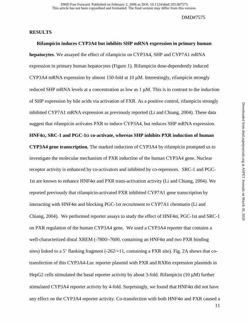
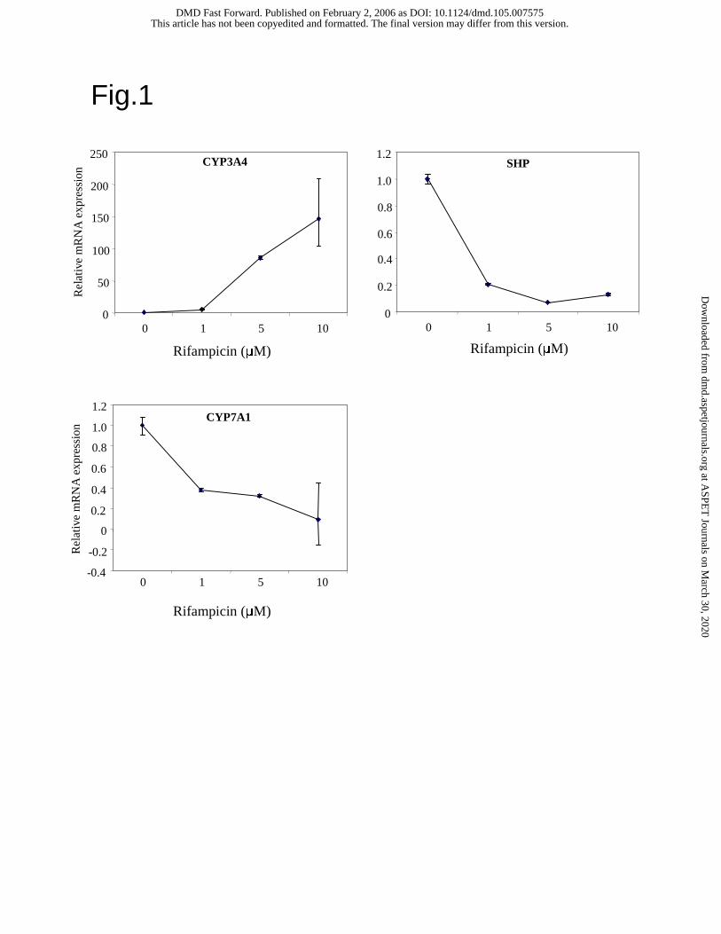
Rifampicin induces CYP3A4 but inhibits SHP mRNA expression in primary human
hepatocytes. We assayed the effect of rifampicin on CYP3A4, SHP and CYP7A1 mRNA
expression in primary human hepatocytes (Figure 1). Rifampicin dose-dependently induced
CYP3A4 mRNA expression by almost 150-fold at 10 µM. Interestingly, rifampicin strongly
reduced SHP mRNA levels at a concentration as low as 1 µM. This is in contrast to the induction
of SHP expression by bile acids via activation of FXR. As a positive control, rifampicin strongly
inhibited CYP7A1 mRNA expression as previously reported (Li and Chiang, 2004). These data
suggest that rifampicin activates PXR to induce CYP3A4, but reduces SHP mRNA expression.
HNF4α, SRC-1 and PGC-1α co-activate, whereas SHP inhibits PXR induction of human
CYP3A4 gene transcription. The marked induction of CYP3A4 by rifampicin prompted us to
investigate the molecular mechanism of PXR induction of the human CYP3A4 gene. Nuclear
receptor activity is enhanced by co-activators and inhibited by co-repressors. SRC-1 and PGC-
1α are known to enhance HNF4α and PXR trans-activation activity (Li and Chiang, 2004). We
reported previously that rifampicin-activated PXR inhibited CYP7A1 gene transcription by
interacting with HNF4α and blocking PGC-1α recruitment to CYP7A1 chromatin (Li and
Chiang, 2004). We performed reporter assays to study the effect of HNF4α, PGC-1α and SRC-1
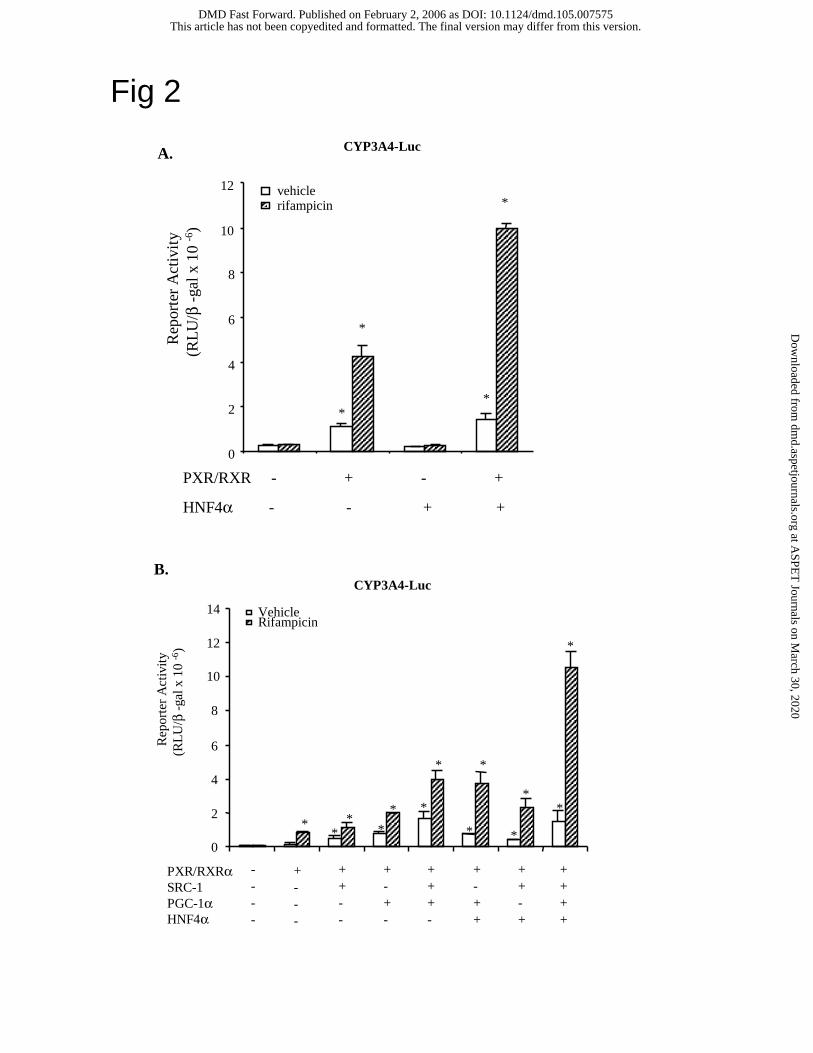
on PXR regulation of the human CYP3A4 gene. We used a CYP3A4 reporter that contains a
well-characterized distal XREM (-7800/-7600, containing an HNF4α and two PXR binding
sites) linked to a 5’ flanking fragment (-262/+11, containing a PXR site). Fig. 2A shows that co-
transfection of this CYP3A4-Luc reporter plasmid with PXR and RXRα expression plasmids in
HepG2 cells stimulated the basal reporter activity by about 3-fold. Rifampicin (10 µM) further
stimulated CYP3A4 reporter activity by 4-fold. Surprisingly, we found that HNF4α did not have
any effect on the CYP3A4 reporter activity. Co-transfection with both HNF4α and PXR caused a
This article has not been copyedited and formatted. The final version may differ from this version.DMD Fast Forward. Published on February 2, 2006 as DOI: 10.1124/dmd.105.007575
at ASPE
T Journals on M
arch 30, 2020dm
d.aspetjournals.orgD
ownloaded from
DMD#7575
12
2.5-fold stimulation of the CYP3A4 reporter activity stimulated by PXR alone when rifampicin
was added. Fig. 2B shows that SRC-1 and/or PGC-1α stimulated PXR induction of CYP3A4
reporter activity by 2-fold in the absence of rifampicin. Addition of rifampicin further stimulated
reporter activity. The maximal induction (about 130-fold) of the CYP3A4 basal reporter activity
by rifampicin was achieved when PXR, HNF4α, SRC-1 and PGC-1α were transfected. It is
noted that inclusion of HNF4α in the assay greatly enhanced rifampicin induction of the reporter
activity suggesting that PXR and HNF4α synergy is important for induction of CYP3A4. These
data suggest that to maximize rifampicin induction of CYP3A4 gene transcription requires
HNF4α and co-activators.
HNF4α does not directly regulate CYP3A4 gene transcription. Our result that HNF4α
alone did not affect CYP3A4 reporter activity is in contrast to a previous report that HNF4α
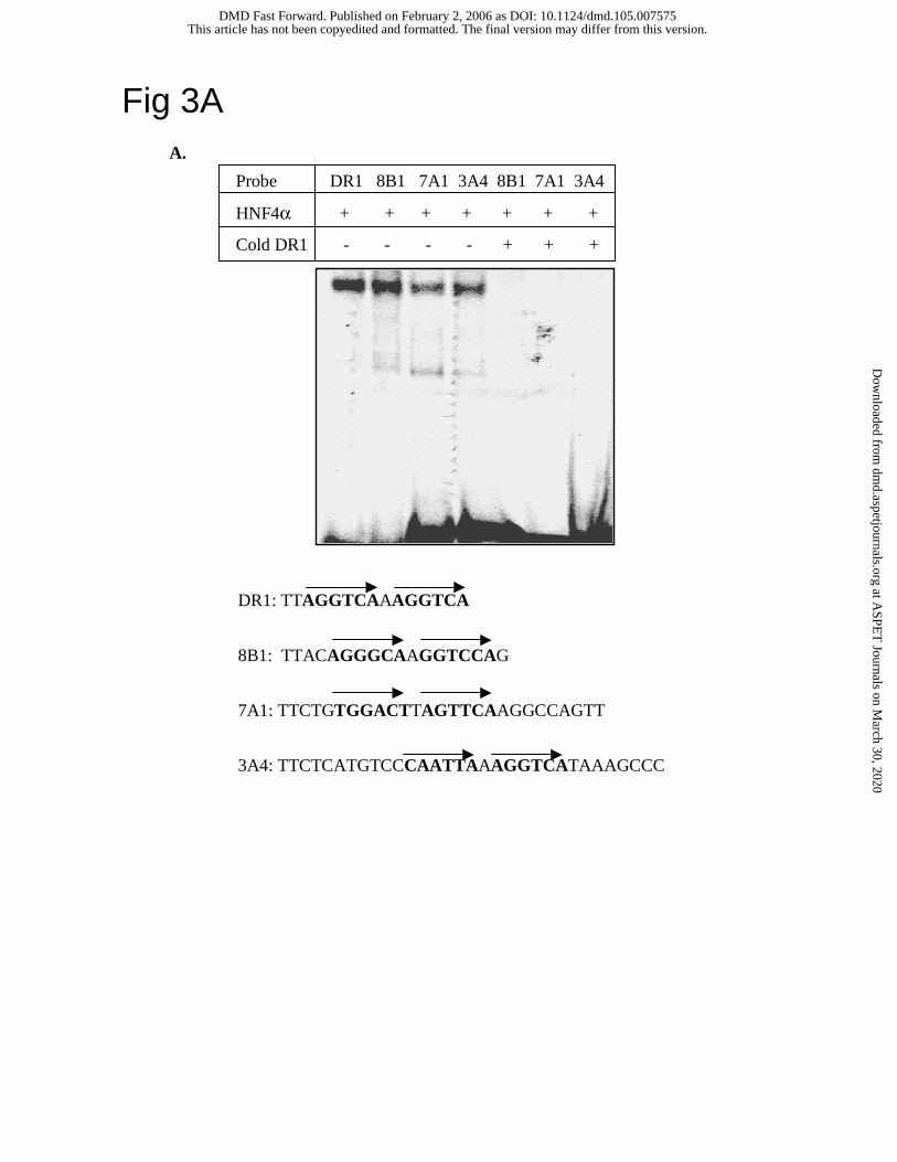
stimulated CYP3A4-Luc reporter activity (Tirona et al., 2003). EMSA shown in Fig. 3A
confirmed that the putative HNF4α binding site in the dXREM of the CYP3A4 gene was able to
bind HNF4α as the oligonucleotide probes designed according to a canonical DR1 motif and the
HNF4α binding sites in the CYP8B1 and CYP7A1 genes. Excess of unlabeled DR1 probe was
able to compete out HNF4α binding to the radiolabeled 8B1, 7A1 and 3A4 probes. We then
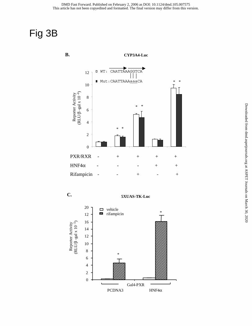
performed site-directed mutagenesis to mutate the putative HNF4α binding site in the CYP3A4
reporter plasmid. The mutant 3A4 probe did not bind HNF4α (data not shown). Fig. 3B shows
that mutations of the HNF4α site in the CYP3A4 reporter plasmid did not affect the basal
reporter activity and the synergistic stimulation of reporter activity by PXR and HNF4α. These
results suggested that HNF4α could stimulate CYP3A4 reporter activity without binding to the
CYP3A4 gene. To further demonstrate that HNF4α could interact with PXR without binding to
DNA, mammalian one-hybrid assay was performed. Fig. 3C demonstrated that HNF4α could
This article has not been copyedited and formatted. The final version may differ from this version.DMD Fast Forward. Published on February 2, 2006 as DOI: 10.1124/dmd.105.007575
at ASPE
T Journals on M
arch 30, 2020dm
d.aspetjournals.orgD
ownloaded from
DMD#7575
13
stimulate a Gal4 reporter (5XUAS-Luc) activity induced by binding of Gal4DBD-PXR fusion
protein to the upstream activation sequence (UAS) of the Gal4 DNA binding domain. All these
results support that HNF4α interaction with PXR synergistically stimulates CYP3A4 gene
transcription and the putative HNF4α binding site in the CYP3A4 gene may not be important in
promoter function.
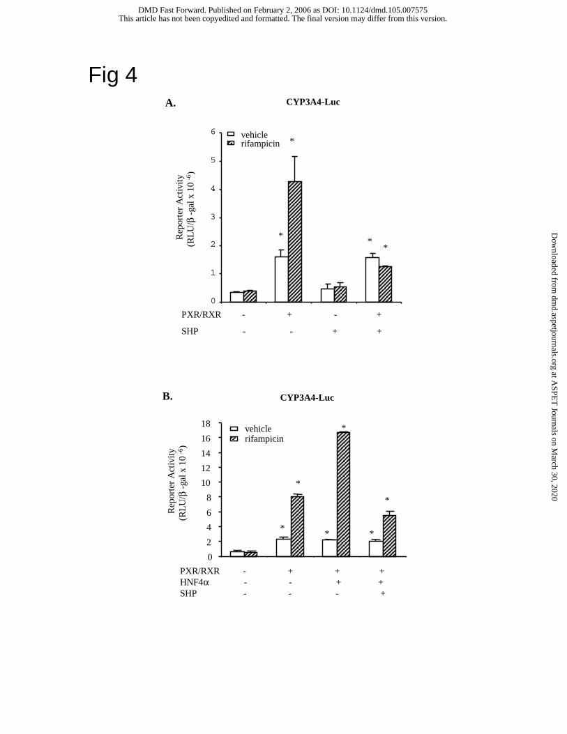
SHP inhibits CYP3A4 reporter activity. A previous study shows that SHP inhibits
CYP3A4 reporter activity and blocks PXR binding to DNA in EMSA assay (Ourlin et al., 2003).
We studied the effect of SHP on CYP3A4 reporter activity stimulated by PXR and HNF4α in
HepG2 cells. Fig. 4A shows that SHP inhibited PXR stimulation of CYP3A4 reporter activity
when activated in the presence of rifampicin. Fig. 4B shows that SHP also inhibited CYP3A4
reporter activity that was synergistically stimulated by PXR and HNF4α in the presence of
rifampicin. However, our EMSA assay failed to confirm that SHP prevents PXR binding to the
PXR binding site in the CYP3A4 gene (data not shown). This discrepancy may be due to the
amount of SHP used in the assay. We used in vitro translated SHP, which typically contains ng
quantity of translated proteins whereas Ourlin et al. used 9 µg GST-SHP fusion protein in EMSA
assays.
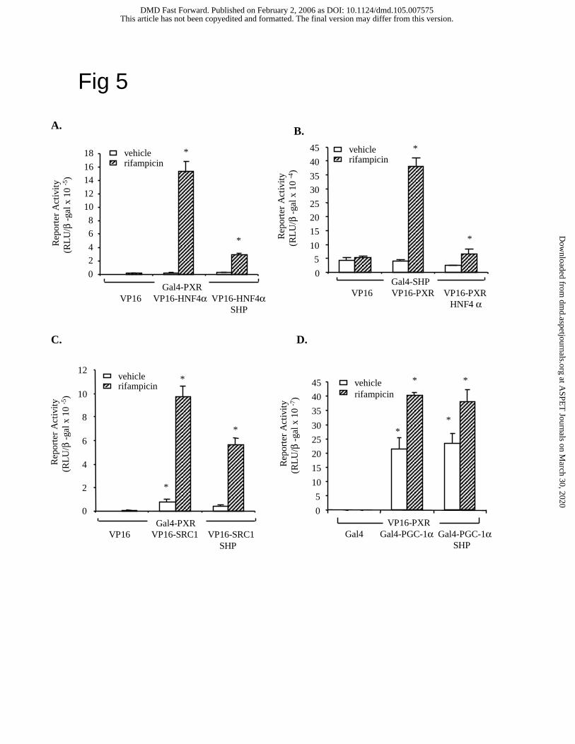
Mammalian two-hybrid assays of SHP inhibition of PXR interaction with co-activators.
We then performed cell-based mammalian two-hybrid assays in HepG2 cells to study the effect
of SHP on PXR interaction with HNF4α, SRC-1 and PGC-1α. Gal4-fusion proteins should bind
to the Gal4 binding site in the reporter plasmid 5XUAS-TK-Luc. If a Gal4-fusion protein
interacts with a VP16 activation domain-fusion protein, the reporter activity should be
stimulated. Addition of a competitor protein may disrupt the interaction and reduce the reporter
activity. This assay is very sensitive in detecting protein-protein interaction without interference
by endogenous transcription factors. Fig. 5A shows that Gal4-PXR strongly interacts with VP16-
This article has not been copyedited and formatted. The final version may differ from this version.DMD Fast Forward. Published on February 2, 2006 as DOI: 10.1124/dmd.105.007575
at ASPE
T Journals on M
arch 30, 2020dm
d.aspetjournals.orgD
ownloaded from
DMD#7575
14
HNF4α, and rifampicin was required for the interaction. Addition of SHP strongly inhibited
PXR/HNF4α interaction. Similarly, Gal4-SHP interacted with VP16-PXR in the presence of
rifampicin, and HNF4α inhibited the interaction (Fig. 5B). Therefore, HNF4α and SHP
competed for interaction with PXR. We did two-hybrid assays of Gal4-PXR interaction with
VP16-SRC-1 or VP16-PGC-1α to test if SHP inhibited PXR interaction with SRC-1 and PGC-
1α. Fig. 5C shows that Gal4-PXR interacted with VP16-SRC-1 but SHP only partially inhibited
the interaction. On the other hand, Gal4-PGC-1α interacted with VP16-PXR, but SHP did not
affect their interaction (Fig. 5D). These results indicated that HNF4α and SHP competed for
binding to PXR. However, SHP was unable to block PXR/PGC-1α interaction and only partially
blocked PXR/SRC-1 interaction.
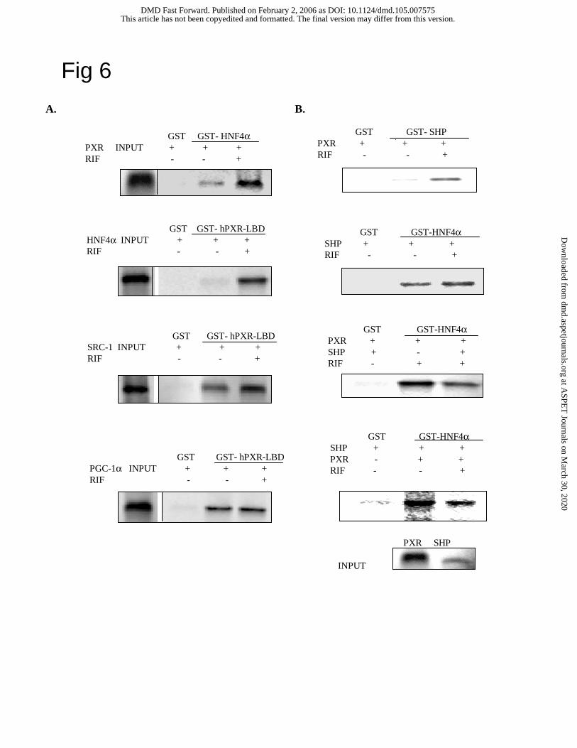
GST pull-down assays of PXR interaction with HNF4α, SRC-1, PGC-1α and SHP. GST
pull down assay is a cell-free assay for studying specific protein-protein interactions. GST-
HNF4α (full-length) fusion protein or GST-hPXR (LBD) fusion protein strongly interacted with
PXR or HNF4α, respectively, only when rifampicin was added to activate PXR (Fig. 6A). PXR
interacted with SRC-1 in the absence of rifampicin, and addition of rifampicin enhanced their
interaction. PXR interaction with PGC-1α does not require rifampicin. The degrees of ligand-
dependency for these co-activators to interact with PXR are consistent with mammalian two-
hybrid assays shown in Fig 5. We next performed GST pull-down assays to study the effect of
rifampicin on SHP interaction with PXR and HNF4α. In vitro translated and 35S labeled-PXR or
SHP was used to interact with GST-SHP or GST-HNF4α, respectively. As shown in Fig. 6B,
rifampicin strongly enhanced PXR and SHP interaction. However, rifampicin did not affect SHP
interaction with HNF4α as expected. We then added in vitro translated unlabeled-SHP or PXR to
study their effect on interaction of 35S-labeled PXR or SHP with HNF4α, respectively. Results
This article has not been copyedited and formatted. The final version may differ from this version.DMD Fast Forward. Published on February 2, 2006 as DOI: 10.1124/dmd.105.007575
at ASPE
T Journals on M
arch 30, 2020dm
d.aspetjournals.orgD
ownloaded from
DMD#7575
15
shown in lower panels of Fig. 6B demonstrated that the amount of PXR or SHP that bound to
GST-HNF4α was decreased. These results provided additional evidence that SHP, PXR and
HNF4α interacted and competed for binding to each other.
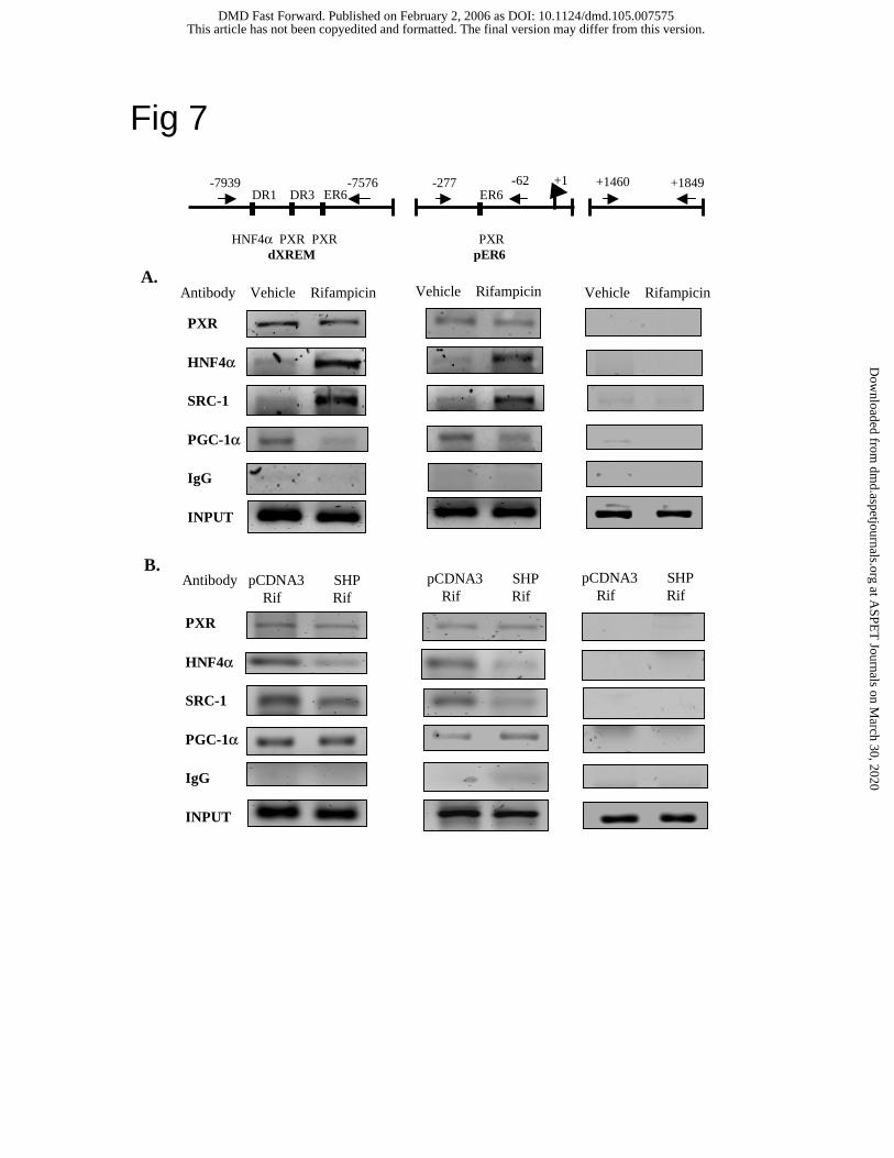
ChIP assays of PXR and co-activator recruitment to CYP3A4 chromatin.
We then performed ChIP assays using HepG2 cells to study the effect of rifampicin on PXR
recruitment of HNF4α, SRC-1 and PGC-1α to the CYP3A4 chromatin that contains PXR
binding sites. ChIP assay is considered as an in vivo assay of transcription factor interaction and
co-activator recruitment in the native promoter context. Since the levels of PGC-1α are low in
HepG2 cells, PGC-1α was overexpressed in HepG2 cells for ChIP assays. An anti-body against
PXR, HNF4α, SRC-1 or PGC-1α was used to immunoprecipitate the cross-linked DNA-protein
complexes in HepG2 cells treated with rifampicin or vehicle (DMSO). PCR primers were
designed to amplify the DNA fragments containing either the distal XREM or the proximal ER6
from these complexes. Fig. 7A shows PCR products generated by the primer pairs for dXREM
(left panels), pER6 (middle panels) and a negative control of the intron 2 region (right panel).
The non-immune IgG was used as a negative control of immunoprecipitation for ChIP assays
(bottom of each panel). The results indicated that PXR was associated with both chromatin and
rifampicin did not affect PXR binding. The levels of HNF4α associated with both dXREM and
dER6 chromatins were very low and similar despite the presence of a HNF4α binding site in
dXREM but not in pER6. Rifampicin strongly induced the recruitment of HNF4α to both
chromatins, most likely via interaction with PXR. Rifampicin also induced PXR or HNF4α
recruitment of SRC-1 to both chromatins. This is consistent with the enhanced binding of
HNF4α and SRC-1 to the PXR in the presence of rifampicin shown in Fig. 6A. It is surprising
that rifampicin reduced the recruitment of PGC-1α to both chromatins. This is in contrast to the
This article has not been copyedited and formatted. The final version may differ from this version.DMD Fast Forward. Published on February 2, 2006 as DOI: 10.1124/dmd.105.007575
at ASPE
T Journals on M
arch 30, 2020dm
d.aspetjournals.orgD
ownloaded from
DMD#7575
16
GST pull down assay results (Fig 6A) that rifampicin had no effect on PGC-1α interaction with
PXR, and the two-hybrid assay results (Fig. 5D) that rifampicin enhanced PXR interaction with
PGC-1α.
We then studied the effect of SHP on PXR recruitment of co-activators to the CYP3A4
chromatin. In experiments shown in Fig. 7B, rifampicin treated HepG2 cells were transfected
with PGC-1α and SHP or pcDNA3 empty plasmid as indicated. DNA-protein complexes were
immunoprecipitated with an antibody against PXR, HNF4α, SRC-1 or PGC-1α for ChIP assay.
Fig. 7B shows that SHP did not affect PXR binding to either chromatin. However, SHP reduced
PXR recruitment of HNF4α and SRC-1 to both chromatins, whereas SHP did not affect PXR
recruitment of PGC-1α. These data are consistent with mammalian two-hybrid assays (Fig. 5)
and GST pull-down assays (Fig. 6) that SHP blocks PXR interaction with HNF4α or SRC-1 but
does not affect PXR and PGC-1α interaction.
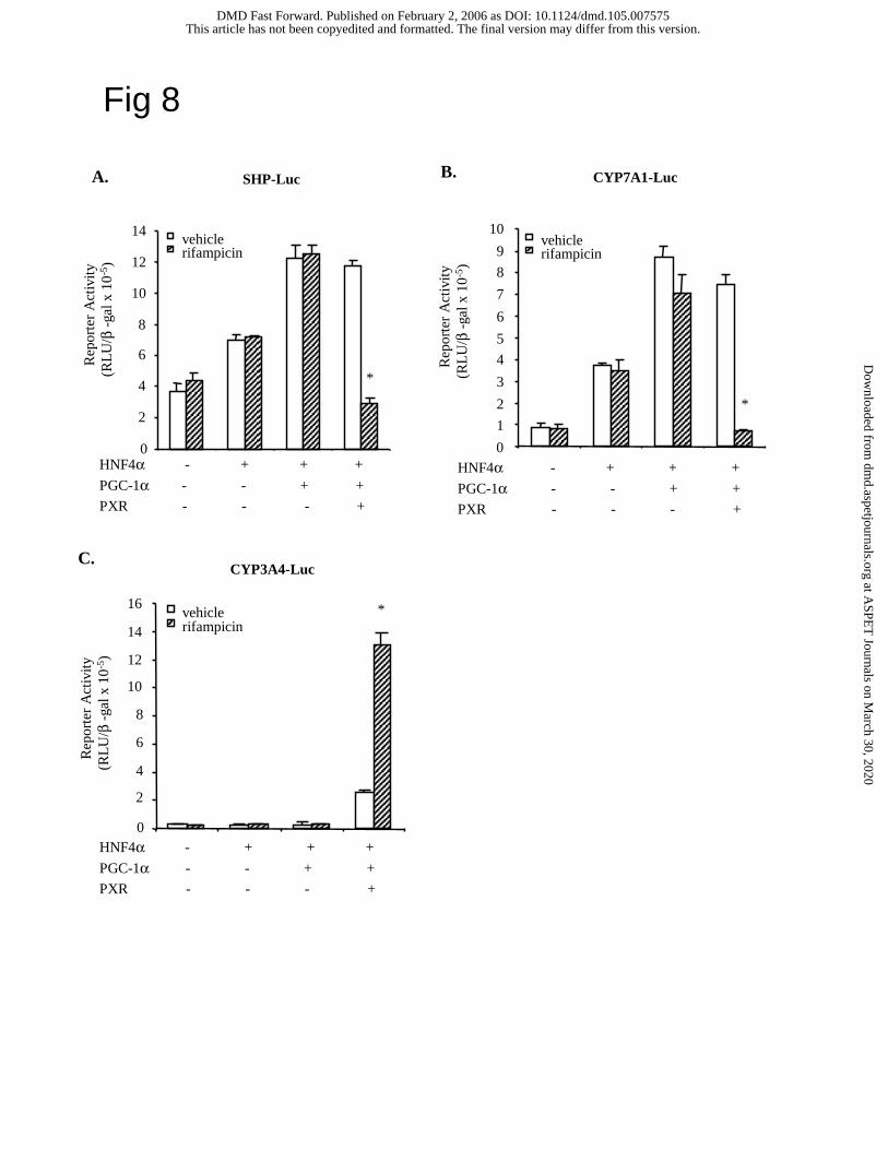
PXR inhibits SHP gene transcription. Since rifampicin strongly reduced SHP mRNA
expression, we hypothesized that PXR might inhibit SHP gene transcription. We and others
reported previously that HNF4α binds to the human SHP promoter and stimulates SHP gene
expression (Jung and Kullak-Ublick, 2003; Jahan and Chiang, 2005). Fig. 8A shows that HNF4α
and PGC-1α stimulated SHP reporter activity in HepG2 cells independent of rifampicin.
However, PXR strongly inhibited the SHP reporter activity stimulated by HNF4α and PGC-1α
only when rifampicin was added. As a positive control, PXR inhibited the CYP7A1 reporter
activity stimulated by HNF4α and PGC-1α in the presence of rifampicin (Fig. 8B) (Li and
Chiang, 2004). In complete contrast, rifampicin-activated PXR markedly stimulated CYP3A4
reporter activity when HNF4α and PGC-1α also were transfected (Fig. 8C). These data support
our previous finding that PXR inhibits the HNF4α target gene CYP7A1 by blocking HNF4α
This article has not been copyedited and formatted. The final version may differ from this version.DMD Fast Forward. Published on February 2, 2006 as DOI: 10.1124/dmd.105.007575
at ASPE
T Journals on M
arch 30, 2020dm
d.aspetjournals.orgD
ownloaded from
DMD#7575
17
recruitment of PGC-1α to CYP7A1 promoter (Li and Chiang, 2004). It appears the same
mechanism also regulates the human SHP promoter. In contrast, PXR stimulates the PXR target
gene CYP3A4. Taken together, our data suggest that HNF4α enhances PXR induction of
CYP3A4 by protein-protein interaction.
This article has not been copyedited and formatted. The final version may differ from this version.DMD Fast Forward. Published on February 2, 2006 as DOI: 10.1124/dmd.105.007575
at ASPE
T Journals on M
arch 30, 2020dm
d.aspetjournals.orgD
ownloaded from
DMD#7575
18
DISCUSSION
The ability to be strongly induced by a large variety of structurally unrelated compounds
allows CYP3A4 to efficiently respond to elevated toxic metabolites such as bile acids and drugs
and protects the liver against cholestasis induced by these compounds. Extensive studies in
recent years have provided convincing evidence that PXR is the key regulator of phase I drug
metabolizing enzymes, phase II bile acid and bilirubin conjugation enzymes and phase III drug
and metabolites transporters (Boyer, 2005). PXR is the most important nuclear receptor that
induces CYP3A4 to hydroxylate bile acids and drugs. However, most studies on PXR regulation
of CYP3A4 were performed in mouse models. In this study we used the primary human
hepatocyte and HepG2 models to study the molecular mechanism of human PXR regulation of
CYP3A4. Our real-time PCR assays revealed a remarkable 150-fold induction of CYP3A4
mRNA expression by rifampicin in human primary hepatocytes. In contrast, rifampicin treatment
reduced SHP and CYP7A1 mRNA expression. These data prompted us to study the mechanism
of PXR regulation of CYP3A4 and SHP gene transcription. Our study revealed that interaction
of PXR with HNF4α and its co-activators PGC-1α and SRC-1 contributes to the strong
induction of CYP3A4 by rifampicin and that concomitant inhibition of SHP gene expression by
PXR minimizes the inhibitory effect of SHP and maximizes PXR induction of CYP3A4. Several
studies suggest the involvement of HNF4α in regulation of the CYP3A4, CYP2C8 and CYP2C9
genes by PXR (Kamiya et al., 2003; Tirona et al., 2003; Chen et al., 2005; Ferguson et al., 2005).
This study suggests that HNF4α may act as a co-activator of PXR to recruit PGC-1α and SRC-1
to the PXR target gene CYP3A4. PXR interacts strongly with HNF4α, SRC-1, PGC-1α, and
SHP. SHP may compete with HNF4α and SRC-1 for binding to PXR and repress CYP3A4 gene
transcription (Fig. 9). Therefore, to maximize the induction of CYP3A4, PXR must inhibit SHP
gene transcription.
This article has not been copyedited and formatted. The final version may differ from this version.DMD Fast Forward. Published on February 2, 2006 as DOI: 10.1124/dmd.105.007575
at ASPE
T Journals on M
arch 30, 2020dm
d.aspetjournals.orgD
ownloaded from
DMD#7575
19
This study also revealed a general mechanism for PXR inhibition of HNF4α-activated
gene transcription. Previous studies suggest that PXR inhibited CYP7A1 by blocking HNF4α
recruitment of PGC-1α (Bhalla et al., 2004; Li and Chiang, 2004). This study revealed the same
mechanism for PXR inhibition of the human SHP gene. It is significant that rifampicin-activated
PXR inhibits both CYP7A1 and SHP expression, whereas bile acid-activated FXR induces SHP
to inhibit CYP7A1. FXR has been shown to modestly regulate the CYP3A4 gene (Gnerre et al.,
2004). It is reported recently that chenodeoxycholic acid and cholic acid induce SHP and repress
PXR-mediated induction of CYP3A4 in human primary hepatocytes and in mice (Ourlin et al.,
2003). Chenodeoxycholic acid and cholic acid are FXR ligands. Bile acid activation of FXR,
PXR, and maybe also the signaling pathways in their study complicated the interpretation of
their results. It is unlikely that FXR activation of CYP3A4 is a significant mechanism for bile
acid induction of CYP3A4 because CYP3A4 can be induced without FXR (Schuetz et al., 2001).
It seems clear that PXR and FXR regulate bile acid metabolism by somewhat different
mechanisms; the secondary bile acid, LCA activates PXR to inhibit bile acid synthesis, increase
bile acid excretion and detoxify bile acids, while the primary bile acid, CDCA activates FXR to
inhibit bile acid synthesis and stimulate biliary bile acid excretion.
The CYP3A4 expression is affected by factors such as genetic polymorphism, hormonal
status, cholestatic liver diseases, and exposure to drugs and xenobiotics. The induction of
CYP3A4 varies widely among individuals and may account for different drug metabolizing
activities and drug-drug interactions. PXR agonists such as rifampicin, dexamethasone,
glucocorticoids, and phenobarbital have been used for treating chronic liver diseases for several
decades without knowing the mechanism of drug action until the discovery of PXR in 1998. A
recent study of gallstone patients taking rifampicin or ursodeoxycholic acid (UDCA) confirmed
that rifampicin and UDCA induce hepatobiliary transport and detoxification systems and
This article has not been copyedited and formatted. The final version may differ from this version.DMD Fast Forward. Published on February 2, 2006 as DOI: 10.1124/dmd.105.007575
at ASPE
T Journals on M
arch 30, 2020dm
d.aspetjournals.orgD
ownloaded from
DMD#7575
20
provided molecular mechanisms for these anti-cholestatic drugs (Marschall et al., 2005). Our
current study provides a molecular mechanism for rifampicin induction of CYP3A4 in
detoxification of bile acids and drugs during cholestasis. Drugs targeted to PXR may have
potential for developing therapeutic drugs for treating cholestatic liver diseases induced by
drugs.
This article has not been copyedited and formatted. The final version may differ from this version.DMD Fast Forward. Published on February 2, 2006 as DOI: 10.1124/dmd.105.007575
at ASPE
T Journals on M
arch 30, 2020dm
d.aspetjournals.orgD
ownloaded from
DMD#7575
21
REFERENCES
Bachs L, Pares A, Elena M, Piera C and Rodes J (1989) Comparison of rifampicin with phenobarbitone for treatment of pruritus in biliary cirrhosis. Lancet 1:574-576.
Bertilsson G, Heidrich J, Svensson K, Asman M, Jendeberg L, Sydow-Backman M, Ohlsson R, Postlind H, Blomquist P and Berkenstam A (1998) Identification of a human nuclear receptor defines a new signaling pathway for CYP3A induction. Proc Natl Acad Sci U S A 95:12208-12213.
Bhalla S, Ozalp C, Fang S, Xiang L and Kemper JK (2004) Ligand-activated PXR interferes with HNF-4 signaling by targeting a common coactivator PGC-1α : Functional implications in hepatic cholesterol and glucose metabolism. J Biol Chem.
Boyer JL (2005) Nuclear receptor ligands: rational and effective therapy for chronic cholestatic liver disease? Gastroenterology 129:735-740.
Chen Y, Kissling G, Negishi M and Goldstein JA (2005) The Nuclear Receptors Constitutive Androstane Receptor and Pregnane X Receptor Cross-Talk with Hepatic Nuclear Factor 4α to Synergistically Activate the Human CYP2C9 Promoter. J Pharmacol Exp Ther 314:1125-1133.
De Fabiani E, Mitro N, Gilardi F, Caruso D, Galli G and Crestani M (2003) Coordinated control of cholesterol catabolism to bile acids and of gluconeogenesis via a novel mechanism of transcription regulation linked to the fasted-to-fed cycle. J Biol Chem 278:39124-39132.
Drocourt L, Ourlin JC, Pascussi JM, Maurel P and Vilarem MJ (2002) Expression of CYP3A4, CYP2B6, and CYP2C9 is regulated by the vitamin D receptor pathway in primary human hepatocytes. J Biol Chem 277:25125-25132.
Ferguson SS, Chen Y, LeCluyse EL, Negishi M and Goldstein JA (2005) Human CYP2C8 is transcriptionally regulated by the nuclear receptors constitutive androstane receptor, pregnane X receptor, glucocorticoid receptor, and hepatic nuclear factor 4alpha. Mol Pharmacol 68:747-757.
Gnerre C, Blattler S, Kaufmann MR, Looser R and Meyer UA (2004) Regulation of CYP3A4 by the bile acid receptor FXR: evidence for functional binding sites in the CYP3A4 gene. Pharmacogenetics 14:635-645.
Goodwin B, Hodgson E and Liddle C (1999) The orphan human pregnane X receptor mediates the transcriptional activation of CYP3A4 by rifampicin through a distal enhancer module. Mol Pharmacol 56:1329-1339.
Goodwin B, Jones SA, Price RR, Watson MA, McKee DD, Moore LB, Galardi C, Wilson JG, Lewis MC, Roth ME, Maloney PR, Willson TM and Kliewer SA (2000) A regulatory cascade of the nuclear receptors FXR, SHP-1, and LRH-1 represses bile acid biosynthesis. Mol Cell 6:517-526.
Goodwin B, Redinbo MR and Kliewer SA (2002) Regulation of cyp3a gene transcription by the pregnane x receptor. Annu Rev Pharmacol Toxicol 42:1-23.
Guengerich FP (1999) Cytochrome P-450 3A4: regulation and role in drug metabolism. Annu Rev Pharmacol Toxicol 39:1-17.
Hayhurst GP, Lee YH, Lambert G, Ward JM and Gonzalez FJ (2001) Hepatocyte nuclear factor 4α (nuclear receptor 2A1) is essential for maintenance of hepatic gene expression and lipid homeostasis. Mol Cell Biol 21:1393-1403.
Hofmann AF (2002) Rifampicin and treatment of cholestatic pruritus. Gut 51:756-757. Jahan A and Chiang JY (2005) Cytokine regulation of human sterol 12α-hydroxylase (CYP8B1)
gene. Am J Physiol Gastrointest Liver Physiol 288:G685-695.
This article has not been copyedited and formatted. The final version may differ from this version.DMD Fast Forward. Published on February 2, 2006 as DOI: 10.1124/dmd.105.007575
at ASPE
T Journals on M
arch 30, 2020dm
d.aspetjournals.orgD
ownloaded from
DMD#7575
22
Jung D and Kullak-Ublick GA (2003) Hepatocyte nuclear factor 1 alpha: a key mediator of the effect of bile acids on gene expression. Hepatology 37:622-631.
Kamiya A, Inoue Y and Gonzalez FJ (2003) Role of the hepatocyte nuclear factor 4α in control of the pregnane X receptor during fetal liver development. Hepatology 37:1375-1384.
Kliewer SA, Goodwin B and Willson TM (2002) The nuclear pregnane X receptor: a key regulator of xenobiotic metabolism. Endocr Rev 23:687-702.
Kliewer SA, Moore JT, Wade L, Staudinger JL, Watson MA, Jones SA, McKee DD, Oliver BB, Willson TM, Zetterstrom RH, Perlmann T and Lehmann JM (1998) An orphan nuclear receptor activated by pregnanes defines a novel steroid signaling pathway. Cell 92:73-82.
Lehmann JM, McKee DD, Watson MA, Willson TM, Moore JT and Kliewer SA (1998) The human orphan nuclear receptor PXR is activated by compounds that regulate CYP3A4 gene expression and cause drug interactions. J Clin Invest 102:1016-1023.
Li T and Chiang JY (2004) Mechanism of Rifampicin and Pregnane X Receptor (PXR) inhibition of Human Cholesterol 7α-Hydroxylase Gene (CYP7A1) Transcription. Am J Physiol Gastrointest Liver Physiol.
Lu TT, Makishima M, Repa JJ, Schoonjans K, Kerr TA, Auwerx J and Mangelsdorf DJ (2000) Molecular basis for feedback regulation of bile acid synthesis by nuclear receptors. Mol Cell 6:507-515.
Marschall HU, Wagner M, Zollner G, Fickert P, Diczfalusy U, Gumhold J, Silbert D, Fuchsbichler A, Benthin L, Grundstrom R, Gustafsson U, Sahlin S, Einarsson C and Trauner M (2005) Complementary stimulation of hepatobiliary transport and detoxification systems by rifampicin and ursodeoxycholic Acid in humans. Gastroenterology 129:476-485.
Ourlin JC, Lasserre F, Pineau T, Fabre JM, Sa-Cunha A, Maurel P, Vilarem MJ and Pascussi JM (2003) The small heterodimer partner interacts with the pregnane X receptor and represses its transcriptional activity. Mol Endocrinol 17:1693-1703.
Puigserver P, Rhee J, Donovan J, Walkey CJ, Yoon JC, Oriente F, Kitamura Y, Altomonte J, Dong H, Accili D and Spiegelman BM (2003) Insulin-regulated hepatic gluconeogenesis through FOXO1-PGC-1α interaction. Nature 423:550-555.
Schuetz EG, Strom S, Yasuda K, Lecureur V, Assem M, Brimer C, Lamba J, Kim RB, Ramachandran V, Komoroski BJ, Venkataramanan R, Cai H, Sinal CJ, Gonzalez FJ and Schuetz JD (2001) Disrupted bile acid homeostasis reveals an unexpected interaction among nuclear hormone receptors, transporters, and cytochrome P450. J Biol Chem 276:39411-39418.
Shin DJ, Campos JA, Gil G and Osborne TF (2003) PGC-1α activates CYP7A1 and bile acid biosynthesis. J Biol Chem 278:50047-50052.
Staudinger JL, Goodwin B, Jones SA, Hawkins-Brown D, MacKenzie KI, LaTour A, Liu Y, Klaassen CD, Brown KK, Reinhard J, Willson TM, Koller BH and Kliewer SA (2001) The nuclear receptor PXR is a lithocholic acid sensor that protects against liver toxicity. Proc Natl Acad Sci U S A 98:3369-3374.
Stroup D and Chiang JY (2000) HNF4 and COUP-TFII interact to modulate transcription of the cholesterol 7α-hydroxylase gene (CYP7A1). J Lipid Res 41:1-11.
Tirona RG, Lee W, Leake BF, Lan LB, Cline CB, Lamba V, Parviz F, Duncan SA, Inoue Y, Gonzalez FJ, Schuetz EG and Kim RB (2003) The orphan nuclear receptor HNF4α determines PXR- and CAR-mediated xenobiotic induction of CYP3A4. Nat Med 9:220-224.
This article has not been copyedited and formatted. The final version may differ from this version.DMD Fast Forward. Published on February 2, 2006 as DOI: 10.1124/dmd.105.007575
at ASPE
T Journals on M
arch 30, 2020dm
d.aspetjournals.orgD
ownloaded from
DMD#7575
23
Xie W, Radominska-Pandya A, Shi Y, Simon CM, Nelson MC, Ong ES, Waxman DJ and Evans RM (2001) An essential role for nuclear receptors SXR/PXR in detoxification of cholestatic bile acids. Proc Natl Acad Sci U S A 98:3375-3380.
This article has not been copyedited and formatted. The final version may differ from this version.DMD Fast Forward. Published on February 2, 2006 as DOI: 10.1124/dmd.105.007575
at ASPE
T Journals on M
arch 30, 2020dm
d.aspetjournals.orgD
ownloaded from
DMD#7575
24
FOOTNOTES
This work was supported by NIH grants DK44442 and DK31584.
This article has not been copyedited and formatted. The final version may differ from this version.DMD Fast Forward. Published on February 2, 2006 as DOI: 10.1124/dmd.105.007575
at ASPE
T Journals on M
arch 30, 2020dm
d.aspetjournals.orgD
ownloaded from
DMD#7575
25
FIGURE LEGEND
Fig. 1. Real time PCR analysis of the effect of rifampicin on CYP3A4, SHP and CYP7A1 mRNA
expression levels in primary human hepatocytes. Relative mRNA expression (un-treated control
is set as 1) levels are plotted vs. concentrations of rifampicin.
Fig. 2. Effects of HNF4α and co-activators on PXR regulation of the human CYP3A4 reporter
gene. CYP3A4 luciferase reporter, p3A4-5’dDR3/dER6/pER6 (0.2 µg) was co-transfected with
PXR/RXRα, SRC-1, HNF4α, and/or PGC-1α (0.1 µg) into HepG2 cells as indicated in A & B.
Cells were treated with vehicle (DMSO) or 10 µM rifampicin for 40 h and harvested for
luciferase activity assays as described under Materials and Methods. The error bars represent the
standard deviation from the mean of triplicate assays of an individual experiment, and “*”
indicates statistically significant difference from control, N=3, p<0.05.
Fig. 3. Synergistically induction of CYP3A4 by HNF4α and PXR does not require HNF4α
binding to the CYP3A4 gene promoter. A. EMSA of HNF4α binding to CYP3A4 dXREM.
Probes containing a consensus HNF4α binding site (DR1) or HNF4α binding sites from
CYP8B1 promoter (8B1), CYP7A1 promoter (7A1) and CYP3A4 dXREM (3A4) were labeled
with S35dATP. Each Probe (5 x 10⁴ cpm) and 5 µl of in vitro synthesized HNF4α protein were
mixed and applied to each lane as indicated. 100-fold excess of unlabeled DR1 probes were used
as cold competitors. B. Luciferase reporter constructs (0.2 µg) of wild type (WT) and mutant
CYP3A4 (Mut) containing mutations in the dXREM HNF4α binding site were transfected into
HepG2 cells. Expression plasmids (0.1 µg) of PXR, RXRα and/or HNF4α were co-transfected
as indicated. Cells were treated with vehicle (DMSO) or 10 µM rifampicin for 40 h and
harvested for luciferase activity assays as described under Experimental Procedures. The error
bars represent the standard deviation from the mean of triplicate assays of an individual
experiment, and “*” indicates statistically significant difference from control, N=3, p<0.05. C.
This article has not been copyedited and formatted. The final version may differ from this version.DMD Fast Forward. Published on February 2, 2006 as DOI: 10.1124/dmd.105.007575
at ASPE
T Journals on M
arch 30, 2020dm
d.aspetjournals.orgD
ownloaded from
DMD#7575
26
Mammalian one-hybrid assay of HNF4α co-activation of PXR. HepG2 cells were transfected
with 5XUAS-TK-Luc reporter (0.2 µg) and 0.1 µg expression plasmids: Gal4-PXR, PCDNA3 or
HNF4α as indicated. Cells were treated with vehicle (DMSO), or 10 µM rifampicin for 40 h.
The error bars represent the standard deviation from the mean of triplicate assays of an
individual experiment, and “*” indicates statistically significant difference from control, N=3,
p<0.05.
Fig. 4. SHP inhibition of CYP3A4 reporter activity stimulated by PXR and HNF4α. Human
CYP3A4 reporter, p3A4-5’dDR3/dER6/pER6 (0.2 µg) was co-transfected with 0.1 µg each of
PXR/RXRα, HNF4α and SHP expression plasmids as indicated in (A) and (B). Reporter assays
were performed and analyzed as in Fig. 2.
Fig. 5. Mammalian two-hybrid assays of the effect of SHP on the PXR interaction with HNF4α,
SRC-1 and PGC-1α. HepG2 cells were transfected with 5XUAS-TK-Luc reporter (0.2 µg) and
0.1 µg expression plasmids: Gal4, Gal4-PXR, Gal4-SHP, Gal4-PGC-1α, VP16, VP16-HNF4α,
VP16-PXR, VP16-SRC1, SHP and HNF4α, as indicated. Cells were treated with vehicle
(DMSO), or 10 µM rifampicin for 40 h. The error bars represent the standard deviation from the
mean of triplicate assays of an individual experiment, and “*” indicates statistically significant
difference from control, N=3, p<0.05.
Fig. 6. GST pull-down assays of PXR interactions with HNF4α, SRC-1, PGC-1α and SHP. A.
Effect of rifampicin on HNF4α, PXR, SRC-1 and PGC-1α interaction. DMSO or 10 µM
rifampicin was added to the incubation mixture. B. Effect of SHP on HNF4α and PXR
interaction. GST-SHP fusion or GST-HNF4α fusion was incubated with 5 µl of 35S-labeled, in
vitro translated PXR or SHP, and/or un-labeled SHP or PXR as indicated. DMSO or 10 µM
rifampicin was added to the incubation mixture as indicated. One µl of the in vitro translated
This article has not been copyedited and formatted. The final version may differ from this version.DMD Fast Forward. Published on February 2, 2006 as DOI: 10.1124/dmd.105.007575
at ASPE
T Journals on M
arch 30, 2020dm
d.aspetjournals.orgD
ownloaded from
DMD#7575
27
protein was used as “input”. GST pull-down assays were performed as described under
Experimental Procedures.
Fig. 7. ChIP assay of the effect of rifampicin on PXR recruitment of HNF4α, SRC-1, and PGC-1
to CYP3A4 chromatin. A (upper panels). HepG2 cells were transfected with 10 µg PGC-1α
expression plasmids. Anti-PXR, anti-HNF4α, anti-PGC-1α, or anti-SRC-1 antibodies was used
to precipitate the DNA-protein complexes. B (lower panels). Effect of SHP on the PXR
recruitment of HNF4α, SRC-1 and PGC-1α to CYP3A4 chromatin. HepG2 cells were
transfected with 10 µg PGC-1α and SHP expression plasmids or pcDNA3 empty plasmid as
control for 40 h. An antibody against PXR, HNF4, SRC-1 or PGC-1 was used to
immunoprecipitate DNA-protein complexes. Cells were treated with 10 µM rifampicin or
vehicle (DMSO) as indicated for 40 h. ChIP assays were performed as described under Material
and Methods. DNA fragments containing distal XREM (left panel), proximal ER6 (middle
panel) or 2nd intron (right panel) was PCR amplified (illustrated on the top of figure) and
analyzed on a 2% agarose gel. Normal IgG was used alone as non-immuno control. 10% of the
total cell lysate was used as “input”.
Fig. 8. PXR regulation of SHP promoter activity. HepG2 cells were cultured to about 80%
confluence. Human SHP-Luc (A), CYP7A1-Luc (B), or CYP3A4-Luc (C) reporter plasmid (0.2
µg) was co-transfected with HNF4α, PGC-1α or PXR expression plasmids (0.1 µg) as indicated.
Cells were treated with vehicle (DMSO), or 10 µM rifampicin for 40 h and harvested for
luciferase activity assays as described under Materials and Methods. The error bars represent the
standard deviation from the mean of triplicate assays of an individual experiment, “*” indicates
statistically significant difference from control, N=3, p<0.05.
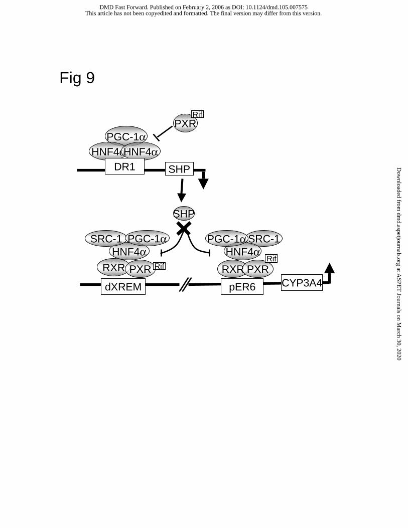
Fig. 9. A model of PXR, HNF4α and co-activator regulation of human CYP3A4 gene in
hepatocytes. PXR/RXRα binds to dXREM and pER6 motifs in the human CYP3A4 promoter.
This article has not been copyedited and formatted. The final version may differ from this version.DMD Fast Forward. Published on February 2, 2006 as DOI: 10.1124/dmd.105.007575
at ASPE
T Journals on M
arch 30, 2020dm
d.aspetjournals.orgD
ownloaded from
DMD#7575
28
Rifampicin induces PXR interaction with HNF4α, which is co-activated by PGC-1α and SRC-1.
HNF4α binding site located upstream of dXREM may not be important in regulation of
CYP3A4. SHP is able to inhibit CYP3A4 gene transcription by blocking PXR and HNF4α
interaction and recruitment of PGC-1α and SRC-1 to CYP3A4 chromatin. SHP gene
transcription is induced by HNF4α, which binds to a DR1 motif in the SHP promoter.
Rifampicin-activated PXR inhibits SHP transcription by blocking PGC-1α recruitment to SHP
chromatin. As a consequence, SHP expression is reduced and CYP3A4 transcription is
maximized.
This article has not been copyedited and formatted. The final version may differ from this version.DMD Fast Forward. Published on February 2, 2006 as DOI: 10.1124/dmd.105.007575
at ASPE
T Journals on M
arch 30, 2020dm
d.aspetjournals.orgD
ownloaded from
0
0.2
0.4
0.6
0.8
1.0
1.2
0 1 5 10
SHP
0
50
100
150
200
250
0 1 5 10
CYP3A4
-0.4
-0.2
0
0.2
0.4
0.6
0.8
1.0
1.2
0 1 5 10
CYP7A1
Rifampicin (µM)
Rifampicin (µM)
Rifampicin (µM)
Rel
ativ
e m
RN
A e
xpre
ssio
nR
elat
ive
mR
NA
exp
ress
ion
Fig.1
This article has not been copyedited and formatted. The final version may differ from this version.DMD Fast Forward. Published on February 2, 2006 as DOI: 10.1124/dmd.105.007575
at ASPE
T Journals on M
arch 30, 2020dm
d.aspetjournals.orgD
ownloaded from
PXR/RXR - + - +
HNF4α - - + +
Rep
orte
r A
ctiv
ity
(RL
U/β
-gal
x 1
0 -6
)
0
2
4
6
8
10
12 vehiclerifampicin
CYP3A4-LucA.
CYP3A4-Luc
Rep
orte
r A
ctiv
ity(R
LU
/β-g
al x
10
-6)
PXR/RXRαSRC-1PGC-1αHNF4α
0
2
4
6
8
10
12
14 VehicleRifampicin
*
*
**
***
* *
*
* *
*
B.
----
+---
++--
+-+-
+++-
+-++
++-+
++++
*
*
**
Fig 2
This article has not been copyedited and formatted. The final version may differ from this version.DMD Fast Forward. Published on February 2, 2006 as DOI: 10.1124/dmd.105.007575
at ASPE
T Journals on M
arch 30, 2020dm
d.aspetjournals.orgD
ownloaded from
Probe DR1 8B1 7A1 3A4 8B1 7A1 3A4
HNF4α + + + + + + +
Cold DR1 - - - - + + +
A.
DR1: TTAGGTCAAAGGTCA
8B1: TTACAGGGCAAGGTCCAG
7A1: TTCTGTGGACTTAGTTCAAGGCCAGTT
3A4: TTCTCATGTCCCAATTAAAGGTCATAAAGCCC
Fig 3A
This article has not been copyedited and formatted. The final version may differ from this version.DMD Fast Forward. Published on February 2, 2006 as DOI: 10.1124/dmd.105.007575
at ASPE
T Journals on M
arch 30, 2020dm
d.aspetjournals.orgD
ownloaded from
Gal4-PXRPCDNA3 HNF4α
Rep
orte
r A
ctiv
ity(R
LU
/β-g
al x
10
-5)
5XUAS-TK-LucC.
0
2
4
6
8
10
12
14
16
12
20 vehiclerifampicin
*
*
PXR/RXR - + + + +
HNF4α - - - + +
Rifampicin - - + - +
Rep
orte
r A
ctiv
ity(R
LU
/β-g
al x
10
-6)
B.
0
2
4
6
8
10
12
CYP3A4-Luc
* *
* *
* *
WT: CAATTAAAGGTCA
Mut:CAATTAAAaaaCA
Fig 3B
This article has not been copyedited and formatted. The final version may differ from this version.DMD Fast Forward. Published on February 2, 2006 as DOI: 10.1124/dmd.105.007575
at ASPE
T Journals on M
arch 30, 2020dm
d.aspetjournals.orgD
ownloaded from
CYP3A4-Luc
0
2
4
6
8
10
12
14
16
18
PXR/RXR - + + +HNF4α - - + + SHP - - - +
Rep
orte
r A
ctiv
ity(R
LU
/β-g
al x
10
-6)
vehiclerifampicin
*
*
*
* *
*
Rep
orte
r A
ctiv
ity(R
LU
/β-g
al x
10
-6)
PXR/RXR - + - +
SHP - - + +
CYP3A4-Luc
B.
0
1
2
3
4
5
6 vehiclerifampicin
A.
*
**
*
Fig 4
This article has not been copyedited and formatted. The final version may differ from this version.DMD Fast Forward. Published on February 2, 2006 as DOI: 10.1124/dmd.105.007575
at ASPE
T Journals on M
arch 30, 2020dm
d.aspetjournals.orgD
ownloaded from
A.
Gal4-SHPVP16 VP16-PXR VP16-PXR
HNF4 α
Rep
orte
r A
ctiv
ity(R
LU
/β-g
al x
10
-4)
0
5
10
15
20
25
30
35
40
45
B.
C. D.
vehiclerifampicin
Rep
orte
r A
ctiv
ity(R
LU
/β-g
al x
10
-5)
Gal4-PXRVP16 VP16-HNF4α VP16-HNF4α
SHP
0
24
68
1012
1416
18 vehiclerifampicin
*
*
*
0
2
4
6
8
10
12
Gal4-PXRVP16 VP16-SRC1 VP16-SRC1
SHP
Rep
orte
r A
ctiv
ity(R
LU
/β-g
al x
10
-5)
vehiclerifampicin
*
*
*
Rep
orte
r A
ctiv
ity(R
LU
/β-g
al x
10
-7)
0
5
10
15
20
25
30
35
40
45
VP16-PXRGal4 Gal4-PGC-1α Gal4-PGC-1α
SHP
vehiclerifampicin
*
*
*
*
*
Fig 5
This article has not been copyedited and formatted. The final version may differ from this version.DMD Fast Forward. Published on February 2, 2006 as DOI: 10.1124/dmd.105.007575
at ASPE
T Journals on M
arch 30, 2020dm
d.aspetjournals.orgD
ownloaded from
A.
GST GST-HNF4αPXR + + +SHP + - +RIF - + +
GST GST- SHPPXR + + +RIF - - +
B.
GST GST-HNF4αSHP + + +PXR - + + RIF - - +
INPUT
PXR SHP
GST GST-HNF4αSHP + + +RIF - - +
GST GST- HNF4αPXR INPUT + + +RIF - - +
GST GST- hPXR-LBDHNF4α INPUT + + +RIF - - +
GST GST- hPXR-LBDPGC-1α INPUT + + +RIF - - +
GST GST- hPXR-LBDSRC-1 INPUT + + +RIF - - +
Fig 6
This article has not been copyedited and formatted. The final version may differ from this version.DMD Fast Forward. Published on February 2, 2006 as DOI: 10.1124/dmd.105.007575
at ASPE
T Journals on M
arch 30, 2020dm
d.aspetjournals.orgD
ownloaded from
HNF4α PXR PXR PXRdXREM pER6
-7939 -7576ER6DR3DR1
-62-277ER6
A.Antibody Vehicle Rifampicin Vehicle Rifampicin
PXR
HNF4α
SRC-1
PGC-1α
IgG
INPUT
B.
PXR
HNF4α
SRC-1
PGC-1α
IgG
INPUT
Antibody pCDNA3 SHPRif Rif
pCDNA3 SHPRif Rif
Vehicle Rifampicin
pCDNA3 SHPRif Rif
+1460 +1849+1
Fig 7
This article has not been copyedited and formatted. The final version may differ from this version.DMD Fast Forward. Published on February 2, 2006 as DOI: 10.1124/dmd.105.007575
at ASPE
T Journals on M
arch 30, 2020dm
d.aspetjournals.orgD
ownloaded from
SHP-Luc
0
2
4
6
8
10
12
14
HNF4α - + + +
PGC-1α - - + +
PXR - - - +
Rep
orte
r A
ctiv
ity(R
LU
/β-g
al x
10-5
)
vehiclerifampicin
A.
0
1
2
3
4
5
6
7
8
9
10
Rep
orte
r A
ctiv
ity(R
LU
/β-g
al x
10-5
)
CYP7A1-LucB.
C.
vehiclerifampicin
HNF4α - + + +
PGC-1α - - + +
PXR - - - +
*
*
Rep
orte
r A
ctiv
ity(R
LU
/β-g
al x
10-5
)
CYP3A4-Luc
0
2
4
6
8
10
12
14
16vehiclerifampicin
HNF4α - + + +
PGC-1α - - + +
PXR - - - +
*
Fig 8
This article has not been copyedited and formatted. The final version may differ from this version.DMD Fast Forward. Published on February 2, 2006 as DOI: 10.1124/dmd.105.007575
at ASPE
T Journals on M
arch 30, 2020dm
d.aspetjournals.orgD
ownloaded from
SHP
SRC-1
PGC-1αPXR
HNF4α
dXREM CYP3A4RXR PXR RXR PXR
pER6
HNF4αHNF4αPGC-1αSRC-1
Rif
RifRif
HNF4αDR1 SHP
PGC-1α
Fig 9
This article has not been copyedited and formatted. The final version may differ from this version.DMD Fast Forward. Published on February 2, 2006 as DOI: 10.1124/dmd.105.007575
at ASPE
T Journals on M
arch 30, 2020dm
d.aspetjournals.orgD
ownloaded from