The Rate-Limiting Step of O Activation in the α ... Rate-Limiting Step of O 2 Activation in the...
8
The Rate-Limiting Step of O 2 Activation in the α‑Ketoglutarate Oxygenase Factor Inhibiting Hypoxia Inducible Factor John A. Hangasky, † Hasand Gandhi, ‡ Meaghan A. Valliere, † Nathaniel E. Ostrom, ‡ and Michael J. Knapp* ,† † Department of Chemistry, University of Massachusetts, Amherst, Massachusetts 01003, United States ‡ Department of Zoology, Michigan State University, East Lansing, Michigan 48824, United States * S Supporting Information ABSTRACT: Factor inhibiting HIF (FIH) is a cellular O 2 - sensing enzyme, which hydroxylates the hypoxia inducible factor-1α. Previously reported inverse solvent kinetic isotope effects indicated that FIH limits its overall turnover through an O 2 activation step (Hangasky, J. A., Saban, E., and Knapp, M. J. (2013) Biochemistry 52, 1594−1602). Here we characterize the rate-limiting step for O 2 activation by FIH using a suite of mechanistic probes on the second order rate constant k cat / K M(O 2 ) . Steady-state kinetics showed that the rate constant for O 2 activation was slow (k cat /K M(O 2 ) app = 3500 M −1 s −1 ) compared with other non-heme iron oxygenases, and solvent viscosity assays further excluded diffusional encounter with O 2 from being rate limiting on k cat /K M(O 2 ) . Competitive oxygen-18 kinetic isotope effect measurements ( 18 k cat /K M(O 2 ) = 1.0114(5)) indicated that the transition state for O 2 activation resembled a cyclic peroxohemiketal, which precedes the formation of the ferryl intermediate observed in related enzymes. We interpret this data to indicate that FIH limits its overall activity at the point of the nucleophilic attack of Fe-bound O 2 on the C-2 carbon of αKG. Overall, these results show that FIH follows the consensus mechanism for αKG oxygenases, suggesting that FIH may be an ideal enzyme to directly access steps involved in O 2 activation among the broad family of αKG oxygenases. M ammalian cells respond to decreased cellular pO 2 levels through the enzyme-catalyzed reaction of O 2 with the hypoxia inducible factor-1α (HIF-1α or HIF). 1 HIF mediates the transcription of hundreds of genes in response to hypoxia 2 with the functions of the gene products ranging from glucose and iron metabolism to cell proliferation and angiogenesis. 3,4 Factor inhibiting HIF (FIH) is a non-heme Fe(II)/αKG oxygenase that turns-off the transcriptional activity of HIF 5,6 by hydroxylating the β-carbon of Asn 803 within the C-terminal activation domain (CTAD) of HIF (Scheme 1). 7−9 Because O 2 activation chemistry is central to hypoxia sensing by HIF, identifying the chemical steps involved in O 2 activation may point the way to methods for perturbing HIF-controlled gene expression. FIH is proposed to follow the consensus mechanism for Fe(II)/αKG oxygenases (Scheme 1) for which the steps are supported to varying degrees by spectroscopic, computational, and kinetic studies. 10−15 VTVH MCD methodologies have been used to spectroscopically identify the release of the aquo ligand upon substrate binding to FIH 16 and other Fe(II)/αKG oxygenases including TauD and CAS. 17,18 O 2 is thought to bind as a ferric superoxide at the open coordination site and then attacks the C-2 carbonyl of αKG to ultimately form succinate and a ferryl intermediate. The molecular details following O 2 activation, including isolation of the ferryl intermediate and observation of HAT have been characterized in the Fe(II)/ αKG oxygenases TauD 15,19−24 and P4H 25 and the related Fe(II)/αKG halogenases CytC3 26 and SyrB2. 27 In contrast to the steps following ferryl formation, those steps of O 2 activation are poorly understood. Computational studies suggest that nucleophilic attack on the C-2 carbonyl of αKG is the rate-limiting step on k cat /K M(O 2 ) , with a cyclic peroxohemiketal proposed as the transition state. 28−30 Although this reaction sequence is supported by pre-steady- state kinetics and an oxygen-18 kinetic isotope effect ( 18 O KIE) study of TauD, 31 insight into O 2 activation is limited because HAT or product release is rate-limiting in TauD and other well- characterized αKG oxygenases. 20,32,33 Consequently, O 2 activation is too rapid to allow for the identification of any intermediates prior to the ferryl. Recent studies showed that the rate-limiting step for FIH differed from that of other characterized αKG oxygenases. 16,34 Upon FIH binding to CTAD, there is partial retention of the aquo ligand 16 suggesting that aquo release may be less facile in FIH than in other enzymes. The inverse SKIE on k cat 34 for FIH Received: October 2, 2014 Revised: November 20, 2014 Published: November 25, 2014 Article pubs.acs.org/biochemistry © 2014 American Chemical Society 8077 dx.doi.org/10.1021/bi501246v | Biochemistry 2014, 53, 8077−8084 This is an open access article published under an ACS AuthorChoice License, which permits copying and redistribution of the article or any adaptations for non-commercial purposes.
-
Upload
trinhduong -
Category
Documents
-
view
213 -
download
0
Transcript of The Rate-Limiting Step of O Activation in the α ... Rate-Limiting Step of O 2 Activation in the...

The Rate-Limiting Step of O2 Activation in the α‑KetoglutarateOxygenase Factor Inhibiting Hypoxia Inducible FactorJohn A. Hangasky,† Hasand Gandhi,‡ Meaghan A. Valliere,† Nathaniel E. Ostrom,‡
and Michael J. Knapp*,†
†Department of Chemistry, University of Massachusetts, Amherst, Massachusetts 01003, United States‡Department of Zoology, Michigan State University, East Lansing, Michigan 48824, United States
*S Supporting Information
ABSTRACT: Factor inhibiting HIF (FIH) is a cellular O2-sensing enzyme, which hydroxylates the hypoxia induciblefactor-1α. Previously reported inverse solvent kinetic isotopeeffects indicated that FIH limits its overall turnover through anO2 activation step (Hangasky, J. A., Saban, E., and Knapp, M. J.(2013) Biochemistry 52, 1594−1602). Here we characterize therate-limiting step for O2 activation by FIH using a suite ofmechanistic probes on the second order rate constant kcat/KM(O2). Steady-state kinetics showed that the rate constant for
O2 activation was slow (kcat/KM(O2)app = 3500 M−1 s−1) compared
with other non-heme iron oxygenases, and solvent viscosity assays further excluded diffusional encounter with O2 from being ratelimiting on kcat/KM(O2). Competitive oxygen-18 kinetic isotope effect measurements (18kcat/KM(O2) = 1.0114(5)) indicated that thetransition state for O2 activation resembled a cyclic peroxohemiketal, which precedes the formation of the ferryl intermediateobserved in related enzymes. We interpret this data to indicate that FIH limits its overall activity at the point of the nucleophilicattack of Fe-bound O2
on the C-2 carbon of αKG. Overall, these results show that FIH follows the consensus mechanism forαKG oxygenases, suggesting that FIH may be an ideal enzyme to directly access steps involved in O2 activation among the broadfamily of αKG oxygenases.
Mammalian cells respond to decreased cellular pO2 levelsthrough the enzyme-catalyzed reaction of O2 with the
hypoxia inducible factor-1α (HIF-1α or HIF).1 HIF mediatesthe transcription of hundreds of genes in response to hypoxia2
with the functions of the gene products ranging from glucoseand iron metabolism to cell proliferation and angiogenesis.3,4
Factor inhibiting HIF (FIH) is a non-heme Fe(II)/αKGoxygenase that turns-off the transcriptional activity of HIF5,6 byhydroxylating the β-carbon of Asn803 within the C-terminalactivation domain (CTAD) of HIF (Scheme 1).7−9 Because O2activation chemistry is central to hypoxia sensing by HIF,identifying the chemical steps involved in O2 activation maypoint the way to methods for perturbing HIF-controlled geneexpression.FIH is proposed to follow the consensus mechanism for
Fe(II)/αKG oxygenases (Scheme 1) for which the steps aresupported to varying degrees by spectroscopic, computational,and kinetic studies.10−15 VTVH MCD methodologies havebeen used to spectroscopically identify the release of the aquoligand upon substrate binding to FIH16 and other Fe(II)/αKGoxygenases including TauD and CAS.17,18 O2 is thought to bindas a ferric superoxide at the open coordination site and thenattacks the C-2 carbonyl of αKG to ultimately form succinateand a ferryl intermediate. The molecular details following O2activation, including isolation of the ferryl intermediate and
observation of HAT have been characterized in the Fe(II)/αKG oxygenases TauD15,19−24 and P4H25 and the relatedFe(II)/αKG halogenases CytC326 and SyrB2.27
In contrast to the steps following ferryl formation, thosesteps of O2 activation are poorly understood. Computationalstudies suggest that nucleophilic attack on the C-2 carbonyl ofαKG is the rate-limiting step on kcat/KM(O2), with a cyclic
peroxohemiketal proposed as the transition state.28−30
Although this reaction sequence is supported by pre-steady-state kinetics and an oxygen-18 kinetic isotope effect (18O KIE)study of TauD,31 insight into O2 activation is limited becauseHAT or product release is rate-limiting in TauD and other well-characterized αKG oxygenases.20,32,33 Consequently, O2
activation is too rapid to allow for the identification of anyintermediates prior to the ferryl.Recent studies showed that the rate-limiting step for FIH
differed from that of other characterized αKG oxygenases.16,34
Upon FIH binding to CTAD, there is partial retention of theaquo ligand16 suggesting that aquo release may be less facile inFIH than in other enzymes. The inverse SKIE on kcat
34 for FIH
Received: October 2, 2014Revised: November 20, 2014Published: November 25, 2014
Article
pubs.acs.org/biochemistry
© 2014 American Chemical Society 8077 dx.doi.org/10.1021/bi501246v | Biochemistry 2014, 53, 8077−8084
This is an open access article published under an ACS AuthorChoice License, which permitscopying and redistribution of the article or any adaptations for non-commercial purposes.
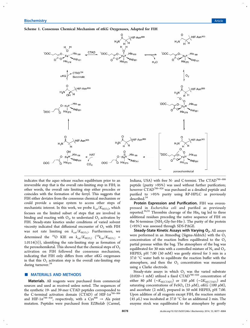
indicates that the aquo release reaches equilibrium prior to anirreversible step that is the overall rate-limiting step in FIH; inother words, the overall rate limiting step either precedes orcoincides with the formation of the ferryl. This suggests thatFIH either deviates from the consensus chemical mechanism orcould provide a unique system to access other steps ofmechanistic interest. In this work, we probe kcat/KM(O2), whichfocuses on the limited subset of steps that are involved inbinding and reacting with O2, to understand O2 activation byFIH. Steady-state kinetics under conditions of varied solventviscosity indicated that diffusional encounter of O2 with FIHwas not rate limiting on kcat/KM(O2). Furthermore, we
determined the 18O KIE on kcat/KM(O2) (18kcat/KM(O2) =1.0114(5)), identifying the rate-limiting step as formation ofthe peroxohemiketal. This showed that the chemical steps of O2activation on FIH followed the consensus mechanism,indicating that FIH only differs from other αKG oxygenasesin that this O2 activation step is the overall rate-limiting stepduring turnover.34
■ MATERIALS AND METHODS
Materials. All reagents were purchased from commercialsources and used as received unless noted. The sequences ofthe synthetic 19- and 39-mer CTAD peptides corresponded tothe C-terminal activation domain (CTAD) of HIF-1α788−806
and HIF-1α788−826, respectively, with a Cys800 → Ala pointmutation. Peptides were purchased from EZBiolab (Carmel,
Indiana, USA) with free N- and C-termini. The CTAD788−806
peptide (purity >95%) was used without further purification;however CTAD788−826 was purchased as a desalted peptide andpurified to >95% purity using RP-HPLC as previouslydescribed.34
Protein Expression and Purification. FIH was overex-pressed in Escherichia coli and purified as previouslyreported.34,35 Thrombin cleavage of the His6 tag led to threeadditional residues preceding the native sequence of FIH onthe N-terminus (NH2-Gly-Ser-His-). The purity of the protein(>95%) was assessed through SDS-PAGE.
Steady-State Kinetic Assays with Varying O2. All assayswere performed in an AtmosBag (Sigma-Aldrich) with the O2
concentration of the reaction buffers equilibrated to the O2
partial pressue within the bag. The atmosphere of the bag wasequilibrated for 30 min with a controlled mixture of N2 and O2.HEPES, pH 7.00 (50 mM) was gently stirred for 5 min in a37.0 °C water bath to equilibrate the reaction buffer with theatmosphere, and then the O2 concentration was measuredusing a Clarke electrode.Steady-state assays in which O2 was the varied substrate
(0.020−1 mM) utilized a fixed CTAD788−826 concentration ofeither 80 μM (∼KM(CTAD)) or 150 μM (∼2KM(CTAD)) andsaturating concentrations of FeSO4 (25 μM), αKG (100 μM),and ascorbate (2 mM), prepared in 50 mM HEPES, pH 7.00.Upon addition of all reagents except FIH, the reaction mixture(45 μL) was incubated at 37.0 °C for an additional 2 min. Theenzyme stock was equilibrated to the atmosphere by gently
Scheme 1. Consensus Chemical Mechanism of αKG Oxygenases, Adapted for FIH
Biochemistry Article
dx.doi.org/10.1021/bi501246v | Biochemistry 2014, 53, 8077−80848078

pipetting the solution down the side of the microcentrifugetube, before injecting an aliquot (5 μL) to initiate the assays.Reaction aliquots were removed throughout a 3 min timecourse, quenched in 75% acetonitrile/0.2% TFA (20 μL)saturated with 3,5-dimethoxy-4-hydroxycinnamic acid andanalyzed for the initial rate of formation of CTADOH using aBruker microFlex MALDI-TOF-MS. Initial rates weredetermined as previously described34 and fit to theMichaelis−Menten equation resulting in the apparent kineticparameters kcat, kcat/KM(O2), and KM(O2).Solvent Viscosity Effect. Assays to test for rate-limiting
diffusional encounter of O2 utilized a fixed CTAD788−826
concentration of 80 μM (∼KM(CTAD)) and saturatingconcentrations of FeSO4 (25 μM), αKG (100 μM), andascorbate (2 mM), with the exception of the addition of sucrose(25% w/w) to the 50 mM HEPES, pH 7.00, to give a relativevisocisty of η/η0 = 2.4.36 Reactions were performed asdescribed above to determine initial rates with O2 as thevaried substrate, which were then fit to the Michaelis−Mentenequation.Steady-State Kinetic Assays with CTAD788−806. Assays in
which CTAD788−806 was varied (0.10−4.6 mM) wereperformed at 37.0 °C in 50 mM HEPES, pH 7.00, andcontained ascorbate (2 mM), αKG (1 mM), FeSO4 (50 μM),and an ambient O2 concentration (217 μM). Assays in whichαKG was varied (0.005−1 mM) were also performed at 37.0°C in 50 mM HEPES, pH 7.00, and contained ascorbic acid (2mM), FeSO4 (50 μM), and CTAD788−806 (750 μM), with anambient O2 concentration (217 μM). Reagents were mixed andincubated at 37.0 °C for 2 min before initiating turnover withenzyme (5−20 μM). At predetermined time points, aliquotswere quenched in 75% acetonitrile/0.2% TFA (20 μL)saturated with α-cyano-4-hydroxycinnamic acid. Initial rateswere detemined as described above and fit to the Michaelis−Menten equation resulting in the apparent kinetic parameterskcat, kcat/KM, and KM.
18O KIE Sample Preparation and Analysis. Assays usedto determine the 18O KIE contained αKG (1.0 mM),CTAD788−806 (250 μM), FeSO4 (50 μM), and O2 (280 μM)in 50 mM HEPES, pH 7.00. Buffer was equilibrated to ambientO2 concentration (280 μM) by gently stirring for 2 days at 21°C. All reagents were prepared freshly using the equilibratedbuffer and gently mixed to make a common reaction mixture.This reaction mixture was injected into a 10 mL crimp vialsealed with a butyl rubber stopper (Geo-Microbial Technolo-gies, Inc.; Ochelata, OK), ensuring all air was removed. After a3 min incubation of the vial at 37.0 °C, each reaction wasinitiated with an injection (20 μL) of a high concentration FIHstock that had been equilibrated to room temperature (21 °C).Reactions were quenched using 6 M HCl, 3.5 M ZnCl2 (40 μL)after an extended reaction time such that the fractionalconversion based on O2 was as high as 35%. An aliquot (5μL) was removed to determine the reaction progress bymeasuring CTADOH formation using a Bruker MALDI-TOF-MS. The quantity of CTADOH produced was used to determinethe fractional conversion of O2 for each quenched reaction. Thesealed crimp vials containing the quenched reactions werestored inverted and submerged in water until analysis byisotope-ratio mass spectrometry (IRMS).The 18O KIE samples were carefully transferred into pre-
evacuated 25 mL glass vessels fitted with a glass high vacuumstopcock (Chemglass). The filling procedure is described in
detail by Emerson et al.37 but briefly consisted of flushing theneck of the bottle with a gentle stream of CO2 to displace airfollowed by introduction of sample water from a small diametertubing (∼3 mm) to the bottleneck. Upon opening the stopcockslowly, sample water is drawn into the evacuated bottle untilthe bottle is approximately half full. The headspace gases andwater are then equilibrated by gently shaking in a 25 °C waterbath overnight. Before IRMS analysis, the sample water wasremoved using aspiration, leaving ∼0.5 mL of sample in thebottle. Headspace gases in the bottle were then analyzed for theδ18O of O2 using a gas chromatograph interfaced to anElementar Isoprime IRMS.38 All δ18O isotopic values werereported using standard delta notation relative to the Viennastandard mean ocean water (VSMOW).39 Equation 1 was usedto convert the δ18O to an R value (18O/16O isotopic ratio),where Rstd is the standard isotopic value for O2 in air(0.0020531)40 and Rf is the 18O/16O isotopic ratio at O2fractional conversion f.
δ= +⎛⎝⎜
⎞⎠⎟R R
O1000
1f
18
std(1)
To determine the 18O/16O isotopic ratio at t = 0 (R0), a samplewas prepared from the common reaction stock containing αKG(1.0 mM), CTAD788−806 (250 μM), FeSO4 (50 μM), and O2(280 μM) in 50 mM HEPES, pH 7.00, and injected into asealed crimp vial. After incubation for 3 min at 37.0 °C, analiquot of 50 mM HEPES, pH 7.00 (20 μL), was injected intothe vial immediately followed by an injection (40 μL) of 6 MHCl, 3.5 M ZnCl2 to quench the reaction.The 18O KIE was determined by fitting Rf/R0 vs f to eq 2,
where f is the fractional conversion of O2 in the reactionaliquot, Rf is the
18O/16O isotope ratio of the aliquot, and R0 isthe 18O/16O isotope ratio of the blank.
= − ‐ −RR
f(1 )f
0
(1/ O KIE) 118
(2)
■ RESULTS AND DISCUSSIONThe O2 activation mechanisms of Fe(II)/αKG oxygenases areof enormous interest due to the biomedical significance of theseenzymes.12,41,42 FIH hydroxylates the HIF transcription factorfor hypoxia sensing, and other members are involved inprocesses such as DNA and RNA repair and histonedemethylation,12,43,44 placing some of these enzymes intobiological roles that are more concerned with regulation thanwith bulk turnover of metabolites. A crucial mechanistic featureof these enzymes is O2 activation to form an active oxidant,identified in several enzymes as a ferryl species.19,25,26 Despitethe importance of the chemical steps leading up to formation ofthe ferryl, these steps remain largely uncharacterized. Becausethe overall rate-limiting step for FIH either precedes orcoincides with ferryl formation,34 FIH could be an excellentenzyme to interrogate steps involved in O2 activation that arecommon to other αKG oxygenases, provided that FIH followsthe consensus mechanism.
Steady-State Kinetics with Varying O2. To characterizethe steps limiting the rate of O2 activation by FIH, steady-statekinetic assays with O2 as the varied substrate were performedusing a fixed CTAD788−826 concentration. Because our assaysused subsaturating CTAD concentration due to reagentexpenses, these steady-state assays used two different
Biochemistry Article
dx.doi.org/10.1021/bi501246v | Biochemistry 2014, 53, 8077−80848079
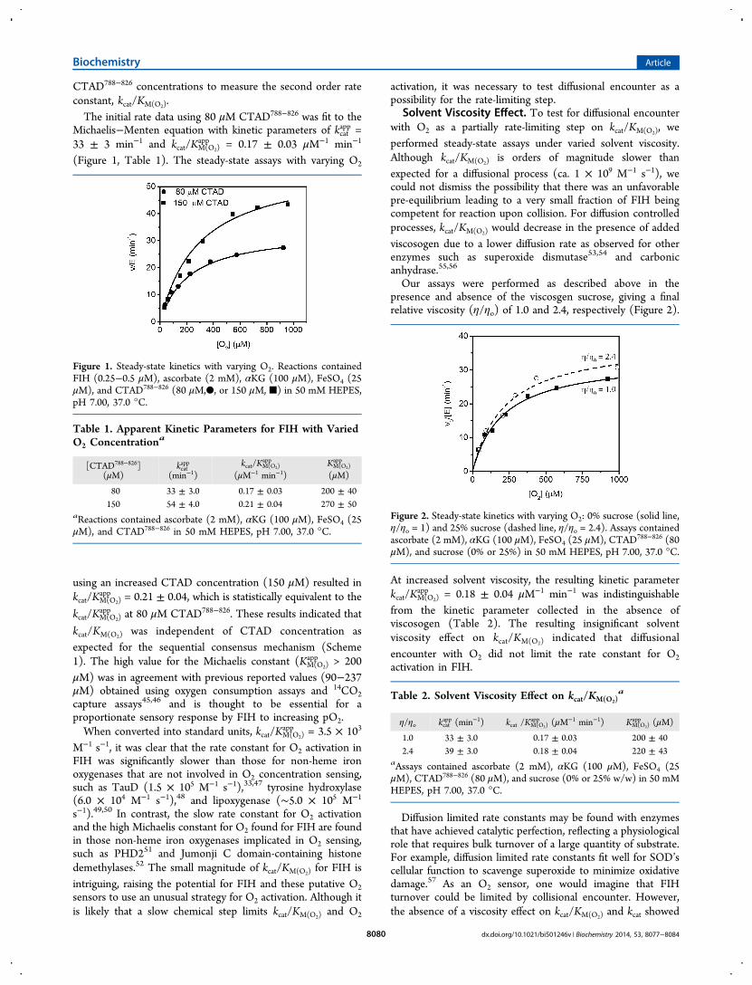
CTAD788−826 concentrations to measure the second order rateconstant, kcat/KM(O2).The initial rate data using 80 μM CTAD788−826 was fit to the
Michaelis−Menten equation with kinetic parameters of kcatapp =
33 ± 3 min−1 and kcat/KM(O2)app = 0.17 ± 0.03 μM−1 min−1
(Figure 1, Table 1). The steady-state assays with varying O2
using an increased CTAD concentration (150 μM) resulted inkcat/KM(O2)
app = 0.21 ± 0.04, which is statistically equivalent to the
kcat/KM(O2)app at 80 μM CTAD788−826. These results indicated that
kcat/KM(O2) was independent of CTAD concentration asexpected for the sequential consensus mechanism (Scheme1). The high value for the Michaelis constant (KM(O2)
app > 200μM) was in agreement with previous reported values (90−237μM) obtained using oxygen consumption assays and 14CO2capture assays45,46 and is thought to be essential for aproportionate sensory response by FIH to increasing pO2.When converted into standard units, kcat/KM(O2)
app = 3.5 × 10³
M−1 s−1, it was clear that the rate constant for O2 activation inFIH was significantly slower than those for non-heme ironoxygenases that are not involved in O2 concentration sensing,such as TauD (1.5 × 10⁵ M−1 s−1),33,47 tyrosine hydroxylase(6.0 × 10⁴ M−1 s−1),48 and lipoxygenase (∼5.0 × 10⁵ M−1
s−1).49,50 In contrast, the slow rate constant for O2 activationand the high Michaelis constant for O2 found for FIH are foundin those non-heme iron oxygenases implicated in O2 sensing,such as PHD251 and Jumonji C domain-containing histonedemethylases.52 The small magnitude of kcat/KM(O2) for FIH isintriguing, raising the potential for FIH and these putative O2sensors to use an unusual strategy for O2 activation. Although itis likely that a slow chemical step limits kcat/KM(O2) and O2
activation, it was necessary to test diffusional encounter as apossibility for the rate-limiting step.
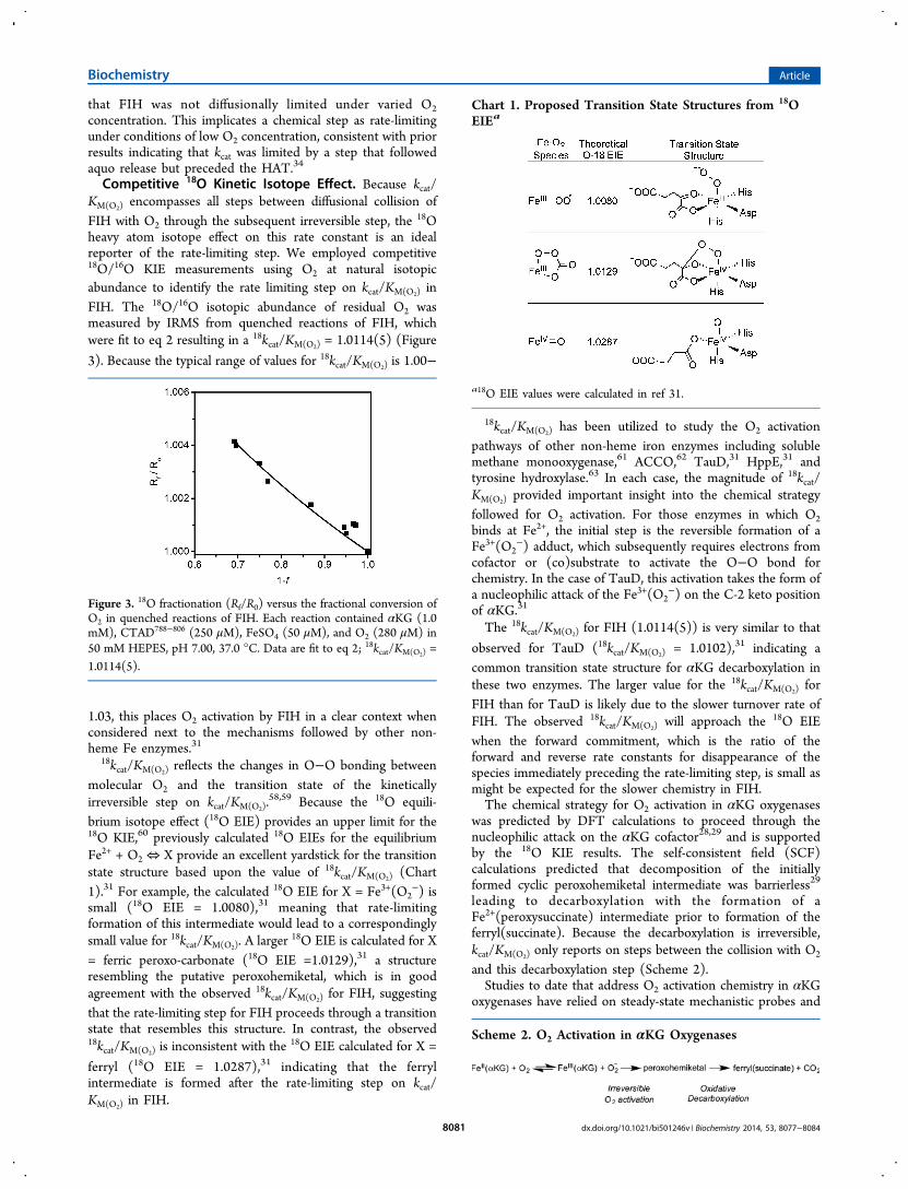
Solvent Viscosity Effect. To test for diffusional encounterwith O2 as a partially rate-limiting step on kcat/KM(O2), weperformed steady-state assays under varied solvent viscosity.Although kcat/KM(O2) is orders of magnitude slower thanexpected for a diffusional process (ca. 1 × 10⁹ M−1 s−1), wecould not dismiss the possibility that there was an unfavorablepre-equilibrium leading to a very small fraction of FIH beingcompetent for reaction upon collision. For diffusion controlledprocesses, kcat/KM(O2) would decrease in the presence of addedviscosogen due to a lower diffusion rate as observed for otherenzymes such as superoxide dismutase53,54 and carbonicanhydrase.55,56
Our assays were performed as described above in thepresence and absence of the viscosgen sucrose, giving a finalrelative viscosity (η/ηo) of 1.0 and 2.4, respectively (Figure 2).
At increased solvent viscosity, the resulting kinetic parameterkcat/KM(O2)
app = 0.18 ± 0.04 μM−1 min−1 was indistinguishablefrom the kinetic parameter collected in the absence ofviscosogen (Table 2). The resulting insignificant solventviscosity effect on kcat/KM(O2) indicated that diffusionalencounter with O2 did not limit the rate constant for O2activation in FIH.
Diffusion limited rate constants may be found with enzymesthat have achieved catalytic perfection, reflecting a physiologicalrole that requires bulk turnover of a large quantity of substrate.For example, diffusion limited rate constants fit well for SOD’scellular function to scavenge superoxide to minimize oxidativedamage.57 As an O2 sensor, one would imagine that FIHturnover could be limited by collisional encounter. However,the absence of a viscosity effect on kcat/KM(O2) and kcat showed
Figure 1. Steady-state kinetics with varying O2. Reactions containedFIH (0.25−0.5 μM), ascorbate (2 mM), αKG (100 μM), FeSO4 (25μM), and CTAD788−826 (80 μM,●, or 150 μM, ■) in 50 mM HEPES,pH 7.00, 37.0 °C.
Table 1. Apparent Kinetic Parameters for FIH with VariedO2 Concentration
a
[CTAD788−826](μM)
kcatapp
(min−1)kcat/KM(O2)
app
(μM−1 min−1)KM(O2)app
(μM)
80 33 ± 3.0 0.17 ± 0.03 200 ± 40150 54 ± 4.0 0.21 ± 0.04 270 ± 50
aReactions contained ascorbate (2 mM), αKG (100 μM), FeSO4 (25μM), and CTAD788−826 in 50 mM HEPES, pH 7.00, 37.0 °C.
Figure 2. Steady-state kinetics with varying O2: 0% sucrose (solid line,η/ηo = 1) and 25% sucrose (dashed line, η/ηo = 2.4). Assays containedascorbate (2 mM), αKG (100 μM), FeSO4 (25 μM), CTAD788−826 (80μM), and sucrose (0% or 25%) in 50 mM HEPES, pH 7.00, 37.0 °C.
Table 2. Solvent Viscosity Effect on kcat/KM(O2)a
η/ηo kcatapp (min−1) kcat /KM(O2)
app (μM−1 min−1) KM(O2)app (μM)
1.0 33 ± 3.0 0.17 ± 0.03 200 ± 402.4 39 ± 3.0 0.18 ± 0.04 220 ± 43
aAssays contained ascorbate (2 mM), αKG (100 μM), FeSO4 (25μM), CTAD788−826 (80 μM), and sucrose (0% or 25% w/w) in 50 mMHEPES, pH 7.00, 37.0 °C.
Biochemistry Article
dx.doi.org/10.1021/bi501246v | Biochemistry 2014, 53, 8077−80848080
that FIH was not diffusionally limited under varied O2concentration. This implicates a chemical step as rate-limitingunder conditions of low O2 concentration, consistent with priorresults indicating that kcat was limited by a step that followedaquo release but preceded the HAT.34
Competitive 18O Kinetic Isotope Effect. Because kcat/KM(O2) encompasses all steps between diffusional collision ofFIH with O2 through the subsequent irreversible step, the 18Oheavy atom isotope effect on this rate constant is an idealreporter of the rate-limiting step. We employed competitive18O/16O KIE measurements using O2 at natural isotopicabundance to identify the rate limiting step on kcat/KM(O2) inFIH. The 18O/16O isotopic abundance of residual O2 wasmeasured by IRMS from quenched reactions of FIH, whichwere fit to eq 2 resulting in a 18kcat/KM(O2) = 1.0114(5) (Figure
3). Because the typical range of values for 18kcat/KM(O2) is 1.00−
1.03, this places O2 activation by FIH in a clear context whenconsidered next to the mechanisms followed by other non-heme Fe enzymes.31
18kcat/KM(O2) reflects the changes in O−O bonding betweenmolecular O2 and the transition state of the kineticallyirreversible step on kcat/KM(O2).
58,59 Because the 18O equili-brium isotope effect (18O EIE) provides an upper limit for the18O KIE,60 previously calculated 18O EIEs for the equilibriumFe2+ + O2 ⇔ X provide an excellent yardstick for the transitionstate structure based upon the value of 18kcat/KM(O2) (Chart1).31 For example, the calculated 18O EIE for X = Fe3+(O2
−) issmall (18O EIE = 1.0080),31 meaning that rate-limitingformation of this intermediate would lead to a correspondinglysmall value for 18kcat/KM(O2). A larger 18O EIE is calculated for X= ferric peroxo-carbonate (18O EIE =1.0129),31 a structureresembling the putative peroxohemiketal, which is in goodagreement with the observed 18kcat/KM(O2) for FIH, suggestingthat the rate-limiting step for FIH proceeds through a transitionstate that resembles this structure. In contrast, the observed18kcat/KM(O2) is inconsistent with the 18O EIE calculated for X =
ferryl (18O EIE = 1.0287),31 indicating that the ferrylintermediate is formed after the rate-limiting step on kcat/KM(O2) in FIH.
18kcat/KM(O2) has been utilized to study the O2 activationpathways of other non-heme iron enzymes including solublemethane monooxygenase,61 ACCO,62 TauD,31 HppE,31 andtyrosine hydroxylase.63 In each case, the magnitude of 18kcat/KM(O2) provided important insight into the chemical strategyfollowed for O2 activation. For those enzymes in which O2binds at Fe2+, the initial step is the reversible formation of aFe3+(O2
−) adduct, which subsequently requires electrons fromcofactor or (co)substrate to activate the O−O bond forchemistry. In the case of TauD, this activation takes the form ofa nucleophilic attack of the Fe3+(O2
−) on the C-2 keto positionof αKG.31
The 18kcat/KM(O2) for FIH (1.0114(5)) is very similar to that
observed for TauD (18kcat/KM(O2) = 1.0102),31 indicating acommon transition state structure for αKG decarboxylation inthese two enzymes. The larger value for the 18kcat/KM(O2) forFIH than for TauD is likely due to the slower turnover rate ofFIH. The observed 18kcat/KM(O2) will approach the 18O EIEwhen the forward commitment, which is the ratio of theforward and reverse rate constants for disappearance of thespecies immediately preceding the rate-limiting step, is small asmight be expected for the slower chemistry in FIH.The chemical strategy for O2 activation in αKG oxygenases
was predicted by DFT calculations to proceed through thenucleophilic attack on the αKG cofactor28,29 and is supportedby the 18O KIE results. The self-consistent field (SCF)calculations predicted that decomposition of the initiallyformed cyclic peroxohemiketal intermediate was barrierless29
leading to decarboxylation with the formation of aFe2+(peroxysuccinate) intermediate prior to formation of theferryl(succinate). Because the decarboxylation is irreversible,kcat/KM(O2) only reports on steps between the collision with O2
and this decarboxylation step (Scheme 2).Studies to date that address O2 activation chemistry in αKG
oxygenases have relied on steady-state mechanistic probes and
Figure 3. 18O fractionation (Rf/R0) versus the fractional conversion ofO2 in quenched reactions of FIH. Each reaction contained αKG (1.0mM), CTAD788−806 (250 μM), FeSO4 (50 μM), and O2 (280 μM) in50 mM HEPES, pH 7.00, 37.0 °C. Data are fit to eq 2; 18kcat/KM(O2) =1.0114(5).
Chart 1. Proposed Transition State Structures from 18OEIEa
a18O EIE values were calculated in ref 31.
Scheme 2. O2 Activation in αKG Oxygenases
Biochemistry Article
dx.doi.org/10.1021/bi501246v | Biochemistry 2014, 53, 8077−80848081

point mutagenesis22,45,64−67 and suggest that hydrogen bondingcontacts to the αKG play an important role in facilitatingdecarboxylation. Further insight into oxidative decarboxylationhas been hampered by the identity of the rate limiting steps inTauD and other αKG oxygenases. In the cases of TauD,20
P4H,25 CytC3,32 and by analogy DAOCS68 and the histonedemethylase KDM4E,69 HAT by the ferryl or product releaseare partially rate-limiting on turnover at elevated O2concentration, preventing the accumulation of any speciesinvolved in O2 activation during the pre-steady-state. A crucialdifference between these other enzymes and FIH is that severallines of evidence indicate that decarboxylation is rate limiting inFIH, suggesting that FIH may allow direct access to stepsinvolved in O2 activation.
■ CONCLUSIONSWe have used multiple kinetic probes to characterize O2activation by FIH. Unlike other previously characterizedFe(II)/αKG enzymes, turnover in FIH is fully limited by therate of O2 activation. This kinetic feature is consistent with thefunction of FIH as an O2 sensor; strong oxidants such as theferryl would be short-lived, ensuring tight coupling between O2activation and CTAD hydroxylation. It may be possible thatother biomedically important Fe(II)/αKG enzymes such as theJmjC and JmjD domain-containing hydroxylases and PHD2employ a similar mechanistic strategy to regulate their function.If so, rate-limited O2 activation may be a more commonmechanistic feature among Fe(II)/αKG oxygenases than iscurrently appreciated.
■ ASSOCIATED CONTENT*S Supporting InformationControl experiments involving steady-state kinetics with theCTAD788−806 peptide, as well as steady-state kinetics in thepresence and absence of ascorbate. The material is available freeof charge via the Internet at http://pubs.acs.org.
■ AUTHOR INFORMATIONCorresponding Author*E-mail: [email protected]. Voice: (413) 545-4001.Fax: (413) 545-4490.FundingThis research was supported by the U.S. National Institutes ofHealth (Grant 1R01-GM077413 to M.J.K.) and the NationalScience Foundation (Grant EAR-1053432) to N.E.O. J.A.H.was supported in part by the NIH Chemistry-Biology InterfacePredoctoral Training Grant T32-GM008515.NotesThe authors declare no competing financial interest.
■ ABBREVIATIONSACCO, 1-aminocyclopropane-1-carboxylic acid oxidase; αKG,α-ketoglutarate; CAS, clavaminate synthase; CTAD, C-terminaltransactivation domain; DFT, density functional theory; EIE,equilibrium isotope effect; FIH, factor-inhibiting HIF; HAT,hydrogen atom transfer; HEPES, 4-(2-hydroxyethyl)-1-piper-azineethanesulfonic acid; HIF, hypoxia inducible factor-1α;HppE, (S)-2-hydroxypropyl-1-phosphonate epoxidase; IRMS,isotope ratio mass spectrometry; KIE, kinetic isotope effect;MALDI-TOF-MS, matrix assisted laser desorption ionization-time-of-flight-mass spectrometry; MCD, magnetic circulardichroism; P4H, prolyl-4-hydroxylase; PHD2, prolyl hydrox-
ylase domain 2; SKIE, solvent kinetic isotope effect; SCF, self-consistent field; SOD, superoxide dismutase; TauD, taurinedioxygenase; TFA, trifluoroacetic acid; VTVH, variable temper-ature variable field
■ REFERENCES(1) Taabazuing, C. Y., Hangasky, J. A., and Knapp, M. J. (2014)Oxygen sensing strategies in mammals and bacteria. J. Inorg. Biochem.133, 63−72.(2) Mole, D. R., Blancher, C., Copley, R. R., Pollard, P. J., Gleadle, J.M., Ragoussis, J., and Ratcliffe, P. J. (2009) Genome-wide Associationof Hypoxia-inducible Factor (HIF)-1a and HIF-2a DNA Binding withExpression Profiling of Hypoxia-inducible Transcripts. J. Biol. Chem.284, 16767−16775.(3) Ke, Q., and Costa, M. (2006) Hypoxia-Inducible Factor-1 (HIF-1). Mol. Pharmacol. 70, 1469−1480.(4) Semenza, G. L. (2003) Targeting HIF-1 for cancer therapy. Nat.Rev. Cancer 3, 721−732.(5) Metzen, E., and Ratcliffe, P. J. (2004) HIF hydroxylation andcellular oxygen sensing. Biol. Chem. 385, 223−230.(6) Schofield, C. J., and Ratchliffe, P. J. (2004) Oxygen sensing byHIF-hydroxylases. Nat. Rev. Mol. Cell Biol. 5, 343−354.(7) Lando, D., Peet, D. J., Gorman, J. J., Whelan, D. A., Whitelaw, M.L., and Bruick, R. K. (2002) FIH-1 is an asparaginyl hydroxylaseenzyme that regulates the transcriptional activity of hypoxia-induciblefactor. Genes Dev. 16, 1466−1471.(8) McNeill, L. A., Hewitson, K. S., Claridge, T. D., Seibel, J. F.,Horsfall, L. E., and Schofield, C. J. (2002) Hypoxia-inducible factorasparaginyl hydroxylase (FIH-1) catalyses hydroxylation at the beta-carbon of asparagine-803. Biochem. J. 367, 571−575.(9) Lando, D., Peet, D. J., Whelan, D. A., Gorman, J. J., and Whitelaw,M. L. (2002) Asparagine hydroxylation of the HIF transactivationdomain a hypoxic switch. Science 295, 858−861.(10) Solomon, E. I., Light, K. M., Liu, L. V., Srnec, M., and Wong, S.D. (2013) Geometric and Electronic Structure Contributions toFunction in Non-heme Iron Enzymes. Acc. Chem. Res. 46, 2725−2739.(11) Blomberg, M. R. A., Borowski, T., Himo, F., Liao, R. Z., andSiegbahn, P. E. M. (2014) Quantum Chemical Studies of Mechanismsfor Metalloenzymes. Chem. Rev. 114, 3601−3658.(12) Hausinger, R. P. (2004) Fe(II)/alpha-Ketoglutarate-dependenthydroxylases and related enzymes. Crit. Rev. Biochem. Mol. Biol. 39,21−68.(13) Aik, W., McDonough, M. A., Thalhammer, A., Chowdhury, R.,and Schofield, C. J. (2012) Role of the jelly-roll fold in substratebinding by 2-oxoglutarate oxygenases. Curr. Opin. Struct. Biol. 22, 691−700.(14) Bollinger, J. M., Price, J. C., Hoffart, L. M., Barr, E. W., andKrebs, C. (2005) Mechanism of Taurine: α-Ketoglutarate Dioxygenase(TauD) from Escherichia coli. Eur. J. Inorg. Chem. 2005, 4245−4254.(15) Grzyska, P. K., Appelman, E. H., Hausinger, R. P., andProshlyakov, D. A. (2010) Insight into the mechanism of an irondioxygenase by resolution of steps following the FeIV=HO species.Proc. Natl. Acad. Sci. U. S. A. 107, 3982−3987.(16) Light, K. M., Hangasky, J. A., Knapp, M. J., and Solomon, E. I.(2013) Spectroscopic Studies of the Mononuclear Non-Heme Fe-IIEnzyme FIH: Second-Sphere Contributions to Reactivity. J. Am. Chem.Soc. 135, 9665−9674.(17) Neidig, M. L., Brown, C. D., Light, K. M., Fujimori, D. G.,Nolan, E. M., Price, J. C., Barr, E. W., Bollinger, J. M., Krebs, C.,Walsh, C. T., and Solomon, E. I. (2007) CD and MCD of CytC3 andtaurine dioxygenase: role of the facial triad in alpha-KG-dependentoxygenases. J. Am. Chem. Soc. 129, 14224−14231.(18) Zhou, J., Kelly, W. L., Bachmann, B. O., Gunsior, M., Townsend,C. A., and Solomon, E. I. (2001) Spectroscopic studies of substrateinteractions with clavaminate synthase 2, a multifunctional alpha-KG-dependent non-heme iron enzyme: Correlation with mechanisms andreactivities. J. Am. Chem. Soc. 123, 7388−7398.
Biochemistry Article
dx.doi.org/10.1021/bi501246v | Biochemistry 2014, 53, 8077−80848082
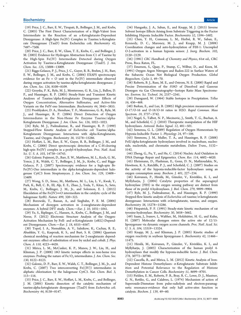
(19) Price, J. C., Barr, E. W., Tirupati, B., Bollinger, J. M., and Krebs,C. (2003) The First Direct Characterization of a High-Valent IronIntermediate in the Reaction of an a-Ketoglutarate-DependentDioxygenase: A High-Spin Fe(IV) Complex in Taurine/a-Ketogluta-rate Dioxygenase (TauD) from Escherichia coli. Biochemistry 42,7497−7508.(20) Price, J. C., Barr, E. W., Glass, T. E., Krebs, C., and Bollinger, J.M. (2003) Evidence for Hydrogen Abstraction from C1 of Taurine bythe High-Spin Fe(IV) Intermediate Detected during OxygenActivation by Taurine:a-Ketoglutarate Dioxygenase (TauD). J. Am.Chem. Soc. 125, 13008−13009.(21) Riggs-Gelasco, P. J., Price, J. C., Guyer, R. B., Brehm, J. H., Barr,E. W., Bollinger, J. M., and Krebs, C. (2004) EXAFS spectroscopicevidence for an Fe = O unit in the Fe(IV) intermediate observedduring oxygen activation by taurine:alpha-ketoglutarate dioxygenase. J.Am. Chem. Soc. 126, 8108−8109.(22) Grzyska, P. K., Ryle, M. J., Monterosso, G. R., Liu, J., Ballou, D.P., and Hausinger, R. P. (2005) Steady-State and Transient KineticAnalyses of Taurine/alpha-Ketoglutarate Dioxygenase: Effects ofOxygen Concentration, Alternative Sulfonates, and Active-SiteVariants on the FeIV-oxo Intermediate. Biochemistry 44, 3845−3855.(23) Proshlyakov, D. A., Henshaw, T. F., Monterosso, G. R., Ryle, M.J., and Hausinger, R. P. (2004) Direct Detection of OxygenIntermediates in the Non-Heme Fe Enzyme Taurine/alpha-Ketoglutarate Dioxygenase. J. Am. Chem. Soc. 126, 1022−1023.(24) Ryle, M. J., Padmakumar, R., and Hausinger, R. P. (1999)Stopped-Flow Kinetic Analysis of Escherichia coli Taurine/alpha-Ketoglutarate Dioxygenase: Interactions with alpha-Ketoglutarate,Taurine, and Oxygen. Biochemistry 38, 15278−15286.(25) Hoffart, L. M., Barr, E. W., Guyer, R. B., Bollinger, J. M., andKrebs, C. (2006) Direct spectroscopic detection of a C-H-cleavinghigh-spin Fe(IV) complex in a prolyl-4-hydroxylase. Proc. Natl. Acad.Sci. U. S. A. 103, 14738−14743.(26) Galonic Fujimori, D., Barr, E. W., Matthews, M. L., Koch, G. M.,Yonce, J. R., Walsh, C. T., Bollinger, J. M., Jr., Krebs, C., and Riggs-Gelasco, P. J. (2007) Spectroscopic evidence for a high-spin Br-Fe(IV)-Oxo intermediate in the alpha-ketoglutarate-dependent halo-genase CytC3 from Streptomyces. J. Am. Chem. Soc. 129, 13408−13409.(27) Wong, S. D., Srnec, M., Matthews, M. L., Liu, L. V., Kwak, Y.,Park, K., Bell, C. B., III, Alp, E. E., Zhao, J., Yoda, Y., Kitao, S., Seto,M., Krebs, C., Bollinger, J. M., Jr., and Solomon, E. I. (2013)Elucidation of the Fe(IV)O intermediate in the catalytic cycle of thehalogenase SyrB2. Nature 499, 320−323.(28) Borowski, T., Bassan, A., and Siegbahn, P. E. M. (2004)Mechanism of dioxygen activation in 2-oxoglutarate-dependentenzymes: A hybrid DFT study. Chem.Eur. J. 10, 1031−1041.(29) Ye, S., Riplinger, C., Hansen, A., Krebs, C., Bollinger, J. M., andNeese, F. (2012) Electronic Structure Analysis of the Oxygen-Activation Mechanism by FeII- and α-Ketoglutarate (αKG)-Depend-ent Dioxygenases. Chem.Eur. J. 18, 6555−6567.(30) Topol, I. A., Nemukhin, A. V., Salnikow, K., Cachau, R. E.,Abashkin, Y. G., Kasprzak, K. S., and Burt, S. K. (2006) Quantumchemical modeling of reaction mechanism for 2-oxoglutarate depend-ent enzymes: effect of substitution of iron by nickel and cobalt. J. Phys.Chem. A 110, 4223−4428.(31) Mirica, L. M., McCusker, K. P., Munos, J. W., Liu, H., andKlinman, J. P. (2008) 18O kinetic isotope effects in non-heme ironenzymes: Probing the nature of Fe/O2 intermediates. J. Am. Chem. Soc.130, 8122−8123.(32) Galonic, D. P., Barr, E. W., Walsh, C. T., Bollinger, J. M., Jr., andKrebs, C. (2007) Two interconverting Fe((IV)) intermediates inaliphatic chlorination by the halogenase CytC3. Nat. Chem. Biol. 3,113−116.(33) Price, J. C., Barr, E. W., Hoffart, L. M., Krebs, C., and Bollinger,J. M. (2005) Kinetic dissection of the catalytic mechanism oftaurine:alpha-ketoglutarate dioxygenase (TauD) from Escherichia coli.Biochemistry 44, 8138−8147.
(34) Hangasky, J. A., Saban, E., and Knapp, M. J. (2013) InverseSolvent Isotope Effects Arising from Substrate Triggering in the FactorInhibiting Hypoxia Inducible Factor. Biochemistry 52, 1594−1602.(35) Chen, Y. H., Comeaux, L. M., Herbst, R. W., Saban, E.,Kennedy, D. C., Maroney, M. J., and Knapp, M. J. (2008)Coordination changes and auto-hydroxylation of FIH-1: UncoupledO-2-activation in a human hypoxia sensor. J. Inorg. Biochem. 102,2120−2129.(36) (1981) CRC Handbook of Chemistry and Physics, 61st ed., CRCPress, Boca Raton, FL.(37) Emerson, S., Quay, P., Stump, C., Wilbur, D., and Knox, M.(1991) Oxygen Argon Nitrogen and Radon-222 in Surface Waters ofthe Subarctic Ocean Net Biological Oxygen Production. GlobalBiogeochem. Cycles 5, 49−70.(38) Roberts, B. J., Russ, M. E., and Ostrom, N. E. (2000) Rapid andPrecise Determination of the δ18O of Dissolved and GaseousDioxygen via Gas Chromatography−Isotope Ratio Mass Spectrome-try. Environ. Sci. Technol. 34, 2337−2341.(39) Dansgaard, W. (1964) Stable Isotopes in Precipitation. Tellus16, 436−468.(40) Barkan, E., and Luz, B. (2005) High precision measurements ofO-17/O-16 and O-18/O-16 ratios in H2O. Rapid Commun. MassSpectrom. 19, 3737−3742.(41) Nagel, S., Talbot, N. P., Mecinovic, J., Smith, T. G., Buchan, A.M., and Schofield, C. J. (2010) Therapeutic manipulation of the HIFhydroxylases. Antioxid. Redox Signal. 12, 481−501.(42) Semenza, G. L. (2009) Regulation of Oxygen Homeostasis byHypoxia-Inducible Factor 1. Physiology 24, 97−106.(43) Simmons, J. M., Muller, T. A., and Hausinger, R. P. (2008)FeII/alpha-ketoglutarate hydroxylases involved in nucleobase, nucleo-side, nucleotide, and chromatin metabolism. Dalton Trans., 5132−5142.(44) Zheng, G., Fu, Y., and He, C. (2014) Nucleic Acid Oxidation inDNA Damage Repair and Epigenetics. Chem. Rev. 114, 4602−4620.(45) Ehrismann, D., Flashman, E., Genn, D. N., Mathioudakis, N.,Hewitson, K. S., Ratcliffe, P. J., and Schofield, C. J. (2007) Studies onthe activity of the hypoxia-inducible-factor hydroxylases using anoxygen consumption assay. Biochem. J. 401, 227−234.(46) Koivunen, P., Hirsila, M., Gunzler, V., Kivirikko, K. I., andMyllyharju, J. (2004) Catalytic properties of the asparaginylhydroxylase (FIH) in the oxygen sensing pathway are distinct fromthose of its prolyl 4-hydroxylases. J. Biol. Chem. 279, 9899−9904.(47) Ryle, M. J., Padmakumar, R., and Hausinger, R. P. (1999)Stopped-flow kinetic analysis of Escherichia coli taurine/α-ketoglutaratedioxygenase: Interactions with α-ketoglutarate, taurine, and oxygen.Biochemistry 38, 15278−15286.(48) Fitzpatrick, P. F. (1991) Steady-state kinetic mechanism of rattyrosine-hydroxylase. Biochemistry 30, 3658−3662.(49) Saam, J., Ivanov, I., Walther, M., Holzhutter, H. G., and Kuhn,H. (2007) Molecular dioxygen enters the active site of 12/15-lipoxygenase via dynamic oxygen access channels. Proc. Natl. Acad. Sci.U. S. A. 104, 13319−13324.(50) Knapp, M. J., and Klinman, J. P. (2003) Kinetic studies ofoxygen reactivity in soybean lipoxygenase-1. Biochemistry 42, 11466−11475.(51) Hirsila, M., Koivunen, P., Gunzler, V., Kivirikko, K. I., andMyllyharju, J. (2003) Characterization of the human prolyl 4-hydroxylases that modify the hypoxia-inducible factor. J. Biol. Chem.278, 30772−30780.(52) Cascella, B., and Mirica, L. M. (2012) Kinetic Analysis of Iron-Dependent Histone Demethylases: α-Ketoglutarate Substrate Inhib-ition and Potential Relevance to the Regulation of HistoneDemethylation in Cancer Cells. Biochemistry 51, 8699−8701.(53) Fielden, E. M., Roberts, P. B., Bray, R. C., Lowe, D. J., Mautner,G. N., Rotilio, G., and Calabres, L. (1974) Mechanism of action ofSuperoxide-Dismutase from pulse-radiolysis and electron-paramag-netic resonance-evidence that only half active-sites function incatalysis. Biochem. J. 139, 49−60.
Biochemistry Article
dx.doi.org/10.1021/bi501246v | Biochemistry 2014, 53, 8077−80848083

(54) Rotilio, G., Bray, R. C., and Fielden, E. M. (1972) A pulseradiolysis study of superoxide dismutase. Biochim. Biophys. Acta,Enzymol. 268, 605−609.(55) Hasinoff, B. B. (1984) Kinetics of Carbonic-Anhydrase inSolvents of Increased Viscoisty - A Partially Diffusion-ControlledReaction. Arch. Biochem. Biophys. 233, 676−681.(56) Pocker, Y., and Janjic, N. (1987) Enyzme-Kinetics in Solvents ofIncreased Viscosity - Dynamic Aspects of Carbonic-AnhydraseCatalysis. Biochemistry 26, 2597−2606.(57) Perry, J. J. P., Shin, D. S., Getzoff, E. D., and Tainer, J. A. (2010)The structural biochemistry of the superoxide dismutases. Biochim.Biophys. Acta, Proteins Proteomics 1804, 245−262.(58) Tian, G., and Klinman, J. P. (1993) Discrimination between 16Oand 18O in Oxygen Binding to the Reversible Oxygen CarriersHemoglobin, Hemerythrin, and Hemocyanin: A new Probe forOxygen Binding and Reductive Activation by Proteins. J. Am. Chem.Soc. 115, 8891−8897.(59) Tian, G., Berry, J., and Klinman, J. (1994) Oxygen-18 KineticIsotope Effects in the Dopamine β-Monooxygenase Reaction:Evidence for a New Chemical Mechanism in Non-Heme Metal-lomonooxygenases. Biochemistry 33, 226−234.(60) Roth, J. P. (2007) Advances in studying bioinorganic reactionmechanisms: Isotopic probes of activated oxygen intermediates inmetalloenzymes. Curr. Opin. Chem. Biol. 11, 142−150.(61) Stahl, S. S., Francisco, W. a, Merkx, M., Klinman, J. P., andLippard, S. J. (2001) Oxygen kinetic isotope effects in soluble methanemonooxygenase. J. Biol. Chem. 276, 4549−4553.(62) Mirica, L. M., and Klinman, J. P. (2008) The nature of O2activation by the ethylene-forming enzyme 1-aminocyclopropane-1-carboxylic acid oxidase. Proc. Natl. Acad. Sci. U. S. A. 105, 1814−1819.(63) Francisco, W. A., Tian, G. C., Fitzpatrick, P. F., and Klinman, J.P. (1998) Oxygen-18 kinetic isotope effect studies of the tyrosinehydroxylase reaction: Evidence of rate limiting oxygen activation. J.Am. Chem. Soc. 120, 4057−4062.(64) Hotopp, J. C. D., and Hausinger, R. P. (2002) Probing the 2,4-dichlorophenoxyacetate/α-ketoglutarate dioxygenase substrate-bindingsite by site-directed mutagenesis and mechanism-based inactivation.Biochemistry 41, 9787−9794.(65) Thornburg, L. D., Lai, M. T., Wishnok, J. S., and Stubbe, J.(1993) A non-heme iron protein with heme tendencies: Aninvestigation of the substrate specificity of thymine hydroxylase.Biochemistry 32, 14023−14033.(66) Saban, E., Chen, Y. H., Hangasky, J., Taabazuing, C., Holmes, B.,and Knapp, M. (2011) The second coordination sphere of FIHcontrols hydroxylation. Biochemistry 50, 4733−4740.(67) Flagg, S. C., Giri, N., Pektas, S., Maroney, M. J., and Knapp, M.J. (2012) Inverse Solvent Isotope Effects Demonstrate Slow AquoRelease from Hypoxia Inducible Factor-Prolyl Hydroxylase (PHD2).Biochemistry 51, 6654−6666.(68) Tarhonskaya, H., Szollossi, A., Leung, I. K. H., Bush, J. T.,Henry, L., Chowdhury, R., Iqbal, A., Claridge, T. D. W., Schofield, C.J., and Flashman, E. (2014) Studies on Deacetoxycephalosporin CSynthase Support a Consensus Mechanism for 2-OxoglutarateDependent Oxygenases. Biochemistry 53, 2483−2493.(69) Sanchez-Fernandez, E. M., Tarhonskaya, H., Al-Qahtani, K.,Hopkinson, R. J., McCullagh, J. S., Schofield, C. J., and Flashman, E.(2013) Investigations on the oxygen dependence of a 2-oxoglutaratehistone demethylase. Biochem. J. 449, 491−496.
Biochemistry Article
dx.doi.org/10.1021/bi501246v | Biochemistry 2014, 53, 8077−80848084