The Natural Estrogenic Compound Diarylheptanoid … Natural Estrogenic Compound Diarylheptanoid...
7
Environmental Health Perspectives • VOLUME 121 | NUMBER 4 | April 2013 433 Research The Natural Estrogenic Compound Diarylheptanoid (D3): In Vitro Mechanisms of Action and in Vivo Uterine Responses via Estrogen Receptor α Wipawee Winuthayanon, 1,2 Pawinee Piyachaturawat, 2 Apichart Suksamrarn, 3 Katherine A. Burns, 1 Yukitomo Arao, 1 Sylvia C. Hewitt, 1 Lars C. Pedersen, 4 and Kenneth S. Korach 1 1 Laboratory of Reproductive and Developmental Toxicology, National Institute of Environmental Health Sciences (NIEHS), National Institutes of Health (NIH), Department of Health and Human Services (DHHS), Research Triangle Park, North Carolina, USA; 2 Department of Physiology, Faculty of Science, Mahidol University, Bangkok, Thailand; 3 Department of Chemistry, Faculty of Science, Ramkhamhaeng University, Bangkok, Thailand; 4 Laboratory of Structural Biology, NIEHS, NIH, DHHS, Research Triangle Park, North Carolina, USA BACKGROUND: Diarylheptanoid (D3) isolated from the medicinal plant, Curcuma comosa, has estrogenic activity. OBJECTIVE: We aimed to elucidate the mechanism(s) of D3 action and compare it with that of 17β-estradiol (E 2 ) using both in vitro and in vivo uterine models. METHODS: We used human uterine (Ishikawa) cells to determine the estrogenic action of D3 on the activation and nuclear translocation of estrogen receptor α (ERα). In addition, we further charac- terized the uterine response to D3 treatment in vivo. RESULTS: D3 activated an estrogen responsive element (ERE) luciferase reporter through ERα, and molecular modeling suggested that D3 could be accommodated in the ERα binding pocket. Using modified ERα to assay ligand-dependent nuclear translocation, we observed D3-dependent ERα interaction and translocation. In mouse uteri, early- and late-phase estrogen-regulated gene responses were increased in D3-treated ovariectomized wild-type animals, in a manner similar to that of E 2 ; no response was seen in ERα knockout animals. We observed a divergence in estrogen responses after D3 treatment: D3 induced robust DNA synthesis in uterine epithelial cells, linked to an increase in cell-cycle–related genes; however, no increase in uterine weight was observed 24 hr after treatment. D3 also affected uterine progesterone receptor expression patterns similar to E 2 . When D3 and E 2 were administered together, we observed no additive or antagonistic effects of D3 on E 2 . Our findings suggest that D3 is a weak estrogenic agonist compound. CONCLUSION: D3 is a weakly acting phytoestrogen that mimics the mitogenic responses produced by E 2 in an ERα-dependent manner, but it is unable to increase uterine weight or enhance or antagonize the effects of estrogen. KEY WORDS: diarylheptanoid, ER-dependent, nuclear translocation, phytoestrogen, uterus. Environ Health Perspect 121:433–439 (2013). http://dx.doi.org/10.1289/ehp.1206122 [Online 18 January 2013] Estrogens play important roles in growth, dif- ferentiation, and maintenance functions of many target tissues in the female reproductive organs (Couse and Korach 1999). The bio- logical actions of estrogen are mediated pri- marily through estrogen receptor (ER) α and β (Couse et al. 1997). ERs are members of the nuclear receptor family of proteins containing multiple functional domains: e A/B domain harbors activation function 1 (AF1); the DNA binding domain is located in the C region of the receptors; the hinge region (D domain) contains nuclear localization sequences (NLS) (Mader et al. 1993); and the E/F domains contain the ligand binding region and AF2 function. AF1 and AF2 portions of the pro- tein facilitate transcriptional activity of the ER (Tora et al. 1989). Upon binding ligand, the ER is localized to the nucleus and initiates gene transcription through multiple pathways, including classical estrogen responsive element (ERE)–dependent pathways and nonclassical pathways (Hall et al. 2001). e uterus is one of the most prominent estrogenic responsive target tissues, predomi- nantly expressing ERα (Couse et al. 1997). Uterine response to estrogen is rapid and eventually leads to a dramatic increase in cell proliferation (Martin et al. 1973). However, the uterotrophic responses to estrogen vary with time after hormone exposure. An early response of water imbibition in uteri is medi- ated through ERα; ERα knockout (αERKO) mice show no water imbibition and no increase in uterine weight after 17β-estradiol (E 2 ) treat- ment (Korach 1994). e genomic responses of the uterus to E 2 have been observed 0.5–96 hr after treatment (Hewitt et al. 2003; Naciff et al. 2007). Some exogenous estrogens (bisphenol A and genistein), as well as one of the endogenous estrogens (estriol), are con- sidered weak estrogens in the uterus. Weak estrogenic compounds are less potent than E 2 ; they exhibit early uterine responses but are less effective in their abilities to cause robust subse- quent uterine responses such as cellular hyper- trophy and hyperplasia (Hewitt and Korach 2011). Stronger estrogens, including E 2 , initi- ate both early and late effects (Anderson et al. 1975). Transcripts that increase 1–2 hr after acute dosing of estrogenic compounds are components of the E 2 responsive “early gene cluster,” which includes Fos and Inhbb (inhibin beta-B) (Hewitt et al. 2003). e late responses include increased and sustained RNA and pro- tein synthesis, which lead to uterine cellular hypertrophy, DNA synthesis, and hyperplasia (Hewitt et al. 2003), as well as an alteration of progesterone receptor (PR) expression patterns (Mote et al. 2006). A second response phase is characterized by a wave of mitosis and DNA synthesis, which occurs 16–24 hr after E 2 treatment and is correlated with the late-phase cell cycle regulators, including Aurkb (aurora kinase B) and Ccnb2 (cyclin B2) (Hewitt et al. 2003; Hewitt and Korach 2011). The early and late events reflect the uterotrophic action of estrogens on uterine tissues and have been widely used to evaluate and compare potency and estrogenic or antagonistic activity of xeno- estrogenic compounds. Diarylheptanoids are phytoestrogens iso- lated from Curcuma comosa, a plant in the Zingiberaceae family. C. comosa has been marketed as a plant-derived dietary supple- ment product traditionally used in indige- nous medicine as an alternative remedy for hormone replacement therapy in menopausal women (Piyachaturawat et al. 1995). Other diarylheptanoids are found in Curcuma and other plants in the ginger family (Keserü and Nógrádi 1995). D3 (Figure 1A), one of the most abundant purified diarylheptanoids from C. comosa rhizome extract (Suksamrarn et al. 2008), exerts the most potent estro- genic activity when administered for 2 or Address correspondence to K.S. Korach, NIEHS, P.O. Box 12233, Research Triangle Park, NC 27709 USA. Telephone: (919) 541-3512. E-mail: korach@ niehs.nih.gov Supplemental Material is available online (http:// dx.doi.org/10.1289/ehp.1206122). We thank A. Binder and Y. Li for critical review of this manuscript and J. Tucker and A. Janoshazi (Fluorescence Microscopy and Imaging Center, Laboratory of Signal Transduction, NIEHS) for their assistance in confocal imaging analysis. Research support was provided by the NIEHS Division of Intramural Research (Z01-ES70065 to K.S.K. and Z01-ES102645 to L.C.P), the ailand Research Fund through the Royal Golden Jubilee PhD program (PHD/0255/2546 to P.P. and W.W.), a Target Research Grant (DBG5180020 to P.P.), and a Strategic Basic Research Grant (DBG5280013 to A.S.). e authors declare they have no actual or potential competing financial interests. Received 10 October 2012; accepted 17 January 2013.
Transcript of The Natural Estrogenic Compound Diarylheptanoid … Natural Estrogenic Compound Diarylheptanoid...
Environmental Health Perspectives • volume 121 | number 4 | April 2013 433
Research
The Natural Estrogenic Compound Diarylheptanoid (D3): In Vitro Mechanisms of Action and in Vivo Uterine Responses via Estrogen Receptor αWipawee Winuthayanon,1,2 Pawinee Piyachaturawat,2 Apichart Suksamrarn,3 Katherine A. Burns,1 Yukitomo Arao,1 Sylvia C. Hewitt,1 Lars C. Pedersen,4 and Kenneth S. Korach1
1Laboratory of Reproductive and Developmental Toxicology, National Institute of Environmental Health Sciences (NIEHS), National Institutes of Health (NIH), Department of Health and Human Services (DHHS), Research Triangle Park, North Carolina, USA; 2Department of Physiology, Faculty of Science, Mahidol University, Bangkok, Thailand; 3Department of Chemistry, Faculty of Science, Ramkhamhaeng University, Bangkok, Thailand; 4Laboratory of Structural Biology, NIEHS, NIH, DHHS, Research Triangle Park, North Carolina, USA
Background: Diarylheptanoid (D3) isolated from the medicinal plant, Curcuma comosa, has estrogenic activity.
oBjective: We aimed to elucidate the mechanism(s) of D3 action and compare it with that of 17β-estradiol (E2) using both in vitro and in vivo uterine models.
Methods: We used human uterine (Ishikawa) cells to determine the estrogenic action of D3 on the activation and nuclear translocation of estrogen receptor α (ERα). In addition, we further charac-terized the uterine response to D3 treatment in vivo.
results: D3 activated an estrogen responsive element (ERE) luciferase reporter through ERα, and molecular modeling suggested that D3 could be accommodated in the ERα binding pocket. Using modified ERα to assay ligand-dependent nuclear translocation, we observed D3-dependent ERα interaction and translocation. In mouse uteri, early- and late-phase estrogen-regulated gene responses were increased in D3-treated ovariectomized wild-type animals, in a manner similar to that of E2; no response was seen in ERα knockout animals. We observed a divergence in estrogen responses after D3 treatment: D3 induced robust DNA synthesis in uterine epithelial cells, linked to an increase in cell-cycle–related genes; however, no increase in uterine weight was observed 24 hr after treatment. D3 also affected uterine progesterone receptor expression patterns similar to E2. When D3 and E2 were administered together, we observed no additive or antagonistic effects of D3 on E2. Our findings suggest that D3 is a weak estrogenic agonist compound.
conclusion: D3 is a weakly acting phyto estrogen that mimics the mitogenic responses produced by E2 in an ERα-dependent manner, but it is unable to increase uterine weight or enhance or antago nize the effects of estrogen.
key words: diarylheptanoid, ER-dependent, nuclear translocation, phytoestrogen, uterus. Environ Health Perspect 121:433–439 (2013). http://dx.doi.org/10.1289/ehp.1206122 [Online 18 January 2013]
Estrogens play important roles in growth, dif-ferentiation, and maintenance functions of many target tissues in the female reproductive organs (Couse and Korach 1999). The bio-logical actions of estrogen are mediated pri-marily through estrogen receptor (ER) α and β (Couse et al. 1997). ERs are members of the nuclear receptor family of proteins containing multiple functional domains: The A/B domain harbors activation function 1 (AF1); the DNA binding domain is located in the C region of the receptors; the hinge region (D domain) contains nuclear localization sequences (NLS) (Mader et al. 1993); and the E/F domains contain the ligand binding region and AF2 function. AF1 and AF2 portions of the pro-tein facilitate transcriptional activity of the ER (Tora et al. 1989). Upon binding ligand, the ER is localized to the nucleus and initiates gene transcription through multiple pathways, including classical estrogen responsive element (ERE)–dependent pathways and non classical pathways (Hall et al. 2001).
The uterus is one of the most prominent estrogenic responsive target tissues, predomi-nantly expressing ERα (Couse et al. 1997). Uterine response to estrogen is rapid and
eventually leads to a dramatic increase in cell proliferation (Martin et al. 1973). However, the utero trophic responses to estrogen vary with time after hormone exposure. An early response of water imbibition in uteri is medi-ated through ERα; ERα knockout (αERKO) mice show no water imbibition and no increase in uterine weight after 17β-estradiol (E2) treat-ment (Korach 1994). The genomic responses of the uterus to E2 have been observed 0.5–96 hr after treatment (Hewitt et al. 2003; Naciff et al. 2007). Some exogenous estrogens (bisphenol A and genistein), as well as one of the endogenous estrogens (estriol), are con-sidered weak estrogens in the uterus. Weak estrogenic compounds are less potent than E2; they exhibit early uterine responses but are less effective in their abilities to cause robust subse-quent uterine responses such as cellular hyper-trophy and hyper plasia (Hewitt and Korach 2011). Stronger estrogens, including E2, initi-ate both early and late effects (Anderson et al. 1975). Transcripts that increase 1–2 hr after acute dosing of estrogenic compounds are components of the E2 responsive “early gene cluster,” which includes Fos and Inhbb (inhibin beta-B) (Hewitt et al. 2003). The late responses
include increased and sustained RNA and pro-tein synthesis, which lead to uterine cellular hypertrophy, DNA synthesis, and hyperplasia (Hewitt et al. 2003), as well as an alteration of progesterone receptor (PR) expression patterns (Mote et al. 2006). A second response phase is charac terized by a wave of mitosis and DNA synthesis, which occurs 16–24 hr after E2 treatment and is correlated with the late-phase cell cycle regulators, including Aurkb (aurora kinase B) and Ccnb2 (cyclin B2) (Hewitt et al. 2003; Hewitt and Korach 2011). The early and late events reflect the utero trophic action of estrogens on uterine tissues and have been widely used to evaluate and compare potency and estrogenic or antagonistic activity of xeno-estrogenic compounds.
Diarylheptanoids are phytoestrogens iso-lated from Curcuma comosa, a plant in the Zingiberaceae family. C. comosa has been marketed as a plant-derived dietary supple-ment product traditionally used in indige-nous medi cine as an alternative remedy for hormone replacement therapy in menopausal women (Piyachaturawat et al. 1995). Other diaryl heptanoids are found in Curcuma and other plants in the ginger family (Keserü and Nógrádi 1995). D3 (Figure 1A), one of the most abundant purified diaryl heptanoids from C. comosa rhizome extract (Suksamrarn et al. 2008), exerts the most potent estro-genic activity when administered for 2 or
Address correspondence to K.S. Korach, NIEHS, P.O. Box 12233, Research Triangle Park, NC 27709 USA. Telephone: (919) 541-3512. E-mail: [email protected]
Supplemental Material is available online (http://dx.doi.org/10.1289/ehp.1206122).
We thank A. Binder and Y. Li for critical review of this manuscript and J. Tucker and A. Janoshazi (Fluorescence Microscopy and Imaging Center, Laboratory of Signal Transduction, NIEHS) for their assistance in confocal imaging analysis.
Research support was provided by the NIEHS Division of Intramural Research (Z01-ES70065 to K.S.K. and Z01-ES102645 to L.C.P), the Thailand Research Fund through the Royal Golden Jubilee PhD program (PHD/0255/2546 to P.P. and W.W.), a Target Research Grant (DBG5180020 to P.P.), and a Strategic Basic Research Grant (DBG5280013 to A.S.).
The authors declare they have no actual or potential competing financial interests.
Received 10 October 2012; accepted 17 January 2013.
Winuthayanon et al.
434 volume 121 | number 4 | April 2013 • Environmental Health Perspectives
3 consecutive days in a rodent uterine bio assay (Winuthayanon et al. 2009a, 2009b). D3 has also been reported to have a vascular relaxa-tive effect in the endothelial cells of rat aortic rings, similar to the effect of estrogen (Intapad et al. 2012). These biological actions of D3 may potentially benefit women without caus-ing adverse side effects such as those caused by current or traditional estrogen replacement therapy (Shifren and Schiff 2010). Because of the high availability of D3 (Suksamrarn et al. 2008), the estrogenic-like bioactivities of D3, and the long-term favorable use of these plant products by daily consumption (in the form of dried fine rhizome power in capsules or as decoctions twice a day), we aimed to charac-terize the in vitro and in vivo mechanism(s) of action of D3, focusing on its effect in uterine cells. We evaluated the estrogenic activities of D3 on wild-type (WT) and ERα-mutant receptor in a human uterine (Ishikawa) cell line as well as evidence of D3 binding to the ERα using a new cellular assay for detecting direct interaction of D3 to the ERα. In addi-tion, we evaluated both early and late biologi-cal responses in the mouse uterus, including any potential effect on modulating the action of E2. This work indicates that—in both a human uterine cell model and in the rodent uterus—D3 has weak estrogenic activity that is mediated through ERα, and that D3 does not synergize or antagonize the effects of E2.
MethodsChemicals. We purchased E2 from Sigma (St. Louis, MO, USA) and ICI 182,780 (ICI) from Tocris Bioscience (Ellisville, MS, USA). All chemicals were dissolved in ethanol unless otherwise indicated. D3 [(3R)-1,7-diphenyl-(4E,6E)-4,6-heptadien-3-ol; Figure 1A] was isolated from C. comosa as described previously (Suksamrarn et al. 2008).
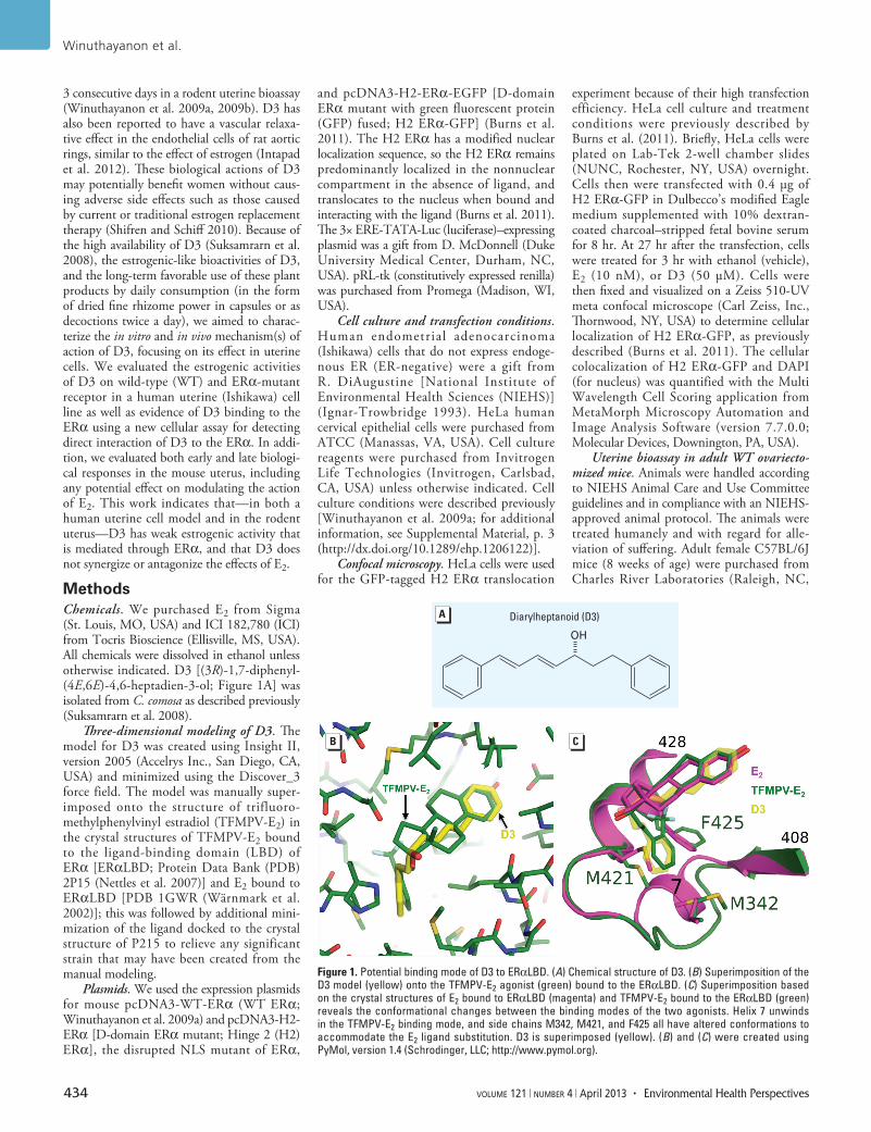
Three-dimensional modeling of D3. The model for D3 was created using Insight II, version 2005 (Accelrys Inc., San Diego, CA, USA) and minimized using the Discover_3 force field. The model was manually super-imposed onto the structure of trifluoro-methyl phenyl vinyl estradiol (TFMPV-E2) in the crystal structures of TFMPV-E2 bound to the ligand-binding domain (LBD) of ERα [ERαLBD; Protein Data Bank (PDB) 2P15 (Nettles et al. 2007)] and E2 bound to ERαLBD [PDB 1GWR (Wärnmark et al. 2002)]; this was followed by additional mini-mization of the ligand docked to the crystal structure of P215 to relieve any significant strain that may have been created from the manual modeling.
Plasmids. We used the expression plasmids for mouse pcDNA3-WT-ERα (WT ERα; Winuthayanon et al. 2009a) and pcDNA3-H2-ERα [D-domain ERα mutant; Hinge 2 (H2) ERα], the disrupted NLS mutant of ERα,
and pcDNA3-H2-ERα-EGFP [D-domain ERα mutant with green fluorescent protein (GFP) fused; H2 ERα-GFP] (Burns et al. 2011). The H2 ERα has a modified nuclear localiza tion sequence, so the H2 ERα remains predominantly localized in the non nuclear compartment in the absence of ligand, and trans locates to the nucleus when bound and inter acting with the ligand (Burns et al. 2011). The 3× ERE-TATA-Luc (luciferase)–expressing plasmid was a gift from D. McDonnell (Duke University Medical Center, Durham, NC, USA). pRL-tk (constitutively expressed renilla) was purchased from Promega (Madison, WI, USA).
Cell culture and transfection conditions. Human endometrial adeno carcinoma (Ishikawa) cells that do not express endoge-nous ER (ER-negative) were a gift from R. DiAugustine [National Institute of Environmental Health Sciences (NIEHS)] (Ignar-Trowbridge 1993). HeLa human cervical epithelial cells were purchased from ATCC (Manassas, VA, USA). Cell culture reagents were purchased from Invitrogen Life Technologies (Invitrogen, Carlsbad, CA, USA) unless otherwise indicated. Cell culture conditions were described previously [Winuthayanon et al. 2009a; for additional information, see Supplemental Material, p. 3 (http://dx.doi.org/10.1289/ehp.1206122)].
Confocal microscopy. HeLa cells were used for the GFP-tagged H2 ERα translocation
experiment because of their high transfection efficiency. HeLa cell culture and treatment conditions were previously described by Burns et al. (2011). Briefly, HeLa cells were plated on Lab-Tek 2-well chamber slides (NUNC, Rochester, NY, USA) overnight. Cells then were transfected with 0.4 µg of H2 ERα-GFP in Dulbecco’s modified Eagle medium supplemented with 10% dextran-coated charcoal–stripped fetal bovine serum for 8 hr. At 27 hr after the transfection, cells were treated for 3 hr with ethanol (vehicle), E2 (10 nM), or D3 (50 µM). Cells were then fixed and visualized on a Zeiss 510-UV meta confocal microscope (Carl Zeiss, Inc., Thornwood, NY, USA) to determine cellular localization of H2 ERα-GFP, as previously described (Burns et al. 2011). The cellular colocalization of H2 ERα-GFP and DAPI (for nucleus) was quantified with the Multi Wavelength Cell Scoring application from MetaMorph Microscopy Automation and Image Analysis Software (version 7.7.0.0; Molecular Devices, Downington, PA, USA).
Uterine bioassay in adult WT ovariec to-mized mice. Animals were handled according to NIEHS Animal Care and Use Committee guidelines and in compliance with an NIEHS-approved animal protocol. The animals were treated humanely and with regard for alle-viation of suffering. Adult female C57BL/6J mice (8 weeks of age) were purchased from Charles River Laboratories (Raleigh, NC,
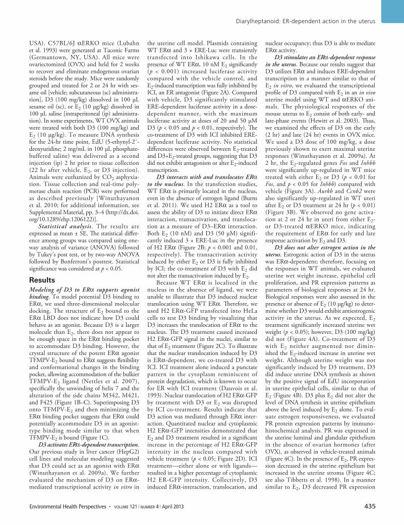
Figure 1. Potential binding mode of D3 to ERαLBD. (A) Chemical structure of D3. (B) Superimposition of the D3 model (yellow) onto the TFMPV-E2 agonist (green) bound to the ERαLBD. (C) Superimposition based on the crystal structures of E2 bound to ERαLBD (magenta) and TFMPV-E2 bound to the ERαLBD (green) reveals the conformational changes between the binding modes of the two agonists. Helix 7 unwinds in the TFMPV-E2 binding mode, and side chains M342, M421, and F425 all have altered conformations to accommodate the E2 ligand substitution. D3 is superimposed (yellow). (B) and (C) were created using PyMol, version 1.4 (Schrodinger, LLC; http://www.pymol.org).
OH
Diarylheptanoid (D3)
Diarylheptanoid: ER-dependent action in the uterus
Environmental Health Perspectives • volume 121 | number 4 | April 2013 435
USA). C57BL/6J αERKO mice (Lubahn et al. 1993) were generated at Taconic Farms (Germantown, NY, USA). All mice were ovariec tomized (OVX) and held for 2 weeks to recover and eliminate endogenous ovarian steroids before the study. Mice were randomly grouped and treated for 2 or 24 hr with ses-ame oil [vehicle; subcutaneous (sc) administra-tion], D3 (100 mg/kg) dissolved in 100 µL sesame oil (sc), or E2 (10 µg/kg) dissolved in 100 µL saline [intra peritoneal (ip) administra-tion). In some experiments, WT OVX animals were treated with both D3 (100 mg/kg) and E2 (10 µg/kg). To measure DNA synthesis for the 24-hr time point, EdU (5-ethynyl-2´-deoxyuridine; 2 mg/mL in 100 µL phosphate-buffered saline) was delivered as a second injection (ip) 2 hr prior to tissue collection (22 hr after vehicle, E2, or D3 injection). Animals were euthanized by CO2 asphyxia-tion. Tissue collection and real-time poly-merase chain reaction (PCR) were performed as described previously [Winuthayanon et al. 2010; for additional information, see Supplemental Material, pp. 3–4 (http://dx.doi.org/10.1289/ehp.1206122)].
Statistical analysis. The results are expressed as mean ± SE. The statistical differ-ence among groups was compared using one-way analysis of variance (ANOVA) followed by Tukey’s post test, or by two-way ANOVA followed by Bonferroni’s posttest. Statistical significance was considered at p < 0.05.
ResultsModeling of D3 to ERα supports agonist binding. To model potential D3 binding to ERα, we used three-dimensional molecular docking. The structure of E2 bound to the ERα LBD does not indicate how D3 could behave as an agonist. Because D3 is a larger molecule than E2, there does not appear to be enough space in the ERα binding pocket to accommodate D3 binding. However, the crystal structure of the potent ERα agonist TFMPV-E2 bound to ERα suggests flexibility and conformational changes in the binding pocket, allowing accommodation of the bulkier TFMPV-E2 ligand (Nettles et al. 2007), specifically the unwinding of helix 7 and the alteration of the side chains M342, M421, and F425 (Figure 1B–C). Superimposing D3 onto TFMPV-E2 and then minimizing the ERα binding pocket suggests that ERα could potentially accommodate D3 in an agonist-type binding mode similar to that when TFMPV-E2 is bound (Figure 1C).
D3 activates ERα-dependent transcription. Our previous study in liver cancer (HepG2) cell lines and molecular modeling suggested that D3 could act as an agonist with ERα (Winuthayanon et al. 2009a). We further evaluated the mechanism of D3 on ERα-mediated transcriptional activity in vitro in
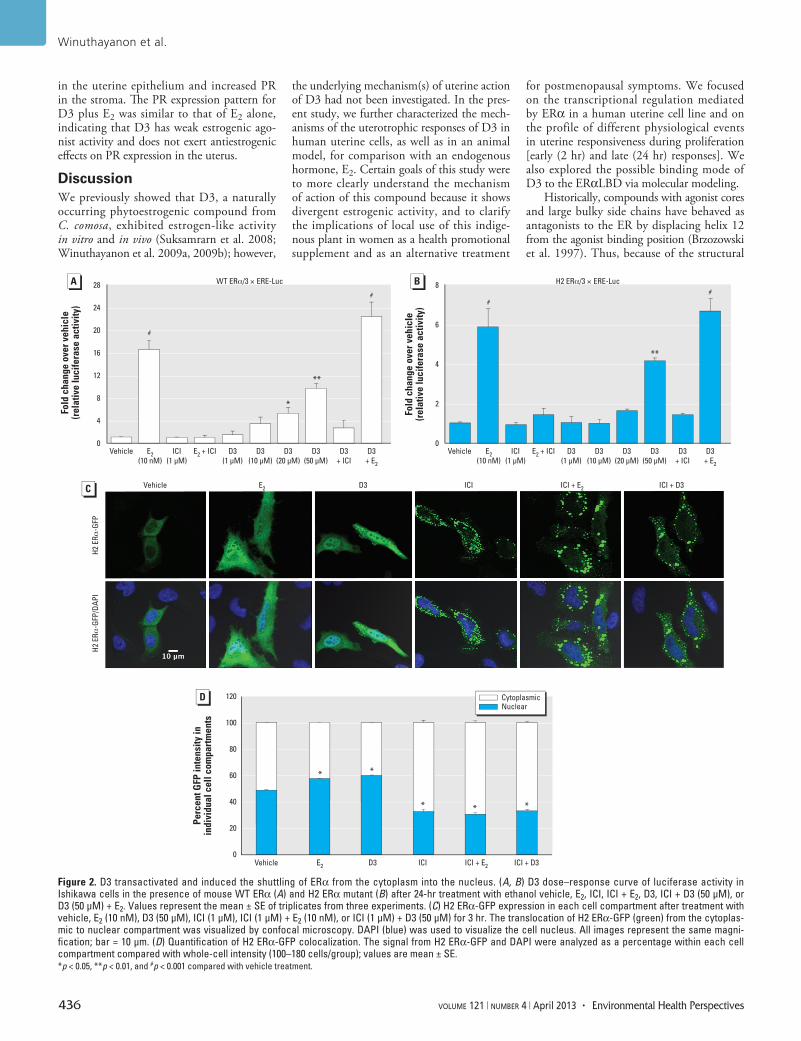
the uterine cell model. Plasmids containing WT ERα and 3 × ERE-Luc were transiently transfected into Ishikawa cells. In the presence of WT ERα, 10 nM E2 significantly (p < 0.001) increased luciferase activity compared with the vehicle control, and E2-induced transcription was fully inhibited by ICI, an ER antago nist (Figure 2A). Compared with vehicle, D3 significantly stimu lated ERE-dependent luciferase activity in a dose-dependent manner, with the maximum luciferase activity at doses of 20 and 50 µM D3 (p < 0.05 and p < 0.01, respectively). The co-treatment of D3 with ICI inhibited ERE-dependent luciferase activity. No statistical differences were observed between E2-treated and D3+E2-treated groups, suggesting that D3 did not exhibit antagonism or alter E2-induced transcription.
D3 interacts with and translocates ERα to the nucleus. In the transfection studies, WT ERα is primarily located in the nucleus, even in the absence of estrogen ligand (Burns et al. 2011). We used H2 ERα as a tool to assess the ability of D3 to initiate direct ERα inter action, trans activation, and transloca-tion as a measure of D3–ERα interaction. Both E2 (10 nM) and D3 (50 µM) signifi-cantly induced 3 × ERE-Luc in the presence of H2 ERα (Figure 2B; p < 0.001 and 0.01, respectively). The transactivation activity induced by either E2 or D3 is fully inhibited by ICI; the co-treatment of D3 with E2 did not alter the transactivation induced by E2.
Because WT ERα is localized in the nucleus in the absence of ligand, we were unable to illustrate that D3 induced nuclear translocation using WT ERα. Therefore, we used H2 ERα-GFP transfected into HeLa cells to test D3 binding by visualizing that D3 increases the translocation of ERα to the nucleus. The D3 treatment caused increased H2 ERα-GFP signal in the nuclei, similar to that of E2 treatment (Figure 2C). To illustrate that the nuclear translocation induced by D3 is ERα-dependent, we co-treated D3 with ICI. ICI treatment alone induced a punctate pattern in the cytoplasm reminiscent of protein degradation, which is known to occur for ER with ICI treatment (Dauvois et al. 1993). Nuclear translocation of H2 ERα-GFP by treatment with D3 or E2 was disrupted by ICI co-treatment. Results indicate that D3 action was mediated through ERα inter-action. Quantitated nuclear and cytoplasmic H2 ERα-GFP intensities demonstrated that E2 and D3 treatment resulted in a significant increase in the percentage of H2 ERα-GFP intensity in the nucleus compared with vehicle treatment (p < 0.05; Figure 2D). ICI treatment—either alone or with ligands—resulted in a higher percentage of cyto plasmic H2 ER-GFP intensity. Collectively, D3 induced ERα-interaction, translocation, and
nuclear occupancy; thus D3 is able to mediate ERα activity.
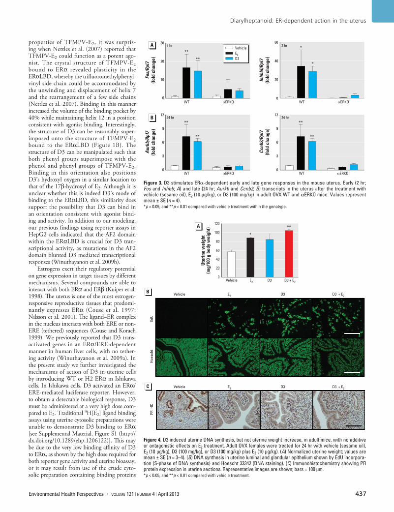
D3 stimulates an ERα-dependent response in the uterus. Because our results suggest that D3 utilizes ERα and induces ERE-dependent transcription in a manner similar to that of E2 in vitro, we evaluated the transcriptional profile of D3 compared with E2 in an in vivo uterine model using WT and αERKO ani-mals. The physio logi cal responses of the mouse uterus to E2 consist of both early- and late-phase events (Hewitt et al. 2003). Thus, we examined the effects of D3 on the early (2 hr) and late (24 hr) events in OVX mice. We used a D3 dose of 100 mg/kg, a dose previously shown to exert maximal uterine responses (Winuthayanon et al. 2009a). At 2 hr, the E2-regulated genes Fos and Inhbb were significantly up-regulated in WT mice treated with either E2 or D3 (p < 0.01 for Fos, and p < 0.05 for Inhbb) compared with vehicle (Figure 3A). Aurkb and Ccnb2 were also significantly up-regulated in WT uteri after E2 or D3 treatment at 24 hr (p < 0.01) (Figure 3B). We observed no gene activa-tion at 2 or 24 hr in uteri from either E2- or D3-treated αERKO mice, indicating the requirement of ERα for early and late response activation by E2 and D3.
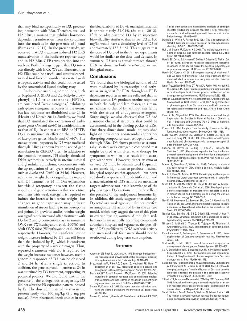
D3 does not alter estrogen action in the uterus. Estrogenic action of D3 in the uterus was ERα-dependent; therefore, focusing on the responses in WT animals, we evaluated uterine wet weight increase, epithelial cell proliferation, and PR expression patterns as parameters of biological responses at 24 hr. Biological responses were also assessed in the presence or absence of E2 (10 µg/kg) to deter-mine whether D3 would exhibit anti estrogenic activity in the uterus. As we expected, E2 treatment significantly increased uterine wet weight (p < 0.05); however, D3 (100 mg/kg) did not (Figure 4A). Co-treatment of D3 with E2 neither augmented nor dimin-ished the E2-induced increase in uterine wet weight. Although uterine weight was not signifi cantly induced by D3 treatment, D3 did induce uterine DNA synthesis as shown by the positive signal of EdU incorporation in uterine epithelial cells, similar to that of E2 (Figure 4B). D3 plus E2 did not alter the level of DNA synthesis in uterine epithelium above the level induced by E2 alone. To eval-uate estrogen responsiveness, we evaluated PR protein expression patterns by immuno-histo chemical analysis. PR was expressed in the uterine luminal and glandular epithelium in the absence of ovarian hormones (after OVX), as observed in vehicle-treated animals (Figure 4C). In the presence of E2, PR expres-sion decreased in the uterine epithelium but increased in the uterine stroma (Figure 4C; see also Tibbetts et al. 1998). In a manner similar to E2, D3 decreased PR expression
Winuthayanon et al.
436 volume 121 | number 4 | April 2013 • Environmental Health Perspectives
in the uterine epithelium and increased PR in the stroma. The PR expression pattern for D3 plus E2 was similar to that of E2 alone, indicating that D3 has weak estrogenic ago-nist activity and does not exert anti estrogenic effects on PR expression in the uterus.
DiscussionWe previously showed that D3, a naturally occurring phyto estrogenic compound from C. comosa, exhibited estrogen-like activity in vitro and in vivo (Suksamrarn et al. 2008; Winuthayanon et al. 2009a, 2009b); however,
the under lying mechanism(s) of uterine action of D3 had not been investigated. In the pres-ent study, we further characterized the mech-anisms of the uterotrophic responses of D3 in human uterine cells, as well as in an animal model, for comparison with an endogenous hormone, E2. Certain goals of this study were to more clearly understand the mechanism of action of this compound because it shows divergent estrogenic activity, and to clarify the implications of local use of this indige-nous plant in women as a health promotional supplement and as an alternative treatment
for post menopausal symptoms. We focused on the transcriptional regulation mediated by ERα in a human uterine cell line and on the profile of different physiological events in uterine responsiveness during prolifera tion [early (2 hr) and late (24 hr) responses]. We also explored the possible binding mode of D3 to the ERαLBD via molecular modeling.
Historically, compounds with agonist cores and large bulky side chains have behaved as antagonists to the ER by displacing helix 12 from the agonist binding position (Brzozowski et al. 1997). Thus, because of the structural
Figure 2. D3 transactivated and induced the shuttling of ERα from the cytoplasm into the nucleus. (A, B) D3 dose–response curve of luciferase activity in Ishikawa cells in the presence of mouse WT ERα (A) and H2 ERα mutant (B) after 24-hr treatment with ethanol vehicle, E2, ICI, ICI + E2, D3, ICI + D3 (50 µM), or D3 (50 µM) + E2. Values represent the mean ± SE of triplicates from three experiments. (C) H2 ERα-GFP expression in each cell compartment after treatment with vehicle, E2 (10 nM), D3 (50 µM), ICI (1 µM), ICI (1 µM) + E2 (10 nM), or ICI (1 µM) + D3 (50 µM) for 3 hr. The translocation of H2 ERα-GFP (green) from the cytoplas-mic to nuclear compartment was visualized by confocal microscopy. DAPI (blue) was used to visualize the cell nucleus. All images represent the same magni-fication; bar = 10 µm. (D) Quantification of H2 ERα-GFP colocalization. The signal from H2 ERα-GFP and DAPI were analyzed as a percentage within each cell compartment compared with whole-cell intensity (100–180 cells/group); values are mean ± SE. *p < 0.05, **p < 0.01, and #p < 0.001 compared with vehicle treatment.
28
24
20
16
12
8
4
0
8
6
4
2
0
Fold
cha
nge
over
veh
icle
(rel
ativ
e lu
cife
rase
act
ivity
)
Fold
cha
nge
over
veh
icle
(rel
ativ
e lu
cife
rase
act
ivity
)
###
#
**
**
*
Vehicle VehicleE2(10 nM)
ICI(1 µM)
E2 + ICI D3(1 µM)
D3(10 µM)
D3(20 µM)
D3(50 µM)
D3+ ICI
D3+ E2
E2(10 nM)
ICI(1 µM)
E2 + ICI D3(1 µM)
D3(10 µM)
D3(20 µM)
D3(50 µM)
D3+ ICI
D3+ E2
Vehicle
Vehicle
H2 E
Rα-G
FPH2
ERα
-GFP
/DAP
I
E2
E2 D3 ICI ICI + E2 ICI + D3
Cytoplasmic Nuclear
ICI + E2 ICI + D3D3
WT ERα/3 × ERE-Luc H2 ERα/3 × ERE-Luc
ICI
120
100
80
60
40
20
0
Perc
ent G
FP in
tens
ity in
indi
vidu
al c
ell c
ompa
rtm
ents
* *
* * *
Diarylheptanoid: ER-dependent action in the uterus
Environmental Health Perspectives • volume 121 | number 4 | April 2013 437
properties of TFMPV-E2, it was surpris-ing when Nettles et al. (2007) reported that TFMPV-E2 could function as a potent ago-nist. The crystal structure of TFMPV-E2 bound to ERα revealed plasticity in the ERαLBD, whereby the trifluoro methyl phenyl-vinyl side chain could be accommodated by the unwinding and displacement of helix 7 and the rearrange ment of a few side chains (Nettles et al. 2007). Binding in this manner increased the volume of the binding pocket by 40% while maintaining helix 12 in a position consistent with agonist binding. Interestingly, the structure of D3 can be reasonably super-imposed onto the structure of TFMPV-E2 bound to the ERαLBD (Figure 1B). The structure of D3 can be manipulated such that both phenyl groups super impose with the phenol and phenyl groups of TFMPV-E2. Binding in this orien ta tion also positions D3’s hydroxyl oxygen in a similar location to that of the 17β-hydroxyl of E2. Although it is unclear whether this is indeed D3’s mode of binding to the ERαLBD, this similiarity does support the possibility that D3 can bind in an orientation consistent with agonist bind-ing and activity. In addition to our modeling, our previous findings using reporter assays in HepG2 cells indicated that the AF2 domain within the ERαLBD is crucial for D3 tran-scriptional activity, as mutations in the AF2 domain blunted D3 mediated transcriptional responses (Winuthayanon et al. 2009b).
Estrogens exert their regulatory potential on gene expression in target tissues by different mechanisms. Several compounds are able to interact with both ERα and ERβ (Kuiper et al. 1998). The uterus is one of the most estrogen-responsive reproductive tissues that predomi-nantly expresses ERα (Couse et al. 1997; Nilsson et al. 2001). The ligand–ER complex in the nucleus inter acts with both ERE or non-ERE (tethered) sequences (Couse and Korach 1999). We previously reported that D3 trans-activated genes in an ERα/ERE-dependent manner in human liver cells, with no tether-ing activity (Winuthayanon et al. 2009a). In the present study we further investigated the mechanisms of action of D3 in uterine cells by introducing WT or H2 ERα in Ishikawa cells. In Ishikawa cells, D3 activated an ERα/ERE-mediated luciferase reporter. However, to obtain a detectable biological response, D3 must be adminis tered at a very high dose com-pared to E2. Traditional 3H[E2] ligand binding assays using uterine cytosolic preparations were unable to demonstrate D3 binding to ERα [see Supplemental Material, Figure S1 (http://dx.doi.org/10.1289/ehp.1206122)]. This may be due to the very low binding affinity of D3 to ERα, as shown by the high dose required for both reporter gene activity and uterine bio assay, or it may result from use of the crude cyto-solic preparation containing binding proteins
Figure 3. D3 stimulates ERα-dependent early and late gene responses in the mouse uterus. Early (2 hr; Fos and Inhbb; A) and late (24 hr; Aurkb and Ccnb2; B) transcripts in the uterus after the treatment with vehicle (sesame oil), E2 (10 µg/kg), or D3 (100 mg/kg) in adult OVX WT and αERKO mice. Values represent mean ± SE (n = 4). *p < 0.05, and **p < 0.01 compared with vehicle treatment within the genotype.
**
**
*
*
2 hr
24 hr
2 hr
24 hr **
**
**
**
30
20
10
0
12
9
6
3
0
12
9
6
3
0
60
40
20
0
Fos/Rpl7
(fold
cha
nge)
InhbbI/Rpl7
(fold
cha
nge)
Ccnb2/Rpl7
(fold
cha
nge)
Aurkb/Rpl7
(fold
cha
nge)
WT αERKO WT αERKO
WT αERKO WT αERKO
VehicleE2 D3
Figure 4. D3 induced uterine DNA synthesis, but not uterine weight increase, in adult mice, with no additive or antagonistic effects on E2 treatment. Adult OVX females were treated for 24 hr with vehicle (sesame oil), E2 (10 µg/kg), D3 (100 mg/kg), or D3 (100 mg/kg) plus E2 (10 µg/kg). (A) Normalized uterine weight; values are mean ± SE (n = 3–4). (B) DNA synthesis in uterine luminal and glandular epithelium shown by EdU incorpora-tion (S-phase of DNA synthesis) and Hoescht 33342 (DNA staining). (C) Immunohistochemistry showing PR protein expression in uterine sections. Representative images are shown; bars = 100 µm. *p < 0.05, and **p < 0.01 compared with vehicle treatment.
Vehicle
Vehicle
Hoes
cht
PR IH
CEd
U
E2 D3 D3 + E2
Vehicle E2 D3 D3 + E2
E2 D3
*
**
D3 + E2
120
100
80
60
40
20
0
Ute
rine
wei
ght
(mg/
100
g bo
dy w
eigh
t)
Winuthayanon et al.
438 volume 121 | number 4 | April 2013 • Environmental Health Perspectives
that may bind non specifically to D3, prevent-ing interaction with ERα. Therefore, we used H2 ERα, a mutant that exhibits hormone- dependent trans location from the cytoplasm into the nucleus in the presence of ligand (Burns et al. 2011). In the present study, we observed that D3 treatment induced H2 ERα transactivation in the luciferase reporter assay and in H2 ERα-GFP translocation into the nucleus. Both findings suggest that D3 inter-acts directly with ERα. We also illustrated that H2 ERα could be a useful and sensitive experi-mental tool for compounds that exerted weak estrogenic activity and that could not be tested by the conventional ligand binding assay.
Endocrine-disrupting compounds, such as bisphenol A (BPA) and 2,2-bis(p-hydroxy-phenyl)-1,1,1-trichloro ethane (HPTE), are considered “weak estrogens,” exhibiting early-phase estrogenic responses in the uterus, but late responses are diminished after 24 hr (Hewitt and Korach 2011). Similarly, we found that D3 stimulated the expression of early-phase genes (Fos and Inhbb) in a fashion similar to that of E2. In contrast to BPA or HPTE, D3 also sustained its effect on the induction of late-phase genes (Aurkb and Ccnb2). The transcriptional responses by D3 were mediated through ERα as shown by the lack of gene stimulation in αERKO uteri. In addition to the genomic responses, D3 clearly stimulated DNA synthesis selectively in uterine luminal and glandular epithelium, concomitant with the up-regulation of cell cycle–related genes, such as Aurkb and Ccnb2 (at 24 hr). However, uterine wet weight did not significantly increase with D3 treatment at 24 hr. One explanation for this discrepancy between the tissue response and gene activation is that a repetitive treatment with weak estrogens is required to induce the increase in uterine weight, but changes in gene expression may indicate stimulation of tissues leading to tissue-response end points. In previous studies, uterine weight was significantly increased after treatment with D3 for 2 and 3 consecutive days in immature OVX rats (Winuthayanon et al. 2009b) and adult OVX mice (Winuthayanon et al. 2009a), respectively. However, the significant uterine weight increase induced by D3 was still lower than that induced by E2, which is consistent with the property of a weak estrogen. Thus, repeated treatment with D3 is required for the weight-increase response; however, uterine genomic responses of D3 can be observed 2 and 24 hr after a single injection. In addition, the gene expression pattern at 24 hr was sustained by D3 treatment, supporting its potential potency. We also found that, in the presence of the endogenous estrogen E2, D3 did not alter the PR expression pattern induced by E2. The dose administered in vivo in the present study was 100 mg/kg (2.5 mg per mouse). From pharmaco kinetic studies in rats,
the bioavailability of D3 via oral administration is approximately 24.01% (Su et al. 2012). If mice administered D3 by ip injection bioavailability similar to that in rats, D3 at 100 mg/kg would have a circulating level of D3 of approxi mately 132.3 µM. This suggests that the dose of D3 used in the in vivo experiments would be similar to the dose used in vitro. In summary, D3 acts as a weak estrogen through ERα, as shown in both in vitro and in vivo biological assays.
ConclusionsWe found that the biological actions of D3 were mediated by its transcriptional activ-ity as an agonist for ERα through an ERE-dependent reporter in uterine cells, and that, in mouse uterus, D3 produces uterine responses in both the early and late phases, in a man-ner similar to that of E2, without inter fering with the effect of endogenous estrogens. Surprisingly, we also observed that D3 had a unique chemical structure that could be accommodated in the binding pocket of ERα. Our three-dimensional modeling may shed light on how other non steroidal endocrine-disrupting compounds exert estrogenic activity through ERα. D3 shows promise as a natu-rally isolated weak estrogenic compound that might be used as an alternative therapy for symptoms in women that result from estro-gen withdrawal. However, either in vitro or in vivo, D3 must be administered frequently at extremely high doses to produce maximal biological responses that approach—but never equal—E2 responses. The identification and charac teriza tion of D3’s actions on molecu lar targets advance our basic knowledge of the phyto estrogen D3’s actions in uterine cells in the presence of the endogenous hormone E2. In addition, this study suggests that although D3 acted as a weak agonist, it did not interfere or antagonize the action of E2 in the in vivo model, which may suggest the use of this plant in ovarian cycling women. Although diaryl-heptanoids are naturally occurring compounds abundant in spices and vege tables, the possibil-ity of D3’s proliferative DNA synthesis activity and increased risk for cancer should not be overlooked during long-term consumption.
RefeRences
Anderson JN, Peck EJ Jr, Clark JH. 1975. Estrogen-induced uter-ine responses and growth: relationship to receptor estrogen binding by uterine nuclei. Endocrinology 96:160–167.
Brzozowski AM, Pike AC, Dauter Z, Hubbard RE, Bonn T, Engström O, et al. 1997. Molecular basis of agonism and antagonism in the oestrogen receptor. Nature 389:753–758.
Burns KA, Li Y, Arao Y, Petrovich RM, Korach KS. 2011. Selective mutations in estrogen receptor α D-domain alters nuclear translocation and non-estrogen response element gene regulatory mechanisms. J Biol Chem 286:12640–12649.
Couse JF, Korach KS. 1999. Estrogen receptor null mice: what have we learned and where will they lead us? Endocr Rev 20:358–417.
Couse JF, Lindzey J, Grandien K, Gustafsson JA, Korach KS. 1997.
Tissue distribution and quantitative analysis of estrogen receptor-α (ERα) and estrogen receptor-β (ERβ) messenger ribonucleic acid in the wild-type and ERα-knockout mouse. Endocrinology 138:4613–4621.
Dauvois S, White R, Parker MG. 1993. The anti estrogen ICI 182780 disrupts estrogen receptor nucleo cyto plasmic shuttling. J Cell Sci 106:1377–1388.
Hall JM, Couse JF, Korach KS. 2001. The multifaceted mecha-nisms of estradiol and estrogen receptor signaling. J Biol Chem 276:36869–36872.
Hewitt SC, Deroo BJ, Hansen K, Collins J, Grissom S, Afshari CA, et al. 2003. Estrogen receptor-dependent genomic responses in the uterus mirror the biphasic physiological response to estrogen. Mol Endocrinol 17:2070–2083.
Hewitt SC, Korach KS. 2011. Estrogenic activity of bisphenol A and 2,2-bis(p-hydroxyphenyl)-1,1,1-trichloroethane (HPTE) demonstrated in mouse uterine gene profiles. Environ Health Perspect 119:63–70.
Ignar-Trowbridge DM, Teng CT, Ross KA, Parker MG, Korach KS, McLachlan JA. 1993. Peptide growth factors elicit estrogen receptor-dependent transcriptional activation of an estrogen-responsive element. Mol Endocrinol 7:992–998.
Intapad S, Saengsirisuwan V, Prasannarong M, Chuncharunee A, Suvitayawat W, Chokchaisiri R, et al. 2012. Long-term effect of phytoestrogens from Curcuma comosa Roxb. on vascu-lar relaxation in ovariectomized rats. J Agric Food Chem 60:758–764.
Keserü GM, Nógrádi M. 1995. The chemistry of natural diaryl-heptanoids. In: Studies in Natural Products Chemistry, Vol 17, Structure and Chemistry (Part D) (Atta-ur-Rahman, ed). Amsterdam:Elsevier, 357–394.
Korach KS. 1994. Insights from the study of animals lacking functional estrogen receptor. Science 266:1524–1527.
Kuiper GGJM, Lemmen JG, Carlsson B, Corton JC, Safe SH, van der Saag PT, et al. 1998. Interaction of estrogenic chemicals and phytoestrogens with estrogen receptor β. Endocrinology 139:4252–4263.
Lubahn DB, Moyer JS, Golding TS, Couse JF, Korach KS, Smithies O. 1993. Alteration of reproductive function but not prenatal sexual development after insertional disruption of the mouse estrogen receptor gene. Proc Natl Acad Sci USA 90:11162–11166.
Mader S, Chambon P, White JH. 1993. Defining a minimal estrogen receptor DNA binding domain. Nucleic Acids Res 21:1125–1132.
Martin L, Finn CA, Trinder G. 1973. Hypertrophy and hyperplasia in the mouse uterus after oestrogen treatment: an autoradio-graphic study. J Endocrinol 56:133–144.
Mote PA, Arnett-Mansfield RL, Gava N, deFazio A, Mulac-Jericevic B, Conneely OM, et al. 2006. Overlapping and distinct expression of progesterone receptors A and B in mouse uterus and mammary gland during the estrous cycle. Endocrinology 147:5503–5512.
Naciff JM, Overmann GJ, Torontali SM, Carr GJ, Khambatta ZS, Tiesman JP, et al. 2007. Uterine temporal response to acute exposure to 17α-ethinyl estradiol in the immature rat. Toxicol Sci 97:467–490.
Nettles KW, Bruning JB, Gil G, O’Neill EE, Nowak J, Guo Y, et al. 2007. Structural plasticity in the oestrogen receptor ligand-binding domain. EMBO Rep 8:563–568.
Nilsson S, Mäkelä S, Treuter E, Tujague M, Thomsen J, Andersson G, et al. 2001. Mechanisms of estrogen action. Physiol Rev 81:1535–1565.
Piyachaturawat P, Ercharuporn S, Suksamrarn A. 1995. Utero-trophic effect of Curcuma comosa in rats. Int J Pharmacogn 33:334–338.
Shifren JL, Schiff I. 2010. Role of hormone therapy in the manage ment of menopause. Obstet Gynecol 115:839–855.
Su A, Sripanidkulchai K, Suksamrarn A, Hu Y, Piyachuturawat P, Sripanidkulchai B. 2012. Pharmacokinetics and organ distri-bution of diarylheptanoid phytoestrogens from Curcuma comosa in rats. J Nat Med 66:468–475.
Suksamrarn A, Ponglikitmongkol M, Wongkrajang K, Chindaduang A, Kittidanairak S, Jankam A, et al. 2008. Diarylheptanoids, new phytoestrogens from the rhizomes of Curcuma comosa: Isolation, chemical modification and estrogenic activity evaluation. Bioorg Med Chem 16:6891–6902.
Tibbetts TA, Mendoza-Meneses M, O’Malley BW, Conneely OM. 1998. Mutual and intercompartmental regulation of estro-gen receptor and progesterone receptor expression in the mouse uterus. Biol Reprod 59:1143–1152.
Tora L, White J, Brou C, Tasset D, Webster N, Scheer E, et al. 1989. The human estrogen receptor has two independent non-acidic transcriptional activation functions. Cell 59:477–487.

Diarylheptanoid: ER-dependent action in the uterus
Environmental Health Perspectives • volume 121 | number 4 | April 2013 439
Wärnmark A, Treuter E, Gustafsson JA, Hubbard RE, Brzozowski AM, Pike AC. 2002. Interaction of transcriptional intermedi-ary factor 2 nuclear receptor box peptides with the coacti-vator binding site of estrogen receptor α. J Biol Chem 277:21862–21868.
Winuthayanon W, Hewitt SC, Orvis GD, Behringer RR, Korach KS. 2010. Uterine epithelial estrogen receptor α
is dispensable for proliferation but essential for complete biological and biochemical responses. Proc Natl Acad Sci USA 107:19272–19277.
Winuthayanon W, Piyachaturawat P, Suksamrarn A, Ponglikitmongkol M, Arao Y, Hewitt SC, et al. 2009a. Diarylheptanoid phytoestrogens isolated from the medici-nal plant Curcuma comosa: Biological actions in vitro and
in vivo indicate ER-dependent mechanisms. Environ Health Perspect 117:1155–1161.
Winuthayanon W, Suksen K, Boonchird C, Chuncharunee A, Ponglikitmongkol M, Suksamrarn A, et al. 2009b. Estrogenic activity of diarylheptanoids from Curcuma comosa Roxb. requires metabolic activation. J Agric Food Chem 57:840–845.