The In Vitro Immune Modulatory Effect of Bisphenol A on...
8
The In Vitro Immune Modulatory Effect of Bisphenol A on Fish Macrophages via Estrogen Receptor α and Nuclear Factor-κB Signaling Ming Yang,* ,†,§ Wenhui Qiu, †,§ Bei Chen, ‡ Jingsi Chen, † Shuai Liu, † Minghong Wu, † and Ke-Jian Wang* ,‡ † School of Environmental and Chemical Engineering, Shanghai University, Shanghai 200444 China ‡ State Key Laboratory of Marine Environmental Science, Xiamen University, Xiamen, Fujian 361005, China * S Supporting Information ABSTRACT: Bisphenol A (BPA) is a well-known environmental endocrine-disrupting chemical. Employing primary macrophages from head kidney of red common carp (Cyprinus carpio), the present study aimed to evaluate the immune modulatory effect of BPA and to explore its potential action mechanism associated with estrogen receptor (ER) and nuclear factor-κB (NF-κB) pathways. A dynamic response process was observed in macrophages upon various concentrations of BPA exposure, which significantly enhanced the antibacterial activity of macrophages at 0.1, 1, or 10 μg/L, but instead induced the apoptosis at 100, 1000, and 10 000 μg/L. A potential pro-inflammatory effect of BPA exposure was suggested, judging from the increased production of nitrite oxide and reactive oxygen species (ROS), the induction of interleukin-1β mRNA and protein, as well as NF-κB and other NF-κB-associated immune gene expression. Following BPA coexposure with the ER or NF-κB antagonist, the induction of ROS, ERα, and NF-κB-associated immune gene expression was significantly inhibited, implying interaction between those two pathways. This study thus indicated that low doses of BPA exposure alone could significantly disturb the immune response of fish primary macrophages in vitro, and for the first time revealed the synergistic action of ERα and NF-κB transcription factors in the BPA effect. ■ INTRODUCTION Bisphenol A (BPA) is a well-known endocrine disrupting chemical (EDC), a constituent of certain plastic products widely used in plastic food and beverage containers, metal can linings, thermal receipt paper, dental sealants, and household paper products. It is listed as one of the highest volume chemicals produced globally, with more than 6 billion pounds of production annually. 1 According to the reported data from the Toxics Release Inventory, U.S.A., the total release of BPA to the environment was about 2.5 million kg in 2007, with the direct release of 13 772 kg to water. 2 As a consequence of its ubiquitous existence in the environment and extensive application in our daily lives, BPA has raised considerable public health concerns since it is widely detected at low nanomolar levels in a variety of human samples such as urine, blood and breast milk. 3,4 Meanwhile, a large number of studies have detected BPA contamination in various aquatic environ- ments at μg/L to even low mg/L or mg/kg and, therefore, its detrimental effects on aquatic animals have also raised great concern about the healthy and sustainable development of aquatic organisms. 5-7 The immune system is crucial to the survival of organisms against pathogens and various environmental stresses. How- ever, due to regulation by sex steroids, it is also a potential target for environmental exogenous hormones. 8,9 BPA has a relatively weak estrogenic potency compared to natural estradiol (E 2 ) based on its binding affinity or activating capacity Received: October 24, 2014 Revised: December 30, 2014 Accepted: January 6, 2015 Published: January 6, 2015 Article pubs.acs.org/est © 2015 American Chemical Society 1888 DOI: 10.1021/es505163v Environ. Sci. Technol. 2015, 49, 1888-1895
Transcript of The In Vitro Immune Modulatory Effect of Bisphenol A on...
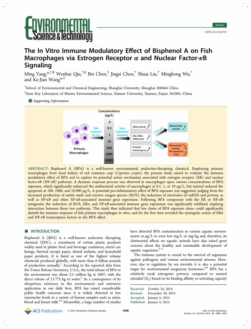
The In Vitro Immune Modulatory Effect of Bisphenol A on FishMacrophages via Estrogen Receptor α and Nuclear Factor-κBSignalingMing Yang,*,†,§ Wenhui Qiu,†,§ Bei Chen,‡ Jingsi Chen,† Shuai Liu,† Minghong Wu,†
and Ke-Jian Wang*,‡
†School of Environmental and Chemical Engineering, Shanghai University, Shanghai 200444 China‡State Key Laboratory of Marine Environmental Science, Xiamen University, Xiamen, Fujian 361005, China
*S Supporting Information
ABSTRACT: Bisphenol A (BPA) is a well-known environmental endocrine-disrupting chemical. Employing primarymacrophages from head kidney of red common carp (Cyprinus carpio), the present study aimed to evaluate the immunemodulatory effect of BPA and to explore its potential action mechanism associated with estrogen receptor (ER) and nuclearfactor-κB (NF-κB) pathways. A dynamic response process was observed in macrophages upon various concentrations of BPAexposure, which significantly enhanced the antibacterial activity of macrophages at 0.1, 1, or 10 μg/L, but instead induced theapoptosis at 100, 1000, and 10 000 μg/L. A potential pro-inflammatory effect of BPA exposure was suggested, judging from theincreased production of nitrite oxide and reactive oxygen species (ROS), the induction of interleukin-1β mRNA and protein, aswell as NF-κB and other NF-κB-associated immune gene expression. Following BPA coexposure with the ER or NF-κBantagonist, the induction of ROS, ERα, and NF-κB-associated immune gene expression was significantly inhibited, implyinginteraction between those two pathways. This study thus indicated that low doses of BPA exposure alone could significantlydisturb the immune response of fish primary macrophages in vitro, and for the first time revealed the synergistic action of ERαand NF-κB transcription factors in the BPA effect.
■ INTRODUCTION
Bisphenol A (BPA) is a well-known endocrine disruptingchemical (EDC), a constituent of certain plastic productswidely used in plastic food and beverage containers, metal canlinings, thermal receipt paper, dental sealants, and householdpaper products. It is listed as one of the highest volumechemicals produced globally, with more than 6 billion poundsof production annually.1 According to the reported data fromthe Toxics Release Inventory, U.S.A., the total release of BPA tothe environment was about 2.5 million kg in 2007, with thedirect release of 13 772 kg to water.2 As a consequence of itsubiquitous existence in the environment and extensiveapplication in our daily lives, BPA has raised considerablepublic health concerns since it is widely detected at lownanomolar levels in a variety of human samples such as urine,blood and breast milk.3,4 Meanwhile, a large number of studies
have detected BPA contamination in various aquatic environ-ments at μg/L to even low mg/L or mg/kg and, therefore, itsdetrimental effects on aquatic animals have also raised greatconcern about the healthy and sustainable development ofaquatic organisms.5−7
The immune system is crucial to the survival of organismsagainst pathogens and various environmental stresses. How-ever, due to regulation by sex steroids, it is also a potentialtarget for environmental exogenous hormones.8,9 BPA has arelatively weak estrogenic potency compared to naturalestradiol (E2) based on its binding affinity or activating capacity
Received: October 24, 2014Revised: December 30, 2014Accepted: January 6, 2015Published: January 6, 2015
Article
pubs.acs.org/est
© 2015 American Chemical Society 1888 DOI: 10.1021/es505163vEnviron. Sci. Technol. 2015, 49, 1888−1895
to estrogenic receptors (ERs) from in vitro assays.10,11
However, the in vivo studies indicate that BPA can promoteestrogen-like activities that are similar to or stronger than E2.
12
Consequently, as a xenoestrogen, BPA has the ability to mimicthe effects of natural estrogens or compete with endogenousestrogens binding to ERs and disrupting the immune defense oforganisms. Previous studies have reported the potential effect ofBPA in modulating the immune response, however, the dataobtained using mammalian models are paradoxical concerningthe immune suppressing or immune stimulating effect of BPAexposure.13−15 In contrast, there are few studies exploring thepotential immunotoxicity of BPA to aquatic animals based onan environmentally relevant scenario.Fish and mammalian immune systems share a number of
similarities. There is evidence that ERs are expressed in piscineimmune cells and participate in the regulation of immuneresponse.16 Thus, xenoestrogens may target fish immunesystem and modulate their immunocompetence.17,18 Previousstudies using fish models imply that the effect of BPA on theimmune response is more likely prone to be pro-inflammatoryat low doses.14 The exposure to BPA at μg/L or low mg/L canlead to functional changes of fish lymphocytes and macro-phages, such as increasing the cellular production of superoxideanions, enhancing cell proliferation and elevating leukocytecounts in vivo.14,19,20 Moreover, our recent in vivo studyindicates that BPA exposure significantly impacts the immuneresponse of zebrafish at environmentally relevant lowconcentrations.21 Several inflammatory mediators includingnitric oxide (NO), cytokines and chemokines in zebrafishembryos are significantly induced upon exposure to BPA at μg/L to low mg/L, which is similar to the patterns induced bylipopolysaccharide stimulation, indicating that BPA exposure atlow doses alone could significantly induce the immuneresponse.Macrophages are responsible for both nonspecific defense
and specific defense. Fish macrophages are morphologically,histochemically, and functionally similar to their mammaliancounterparts.22 They display phagocytosis, produce oxygenradicals, synthesize cytokines/chemokines, and also havebactericidal activity. Fish macrophages express ERs and areregulated by estrogens.23 Common carp (Cyprinus carpio) isone of the most important aquaculture species in the word.Widespread in Asia, red common carp is popular for food andalso for decorative purposes. In the present study, the primarymacrophages from head kidney of red common carp wereisolated and cultured, and effects of BPA were investigated onmacrophages upon a series of concentrations of BPA exposure.We determined their antibacterial function, oxygen radicalproduction, as well as gene expression of an antimicrobialpeptide hepcidin24 and several cytokines including pro-inflammatory interleukin-1β (IL-1β), interleukin-6 subfamily likecytokine (M17), and anti-inflammatory IL-10, which wereimportant immune parameters associated with the activationof macrophages. Besides, the apoptosis level of macrophageswas assayed. It has been reported recently that BPA affectedcytokine expression in human macrophages through ER-dependent mechanism associated with extracellular regulatedprotein kinases/nuclear factor-κB (NF-κB) signaling.25 Sim-ilarly, we investigated the potential roles of ERα and NF-κBtranscription factors in BPA effects on fish macrophages. Ourstudy, thereby, further testified that BPA exposure at low dosessignificantly modulated the immune response of primarymacrophages in vitro, and for the first time revealed the
synergistic action of ERα and NF-κB transcription factors in theBPA immune modulating effect.
■ MATERIALS AND METHODSChemicals. BPA (CAS number 80−05−7, 99+%) was
purchased from Sigma-Aldrich (St. Louis, MO). The stocksolution was prepared by dissolving the chemical in dimethylsulfoxide (DMSO) to a concentration at 10 g/L. Fresh stocksolution was made every week. All other chemicals used were ofanalytical grade and purchased from Sigma−Aldrich (St. Louis,MO) and Sangon (Shanghai, China).
Primary Macrophages. Red common carp weighing 300−500 g, were purchased from a fish farm in Qingpu District ofShanghai, China. Fish were acclimated to laboratory conditionsin dechlorinated tap water at room temperature for at least 2weeks and then healthy fish were chosen for the experiments.The method for isolation of primary macrophages from thehead kidney of the carp was modified based on previousstudies26,27 and the experimental details were shown inSupporting Information (SI). The cells were then adjusted toa density of 1 × 10⁷ cells/mL and plated in 96-well microplates(Corning) at 100 μL/well or in 25 cm2 cell culture flasks(Corning) at 3 mL/flask. After overnight incubation at 26 °C,monolayers of adherent cells (about 1% of the seeded cells) inthe microplates and culture flasks were washed twice to removeunattached cells, and then they were used for the chemicalexposure. The viability of cells was assessed using the trypanblue (Sigma-Analytical) exclusion test. Primary macrophageswere identified using nonspecific esterase staining and Wright-Giemsa staining methods (SI Figure S1).28
Experimental Design. Primary macrophages were exposedto BPA solutions at 0.1, 1, 10, 100, 1000, and 10 000 μg/L inphenol red-free medium for 6 h. After treatment, cells werewashed three times and separately collected for the subsequentbioassays. An ER inhibition experiment was designed separatelyusing coexposure of macrophages for 6 h to 5 mg/L of BPAwith an ER antagonist ICI 182,780 (Tocris Bioscience, UnitedKingdom) at 50 μM.11 Acute exposure of primary macrophagesto 1000 μg/L of BPA for 48 h was performed, theconcentration chosen based on the response of cells to 6 hexposure. The time-course response of primary macrophageswas then measured at 6, 24, and 48 h. Two nuclear receptorinhibition groups were designed using coexposure either to1000 μg/L of BPA with ICI 182,780 at 50 μM, or to 1000 μg/Lof BPA with an NF-κB antagonist pyrrolidine dithiocarbamate(PDTC, Sigma) at 50 μM.29 A blank control group and avehicle control group (0.002% DMSO in culture medium) wereset up in parallel with these experiments and at least threereplicates were set for each treatment group. The exposuresolution was replaced completely every 24 h.
Bactericidal Activity and Phagocytic Capability ofPrimary Macrophages. After 6 h exposure to BPA,macrophages were washed several times with antibiotic-freeculture medium to completely remove BPA residue, and thenthey were inoculated with freshly prepared bacterial culture todetermine the bactericidal activity and the phagocytic capabilityagainst Gram-positive Staphylococcus aureus (CGMCC 1.363)and Gram-negative Escherichia coli (CGMCC 1.2389), andVibrio parahaemolyticus (CGMCC 1.1615) following modifiedprotocols described by previous studies.30 The detailedmethods are shown in SI.
Biochemical Assays. Cytotoxicity was measured usingCellTiter 96 AQueous Non-Radioactive Cell Proliferation
Environmental Science & Technology Article
DOI: 10.1021/es505163vEnviron. Sci. Technol. 2015, 49, 1888−1895
1889
Assay (Promega) following the manufacturer’s instructions.The protein level of each sample was determined usingBradford assay (Beyotime Institute of Biotechnology, China).Lysozyme activity, total NO level, and the activities of total NOsynthase (TNOS) and induced NO synthase (iNOS) weremeasured following the protocols of commercially available kits(Nanjing Jiancheng Bioengineering Institute). Intracellularreactive oxygen species (ROS) content was determined bymeasuring the oxidative conversion of 2′,7′-dichlorofluoresceindiacetate (Beyotime Institute of Biotechnology, China) tofluorescent dichlorofluorescein.31
RNA Isolation, Reverse-Transcriptase, And Quantita-tive Polymerase Chain Reaction. Total RNA was extractedusing an RNA prep pure tissue kit (Tiangen Biotech, China).Reverse-transcriptase reactions were performed on 1 μg of totalRNA using a Transcriptor First Strand cDNA Synthesis Kit(Roche). Real-time quantitative PCR was performed using theFastStart Universal SYBR Green Master (ROX) kit (Roche)and the iQ5Multicolor Real-Time PCR Detection System (Bio-Rad). Melting curve analysis and agarose gel electrophoresiswere performed to validate the specificity of PCR amplicons.The subunit S11 of the ribosomal gene 40S was selected asreference gene based on a previous study.32 The primersspecific for target genes and the endogenous 40S ribosomalprotein S11 are listed in Table S1 (SI). A Ct-based relativequantification with efficiency correction normalized to 40Sribosomal protein S11 was calculated using the 2−△△Ct method.TUNEL Staining. The apoptosis level of primary macro-
phages after exposure to various concentrations of BPA was
determined based on the labeling of DNA strand breaks(TUNEL technology) by fluorescein using an in situ cell deathdetection kit (Roche) following the manufacturer’s instructions.Cells were costained with 4′,6′-diamidino-2-phenylindole toidentify the nuclei. TUNEL-positive cells were analyzed with aconfocal fluorescence microscope (LSM 780 NLO, ZEISS) andthe percentages of TUNEL-positive cells were then calculated.
Enzyme-Linked Immunosorbent Assay for Interleukin1β. Interleukin 1β levels in the cell homogenates weremeasured using a Fish Interleukin 1β ELISA Kit (MyBio-Source) employing the quantitative sandwich enzyme immuno-assay technique. Antibody specific for fish IL-1β was precoatedonto a 96-well microplate. A series of gradient dilutions of IL-1β standard and the concentrations of IL-1β in the sampleswere then interpolated from the standard curve.
Statistical Analysis. All data were normalized using theKolmogorov−Smirnov one-sample test and Levene’s test.Statistical analyses were performed using SPSS Statistics 18.0(SPSS Inc., Chicago, IL). For all the tested parameters, therewas no significant difference between the blank control groupand the vehicle control group, and thus the vehicle control wasset as the control group for the statistical analysis that followed.
■ RESULTSBactericidal Activity, Phagocytic Capability and
Lysozyme Activity. The viability of primary macrophageswas not impaired after 6 h exposure to BPA based on thecytotoxicity assay (SI Figure S2). In contrast, the bactericidalactivity of primary macrophages was induced in a U-shaped
Figure 1. Changes in antibacterial activities of the primary macrophages after 6 h-exposure to bisphenol A (BPA). (A) The bactericidal activity wasassayed by measuring the absorbance of extracellular bacteria at 600 nm (the black lines, n = 6), and the phagocytosis was measured by counting theintracellular bacteria (the blue lines, n = 6). (B) The representative images comparing the phagocytic activities between the control group and the 10μg/L of BPA group (scale bar = 5 μm). (C) The lysozyme activity (n = 3). Values are means ± standard deviation. Significant differences versuscontrol are indicated as *p < 0.05 (ANOVA, Dunnett’s test).
Environmental Science & Technology Article
DOI: 10.1021/es505163vEnviron. Sci. Technol. 2015, 49, 1888−1895
1890
pattern judging from the decrease of bacterial density based onthe OD 600 value (Figure 1A I, III, V). Especially, in some BPAtreatment groups at environmentally relevant concentrations of1 or 10 μg/L, the bactericidal activity was significantly highlypromoted compared to the control (Dunnett’s test, p < 0.05).Correspondingly, the phagocytic capability of primary macro-phages was changed in an inverse U-shaped pattern as shown inFigure 1A II, IV, VI. Consistent with the bactericidal activity,the calculated phagocytic index of primary macrophages washighest in the 1 or 10 μg/L BPA treatment group. Theenhanced phagocytosis of cells exposed to 10 μg/L of BPA isshown in Figure 1B. Consistently, as shown in Figure 1C, thecellular lysozyme activity was also changed in an inverse U-shaped pattern after BPA exposure, and the highest inductionwas presented in the 1 μg/L BPA treatment group compared tothe control (Dunnett’s test, p < 0.05).NO Level, and iNOS and TNOS Activity. A roughly
concentration-dependent increase of NO level, and iNOS andTNOS activity was observed in macrophages upon 6 hexposure to BPA, as shown in Figure 2A. The NO level andTNOS activity significantly increased in the 1000 and 10 000μg/L groups. However, the iNOS activity significantly increasedin the 100, 1000, and 10 000 μg/L groups (Dunnett’s test, p <0.05).Expression of Immune Related Genes. Gene expression
of a broad-spectrum antimicrobial peptide and an acute phaseprotein, hepcidin, as well as several pro-inflammatory mediatorswas assessed to evaluate the effects of BPA on primarymacrophages after 6-h exposure. Generally, the mRNA levels ofthe tested genes including hepcidin, IL-10, M17, and IL-1β weredramatically induced in a roughly concentration-dependentmanner after BPA exposure (Figure 2B). The mRNA levels ofhepcidin and IL-10 increased significantly in the 100, 1000, and10 000 μg/L groups (Dunnett’s test, p < 0.05); the mRNA levelof M17 was significantly induced by exposure to 10, 100, 1000,and 10 000 μg/L of BPA (Dunnett’s test, p < 0.05); and themRNA level of IL-1β in the macrophages was significantlyinduced by exposure to 1000 and 10 000 μg/L.
ROS Content, NF-κB Expression and the ApoptosisLevel. To evaluate the damage of acute BPA exposure on themacrophages, the cellular ROS content was examined after 6 hexposure, and it was significantly induced in a concentrationdependent manner in the 100, 1000, and 10 000 μg/Ltreatment groups compared to the control (LSD test, p <0.05) as shown in Figure 3A. Correspondingly, a significantincrease of NF-κB (p65) expression was also observed in
Figure 2. Induction of nitric oxide production, induced and the total nitric oxide synthase activities (A), as well as the expression of immune relatedgenes (B) in the primary macrophages upon 6 h-exposure of bisphenol A. Values are means ± standard deviation (n = 3). Significant differencesversus control are indicated as *p < 0.05 (ANOVA, Dunnett’s test).
Figure 3. Bisphenol A induced the production of reactive oxygenspecies (ROS) (A), the expression of NF-κB (B) and the apoptosis(C) in primary macrophages upon 6 h-exposure. Values are means ofthe standard error of means relative to the control (n = 6 for ROS andn = 3 for the others). Significant differences versus control areindicated as *p < 0.05 ((ANOVA, LSD’s, or Dunnett’s test)).
Environmental Science & Technology Article
DOI: 10.1021/es505163vEnviron. Sci. Technol. 2015, 49, 1888−1895
1891
primary macrophages upon 6-h exposure to BPA at 100, 1000,and 10 000 μg/L (Figure 3B). A concentration-dependentincrease of apoptosis level was observed in primary macro-phages upon 6 h exposure to BPA, as shown in Figure 3C andFigure S3 in SI. A significant increase of the cell number ofTUNEL positive nuclei was shown in the 100, 1000, and10 000 μg/L BPA treatment groups (Dunnett’s test, p < 0.05).Gene Expression of ERα. To understand the potential
activity of ERs in the immune response upon BPA exposure,the expression level of ERα was examined in primarymacrophages. As shown in Figure 4A, the mRNA level of
ERα increased significantly (Dunnett’s test, p < 0.05) as BPAconcentrations were raised to 100, 1000, and 10 000 μg/L in aconcentration-dependent manner. After correlation analysisbetween the tested parameters using Spearman’s test (SI TableS2), it was notable that the elevated ERα expression levels inmacrophages upon BPA exposure were significantly correlatedwith the changes of lysozyme activity, iNOS and TNOSactivities, as well as tested immune related gene expression andapoptosis levels in our study, which implied an important roleof ERα in the immune regulation upon acute BPA exposure.The Effect of an Estrogen Receptor Antagonist. To
determine the involvement of ERα in the immunotoxicity ofBPA exposure, coexposure of the cultured macrophages to arelatively high concentration of BPA with an ER inhibitor ICI182 780 was performed and the mRNA levels of ERα and IL-1β, as well as the protein level of IL-1β in macrophages, weredetected after exposure (Figure 4B, C, and D). The resultsshowed that coexposure to BPA with ICI 182 780 efficientlyinhibited the induction of ERα expression. The expression ofIL-1β and IL-1β protein level in macrophages were bothinduced after BPA exposure, which was in parallel with theincreased gene expression of ERα. However, after coexposure
to BPA with ICI 182 780, the induction levels of IL-1β mRNAand IL-1β protein in macrophages were both significantlydecreased compared to the BPA exposure group (LSD test, p <0.05), indicating an involvement of the ER pathway in theimmune modulation upon BPA exposure.
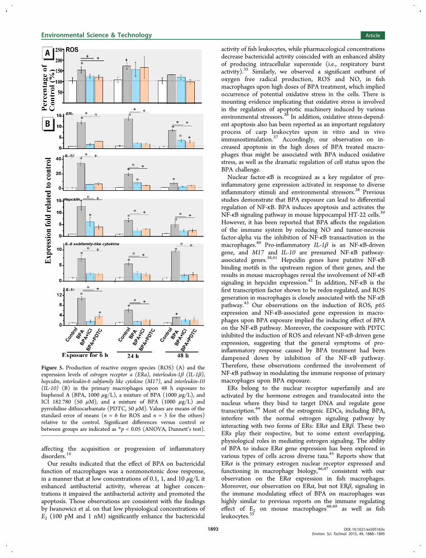
The Time-Course Response of Macrophages uponCoexposure to BPA with ER or NF-κB Antagonist. Theviability of primary macrophages was not significantly impairedduring 48 h exposure to BPA and coexposure to BPA with ERor NF-κB antagonist compared to the vehicle control (SI FigureS4). As shown in Figure 5A, the cellular ROS content wassignificantly induced upon exposure to BPA after 6 and 24, butnot after 48 h. However, upon coexposure to BPA with ICI182 780 or PDTC, the induction level of ROS content wassignificantly inhibited after 6 h by the antagonist effects of theER or NF-κB inhibitor. Similar to the results of ROS content,increased mRNA levels of ERα, IL-1β, hepcidin, M17, and IL-10were observed in the BPA treatment group after 6, 24, and also48 h (Figure 5B). However, the induction levels of those geneswere all significantly decreased (Dunnett’s test, p < 0.05) bycoexposure with ICI 182,780 at all the time points or PDTCafter 6 and 24 h. The results indicated that ER inhibitor ICI182 780 significantly inhibited the induction of the ROScontent and the immune gene expression in macrophages uponexposure to BPA. In addition, the immune response mediatorNF-κB antagonist PDTC in turn could significantly restrain theER induction in macrophages upon BPA exposure.
■ DISCUSSION
Using fish primary macrophages as a model, our studypresented a dynamic response process of immunologicalparameters in macrophages upon various concentrations ofBPA exposure. With increased BPA exposure, ROS and NOlevels, iNOS and TNOS activities, as well as immune geneexpression such as IL-1β, IL-10 in the macrophages wassignificantly induced in a concentration-dependent manner,consistent with our previous in vivo observations on zebrafishembryos upon 7 day BPA treatment.21 Moreover, thealterations of most tested parameters were significantlycorrelated (SI Table S2), indicating a coordinated modulationprocess in macrophages responded to increased BPA exposure.Consistent with our observation on M17 (IL-6 like), Liu et al.25
have showed that BPA treatment increased pro-inflammatorycytokines IL-6 and tumor necrosis factor-α (TNF-α)production in THP1 macrophages and primary humanmacrophages at BPA concentrations of 0.01 and 0.1 μM, thatis, about 2 and 20 μg/L. Also, Cabas et al.33 have reported that17α-ethynylestradiol, an environmental estrogen, at lowconcentrations increase the mRNA levels of IL-1β, IL-6, andTNF-α in fish macrophages, which is similar to our observationson that BPA exposure alone has a pro-inflammatory effect invitro. Tissue macrophages, as well as newly recruitedmonocytes, are subject to a hierarchy of activation states,such as phagocytosis, cellular activation and the release ofcytokines, chemokines and growth factors. The activatedmacrophages and their released pro-inflammatory productsparticipate in both beneficial and detrimental outcomes duringthe inflammatory process and lead to the pathogenesis ofinflammatory and degenerative diseases.34 Therefore, theactivated and promoted status of macrophages upon BPAexposure would be adverse to organisms in the long term, andmay lead to an increase of local inflammatory burden, thus
Figure 4. Bisphenol A (BPA) induced estrogen receptor α (ERα)expression in the primary macrophages upon 6 h exposure (A). Theestrogen inhibitor ICI 182,780 (50 μM) significant decreased theinduction of ERa (B) and interleukin 1β (IL-1β) expression (C) andthe protein level of IL-1β (D). Values are means of the standard errorof means (n = 3) relative to the control. Significant differences versuscontrol or between groups are indicated as *p < 0.05 ((ANOVA,LSD’s or Dunnett’s test)).
Environmental Science & Technology Article
DOI: 10.1021/es505163vEnviron. Sci. Technol. 2015, 49, 1888−1895
1892
affecting the acquisition or progression of inflammatorydisorders.14
Our results indicated that the effect of BPA on bactericidalfunction of macrophages was a nonmonotonic dose response,in a manner that at low concentrations of 0.1, 1, and 10 μg/L itenhanced antibacterial activity, whereas at higher concen-trations it impaired the antibacterial activity and promoted theapoptosis. Those observations are consistent with the findingsby Iwanowicz et al. on that low physiological concentrations ofE2 (100 pM and 1 nM) significantly enhance the bactericidal
activity of fish leukocytes, while pharmacological concentrationsdecrease bactericidal activity coincided with an enhanced abilityof producing intracellular superoxide (i.e., respiratory burstactivity).35 Similarly, we observed a significant outburst ofoxygen free radical production, ROS and NO, in fishmacrophages upon high doses of BPA treatment, which impliedoccurrence of potential oxidative stress in the cells. There ismounting evidence implicating that oxidative stress is involvedin the regulation of apoptotic machinery induced by variousenvironmental stressors.36 In addition, oxidative stress-depend-ent apoptosis also has been reported as an important regulatoryprocess of carp leukocytes upon in vitro and in vivoimmunostimulation.37 Accordingly, our observation on in-creased apoptosis in the high doses of BPA treated macro-phages thus might be associated with BPA induced oxidativestress, as well as the dramatic regulation of cell status upon theBPA challenge.Nuclear factor-κB is recognized as a key regulator of pro-
inflammatory gene expression activated in response to diverseinflammatory stimuli and environmental stressors.38 Previousstudies demonstrate that BPA exposure can lead to differentialregulation of NF-κB. BPA induces apoptosis and activates theNF-κB signaling pathway in mouse hippocampal HT-22 cells.39
However, it has been reported that BPA affects the regulationof the immune system by reducing NO and tumor-necrosisfactor-alpha via the inhibition of NF-κB transactivation in themacrophages.40 Pro-inflammatory IL-1β is an NF-κB-drivengene, and M17 and IL-10 are presumed NF-κB pathway-associated genes.38,41 Hepcidin genes have putative NF-κBbinding motifs in the upstream region of their genes, and theresults in mouse macrophages reveal the involvement of NF-κBsignaling in hepcidin expression.42 In addition, NF-κB is thefirst transcription factor shown to be redox-regulated, and ROSgeneration in macrophages is closely associated with the NF-κBpathway.43 Our observations on the induction of ROS, p65expression and NF-κB-associated gene expression in macro-phages upon BPA exposure implied the inducing effect of BPAon the NF-κB pathway. Moreover, the coexposure with PDTCinhibited the induction of ROS and relevant NF-κB-driven geneexpression, suggesting that the general symptoms of pro-inflammatory response caused by BPA treatment had beendampened down by inhibition of the NF-κB pathway.Therefore, these observations confirmed the involvement ofNF-κB pathway in modulating the immune response of primarymacrophages upon BPA exposure.ERs belong to the nuclear receptor superfamily and are
activated by the hormone estrogen and translocated into thenucleus where they bind to target DNA and regulate genetranscription.44 Most of the estrogenic EDCs, including BPA,interfere with the normal estrogen signaling pathway byinteracting with two forms of ERs: ERα and ERβ. These twoERs play their respective, but to some extent overlapping,physiological roles in mediating estrogen signaling. The abilityof BPA to induce ERα gene expression has been explored invarious types of cells across diverse taxa.45 Reports show thatERα is the primary estrogen nuclear receptor expressed andfunctioning in macrophage biology,46,47 consistent with ourobservation on the ERα expression in fish macrophages.Moreover, our observation on ERα, but not ERβ, signaling inthe immune modulating effect of BPA on macrophages washighly similar to previous reports on the immune regulatingeffect of E2 on mouse macrophages48,49 as well as fishleukocytes.35
Figure 5. Production of reactive oxygen species (ROS) (A) and theexpression levels of estrogen receptor a (ERa), interleukin-1β (IL-1β),hepcidin, interleukin-6 subfamily like cytokine (M17), and interleukin-10(IL-10) (B) in the primary macrophages upon 48 h exposure tobisphenol A (BPA, 1000 μg/L), a mixture of BPA (1000 μg/L), andICI 182 780 (50 μM), and a mixture of BPA (1000 μg/L) andpyrrolidine dithiocarbamate (PDTC, 50 μM). Values are means of thestandard error of means (n = 6 for ROS and n = 3 for the others)relative to the control. Significant differences versus control orbetween groups are indicated as *p < 0.05 (ANOVA, Dunnett’s test).
Environmental Science & Technology Article
DOI: 10.1021/es505163vEnviron. Sci. Technol. 2015, 49, 1888−1895
1893
In mammalian models, the functional interaction betweenER and NF-κB can serve as an important regulatory linkbetween the endocrine and immune systems.50 From ourresults, after the coexposure of macrophages to BPA with eitherER or NF-κB inhibitor, the induction of cellular ROS levels,ERα expression, as well as hepcidin and IL-1β, IL-10, M17expression were almost all significantly inhibited during the 48h time course, suggesting a potential interaction of ER and NF-κB signaling pathways in immunomodulation upon BPAtreatments. It should be noted that the interaction betweenER and NF-κB signaling pathways was bidirectional inmacrophages upon BPA exposure: promoted cellular immuneregulation and NF-κB-associated gene expression wereaccompanied by the induction of ERα expression; but thesuppression of NF-κB signaling restrained not only theimmune-related parameters but also ERα expression; andblocking of the ER pathway inhibited not only the induction ofERα expression but also the symptoms of the pro-inflammatoryresponse. Therefore, our results demonstrated positivesynergism between ER and NF-κB signaling pathways in theimmune modulating effect of BPA on fish macrophages. Incontrast to the negative crosstalk between ER and NF-κB, thepositive interaction between these two pathways has rarely beenreported and is proposed to be highly promoter-specific.38,51 Asto the molecular basis for the synergistic action of ER and NF-κB pathways underlined in the immunomodulatory mechanismof BPA exposure, this still remains elusive and needs to befurther explored.In short, this study revealed the immunomodulatory effect of
BPA on fish macrophages in a concentration-dependentmanner within a relatively low and environmentally relevantconcentration range. A pro-inflammatory effect of BPA wasobserved, and the involvement of ERα and NF-κB pathwayswas suggested in its action mechanism. Our findings providefurther evidence that BPA interferes with the immuneregulation of fish macrophages under in vitro conditions. Thedata provided in the study shall strengthen our understandingof BPA immunotoxicity in fish.
■ ASSOCIATED CONTENT*S Supporting InformationAdditional details and description of some of the methods usedas well as supporting figures and tables referred to in the text.This material is available free of charge via the Internet athttp://pubs.acs.org.
■ AUTHOR INFORMATIONCorresponding Authors*Phone/fax: +86 21 66137507; e-mail: [email protected].*Phone/fax: +86 592 2184658; e-mail: [email protected] Contributions§These authors contributed equally.NotesThe authors declare no competing financial interest.
■ ACKNOWLEDGMENTSThis work was sponsored by the National Natural ScienceFoundation of China (grants 31100376, 31470554, 41276102),the Program for Innovative Research Team in University (IRT13078), the Shanghai Municipal Education Commission (grant14YZ001) and the MEL Visiting Fellowship Program. ProfessorJohn Hodgkiss of The University of Hong Kong is thanked for
his assistance in polishing the English. We thank Chen Hui-Yunand Liu Jie (MEL at Xiamen University) for their assistance inthe confocal imaging.
■ REFERENCES(1) Bailin, P. D.; Byrne, M.; Lewis, S.; Liroff, R. Public awarenessdrives market for safer alternatives: Bisphenol A market analysis report,2008. http://www.iehn.org/publications.reports.bpa.php(2) USEPA. Bisphenol A Action Plan. (CASRN 80−05−7) CA IndexName: Phenol, 4,4′-(1-methylethylidene) bis-. 2010−3-29.(3) Vandenberg, L. N.; Chahoud, I.; Heindel, J. J.; Padmanabhan, V.;Paumgartten, F. J.; Schoenfelder, G. Urinary, circulating, and tissuebiomonitoring studies indicate widespread exposure to bisphenol A.Environ. Health Perspect. 2010, 118, 1055−1070.(4) Zimmers, S. M.; Browne, E. P.; O’Keefe, P. W.; Anderton, D. L.;Kramer, L.; Reckhow, D. A.; Arcaro, K. F. Determination of freeBisphenol A (BPA) concentrations in breast milk of U.S. women usinga sensitive LC/MS/MS method. Chemosphere 2014, 104, 237−243.(5) Flint, S.; Markle, T.; Thompson, S.; Wallace, E. Bisphenol Aexposure, effects, and policy: A wildlife perspective. J. Environ. Manage.2012, 104, 19−34.(6) Huang, Y. G.; Wong, C. K. C.; Zheng, J. S.; Bouwman, H.; Barra,R.; Wahlstrom, B.; Neretin, L.; Wong, M. H. Bisphenol A (BPA) inChina: A review of sources, environmental levels, and potential humanhealth impacts. Environ. Int. 2012, 42, 91−99.(7) Kang, J.-H.; Aasi, D.; Katayama, Y. Bisphenol A in the aquaticenvironment and its endocrine-disruptive effects on aquatic organisms.Crit. Rev. Toxicol. 2007, 37, 607−625.(8) Ansar Ahmed, S. The immune system as a potential target forenvironmental estrogens (endocrine disrupters): A new emergingfield. Toxicology 2000, 150, 191−206.(9) Milla, S.; Depiereux, S.; Kestemont, P. The effects of estrogenicand androgenic endocrine disruptors on the immune system of fish: Areview. Ecotoxicology 2011, 20, 305−319.(10) Gaido, K. W.; Leonard, L. S.; Lovell, S.; Gould, J. C.; Babaï, D.;Portier, C. J.; McDonnell, D. P. Evaluation of chemicals withendocrine modulating activity in a yeast-based steroid hormonereceptor gene transcription assay. Toxicol. Appl. Pharmacol. 1997, 143,205−212.(11) Marques, M.; Laflamme, L.; Benassou, I.; Cissokho, C.;Guillemette, B.; Gaudreau, L. Low levels of 3,3′-diindolylmethaneactivate estrogen receptor α and induce proliferation of breast cancercells in the absence of estradiol. BMC Cancer 2014, 14, 524.(12) Alonso-Magdalena, P.; Ropero, A. B.; Soriano, S.; García-Arevalo, M.; Ripoll, C.; Fuentes, E.; Quesada, I.; Nadal, A. Bisphenol-Aacts as a potent estrogen via non-classical estrogen triggered pathways.Mol. Cell. Endocrinol. 2012, 355, 201−207.(13) Straub, R. H. The complex role of estrogens in inflammation.Endocr. Rev. 2007, 28 (5), 521−574.(14) Rogers, J. A.; Metz, L.; Yong, V. W. Review: Endocrinedisrupting chemicals and immune responses: A focus on bisphenol-Aand its potential mechanisms. Mol. Immunol. 2013, 53, 421−430.(15) Schug, T. T.; Birnbaum, L. S. Human Health Effects of BisphenolA. In Toxicants in Food Packaging and Household Plastics: Exposure andHealth Risks to Consumers; Snedeker, S. M., Eds; Springer: London,2014; 1−29.(16) Casanova-Nakayama, A.; Wenger, M.; Burki, R.; Eppler, E.;Krasnov, A.; Segner, H. Endocrine disrupting compounds: Can theytarget the immune system of fish? Mar. Pollut. Bull. 2011, 63, 412−416.(17) Massart, S.; Milla, S.; Kestemont, P. Expression of gene, proteinand immunohistochemical localization of the estrogen receptorisoform ERα1 in male rainbow trout lymphoid organs; Indication ofthe role of estrogens in the regulation of immune mechanisms. Comp.Biochem. Physiol. 2014, 174B, 53−61.(18) Shelley, L. K.; Osachoff, H. L.; van Aggelen, G. C.; Ross, P. S.;Kennedy, C. J. Alteration of immune function endpoints anddifferential expression of estrogen receptor isoforms in leukocytes
Environmental Science & Technology Article
DOI: 10.1021/es505163vEnviron. Sci. Technol. 2015, 49, 1888−1895
1894
from 17β-estradiol exposed rainbow trout (Oncorhynchus mykiss). Gen.Comp. Endocrinol. 2013, 180, 24−32.(19) Gushiken, Y.; W, H.; Sakai, M. In vitro effect of carp phagocyticcells by bisphenol A and nonylphenol. Fish. Sci. 2002, 68, 178−183.(20) Yin, D.; Hu, S.; Gu, Y.; Wei, L.; Liu, S.; Zhang, A.Immunotoxicity of bisphenol A to Carassius auratus lymphocytesand macrophages following in vitro exposure. J. Environ. Sci. 2007, 19,232−237.(21) Xu, H.; Yang, M.; Qiu, W.; Pan, C.; Wu, M. The impact ofendocrine-disrupting chemicals on oxidative stress and innate immuneresponse in zebrafish embryos. Environ. Toxicol. Chem. 2013, 32,1793−1799.(22) Pijanowski, L.; Scheer, M.; Verburg-van Kemenade, B. M. L.;Chadzinska, M. Production of inflammatory mediators and extrac-ellular traps by carp macrophages and neutrophils in response tolipopolysaccharide and/or interferon-γ2. Fish Shellfish Immunol. 2014,DOI: 10.1016/j.fsi.2014.11.019.(23) Liarte, S.; Chaves-Pozo, E.; Abellan, E.; Meseguer, J.; Mulero,V.; García-Ayala, A. 17β-estradiol regulates gilthead seabreamprofessional phagocyte responses through macrophage activation.Dev. Comp. Immunol. 2011, 35, 19−27.(24) Yang, M.; Wang, K.-J.; Chen, J.-H.; Qu, H.-D.; Li, S.-J. Genomicorganization and tissue-specific expression analysis of hepcidin-likegenes from black porgy (Acanthopagrus schlegelii B.). Fish ShellfishImmunol. 2007, 23, 1060−1071.(25) Liu, Y.; Mei, C.; Liu, H.; Wang, H.; Zeng, G.; Lin, J.; Xu, M.Modulation of cytokine expression in human macrophages byendocrine-disrupting chemical bisphenol A. Biochem. Biophys. Res.Commun. 2014, 451, 592−598.(26) Elkamel, A. A.; Hawke, J. P.; Henk, W. G.; Thune, R. L.Photobacterium damselae subsp. piscicida is capable of replicating inhybrid striped bass macrophages. J. Aquat. Anim. Health 2003, 15,175−183.(27) Miller, N. W.; McKinney, E. C. In vitro culture of fishleukocytes. Biochem. Mol. Biol. Fishes 1994, 3, 341−353.(28) Ishida, Y.; Tertinegg, I.; Heersche, J. N. Progesterone anddexamethasone stimulate proliferation and differentiation of osteopro-genitors and progenitors for adipocytes and macrophages in cellpopulations derived from adult rat vertebrae. J. Bone Miner. Res. 1996,11, 921−930.(29) Bessho, R.; Matsubara, K.; Kubota, M.; Kuwakado, K.; Hirota,H.; Wakazono, Y.; Lin, Y. W.; Okuda, A.; Kawai, M.; Nishikomori, R.;Heike, T. Pyrrolidine dithiocarbamate, a potent inhibitor of nuclearfactor κB (NF-κB) activation, prevents apoptosis in humanpromyelocytic leukemia HL-60 cells and thymocytes. Biochem.Pharmacol. 1994, 48, 1883−1889.(30) Campbell, P. A.; Canono, B. P.; Drevets, D. A. Measurement ofbacterial ingestion and killing by macrophages. Curr. Protoc. Immunol.2001, 14.16 (11−14.16), 13.(31) Ali, S. F.; LeBel, C. P.; Bondy, S. C. Reactive oxygen speciesformation as a biomarker of methylmercury and trimethyltinneurotoxicity. Neurotoxicology 1992, 13, 637−648.(32) Gonzalez, S. F.; Buchmanna, K.; Nielsen, M. E. Real-time geneexpression analysis in carp (Cyprinus carpio L.) skin: Inflammatoryresponses caused by the ectoparasite Ichthyophthirius multif iliis. FishShellfish Immunol. 2007, 22 (6), 641−650.(33) Cabas, I.; Liarte, S.; García-Alcazar, A.; Meseguer, J.; Mulero, V.;García-Ayala, A. 17α-ethynylestradiol alters the immune response ofthe teleost gilthead seabream (Sparus aurata L.) both in vivo and invitro. Dev. Comp. Immunol. 2012, 36, 547−556.(34) Chawla, A.; Nguyen, K. D.; Goh, Y. P. S. Macrophage-mediatedinflammation in metabolic disease. Nat. Rev. Immunol. 2011, 11, 738−749.(35) Iwanowicz, L. R.; Stafford, J. L.; Patino, R.; Bengten, E.; Miller,N. W.; Blazer, V. S. Channel catfish (Ictalurus punctatus) leukocytesexpress estrogen receptor isoforms ERα and ERβ2 and are functionallymodulated by estrogens. Fish Shellfish Immunol. 2014, 40, 109−119.
(36) Franco, R.; Sanchez-Olea, R.; Reyes-Reyes, E. M.; Panayiotidis,M. I. Environmental toxicity, oxidative stress and apoptosis: Menage a Trois. Mutat. Res. 2009, 674, 3−22.(37) Kepka, M.; Verburg-van Kemenade, B. M. L.; Homa, J.;Chadzinska, M. Mechanisms involved in apoptosis of carp leukocytesupon in vitro and in vivo immunostimulation. Fish Shellfish Immunol.2014, 39, 386−395.(38) De Bosscher, K.; Berghe, W. V.; Haegeman, G. Cross-talkbetween nuclear receptors and nuclear factor κb. Oncogene 2006, 25,6868−6886.(39) Lee, S.; Suk, K.; Kim, I. K.; Jang, I. S.; Park, J. W.; Johnson, V. J.;Kwon, T. K.; Choi, B. J.; Kim, S. H. Signaling pathways of bisphenolA-induced apoptosis in hippocampal neuronal cells: Role of calcium-induced reactive oxygen species, mitogen-activated protein kinases,and nuclear factor-kappaB. J. Neurosci. Res. 2008, 86, 2932−2942.(40) Kim, J. Y.; Jeong, H. G. Down-regulation of inducible nitricoxide synthase and tumor necrosis factor-α expression by bisphenol avia nuclear factor-κb inactivation in macrophages. Cancer Lett. 2003,196, 69−76.(41) Fujiki, K.; Nakao, M.; Dixon, B. Molecular cloning andcharacterisation of a carp (Cyprinus carpio) cytokine-like cDNA thatshares sequence similarity with IL-6 subfamily cytokines CNTF, OSMand LIF. Dev. Comp. Immunol. 2003, 27, 127−136.(42) Sow, F. B.; Alvarez, G. R.; Gross, R. P.; Satoskar, A. R.;Schlesinger, L. S.; Zwilling, B. S.; Lafuse, W. P. Role of STAT1, NF-κB,and C/EBPβ in the macrophage transcriptional regulation of hepcidinby mycobacterial infection and IFN-γ. J. Leukoc. Biol. 2009, 86, 1247−1258.(43) Gloire, G.; Legrand-Poels, S.; Piette, J. NF-κB activation byreactive oxygen species: Fifteen years later. Biochem. Pharmacol. 2006,72, 1493−1505.(44) Dahlman-Wright, K.; Cavailles, V.; Fuqua, S. A.; Jordan, V. C.;Katzenellenbogen, J. A.; Korach, K. S.; Maggi, A.; Muramatsu, M.;Parker, M. G.; Gustafsson, J. A. International union of pharmacology.LXIV. Estrogen receptors. Pharmacol. Rev. 2006, 58, 773−781.(45) Bhandari, R. K.; Deem, S. L.; Holliday, D. k.; Jandegian, C. M.;Kassotis, C. D.; Nagel, S. C.; Tillitt, D. E.; vom Saal, F. S.; Rosenfeld,C. S. Effect of the environmental estrogenic contaminants bisphenol Aand 17α-ethinyl estradiol on sexual development and adult behaviorsin aquatic wildlife species. Gen. Comp. Endrocrinol. 2014, http://dx.doi.org/10.1016/j.ygcen.2014.09.014.(46) Barish, G. D.; Downes, M.; Alaynick, W. A.; Yu, R. T.; Ocampo,C. B.; Bookout, A. L.; Mangelsdorf, D. J.; Evans, R. M. A nuclearreceptor atlas: Macrophage activation. Mol. Endocrinol. 2005, 19,2466−2477.(47) Valledor, A. F.; Ricote, M. Nuclear receptor signaling inmacrophages. Biochem. Pharmacol. 2004, 67, 201−212.(48) Calippe, B.; Douin-Echinard, V.; Delpy, L.; Laffargue, M.; Lelu,K.; Krust, A.; Pipy, B.; Bayard, F.; Arnal, J.-F.; Guery, J.-C. 17β-estradiol promotes TLR4-triggered proinflammatory mediator pro-duction through direct estrogen receptor α signaling in macrophagesin vivo. J. Immunol. 2010, 185, 1169−1176.(49) Vegeto, E.; Belcredito, S.; Etteri, S.; Ghisletti, S.; Brusadelli, A.;Meda, C.; Krust, A.; Dupont, S.; Ciana, P.; Chambon, P. Estrogenreceptor-α mediates the brain antiinflammatory activity of estradiol.Proc. Natl. Acad. Sci. U. S. A. 2003, 100, 9614−9619.(50) Kalaitzidis, D.; Gilmore, T. D. Transcription factor cross-talk:The estrogen receptor and NF-κB. Trends Endocrinol. Metab. 2005, 16,46−52.(51) Wissink, S.; van der Burg, B.; Katzenellenbogen, B. S.; van derSaag, P. T. Synergistic activation of the serotonin-1A receptor bynuclear factor-κB and estrogen. Mol. Endocrinol. 2001, 15, 543−552.
Environmental Science & Technology Article
DOI: 10.1021/es505163vEnviron. Sci. Technol. 2015, 49, 1888−1895
1895


![Bisphenol A in “BPA O free” baby feeding bottlesjrms.mui.ac.ir/files/journals/1/articles/8754/public/... · 2013-02-26 · harmful properties[1,2] As antioxidant ingredient, BPA](https://static.fdocument.org/doc/165x107/5ebbf304cf89a0794f45be8b/bisphenol-a-in-aoebpa-o-freea-baby-feeding-2013-02-26-harmful-properties12.jpg)