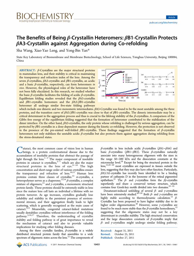
The Benefits of Being β-Crystallin Heteromers: βB1-Crystallin Protects βA3-Crystallin against...
11
The Benefits of Being β-Crystallin Heteromers: βB1-Crystallin Protects βA3-Crystallin against Aggregation during Co-refolding Sha Wang, Xiao-Yao Leng, and Yong-Bin Yan* State Key Laboratory of Biomembrane and Membrane Biotechnology, School of Life Sciences, Tsinghua University, Beijing 100084, China ABSTRACT: β-Crystallins are the major structural proteins in mammalian lens, and their stability is critical in maintaining the transparency and refraction index of the lens. Among the seven β-crystallins, βA3-crystallin and βB1-crystallin, an acidic and a basic β-crystallin, respectively, can form heteromers in vivo. However, the physiological roles of the heteromer have not been fully elucidated. In this research, we studied whether the basic β-crystallin facilitates the folding of acidic β-crystallin. Equilibrium folding studies revealed that the βA3-crystallin and βB1-crystallin homomers and the βA3/βB1-crystallin heteromer all undergo similar five-state folding pathways which include one dimeric and two monomeric intermediates. βA3-Crystallin was found to be the most unstable among the three proteins, and the transition curve of βA3/βB1-crystallin was close to that of βB1-crystallin. The dimeric intermediate may be a critical determinant in the aggregation process and thus is crucial to the lifelong stability of the β-crystallins. A comparison of the Gibbs free energy of the equilibrium folding suggested that the formation of heteromer contributed to the stabilization of the dimer interface. On the other hand, βA3-crystallin, the only protein whose refolding is challenged by serious aggregation, can be protected by βB1-crystallin in a dose-dependent manner during the kinetic co-refolding. However, the protection is not observed in the presence of the pre-existed well-folded βB1-crystallin. These findings suggested that the formation of β-crystallin heteromers not only stabilizes the unstable acidic β-crystallin but also protects them against aggregation during refolding from the stress-denatured states. C ataract, the most common cause of vision loss in human beings, is a protein conformational disease due to the accumulation of insoluble proteins that obstruct the passage of light through the lens. 1−3 The major component of insoluble proteins in cataract is crystallins, 3−7 which are also the major structural proteins in the lens of eye. 8−10 The high concentration and short-range order of various crystallins ensure the transparency and refraction of lens. 10,11 Human lens proteins contain three classes of crystallin: 12 α-crystallin, a heteropolymer serves as a chaperone; 13,14 β-crystallin, a complex mixture of oligomers; 15 and γ-crystallin, a monomeric structural protein family. These proteins should be extremely stable in lens since the mature lens cell lasts an individual a lifetime with no protein turnover. As age increases, crystallins gradually lose solubility and stability in the persistent presence of environ- mental stresses, and their aggregation finally leads to light scattering, which is generally recognized as the main cause of cataract. 3,11 Meanwhile, congenital cataract-linked mutations usually destabilize crystallins without interference of the folding pathway. 3,16,17 Therefore, the understanding of crystallin stability and folding pathway is of great importance to reveal the mechanism of cataract and also to provide valuable implications for studying other folding diseases. Among the three crystallin families, β-crystallin is a widely distributed structural protein family that assembles to a wide spectrum of oligomeric states across the lens. 12 The components of β-crystallin in lens include acidic β-crystallins (βA1−βA4) and basic β-crystallins (βB1−βB3). These β-crystallins naturally associate into many heterogeneous oligomers with the sizes in the range 50−200 kDa and the dissociation constants at the micromolar level. 18 Except for being the structural protein in the lens, 15,18−20 most crystallins are expressed in tissues outside the lens, suggesting that they may also have other function. Particularly, βA3/A1-crystallin has recently been identified to be a binding partner of cathepsin D in the lysosome of the retinal pigmented epithelium. 21 The β- and γ-crystallins form the βγ-crystallin superfamily and share a conserved tertiary structure, which contains four Greek-key motifs divided into two domains. 22−25 Denaturant-induced unfolding of several β- and γ-crystallins have been extensively studied. 26−29 Both β- and γ-crystallins are highly stable according to thermodynamic calculation. 12 β- Crystallin has been proposed to have higher stability due to its higher order oligomerization. 26 However, some γ-crystallins are found to be much more stable than the β-crystallin homodimers, 12 suggesting that the oligomeric states may not be a key determinant in crystallin stability. The high structural conservation and the large dissociation constants of β-crystallin imply that β- and γ-crystallins might undergo similar folding pathway. Received: August 31, 2011 Revised: October 25, 2011 Published: October 27, 2011 Article pubs.acs.org/biochemistry © 2011 American Chemical Society 10451 dx.doi.org/10.1021/bi201375p | Biochemistry 2011, 50, 10451−10461
Transcript of The Benefits of Being β-Crystallin Heteromers: βB1-Crystallin Protects βA3-Crystallin against...
The Benefits of Being β-Crystallin Heteromers: βB1-Crystallin ProtectsβA3-Crystallin against Aggregation during Co-refoldingSha Wang, Xiao-Yao Leng, and Yong-Bin Yan*
State Key Laboratory of Biomembrane and Membrane Biotechnology, School of Life Sciences, Tsinghua University, Beijing 100084,China
ABSTRACT: β-Crystallins are the major structural proteinsin mammalian lens, and their stability is critical in maintainingthe transparency and refraction index of the lens. Among theseven β-crystallins, βA3-crystallin and βB1-crystallin, an acidicand a basic β-crystallin, respectively, can form heteromers invivo. However, the physiological roles of the heteromer havenot been fully elucidated. In this research, we studied whetherthe basic β-crystallin facilitates the folding of acidic β-crystallin.Equilibrium folding studies revealed that the βA3-crystallinand βB1-crystallin homomers and the βA3/βB1-crystallinheteromer all undergo similar five-state folding pathwayswhich include one dimeric and two monomeric intermediates. βA3-Crystallin was found to be the most unstable among the threeproteins, and the transition curve of βA3/βB1-crystallin was close to that of βB1-crystallin. The dimeric intermediate may be acritical determinant in the aggregation process and thus is crucial to the lifelong stability of the β-crystallins. A comparison of theGibbs free energy of the equilibrium folding suggested that the formation of heteromer contributed to the stabilization of thedimer interface. On the other hand, βA3-crystallin, the only protein whose refolding is challenged by serious aggregation, can beprotected by βB1-crystallin in a dose-dependent manner during the kinetic co-refolding. However, the protection is not observedin the presence of the pre-existed well-folded βB1-crystallin. These findings suggested that the formation of β-crystallinheteromers not only stabilizes the unstable acidic β-crystallin but also protects them against aggregation during refolding fromthe stress-denatured states.
Cataract, the most common cause of vision loss in humanbeings, is a protein conformational disease due to the
accumulation of insoluble proteins that obstruct the passage oflight through the lens.1−3 The major component of insolubleproteins in cataract is crystallins,3−7 which are also the majorstructural proteins in the lens of eye.8−10 The highconcentration and short-range order of various crystallins ensurethe transparency and refraction of lens.10,11 Human lensproteins contain three classes of crystallin:12 α-crystallin, aheteropolymer serves as a chaperone;13,14 β-crystallin, a complexmixture of oligomers;15 and γ-crystallin, a monomeric structuralprotein family. These proteins should be extremely stable in lenssince the mature lens cell lasts an individual a lifetime with noprotein turnover. As age increases, crystallins gradually losesolubility and stability in the persistent presence of environ-mental stresses, and their aggregation finally leads to lightscattering, which is generally recognized as the main cause ofcataract.3,11 Meanwhile, congenital cataract-linked mutationsusually destabilize crystallins without interference of the foldingpathway.3,16,17 Therefore, the understanding of crystallinstability and folding pathway is of great importance to revealthe mechanism of cataract and also to provide valuableimplications for studying other folding diseases.Among the three crystallin families, β-crystallin is a widely
distributed structural protein family that assembles to a widespectrum of oligomeric states across the lens.12 The components of
β-crystallin in lens include acidic β-crystallins (βA1−βA4) andbasic β-crystallins (βB1−βB3). These β-crystallins naturallyassociate into many heterogeneous oligomers with the sizes inthe range 50−200 kDa and the dissociation constants at themicromolar level.18 Except for being the structural protein in thelens,15,18−20 most crystallins are expressed in tissues outside thelens, suggesting that they may also have other function. Particularly,βA3/A1-crystallin has recently been identified to be a bindingpartner of cathepsin D in the lysosome of the retinal pigmentedepithelium.21 The β- and γ-crystallins form the βγ-crystallinsuperfamily and share a conserved tertiary structure, whichcontains four Greek-key motifs divided into two domains.22−25
Denaturant-induced unfolding of several β- and γ-crystallinshave been extensively studied.26−29 Both β- and γ-crystallins arehighly stable according to thermodynamic calculation.12 β-Crystallin has been proposed to have higher stability due to itshigher order oligomerization.26 However, some γ-crystallins arefound to be much more stable than the β-crystallin homodimers,12
suggesting that the oligomeric states may not be a keydeterminant in crystallin stability. The high structural conservationand the large dissociation constants of β-crystallin imply thatβ- and γ-crystallins might undergo similar folding pathway.
Received: August 31, 2011Revised: October 25, 2011Published: October 27, 2011
Article
pubs.acs.org/biochemistry
© 2011 American Chemical Society 10451 dx.doi.org/10.1021/bi201375p |Biochemistry 2011, 50, 10451−10461
The urea- or guanidine hydrochloride (GdnHCl)-inducedunfolding of β- and γ-crystallins is suggested to follow eithera two-state or a three-state model,12 but the unfolding pathwayof βB1-crystallin is proposed to contain more than oneintermediate.28 It is unclear yet whether the difference in thefolding mechanism is caused by the dissimilarity in the proteinsor the accuracy of experiments. Moreover, the urea-inducedunfolding of βA3-crystallin was found to have a relatively lowerthermodynamic stability and a higher aggregation tendencythan βB1-crystallin.22 However, although all β-crystallins,except for βB2-crystallin, form heteromers in the lens, little isknown about the folding of naturally formed heterogeneousoligomers of β-crystallin. The details of the structural changesduring β-crystallin folding and unfolding also remain elusive.In human lens, both α- and β-crystallins mainly exist as
heteromers, and several previous studies have tried to explore thebenefits of the formation of the evolution-selected β-crystallinheteromers. It has been shown that the acidic β-crystallins arestabilized in the heteromers when subjected to heat- or urea-induced denaturation.19,22,30,31 Moreover, βB1- and βB3-crystallingenes are expressed early during lens development.12 Aninteresting question is whether the pre-existed basic β-crystallinsfavors the folding of acidic β-crystallins. In this research, thisquestion was addressed by comparing the GdnHCl-inducedunfolding and refolding pathways of βA3- and βB1-crystallins aswell as their heteromer βA3/βB1-crystallin. The βA3- and βB1-crystallins were chosen as the model system according to theirhigh expression level in human lens and their abundance in theβ-crystallin heteromers. Our results clearly showed that the threeβ-crystallins all follow multistate folding processes, among whichthe folding process of βA3/βB1-crystallin is similar to that of βB1.Interestingly, we found that βB1-crystallin can successfully protectβA3-crystallin against aggregation during co-refolding, but thepre-existed well-folded βB1-crystallin could not.
■ MATERIALS AND METHODSChemicals. GdnHCl, 8-anilino-1-naphthalenesulfonic acid
(ANS), and sodium dodecyl sulfate (SDS) were purchasedform Sigma Chemical Co. All other chemicals were localproducts of analytical grade.Protein Expression and Purification. The His-tagged
recombinant βB1- and βA3-crystallins were overexpressed inEscherichia coli BL21 with pET28a plasmid and purified by Ni-NTA affinity column and gel filtration chromatography asdescribed previously.31 The βA3/βB1-crystallin heteromer wasprepared by incubating equimolar of βB1-crystallin and βA3-crystallin at 37 °C for 2 h, and the formation of heteromerβA3/βB1-crystallin was identified by size-exclusion chromatog-raphy (SEC) and SDS-PAGE analysis. The protein concen-tration was determined according to the Bradford method usingbovine serum albumin as a standard.32
Protein Unfolding and Refolding. The unfolding experi-ments of the three proteins were performed by incubating thepurified proteins in buffer A (20 mM sodium phosphate buffer,0.15 M NaCl, 1 mM EDTA, 1 mM DTT, pH 7.2) containingdifferent concentrations of GdnHCl for 16 h at 4 °C. As for therefolding experiments, the purified proteins were fullydenatured in 6 M GdnHCl for 12 h. Then the equilibriumrefolding was carried out by dilution of the denatured samplesin buffer A with the final concentrations of GdnHCl variedfrom 0.2 to 6 M at 25 °C for 24 h. The final proteinconcentration was 0.2 mg/mL for both the unfolding andrefolding experiments.
Spectroscopic Experiments. All spectroscopic experimentswere performed at 25 °C with a protein concentration of 0.2 mg/mL.The fluorescence spectra were measured on an F-2500fluorescence spectrophotometer (Hitachi Ltd., Tokyo, Japan)with a 5 nm slit width for both excitation and emission. Theintrinsic fluorescence was measured with an excitation wavelengthof 295 nm and an emission wavelength ranging from 300 to 400nm. The extrinsic ANS fluorescence of crystallins at variousconcentration of GdnHCl was monitored with an excitationwavelength of 380 nm and an emission wavelength ranging from400 to 600 nm. The final concentration of ANS was 20 μM. Thecircular dichroism (CD) spectra were recorded on a Jasco J-715spectropolarimeter (Jasco Corp., Tokyo, Japan) with a 1 mm pathlength cell over a wavelength range of 190−250 nm. The resultantspectra were obtained by the subtraction of the spectra of thecorresponding buffers. The parameter A, which is the ratio of theintensity at 320 nm to that at 365 nm of the intrinsic fluorescence(I320/I365), is the characteristic of the shape and position of thefluorescence spectrum.33,34 The phase diagram analysis of theintrinsic fluorescence, which is a sensitive tool to detect the foldingintermediates,35 was carried out as described previously.36,37
Aggregation Experiments. Details regarding the aggrega-tion experiments were the same as those described previously.38
The time-course aggregation during refolding was recordedimmediately after the refolding was initiated by a fast manualdilution of the GdnHCl-denatured β-crystallins into buffer A at25 °C. It has been proposed that the light scattering intensity isproportional to the amount of the protein in the aggregatedform,39 thus the turbidity of the proteins during unfolding wasmonitored by recording the absorbance at 400 nm on anUltraspec 4300 pro UV/vis spectrophotometer from AmershamPharmacia Biotech (Uppsala, Sweden). The final proteinconcentration was 0.1 or 0.2 mg/mL.Size-Exclusion Chromatography. The size-exclusion
chromatography (SEC) analysis of the samples was the same asdescribed previously.40 In brief, the gel filtration experiments werecarried out on a Superdex 75HR 10/30 column on an AKTAFPLC (Amersham Pharmacia Biotech, Sweden). The column waspre-equilibrated with buffer A containing the given concentrationsof GdnHCl, and then about 100 μL protein solutions were injectedinto the column. All samples were run at a flow rate of 0.5 mL/minat 16 °C. The elution peaks were collected for further identificationby SDS−polyacrylamide gel electrophoresis (SDS-PAGE) using12.5% separating gel in the reducing conditions. Silver staining wasperformed according to the standard protocol.41
Data Analysis. The equilibrium folding transition curvesmonitored by parameter A was analyzed according to a three-state model as characterized previously.26,42
(1)
where N2 is the native protein, I is the monomeric intermediatestate, and U is the fully unfolded state. The equilibriumconstants for the three transitions in eq 1 are
(2)
and the mole fractions of each species are
(3)
(4)
Biochemistry Article
dx.doi.org/10.1021/bi201375p |Biochemistry 2011, 50, 10451−1046110452
The quadratic equation (eq 4) is solved, which yield f I:
(5)
in which
The free energy changes can be expressed as a function of theGdnHCl concentration:
(6)
(7)
(8)
The parameter A data are described as the following equation:
(9)
where y is the global parameter A data; yN and yU are the interceptof the initial, intermediate, and final baselines, respectively. mN andmU are the slopes of the initial, intermediate, and final transitions,respectively. yI represents the fraction of the intermediate calculatedby the data, which is assumed not to vary within the transition zoneto minimize the number of parameters in the fit. The foldingprofiles were fitted to eq 9 with the regression wizard of SigmaPlot,followed by the nonlinear least-squares algorithm. The initial
parameters were provided, and the regression was run until aminimum R 2 was reached. A fit was judged to be satisfactory bypassing of both the normality and constant variance tests.
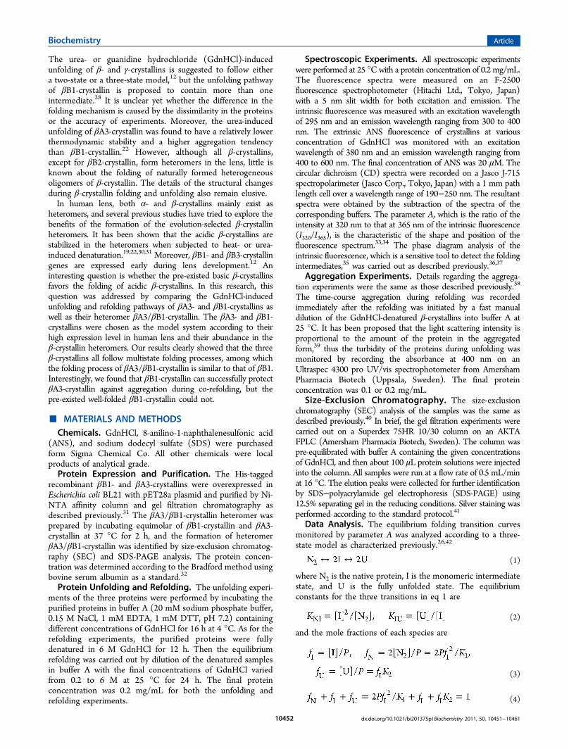
■ RESULTSMultistate Equilibrium Unfolding of βA3-, βB1-, and
βA3/βB1-Crystallins. β-Crystallin samples with increasingconcentration of GdnHCl were incubated at 4 °C overnight toreach the equilibrium. CD, Trp fluorescence, and ANS were usedto monitor the secondary and tertiary structural change duringunfolding of βA3-, βB1-, and βA3/βB1-crystallins. The folding ofthe three proteins was independent of protein concentration, asindicated by the almost indistinguishable transition curves fromsamples with a protein concentration of 0.2 and 1 mg/mL (datanot shown). As shown in Figure 1A, the absolute value of theCD signals increased slightly for all three proteins when theGdnHCl concentration rose from 0 to 1.6 M, which implied thatlow concentration of GdnHCl could rearrange the structure andinduce non-native regular secondary structures. Similar observa-tions have also been reported for the unfolding of the otherproteins.40,43,44 Gradual loss of the regular secondary structuresbegan from 1.6 M GdnHCl and reached the fully denatured stateat around 5 M. The transition curve of βA3/βB1-crystallin wasvery similar to that of βA3-crystallin, except for a faster decreaseof the ellipticity at high concentration of GdnHCl.Each monomer of βA3- and βB1-crystallin contains 10 and 8
Trp residues, respectively, rendering the change of intrinsic Trpfluorescence a sensitive probe of the tertiary structural changes ofβ-crystallin.45−47 The maximum emission wavelengths of the Trp
Figure 1. Unfolding transition curves of βA3- (filled cycles), βB1- (open square), and βA3/βB1-crystallins (open triangle) monitored by theellipticity at 222 nm (A), the emission maximum wavelength (B), and the intensity (C) of the intrinsic Trp fluorescence and the ANS fluorescenceintensity at 470 nm (D). For the unfolding experiments, the protein was incubated in buffer A in the presence of various concentrations of GdnHClfor 12 h at 4 °C. The final protein concentration was 0.2 mg/mL. All spectroscopic experiments were carried out at 25 °C.
Biochemistry Article
dx.doi.org/10.1021/bi201375p |Biochemistry 2011, 50, 10451−1046110453
fluorescence (Emax) of the native βB1- and βA3-crystallins werelocated at 334.5 and 336 nm, while the formation of theheteromer slightly blue-shifted the Emax value to about 333 nm.This observation is quite consistent with those in previouspublications,22,31 which suggested that some of the exposed Trpfluorophores became buried in the heteromer. The dependenceof the Emax on the concentration of GdnHCl clearly revealed therelative stability of the three proteins: βB1-crystallin > βA3/βB1-crystallins > βA3-crystallin (Figure 1B). Although the heteromerhad a similar behavior to βB1-crystallin at low denaturantconcentrations, it experienced a much more abrupt transitionwhen GdnHCl concentration rose above 3 M. On the other hand,the heteromer also shows a completely different pattern in thechange of emission intensity upon unfolding. The emissionintensities of the intrinsic fluorescence of βA3- and βB1-crystallinsslightly decreased and reached a minimum at 1.2 M GdnHCl,while that of βA3/βB1-crystallin increases by 10% at lowGdnHCl concentrations (Figure 1C). The main transitionoccurred at similar GdnHCl concentrations when monitored byintrinsic fluorescence position or intensity. Interestingly, anadditional transition with a significant increase of the Trpfluorescence was observed for all three proteins at high GdnHClconcentrations above 5 M. This phenomenon is seldom observedin previous folding studies. The unfolding states exhibited themaximum emission intrinsic fluorescence intensity, indicating thata significant quenching of the intrinsic fluorescence in the nativestates of β-crystallins. These observations implied that severaldistinct intermediates might be populated during the unfolding ofthe three proteins and the unfolding behavior of the heterodimerβA3/βB1-crystallin could not be simply explained by the sum ofits two component monomers.
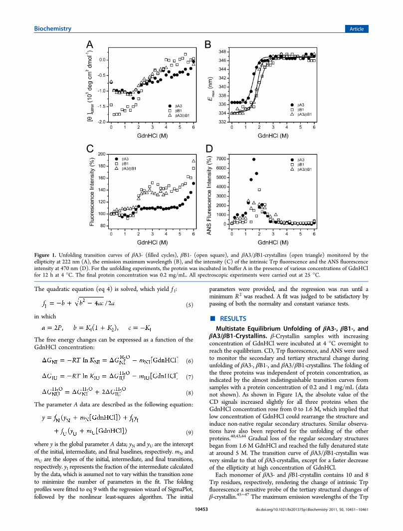
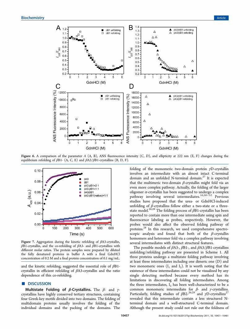
ANS is a widely used fluorescence probe to monitor thehydrophobic exposure of the proteins.48 As presented in Figure1D, the ANS intensity of βA3-crystallin reached its maximum at1.6 M GdnHCl, indicating the existence of an intermediate statewith a significant increase in hydrophobic exposure. Notably, theCD signals had only minor changes at 1.6 M GdnHCl, implyingthe molten globular (MG)-like property of this intermediate state.In contrast, the ANS intensities of βB1- and βA3/βB1-crystallins(especially βA3/βB1-crystallin) increased to a much smaller extentthan that of βA3-crystallin. Moreover, the ANS intensity peaks ofβB1- and βA3/βB1-crystallins do not agree with a simpletransition containing only one intermediate. In fact, more thanone intermediate with dissimilar hydrophobic exposure propertiesmight be accumulated at around 1−2 M GdnHCl. Parameter A isa precise method to analyze the change in the position and shapeof the intrinsic Trp fluorescence spectra.34 As shown in Figure 2A,the transition curve from parameter A of βA3-crystallin indicatedthat the change of the intrinsic fluorescence was a two-stageprocess involving one intermediate, while βB1- and βA3/βB1-crystallins showed a much more complex pattern. The phasediagram analysis of Trp fluorescence, a sensitive tool tocharacterize the folding intermediate(s),35−37,49 was also used todetermine the position where the intermediate state(s) appeared.The joint position of lines in the phase diagram indicates theappearance of an intermediate at the corresponding condition. Asshown in Figure 2B−D, all of the three proteins showed nonlineardiagrams. Consistent with the above analysis, the folding pathwayof the heteromer was similar to that of βB1-crystallin but was quitedifferent from that of βA3-crystallin. All three proteins had twocommon intermediates appeared at around 1.0 and 3.4 MGdnHCl, respectively. The major difference was that βA3-crystallin
Figure 2. Parameter A (A) and phase diagram (B, C, D) analysis of the intrinsic fluorescence spectra of βA3- (cycle), βB1- (square), and βA3/βB1-crystallins (triangle) during unfolding. The parameter A was obtained by dividing the fluorescence intensity at 320 nm (I320) by that at 365 nm (I365).The phase diagram was constructed by monitoring the changes of I365 as a function of I320.
Biochemistry Article
dx.doi.org/10.1021/bi201375p |Biochemistry 2011, 50, 10451−1046110454
lacked the accumulation of the intermediate between 0.8 and3.2 M GdnHCl. The absence of this intermediate in the phasediagram of βA3-crystallin might be caused the failure of thismethod in analyzing irreversible transitions, as suggested byprevious researches.35 Actually, an intermediate of βA3-crystallin appeared at around 1.6 M with the highest ANSfluorescence intensity, the lowest Trp fluorescence intensity,the nearly fully solvent-exposed Trp fluorophores, and thelargest CD signal (Figure 1).To determine the quaternary structural change and the
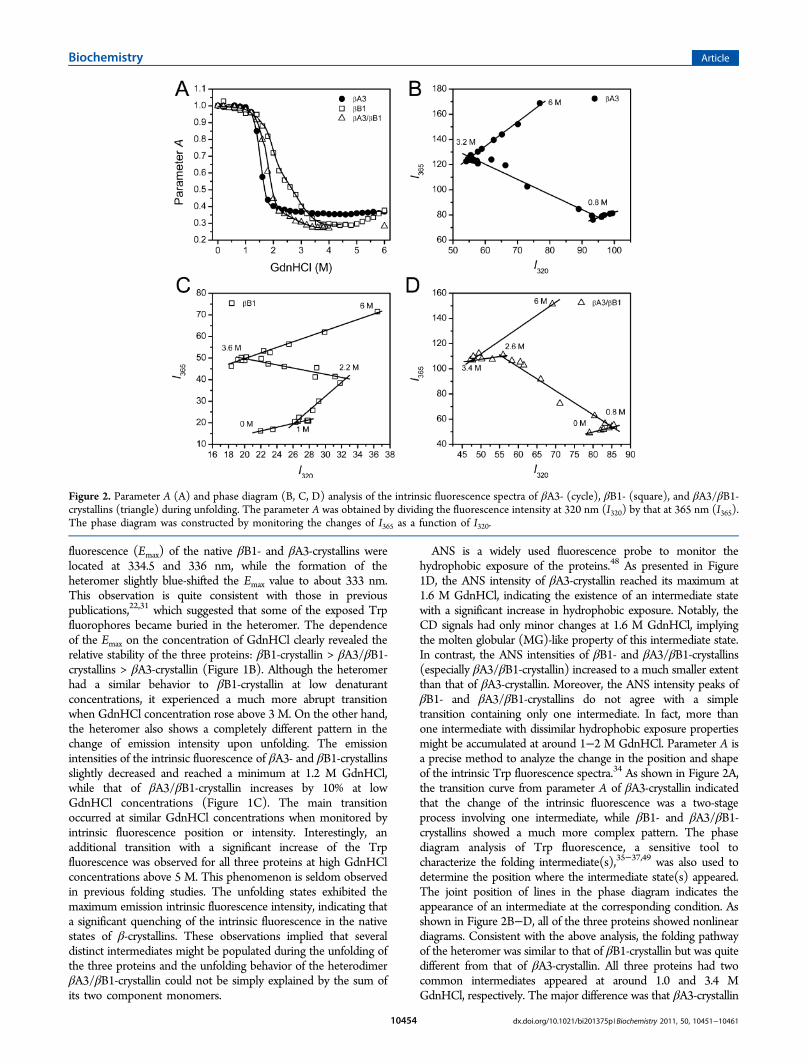
association forms of the intermediates during GdnHCl-inducedunfolding, SEC analysis was performed for samples denaturedunder different concentrations of GdnHCl. The native βA3-and βA3/βB1-crystallin were dominated by the dimeric state indilute solutions according to the elution volumes, while βB1-crystallin existed in a dimer−monomer equilibrium (Figure 3).
This is in good agreement with the previous observations. TheSEC analysis was performed under the same conditions for thethree proteins to facilitate the identifying of the dissociation ofthe heteromer. As shown in Figure 3C, both of the 1.0 and 1.6M profiles of βA3/βB1-crystallin contained two peaks. . As theconcentration of GdnHCl rose to 2.4 M, only one elution peakcould be observed for βA3/βB1-crystallin. The target peakswere collected and analyzed by SDS-PAGE and silver staining41
to verify the constitutions of the peaks (inset of Figure 3C).Although the band for peak M was too weak to be detected dueto the very low protein concentration, both of the peaks D andM in the 1.0 M profile contained βB1- and βA3-crystallins.Since the elution volume of the native βA3/βB1-crystallinheterodimer was between that of D and M, these two peakscould be assigned as heterodimer and a mixture of βA3- andβB1-crystallin monomer, respectively. As for the 1.6 M profile,the peak eluted at 14 mL occupied at the same position as thoseof βB1- and βA3-crystallins, indicating that this peak originatedfrom the dissociated βB1- and βA3-crystallin monomers. Thesingle peak in the 2.4 M GdnHCl profile was a mixture of βB1-and βA3-crystallin monomers, suggesting a complete dissoci-ation at this concentration of GdnHCl for all of the threeproteins. Similar to previous results,26,40 herein we also foundthe gradual decreases of elution volume during unfolding ofβ-crystallins. This might be caused by the competing effectsbetween dissociation and unfolding, of which one decreasedand the other increased the apparent molecular size of theproteins. Nonetheless, βB1- and βA3-crystallins clearlydissociated into monomers at GdnHCl concentrations above1.6 M, while βA3/βB1-crystallin fully dissociated in 2.4 MGdnHCl.Partial-Reversible Refolding of βA3-, βB1-, and βA3/
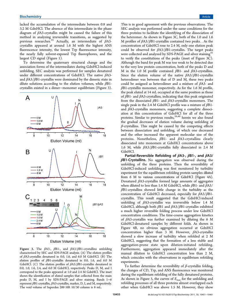
βB1-Crystallins. No aggregation was observed during theunfolding of the three proteins. Then the reversibility ofGdnHCl-induced unfolding was first monitored by turbidityexperiment for the equilibrium refolding protein samples dilutedfrom 6 M to various concentrations of GdnHCl (Figure 4A).Denatured βA3-crystallin formed large amounts of aggregateswhen diluted to less than 1.4 M GdnHCl, while βB1- and βA3/βB1-crystallins showed little change in the turbidity as theconcentration of GdnHCl decreased, especially for βA3/βB1-crystallin. This result suggested that the GdnHCl-inducedunfolding of βA3-crystallin was irreversible below 1.4 MGdnHCl, although both βB1 and βA3/βB1-crystallin exhibiteda much higher reversible folding process under low GdnHClconcentration conditions. The time-course aggregation kineticsof βA3-crystallin was further examined by diluting the 6 MGdnHCl-denatured samples by different folds. As shown inFigure 4B, no obvious aggregation occurred at GdnHClconcentration higher than 3 M. However, βA3-crystallinshowed a slow increase of turbidity when refolded at 2 MGdnHCl, suggesting that the formation of a less stable andaggregation-prone state upon dilution-initiated refolding.Furthermore, aggregation appeared immediately after themanual dilution to GdnHCl concentration less than 2 M,which coincides with the observations in equilibrium refoldingexperiments.To further determine the reversibility of β-crystallin folding,
the changes of CD, Trp, and ANS fluorescence was monitoredduring the equilibrium refolding of the fully denatured proteins.As shown in Figure 5, the curves of Emax for the unfolding andrefolding processes of all three proteins almost overlapped eachother when GdnHCl was above 1.5 M. However, they show
Figure 3. The βA3-, βB1-, and βA3/βB1-crystallins unfoldingcharacterized by SEC and SDS-PAGE analysis. (A) The elution profilesof βA3-crystallin denatured in 0.0, 1.6, and 6.0 M GdnHCl. (B) Theelution profiles of βB1-crystallin denatured in 0.0, 1.6, and 6.0 MGdnHCl. (C) The elution profiles of βA3/βB1-crystallin denatured in0.0, 1.0, 1.6, 2.4, and 6.0 M GdnHCl, respectively. Peaks D, M, and Icorrespond to the peaks appeared at 1.0 and 2.4 M GdnHCl. The insetshows the identification of eluted samples that collected from the mainpeaks D, M, and I by SDS-PAGE and silver staining. Bands 1−6represent βB1-crystallin, βA3-crystallin, marker, D, I, and M, respectively.The void volume of Superdex 200 HR 10/30 column is 8 mL.
Biochemistry Article
dx.doi.org/10.1021/bi201375p |Biochemistry 2011, 50, 10451−1046110455
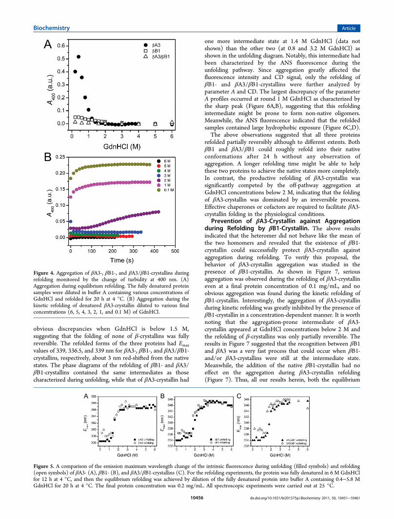
obvious discrepancies when GdnHCl is below 1.5 M,suggesting that the folding of none of β-crystallins was fullyreversible. The refolded forms of the three proteins had Emaxvalues of 339, 336.5, and 339 nm for βA3-, βB1-, and βA3/βB1-crystallins, respectively, about 3 nm red-shifted from the nativestates. The phase diagrams of the refolding of βB1- and βA3/βB1-crystallins contained the same intermediates as thosecharacterized during unfolding, while that of βA3-crystallin had
one more intermediate state at 1.4 M GdnHCl (data notshown) than the other two (at 0.8 and 3.2 M GdnHCl) asshown in the unfolding diagram. Notably, this intermediate hadbeen characterized by the ANS fluorescence during theunfolding pathway. Since aggregation greatly affected thefluorescence intensity and CD signal, only the refolding ofβB1- and βA3/βB1-crystallins were further analyzed byparameter A and CD. The largest discrepancy of the parameterA profiles occurred at round 1 M GdnHCl as characterized bythe sharp peak (Figure 6A,B), suggesting that this refoldingintermediate might be prone to form non-native oligomers.Meanwhile, the ANS fluorescence indicated that the refoldedsamples contained large hydrophobic exposure (Figure 6C,D).The above observations suggested that all three proteins
refolded partially reversibly although to different extents. BothβB1 and βA3/βB1 could roughly refold into their nativeconformations after 24 h without any observation ofaggregation. A longer refolding time might be able to helpthese two proteins to achieve the native states more completely.In contrast, the productive refolding of βA3-crystallin wassignificantly competed by the off-pathway aggregation atGdnHCl concentrations below 2 M, indicating that the foldingof βA3-crystallin was dominated by an irreversible process.Effective chaperones or cofactors are required to facilitate βA3-crystallin folding in the physiological conditions.Prevention of βA3-Crystallin against Aggregation
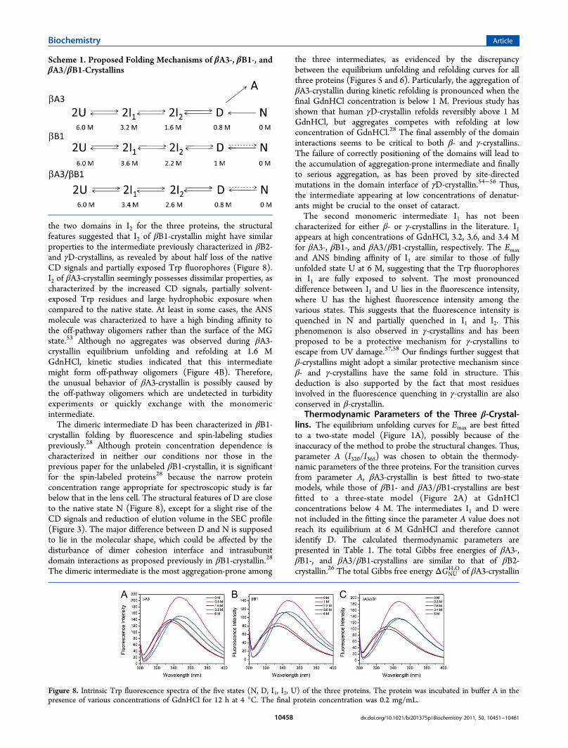
during Refolding by βB1-Crystallin. The above resultsindicated that the heteromer did not behave like the mean ofthe two homomers and revealed that the existence of βB1-crystallin could successfully protect βA3-crystallin againstaggregation during refolding. To verify this proposal, thebehavior of βA3-crystallin aggregation was studied in thepresence of βB1-crystallin. As shown in Figure 7, seriousaggregation was observed during the refolding of βA3-crystallineven at a final protein concentration of 0.1 mg/mL, and noobvious aggregation was found during the kinetic refolding ofβB1-crystallin. Interestingly, the aggregation of βA3-crystallinduring kinetic refolding was greatly inhibited by the presence ofβB1-crystallin in a concentration-dependent manner. It is worthnoting that the aggregation-prone intermediate of βA3-crystallin appeared at GdnHCl concentrations below 2 M andthe refolding of β-crystallins was only partially reversible. Theresults in Figure 7 suggested that the recognition between βB1and βA3 was a very fast process that could occur when βB1-and/or βA3-crystallins were still at the intermediate state.Meanwhile, the addition of the native βB1-crystallin had noeffect on the aggregation during βA3-crystallin refolding(Figure 7). Thus, all our results herein, both the equilibrium
Figure 4. Aggregation of βA3-, βB1-, and βA3/βB1-crystallins duringrefolding monitored by the change of turbidity at 400 nm. (A)Aggregation during equilibrium refolding. The fully denatured proteinsamples were diluted in buffer A containing various concentrations ofGdnHCl and refolded for 20 h at 4 °C. (B) Aggregation during thekinetic refolding of denatured βA3-crystallin diluted to various finalconcentrations (6, 5, 4, 3, 2, 1, and 0.1 M) of GdnHCl.
Figure 5. A comparison of the emission maximum wavelength change of the intrinsic fluorescence during unfolding (filled symbols) and refolding(open symbols) of βA3- (A), βB1- (B), and βA3/βB1-crystallins (C). For the refolding experiments, the protein was fully denatured in 6 M GdnHClfor 12 h at 4 °C, and then the equilibrium refolding was achieved by dilution of the fully denatured protein into buffer A containing 0.4−5.8 MGdnHCl for 20 h at 4 °C. The final protein concentration was 0.2 mg/mL. All spectroscopic experiments were carried out at 25 °C.
Biochemistry Article
dx.doi.org/10.1021/bi201375p |Biochemistry 2011, 50, 10451−1046110456
and the kinetic refolding, suggested the essential role of βB1-crystallin in efficient refolding of βA3-crystallin and the ratiodependence of this co-refolding.
■ DISCUSSIONMultistate Folding of β-Crystallins. The β- and γ-
crystallins have highly conserved tertiary structures, containingfour Greek-key motifs divided into two domains. The folding ofmultidomain proteins usually involves the folding of theindividual domains and the packing of the domains. The
folding of the monomeric two-domain protein γD-crystallininvolves an intermediate with an almost intact C-terminaldomain and an unfolded N-terminal domain.27 It is expectedthat the multimeric two-domain β-crystallin might fold via aneven more complex pathway. Actually, the folding of the largeroligomer α-crystallin has been suggested to undergo a complexpathway involving several intermediates.16,50−52 Previousstudies have proposed that the urea- or GdnHCl-inducedunfolding of β-crystallins follow either a two-state or a three-state model.26,29 The folding process of βB1-crystallin has beenreported to contain more than one intermediate using spin andfluorescence labeling as probes, respectively. However, theprobes would also affect the observed folding pathway ofproteins.28 In this research, we used comprehensive spectro-scopic analysis and found that both of the β-crystallinhomomers and heteromer fold via a complex pathway involvingseveral intermediates with distinct structural features.The possible models of βA3-, βB1-, and βA3/βB1-crystallins
unfolding/refolding pathway are presented in Scheme 1. Allthree proteins undergo a multistate folding pathway involvingat least three intermediates including one dimeric one (D) andtwo monomeric ones (I1 and I2). It is worth noting that theexistence of these intermediates could not be visualized by anysingle detecting method because every method has itslimitations in discovering all folding intermediates. Amongthe three intermediates, I2 has been well-characterized to be acommon monomeric intermediate for β- and γ-crystallins.Particularly, folding studies of βB2-26,29 and γD-crystallins27
revealed that this intermediate contain a less structured N-terminal domain and a well-structured C-terminal domain.Although the present study could not rule out the foldness of
Figure 6. A comparison of the parameter A (A, B), ANS fluorescence intensity (C, D), and ellipticity at 222 nm (E, F) changes during theequilibrium refolding of βB1- (A, C, E) and βA3/βB1-crystallins (B, D, F).
Figure 7. Aggregation during the kinetic refolding of βA3-crystallin,βB1-crystallin, and the co-refolding of βA3- and βB1-crystallins withdifferent molar ratios. The protein samples were prepared by dilutedthe fully denatured proteins in buffer A with a final GdnHClconcentration of 0.2 M and a final protein concentration of 0.1 mg/mL.
Biochemistry Article
dx.doi.org/10.1021/bi201375p |Biochemistry 2011, 50, 10451−1046110457
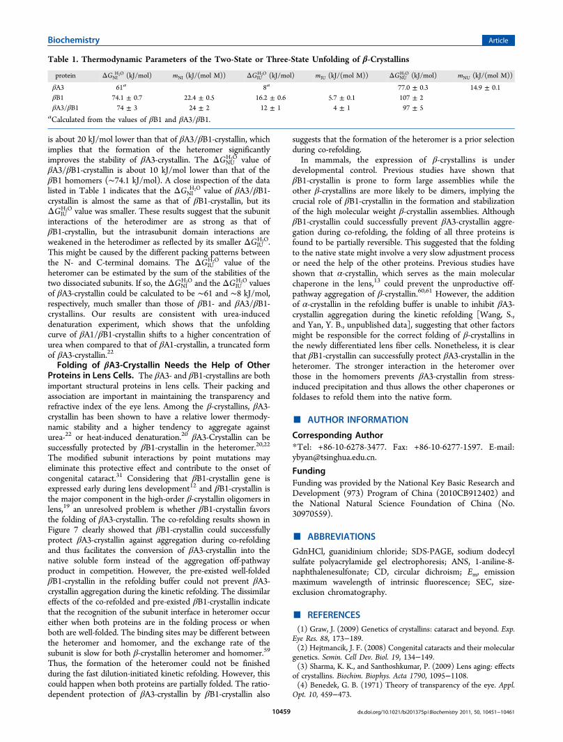
the two domains in I2 for the three proteins, the structuralfeatures suggested that I2 of βB1-crystallin might have similarproperties to the intermediate previously characterized in βB2-and γD-crystallins, as revealed by about half loss of the nativeCD signals and partially exposed Trp fluorophores (Figure 8).I2 of βA3-crystallin seemingly possesses dissimilar properties, ascharacterized by the increased CD signals, partially solvent-exposed Trp residues and large hydrophobic exposure whencompared to the native state. At least in some cases, the ANSmolecule was characterized to have a high binding affinity tothe off-pathway oligomers rather than the surface of the MGstate.53 Although no aggregates was observed during βA3-crystallin equilibrium unfolding and refolding at 1.6 MGdnHCl, kinetic studies indicated that this intermediatemight form off-pathway oligomers (Figure 4B). Therefore,the unusual behavior of βA3-crystallin is possibly caused bythe off-pathway oligomers which are undetected in turbidityexperiments or quickly exchange with the monomericintermediate.The dimeric intermediate D has been characterized in βB1-
crystallin folding by fluorescence and spin-labeling studiespreviously.28 Although protein concentration dependence ischaracterized in neither our conditions nor those in theprevious paper for the unlabeled βB1-crystallin, it is significantfor the spin-labeled proteins28 because the narrow proteinconcentration range appropriate for spectroscopic study is farbelow that in the lens cell. The structural features of D are closeto the native state N (Figure 8), except for a slight rise of theCD signals and reduction of elution volume in the SEC profile(Figure 3). The major difference between D and N is supposedto lie in the molecular shape, which could be affected by thedisturbance of dimer cohesion interface and intrasubunitdomain interactions as proposed previously in βB1-crystallin.28
The dimeric intermediate is the most aggregation-prone among
the three intermediates, as evidenced by the discrepancybetween the equilibrium unfolding and refolding curves for allthree proteins (Figures 5 and 6). Particularly, the aggregation ofβA3-crystallin during kinetic refolding is pronounced when thefinal GdnHCl concentration is below 1 M. Previous study hasshown that human γD-crystallin refolds reversibly above 1 MGdnHCl, but aggregates competes with refolding at lowconcentration of GdnHCl.28 The final assembly of the domaininteractions seems to be critical to both β- and γ-crystallins.The failure of correctly positioning of the domains will lead tothe accumulation of aggregation-prone intermediate and finallyto serious aggregation, as has been proved by site-directedmutations in the domain interface of γD-crystallin.54−56 Thus,the intermediate appearing at low concentrations of denatur-ants might be crucial to the onset of cataract.The second monomeric intermediate I1 has not been
characterized for either β- or γ-crystallins in the literature. I1appears at high concentrations of GdnHCl, 3.2, 3.6, and 3.4 Mfor βA3-, βB1-, and βA3/βB1-crystallin, respectively. The Emaxand ANS binding affinity of I1 are similar to those of fullyunfolded state U at 6 M, suggesting that the Trp fluorophoresin I1 are fully exposed to solvent. The most pronounceddifference between I1 and U lies in the fluorescence intensity,where U has the highest fluorescence intensity among thevarious states. This suggests that the fluorescence intensity isquenched in N and partially quenched in I1 and I2. Thisphenomenon is also observed in γ-crystallins and has beenproposed to be a protective mechanism for γ-crystallins toescape from UV damage.57,58 Our findings further suggest thatβ-crystallins might adopt a similar protective mechanism sinceβ- and γ-crystallins have the same fold in structure. Thisdeduction is also supported by the fact that most residuesinvolved in the fluorescence quenching in γ-crystallin are alsoconserved in β-crystallin.Thermodynamic Parameters of the Three β-Crystal-
lins. The equilibrium unfolding curves for Emax are best fittedto a two-state model (Figure 1A), possibly because of theinaccuracy of the method to probe the structural changes. Thus,parameter A (I320/I365) was chosen to obtain the thermody-namic parameters of the three proteins. For the transition curvesfrom parameter A, βA3-crystallin is best fitted to two-statemodels, while those of βB1- and βA3/βB1-crystallins are bestfitted to a three-state model (Figure 2A) at GdnHClconcentrations below 4 M. The intermediates I1 and D werenot included in the fitting since the parameter A value does notreach its equilibrium at 6 M GdnHCl and therefore cannotidentify D. The calculated thermodynamic parameters arepresented in Table 1. The total Gibbs free energies of βA3-,βB1-, and βA3/βB1-crystallins are similar to that of βB2-crystallin.26 The total Gibbs free energy ΔGNU
H2O of βA3-crystallin
Scheme 1. Proposed Folding Mechanisms of βA3-, βB1-, andβA3/βB1-Crystallins
Figure 8. Intrinsic Trp fluorescence spectra of the five states (N, D, I1, I2, U) of the three proteins. The protein was incubated in buffer A in thepresence of various concentrations of GdnHCl for 12 h at 4 °C. The final protein concentration was 0.2 mg/mL.
Biochemistry Article
dx.doi.org/10.1021/bi201375p |Biochemistry 2011, 50, 10451−1046110458
is about 20 kJ/mol lower than that of βA3/βB1-crystallin, whichimplies that the formation of the heteromer significantlyimproves the stability of βA3-crystallin. The ΔGNU
H2O value ofβA3/βB1-crystallin is about 10 kJ/mol lower than that of theβB1 homomers (∼74.1 kJ/mol). A close inspection of the datalisted in Table 1 indicates that the ΔGNI
H2O value of βA3/βB1-crystallin is almost the same as that of βB1-crystallin, but itsΔGIU
H2O value was smaller. These results suggest that the subunitinteractions of the heterodimer are as strong as that ofβB1-crystallin, but the intrasubunit domain interactions areweakened in the heterodimer as reflected by its smaller ΔGIU
H2O.This might be caused by the different packing patterns betweenthe N- and C-terminal domains. The ΔGIU
H2O value of theheteromer can be estimated by the sum of the stabilities of thetwo dissociated subunits. If so, the ΔGNI
H2O and the ΔGIUH2O values
of βA3-crystallin could be calculated to be ∼61 and ∼8 kJ/mol,respectively, much smaller than those of βB1- and βA3/βB1-crystallins. Our results are consistent with urea-induceddenaturation experiment, which shows that the unfoldingcurve of βA1/βB1-crystallin shifts to a higher concentration ofurea when compared to that of βA1-crystallin, a truncated formof βA3-crystallin.22
Folding of βA3-Crystallin Needs the Help of OtherProteins in Lens Cells. The βA3- and βB1-crystallins are bothimportant structural proteins in lens cells. Their packing andassociation are important in maintaining the transparency andrefractive index of the eye lens. Among the β-crystallins, βA3-crystallin has been shown to have a relative lower thermody-namic stability and a higher tendency to aggregate againsturea-22 or heat-induced denaturation.20 βA3-Crystallin can besuccessfully protected by βB1-crystallin in the heteromer.20,22
The modified subunit interactions by point mutations mayeliminate this protective effect and contribute to the onset ofcongenital cataract.31 Considering that βB1-crystallin gene isexpressed early during lens development12 and βB1-crystallin isthe major component in the high-order β-crystallin oligomers inlens,19 an unresolved problem is whether βB1-crystallin favorsthe folding of βA3-crystallin. The co-refolding results shown inFigure 7 clearly showed that βB1-crystallin could successfullyprotect βA3-crystallin against aggregation during co-refoldingand thus facilitates the conversion of βA3-crystallin into thenative soluble form instead of the aggregation off-pathwayproduct in competition. However, the pre-existed well-foldedβB1-crystallin in the refolding buffer could not prevent βA3-crystallin aggregation during the kinetic refolding. The dissimilareffects of the co-refolded and pre-existed βB1-crystallin indicatethat the recognition of the subunit interface in heteromer occureither when both proteins are in the folding process or whenboth are well-folded. The binding sites may be different betweenthe heteromer and homomer, and the exchange rate of thesubunit is slow for both β-crystallin heteromer and homomer.59
Thus, the formation of the heteromer could not be finishedduring the fast dilution-initiated kinetic refolding. However, thiscould happen when both proteins are partially folded. The ratio-dependent protection of βA3-crystallin by βB1-crystallin also
suggests that the formation of the heteromer is a prior selectionduring co-refolding.In mammals, the expression of β-crystallins is under
developmental control. Previous studies have shown thatβB1-crystallin is prone to form large assemblies while theother β-crystallins are more likely to be dimers, implying thecrucial role of βB1-crystallin in the formation and stabilizationof the high molecular weight β-crystallin assemblies. AlthoughβB1-crystallin could successfully prevent βA3-crystallin aggre-gation during co-refolding, the folding of all three proteins isfound to be partially reversible. This suggested that the foldingto the native state might involve a very slow adjustment processor need the help of the other proteins. Previous studies haveshown that α-crystallin, which serves as the main molecularchaperone in the lens,13 could prevent the unproductive off-pathway aggregation of β-crystallin.60,61 However, the additionof α-crystallin in the refolding buffer is unable to inhibit βA3-crystallin aggregation during the kinetic refolding [Wang, S.,and Yan, Y. B., unpublished data], suggesting that other factorsmight be responsible for the correct folding of β-crystallins inthe newly differentiated lens fiber cells. Nonetheless, it is clearthat βB1-crystallin can successfully protect βA3-crystallin in theheteromer. The stronger interaction in the heteromer overthose in the homomers prevents βA3-crystallin from stress-induced precipitation and thus allows the other chaperones orfoldases to refold them into the native form.
■ AUTHOR INFORMATION
Corresponding Author*Tel: +86-10-6278-3477. Fax: +86-10-6277-1597. E-mail:[email protected].
FundingFunding was provided by the National Key Basic Research andDevelopment (973) Program of China (2010CB912402) andthe National Natural Science Foundation of China (No.30970559).
■ ABBREVIATIONS
GdnHCl, guanidinium chloride; SDS-PAGE, sodium dodecylsulfate polyacrylamide gel electrophoresis; ANS, 1-aniline-8-naphthalenesulfonate; CD, circular dichroism; Em, emissionmaximum wavelength of intrinsic fluorescence; SEC, size-exclusion chromatography.
■ REFERENCES(1) Graw, J. (2009) Genetics of crystallins: cataract and beyond. Exp.
Eye Res. 88, 173−189.(2) Hejtmancik, J. F. (2008) Congenital cataracts and their molecular
genetics. Semin. Cell Dev. Biol. 19, 134−149.(3) Sharma, K. K., and Santhoshkumar, P. (2009) Lens aging: effects
of crystallins. Biochim. Biophys. Acta 1790, 1095−1108.(4) Benedek, G. B. (1971) Theory of transparency of the eye. Appl.
Opt. 10, 459−473.
Table 1. Thermodynamic Parameters of the Two-State or Three-State Unfolding of β-Crystallins
protein ΔGNIH2O (kJ/mol) mNI (kJ/(mol M)) ΔGIU
H2O (kJ/mol) mIU (kJ/(mol M)) ΔGNUH2O (kJ/mol) mNU (kJ/(mol M))
βA3 61a 8a 77.0 ± 0.3 14.9 ± 0.1βB1 74.1 ± 0.7 22.4 ± 0.5 16.2 ± 0.6 5.7 ± 0.1 107 ± 2βA3/βB1 74 ± 3 24 ± 2 12 ± 1 4 ± 1 97 ± 5
aCalculated from the values of βB1 and βA3/βB1.
Biochemistry Article
dx.doi.org/10.1021/bi201375p |Biochemistry 2011, 50, 10451−1046110459
(5) Kuszak, J. R., Bertram, B. A., Macsai, M. S., and Rae, J. L. (1984)Sutures of the crystalline lens: a review. Scanning Electron Microsc.,1369−1378.(6) Bron, A. J., Vrensen, G. F., Koretz, J., Maraini, G., and Harding, J. J.
(2000) The ageing lens. Ophthalmologica 214, 86−104.(7) Ueda, Y., Duncan, M. K., and David, L. L. (2002) Lens
proteomics: The accumulation of Crystallin modifications in themouse lens with age. Invest. Ophth. Vis. Sci. 43, 205−215.(8) Piatigorsky, J. (1989) Lens crystallins and their genes - diversity
and tissue-specific expression. FASEB J. 3, 1933−1940.(9) Graw, J. (1997) The crystallins: genes, proteins and diseases. Biol.
Chem. 378, 1331−1348.(10) Andley, U. P. (2007) Crystallins in the eye: Function and
pathology. Prog. Retin. Eye Res. 26, 78−98.(11) Tholozan, F. M. D., and Quinlan, R. A. (2007) Lens cells: More
than meets the eye. Int. J. Biochem. Cell Biol. 39, 1754−1759.(12) Bloemendal, H., de Jong, W., Jaenicke, R., Lubsen, N. H.,
Slingsby, C., and Tardieu, A. (2004) Ageing and vision: structure,stability and function of lens crystallins. Prog. Biophys. Mol. Biol. 86,407−485.(13) Horwitz, J. (1992) α-Crystallin can function as a molecular
chaperone. Proc. Natl. Acad. Sci. U. S. A. 89, 10449−10453.(14) Horwitz, J. (2003) α-Crystallin. Exp. Eye Res. 76, 145−153.(15) Hejtmancik, J. F., Wingfield, P. T., and Sergeev, Y. V. (2004)
β-Crystallin association. Exp. Eye Res. 79, 377−383.(16) Pang, M., Su, J. T., Feng, S., Tang, Z. W., Gu, F., Zhang, M., Ma,
X., and Yan, Y. B. (2010) Effects of congenital cataract mutationR116H on alphaA-Crystallin structure, function and stability. Biochim.Biophys. Acta 1804, 948−956.(17) Zhang, W., Cai, H.-C., Li, F.-F., Xi, Y.-B., Ma, X., and Yan, Y.-B.
(2011) The congenital cataract-linked G61C mutation destabilizes γD-Crystallin and promotes non-native aggregation. PLoS One 6, e20564.(18) Chan, M. P., Dolinska, M., Sergeev, Y. V., Wingfield, P. T., and
Hejtmancik, J. F. (2008) Association properties of βB1- and βA3-crystallins: ability to form heterotetramers. Biochemistry 47, 11062−11069.(19) Bateman, O. A., Lubsen, N. H., and Slingsby, C. (2001)
Association behaviour of human βB1-Crystallin and its truncatedforms. Exp. Eye Res. 73, 321−331.(20) Srivastava, K., Gupta, R., Chaves, J. M., and Srivastava, O. P.
(2009) Truncated human βB1-Crystallin shows altered structuralproperties and interaction with human βA3-Crystallin. Biochemistry 48,7179−7189.(21) Zigler, J. S. Jr., Zhang, C., Grebe, R., Sehrawat, G., Hackler, L.
Jr., Adhya, S., Hose, S., McLeod, D. S., Bhutto, I., Barbour, W.,Parthasarathy, G., Zack, D. J., Sergeev, Y., Lutty, G. A., Handa, J. T.,and Sinha, D. (2011) Mutation in the βA3/A1-Crystallin gene impairsphagosome degradation in the retinal pigmented epithelium of the rat.J. Cell. Sci. 124, 523−531.(22) Bateman, O. A., Sarra, R., van Genesen, S. T., Kappe, G.,
Lubsen, N. H., and Slingsby, C. (2003) The stability of human acidicβ-Crystallin oligomers and hetero-oligomers. Exp. Eye Res. 77, 409−422.(23) Bax, B., Lapatto, R., Nalini, V., Driessen, H., Lindley, P. F.,
Mahadevan, D., Blundell, T. L., and Slingsby, C. (1990) X-ray analysisof βB2-Crystallin and evolution of oligomeric lens proteins. Nature347, 776−780.(24) Jung, J., Byeon, I. J., Wang, Y., King, J., and Gronenborn, A. M.
(2009) The structure of the cataract-causing P23T mutant of humanγD-Crystallin exhibits distinctive local conformational and dynamicchanges. Biochemistry 48, 2597−2609.(25) Van Montfort, R. L., Bateman, O. A., Lubsen, N. H., and
Slingsby, C. (2003) Crystal structure of truncated human βB1-Crystallin. Protein Sci. 12, 2606−2612.(26) Fu, L., and Liang, J. J. (2002) Unfolding of human lens
recombinant βB2- and γC-crystallins. J. Struct. Biol. 139, 191−198.(27) Kosinski-Collins, M. S., and King, J. (2003) In vitro unfolding,
refolding, and polymerization of human γD Crystallin, a proteininvolved in cataract formation. Protein Sci. 12, 480−490.
(28) Koteiche, H. A., Kumar, M. S., and McHaourab, H. S. (2007)Analysis of βB1-Crystallin unfolding equilibrium by spin andfluorescence labeling: evidence of a dimeric intermediate. FEBS Lett.581, 1933−1938.(29) Wieligmann, K., Mayr, E. M., and Jaenicke, R. (1999) Folding
and self-assembly of the domains of βB2-Crystallin from rat eye lens.J. Mol. Biol. 286, 989−994.(30) Gupta, R., Chen, J., and Srivastava, O. P. (2010) A serine-type
protease activity of human lens βA3-Crystallin is responsible for itsautodegradation. Mol. Vis. 16, 2242−2252.(31) Wang, K. J., Wang, S., Cao, N. Q., Yan, Y. B., and Zhu, S. Q.
(2011) A novel mutation in CRYBB1 associated with congenitalcataract-microcornea syndrome: the p.Ser129Arg mutation destabilizesthe βB1/βA3-Crystallin heteromer but not the βB1-Crystallinhomomer. Hum. Mutat. 32, E2050−E2060.(32) Bradford, M. M. (1976) A rapid and sensitive method for the
quantitation of microgram quantities of protein utilizing the principleof protein-dye binding. Anal. Biochem. 72, 248−254.(33) Turoverov, K. K., Haitlina, S. Y., and Pinaev, G. P. (1976) Ultra-
violet fluorescence of actin. Determination of native actin content inactin preparations. FEBS Lett. 62, 4−6.(34) Miller, D. L., and Schildbach, J. F. (2003) Evidence for a
Monomeric Intermediate in the Reversible Unfolding of F FactorTraM. J. Biol. Chem. 278, 10400−10407.(35) Bushmarina, N. A., Kuznetsova, I. M., Biktashev, A. G.,
Turoverov, K. K., and Uversky, V. N. (2001) Partially foldedconformations in the folding pathway of bovine carbonic anhydraseII: a fluorescence spectroscopic analysis. ChemBioChem 2, 813−821.(36) He, H.-W., Zhang, J., Zhou, H.-M., and Yan, Y.-B. (2005)
Conformational Change in the C-Terminal Domain Is Responsible forthe Initiation of Creatine Kinase Thermal Aggregation. Biophys. J. 89,2650−2658.(37) Su, J.-T., Kim, S.-H., and Yan, Y.-B. (2007) Dissecting the
pretransitional conformational changes in aminoacylase I thermaldenaturation. Biophys. J. 92, 578−587.(38) Jiang, Y., Yan, Y.-B., and Zhou, H.-M. (2006) Polyvinylpyrro-
lidone 40 assists the refolding of bovine carbonic anhydrase B byaccelerating the refolding of the first molten globule intermediate.J. Biol. Chem. 281, 9058−9065.(39) Kurganov, B. I. (2002) Kinetics of protein aggregation.
Quantitative estimation of the chaperone-like activity in test-systemsbased on suppression of protein aggregation. Biochemistry (Moscow)67, 409−422.(40) Wang, S., Liu, W. F., He, Y. Z., Zhang, A., Huang, L., Dong, Z. Y.,
and Yan, Y. B. (2008) Multistate folding of a hyperthermostable Fe-superoxide dismutase (TcSOD) in guanidinium hydrochloride: Theimportance of the quaternary structure. Biochim. Biophys. Acta 1784,445−454.(41) Switzer, R. C. III, Merril, C. R., and Shifrin, S. (1979) A highly
sensitive silver stain for detecting proteins and peptides inpolyacrylamide gels. Anal. Biochem. 98, 231−237.(42) Ke, H., Zhang, S., Li, J., Howlett, G. J., and Wang, C. C. (2006)
Folding of Escherichia coli DsbC: characterization of a monomericfolding intermediate. Biochemistry 45, 15100−15110.(43) Hagihara, Y., Aimoto, S., Fink, A. L., and Goto, Y. (1993)
Guanidine hydrochloride-induced folding of proteins. J. Mol. Biol. 231,180−184.(44) He, G.-J., Zhang, A., Liu, W.-F., Cheng, Y., and Yan, Y.-B.
(2009) Conformational stability and multistate unfolding of poly(A)-specific ribonuclease. FEBS J. 276, 2849−2860.(45) Burstein, E. A., Abornev, S. M., and Reshetnyak, Y. K. (2001)
Decomposition of protein tryptophan fluorescence spectra into log-normal components. I. Decomposition algorithms. Biophys. J. 81,1699−1709.(46) Reshetnyak, Y. K., and Burstein, E. A. (2001) Decomposition of
protein tryptophan fluorescence spectra into log-normal components.II. The statistical proof of discreteness of tryptophan classes inproteins. Biophys. J. 81, 1710−1734.
Biochemistry Article
dx.doi.org/10.1021/bi201375p |Biochemistry 2011, 50, 10451−1046110460
(47) Reshetnyak, Y. K., Koshevnik, Y., and Burstein, E. A. (2001)Decomposition of protein tryptophan fluorescence spectra into log-normal components. III. Correlation between fluorescence andmicroenvironment parameters of individual tryptophan residues.Biophys. J. 81, 1735−1758.(48) Rosen, C. G., and Weber, G. (1969) Dimer formation from
1-amino-8-naphthalenesulfonate catalyzed by bovine serum albumin.A new fluorescent molecule with exceptional binding properties.Biochemistry 8, 3915−3920.(49) Georlette, D., Blaise, V., Dohmen, C., Bouillenne, F., Damien,
B., Depiereux, E., Gerday, C., Uversky, V. N., and Feller, a. G. (2003)Cofactor Binding Modulates the Conformational Stabilities andUnfolding Patterns of NAD-dependent DNA Ligases from Escherichiacoli and Thermus scotoductus. J. Biol. Chem. 278, 49945−49953.(50) Raman, B., Ramakrishna, T., and Rao, C. M. (1995) Rapid
refolding studies on the chaperone-like α-Crystallin. Effect of α-Crystallin on refolding of β- and γ-crystallins. J. Biol. Chem. 270,19888−19892.(51) Doss-Pepe, E. W., Carew, E. L., and Koretz, J. F. (1998) Studies
of the denaturation patterns of bovine α-Crystallin using an ionicdenaturant, guanidine hydrochloride and a non-ionic denaturant, urea.Exp. Eye Res. 67, 657−679.(52) Sun, T.-X., Akhtar, N. J., and Liang, J. J. N. (1999)
Thermodynamic stability of human lens recombinant αA- and αB-crystallins. J. Biol. Chem. 274, 34067−34071.(53) Povarova, O. I., Kuznetsova, I. M., and Turoverov, K. K. (2010)
Differences in the pathways of proteins unfolding induced by urea andguanidine hydrochloride: molten globule state and aggregates. PLoSOne 5, e15035.(54) Flaugh, S. L., Kosinski-Collins, M. S., and King, J. (2005)
Interdomain side-chain interactions in human γD Crystallininfluencing folding and stability. Protein Sci. 14, 2030−2043.(55) Flaugh, S. L., Kosinski-Collins, M. S., and King, J. (2005)
Contributions of hydrophobic domain interface interactions to thefolding and stability of human γD-Crystallin. Protein Sci. 14, 569−581.(56) Das, P., King, J. A., and Zhou, R. (2010) β-strand interactions at
the domain interface critical for the stability of human lens γD-Crystallin. Protein Sci. 19, 131−140.(57) Chen, J., Toptygin, D., Brand, L., and King, J. (2008)
Mechanism of the efficient tryptophan fluorescence quenching inhuman γD-Crystallin studied by time-resolved fluorescence. Bio-chemistry 47, 10705−10721.(58) Chen, J., Callis, P. R., and King, J. (2009) Mechanism of the
very efficient quenching of tryptophan fluorescence in human γD- andγS-crystallins: the γ-Crystallin fold may have evolved to protecttryptophan residues from ultraviolet photodamage. Biochemistry 48,3708−3716.(59) Sergeev, Y. V., Dolinska, M. B., Chan, M. P., Palmer, I., and
Wingfield, P. T. (2009) N-Terminal Extension of βB1-Crystallin:Identification of a Critical Region That Modulates Protein Interactionwith βA3-Crystallin. Biochemistry 48, 9684−9695.(60) Wang, K. Y., and Spector, A. (1994) The chaperone activity of
bovine α-Crystallin - interaction with other lens crystallins in nativeand denatured states. J. Biol. Chem. 269, 13601−13608.(61) Michiel, M., Duprat, E., Skouri-Panet, F., Lampi, J. A., Tardieu,
A., Lampi, K. J., and Finet, S. (2010) Aggregation of deamidatedhuman βB2-Crystallin and incomplete rescue by α-Crystallinchaperone. Exp. Eye Res. 90, 688−698.
Biochemistry Article
dx.doi.org/10.1021/bi201375p |Biochemistry 2011, 50, 10451−1046110461