Table S1: Amino acid changes to the HexA α-subunit to ... S1: Amino acid changes to the HexA...
6
Table S1: Amino acid changes to the HexA α -subunit to convert the dimer interface from α to β and to introduce the putative GM2A binding surface from β - onto the α - subunit Residue position (α-numbering) Change (α to β) Reason 184 Ser (S) to Lys (K) Generate β dimer Interface 209 Pro (P) to Gln (Q) Generate β dimer Interface 228 Asn (N) to Ser (S) Generate β dimer Interface 229 Pro deleted Generate β dimer Interface 230 Val (V) to Leu (L) Generate β dimer Interface 231 Thr (T) to Ser (S) Generate β dimer Interface 429 Pro (P) to Gln (Q) Generate β dimer Interface & GM2AP binding site 432 Lys (K) to Arg (R) GM2AP binding site 433 Asp (D) to Lys (K) GM2AP binding site 436 Ile (I) to Lys (K) GM2AP binding site 466 Asn (N) to Ala (A) Generate beta dimer Interface 491 Ser (S) to Arg (R) GM2AP binding site
Transcript of Table S1: Amino acid changes to the HexA α-subunit to ... S1: Amino acid changes to the HexA...
Table S1: Amino acid changes to the HexA α-subunit to convert the dimer interface from α
to β and to introduce the putative GM2A binding surface from β- onto the α- subunit
Residue position
(α-numbering)
Change (α to β) Reason
184 Ser (S) to Lys (K) Generate β dimer Interface
209 Pro (P) to Gln (Q) Generate β dimer Interface
228 Asn (N) to Ser (S) Generate β dimer Interface
229 Pro deleted Generate β dimer Interface
230 Val (V) to Leu (L) Generate β dimer Interface
231 Thr (T) to Ser (S) Generate β dimer Interface
429 Pro (P) to Gln (Q) Generate β dimer Interface & GM2AP binding site
432 Lys (K) to Arg (R) GM2AP binding site
433 Asp (D) to Lys (K) GM2AP binding site
436 Ile (I) to Lys (K) GM2AP binding site
466 Asn (N) to Ala (A) Generate beta dimer Interface
491 Ser (S) to Arg (R) GM2AP binding site
493 Leu (L) to Met (M) GM2AP binding site
494 Thr (T) to Asp (D) GM2AP binding site
495 Phe (F) to Asp (D) GM2A binding site
498 Glu (E) to Asp (D) GM2AP binding site
508 Leu (L) to Val (V) Generate β dimer Interface
513 Gln (Q) to Ala (A) Generate β dimer Interface
518 Asn (N) to Tyr (Y) Generate β dimer Interface
519 Val (V) to Ala (A) Generate β dimer Interface
521 Phe (F) to Tyr (Y) Generate β dimer Interface
523 Glu (E) to Asn (N) Generate β dimer Interface
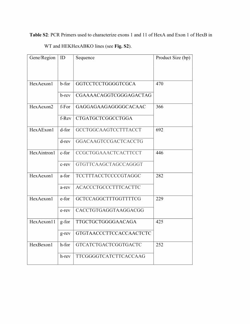
Table S2: PCR Primers used to characterize exons 1 and 11 of HexA and Exon 1 of HexB in
WT and HEKHexABKO lines (see Fig. S2).
Gene/Region ID Sequence Product Size (bp)
b-for GGTCCTCCTGGGGTCGCA HexAexon1
b-rev CGAAAACAGGTCGGGAGACTAG
470
f-For GAGGAGAAGAGGGGCACAAC HexAexon2
f-Rev CTGATGCTCGGCCTGGA
366
d-for GCCTGGCAAGTCCTTTACCT HexAExon1
d-rev GGACAAGTCCGACTCACCTG
692
c-for CCGCTGGAAACTCACTTCCT HexAintron1
c-rev GTGTTCAAGCTAGCCAGGGT
446
a-for TCCTTTACCTCCCCGTAGGC HexAexon1
a-rev ACACCCTGCCCTTTCACTTC
282
e-for GCTCCAGGCTTTGGTTTTCG HexAexon1
e-rev CACCTGTGAGGTAAGGACGG
229
g-for TTGCTGCTGGGGAACAGA HexAexon11
g-rev GTGTAACCCTTCCACCAACTCTC
425
h-for GTCATCTGACTCGGTGACTC HexBexon1
h-rev TTCGGGGTCATCTTCACCAAG
252
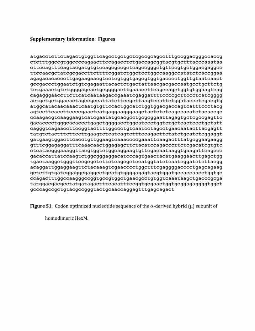
SupplementaryInformation:Figures
atgacctcttctagactgtggttcagcctgctgctcgccgcagcctttgccggacgggccaccgctctttggccgtggccccagaacttccagacctctgaccagcggtacgtgctttacccaaataacttccagtttcagtacgatgtgtccagcgccgctcagccgggctgttccgtgctggacgaggccttccaacgctatcgcgaccttcttttcggatctggctcctggccaaggccatatctcaccggaaagagacacacccttgagaagaacgtcctcgtggtgagcgtggtgacccctggttgtaatcaactgccgaccctggaatctgtcgagaattacactctgactattaacgacgaccaatgcctgcttctgtctgaaactgtctggggagcactgcggggacttgaaaccttcagccagctggtgtggaagtcagcagagggaaccttcttcatcaataagaccgaaatcgaggattttccccgcttccctcatcggggactgctgctggacactagccgccattatcttccgcttaagtccattctggataccctcgacgtgatggcatacaacaaactcaatgtgttccactggcatctggtggacgaccagtcatttccctacgagtccttcaccttccccgaactcatgaggaagggaagctactctctcagccacatctacaccgcccaagacgtcaaggaagtcatcgaatatgcacgcctgcgcggaattagagtgctcgccgagttcgacacccctgggcacaccctgagctggggacctggcatccctggtctgctcactccctgctattcagggtcagaaccttccggtacttttggccctgtcaatcctagcctgaacaatacttacgagtttatgtctactttcttccttgaagtctcatcagtctttccagacttctatctgcatctcggaggtgatgaagtggacttcacctgttggaagtcaaaccccgaaattcaagactttatgcggaagaagggtttcggagaggatttcaaacaactggagagcttctacatccagacccttctcgacatcgtgtcctcatacgggaaaggttacgtggtctggcaggaagtgttcgacaataaggtgaagattcagcccgacaccattatccaagtctggcgggaggacatcccagtgaactacatgaaggaacttgagctggtgactaaggctgggttccgcgctcttctcagcgctccatggtatctcaatcggatctcttacggacaggattggaggaagttctacaaagtcgaacccctggctttcgaggggacccctgagcagaaggctcttgtgatcggaggcgaggcctgcatgtggggagagtacgtggatgccaccaacctggtgcccagactttggccaagggccggtgccgtggctgaacgcctgtggtcaaataagctgacccgcgatatggacgacgcctatgatagactttcacatttccggtgcgaactggtgcggagaggggtggctgcccagccgctgtacgccgggtactgcaaccaggagtttgagcagact
FigureS1.Codonoptimizednucleotidesequenceoftheα-derivedhybrid(µ)subunitof
homodimericHexM.
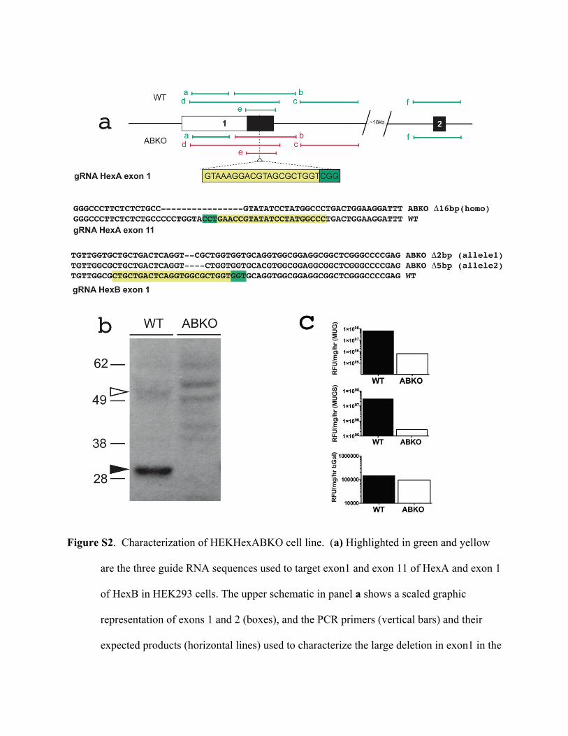
Figure S2. Characterization of HEKHexABKO cell line. (a) Highlighted in green and yellow
are the three guide RNA sequences used to target exon1 and exon 11 of HexA and exon 1
of HexB in HEK293 cells. The upper schematic in panel a shows a scaled graphic
representation of exons 1 and 2 (boxes), and the PCR primers (vertical bars) and their
expected products (horizontal lines) used to characterize the large deletion in exon1 in the
ABKO line. Primers that resulted in products in the WT and ABKO lines are highlighted
in green, those that only produced a product in the WT line are in red. Sequence of
primers used to generate the PCR products are described in Table S2 with the
corresponding labels. Sequences surrounding the remaining guide RNAs in exon 11 of
HexA and exon1 of HexB show the 16bp deletion that was found in exon 11 of both
HexA alleles, and the 2bp or 5bp deletion found in exon1 of the two HexB alleles. Each
of the deletions would result in a frame-shift followed by a prematurely terminated
codon. (b) Comparison of the major α- and β- polypeptide levels in the WT HEK cells
(20 µg) versus the HEXABKO line (ABKO, 40 µg). Western blot of lysates from WT
and ABKO lines were probed with antibody against human Hex A (recognized bot the α-
and β- polypeptides). Even though five times the amount of protein was loaded in the
case of ABKO lysates, bands corresponding to the polypeptides from either of the HexA
subunits were only observed in WT lysate. (c). Comparison of Hex activity levels (log
scale, Y-axis) in WT versus ABKO line. Specific activity of HexA/B and bGal enzymes
in WT and ABKO lines were determined using the fluorogenic substrates MUGS, MUG
and MUbGal, respectively. Bgal activity levels in WT and ABKO lines are unchanged
whereas MUGS and MUG activity in the ABKO line is reduced by more than 3 log
orders in comparison to WT cells.


![Der Einfluß von Packungseffekten auf die ... · in-vitro-Umsetzung von N-Acetoxyanilin mit Desoxyguanosin und DNA ... aus 3,4-Dilithio-2,5-dimethyl-2,4-hexa-dien; das erste „Hetero[6]radialen"](https://static.fdocument.org/doc/165x107/5b1540ab7f8b9adc528b6487/der-einfluss-von-packungseffekten-auf-die-in-vitro-umsetzung-von-n-acetoxyanilin.jpg)