Synthesis, Solution Structure and -selectivity oPhylaf a ... · Synthesis, Solution Structure and...
26
Synthesis, Solution Structure and Phyla-selectivity of a Spider δ-Toxin That Slows Inactivation of Specific Voltage-gated Sodium Channel Subtypes* Nahoko Yamaji ‡1 , Michelle J. Little §1 , Hideki Nishio ¶ , Bert Billen ║ , Elba Villegas ** , Yuji Nishiuchi ¶ , Jan Tytgat ║ , Graham M. Nicholson §2 , and Gerardo Corzo #3 From the ‡ Suntory Institute for Bioorganic Research, Mishima-gun, Shimamoto-cho, Wakayamadai 1-1-1, Osaka 618-8503, Japan, the § Neurotoxin Research Group, Department of Medical & Molecular Biosciences, University of Technology, Sydney, Broadway, NSW 2007, Australia, the ¶ Peptide Institute Inc., Protein Research Foundation, Osaka, Japan, the ║ Laboratory of Toxicology, University of Leuven, Campus Gasthuisberg, O&N2, PO Box 922, 3000 Leuven, Belgium, the ** Centro de Investigacion en Biotecnologia UAEM, Av. Universidad 2001, Cuernavaca, Morelos 62210, Mexico, and the # Instituto de Biotecnologia-UNAM, Av. Universidad 2001, Cuernavaca, Morelos 62210, Mexico 2 To whom correspondence should be addressed. Tel.: 61-2-9514-2230; Fax: 61-2-9514-8206; E-mail: [email protected] 3 To whom correspondence should be addressed. Tel.: 52-777-329-1600; Fax: 52-777-317-2388; E-mail: [email protected] Magi 4, now renamed δ-hexatoxin-Mg1a, is a 43-residue neurotoxic peptide from the venom of the hexathelid Japanese funnel-web spider (Macrothele gigas) with homology to δ-hexatoxins from Australian funnel-web spiders. It binds with high affinity to receptor site 3 on insect voltage-gated sodium (Na V ) channels but, unlike δ-hexatoxins, does not compete for the related site 3 in rat brain despite being previously shown to be lethal by intracranial injection. To elucidate differences in Na V channel selectivity we have undertaken the first characterisation of a peptide toxin on a broad range of mammalian and insect Na V channel subtypes showing that δ-hexatoxin-Mg1a selectively slows channel inactivation of mammalian Na V 1.1, Na V 1.3, and Na V 1.6 but more importantly shows higher affinity for insect Na V 1 (para) channels. Consequently, δ-hexatoxin-Mg1a induces tonic repetitive firing of nerve impulses in insect neurons accompanied by plateau potentials. In addition, we have chemically synthesised and folded δ-hexatoxin-Mg1a, ascertained the bonding pattern of the four disulfides and determined its three dimensional solution structure using NMR spectroscopy. Despite modest sequence homology, we show that key residues important for the activity of scorpion α-toxins and δ-hexatoxins are distributed in a topologically similar manner in δ-hexatoxin-Mg1a. However subtle differences in the toxin surfaces are important for the novel selectivity of δ-hexatoxin-Mg1a for certain mammalian and insect Na V channel subtypes. As such, δ-hexatoxin-Mg1a provides us with a specific tool with which to study channel structure and function and determinants for phyla- and tissue-specific activity. Voltage-gated sodium (Na V ) 4 channels are responsible for the generation and propagation of electrical signals in excitable cells. At least nine different genes encoding distinct Na V channels isoforms have been identified, and functionally expressed, in mammals (1). They are characterized by their sensitivity to TTX, with Na V 1.5, Na V 1.8 and Na V 1.9 being TTX-insensitive or TTX-resistant and the remaining subtypes being sensitive to nanomolar concentrations of TTX. In addition, localization of the subtypes also varies, with Na V 1.1–1.3 mostly distributed in the central nervous system, Na V 1.6–1.9 principally located in the peripheral nervous system, and Na V 1.4 and Na V 1.5 found in skeletal and cardiac muscle, respectively. The structural diversity of Na V channels also coincides with variations in physiological and pharmacological properties (2). In contrast, insects express only one gene (para) that undergoes extensive alternative splicing and RNA editing (3). The para-encoded Na V channel is exceptionally well conserved across diverse orders of insects, with the level of identity ranging from 87 to 98% (3). This is one reason why insecticides that target insect Na V channels have broad activity
Transcript of Synthesis, Solution Structure and -selectivity oPhylaf a ... · Synthesis, Solution Structure and...
Synthesis, Solution Structure and Phyla-selectivity of a Spider δ-Toxin That Slows Inactivation of Specific Voltage-gated Sodium Channel Subtypes* Nahoko Yamaji‡1, Michelle J. Little§1, Hideki Nishio¶, Bert Billen, Elba Villegas**, Yuji Nishiuchi¶, Jan Tytgat, Graham M. Nicholson§2, and Gerardo Corzo#3 From the ‡Suntory Institute for Bioorganic Research, Mishima-gun, Shimamoto-cho, Wakayamadai 1-1-1, Osaka 618-8503, Japan, the §Neurotoxin Research Group, Department of Medical & Molecular Biosciences, University of Technology, Sydney, Broadway, NSW 2007, Australia, the ¶Peptide Institute Inc., Protein Research Foundation, Osaka, Japan, the Laboratory of Toxicology, University of Leuven, Campus Gasthuisberg, O&N2, PO Box 922, 3000 Leuven, Belgium, the **Centro de Investigacion en Biotecnologia UAEM, Av. Universidad 2001, Cuernavaca, Morelos 62210, Mexico, and the #Instituto de Biotecnologia-UNAM, Av. Universidad 2001, Cuernavaca, Morelos 62210, Mexico 2 To whom correspondence should be addressed. Tel.: 61-2-9514-2230; Fax: 61-2-9514-8206; E-mail: [email protected]
3 To whom correspondence should be addressed. Tel.: 52-777-329-1600; Fax: 52-777-317-2388; E-mail: [email protected]
Magi 4, now renamed δ-hexatoxin-Mg1a, is a 43-residue neurotoxic peptide from the venom of the hexathelid Japanese funnel-web spider (Macrothele gigas) with homology to δ-hexatoxins from Australian funnel-web spiders. It binds with high affinity to receptor site 3 on insect voltage-gated sodium (NaV) channels but, unlike δ-hexatoxins, does not compete for the related site 3 in rat brain despite being previously shown to be lethal by intracranial injection. To elucidate differences in NaV channel selectivity we have undertaken the first characterisation of a peptide toxin on a broad range of mammalian and insect NaV channel subtypes showing that δ-hexatoxin-Mg1a selectively slows channel inactivation of mammalian NaV1.1, NaV1.3, and NaV1.6 but more importantly shows higher affinity for insect NaV1 (para) channels. Consequently, δ-hexatoxin-Mg1a induces tonic repetitive firing of nerve impulses in insect neurons accompanied by plateau potentials. In addition, we have chemically synthesised and folded δ-hexatoxin-Mg1a, ascertained the bonding pattern of the four disulfides and determined its three dimensional solution structure using NMR spectroscopy. Despite modest sequence homology, we show that key residues important for the activity of scorpion α-toxins and δ-hexatoxins are distributed in a topologically similar manner in δ-hexatoxin-Mg1a. However subtle differences in the toxin surfaces are important for the novel
selectivity of δ-hexatoxin-Mg1a for certain mammalian and insect NaV channel subtypes. As such, δ-hexatoxin-Mg1a provides us with a specific tool with which to study channel structure and function and determinants for phyla- and tissue-specific activity.
Voltage-gated sodium (NaV)4 channels are
responsible for the generation and propagation of electrical signals in excitable cells. At least nine different genes encoding distinct NaV channels isoforms have been identified, and functionally expressed, in mammals (1). They are characterized by their sensitivity to TTX, with NaV1.5, NaV1.8 and NaV1.9 being TTX-insensitive or TTX-resistant and the remaining subtypes being sensitive to nanomolar concentrations of TTX. In addition, localization of the subtypes also varies, with NaV1.1–1.3 mostly distributed in the central nervous system, NaV1.6–1.9 principally located in the peripheral nervous system, and NaV1.4 and NaV1.5 found in skeletal and cardiac muscle, respectively. The structural diversity of NaV channels also coincides with variations in physiological and pharmacological properties (2). In contrast, insects express only one gene (para) that undergoes extensive alternative splicing and RNA editing (3). The para-encoded NaV channel is exceptionally well conserved across diverse orders of insects, with the level of identity ranging from 87 to 98% (3). This is one reason why insecticides that target insect NaV channels have broad activity
Structure of a spider toxin that targets sodium channel subtypes
2
across many insect orders. In contrast, para-type NaV channels have significantly lower levels of identity with the various types of mammalian NaV channels with the level of identity typically around 50–60% (3). This explains why a high degree of phylogenetic specificity can be achieved with both NaV channel toxins and insecticides that target the NaV channel.
At least seven distinct toxin binding sites have been identified by radioligand binding and electrophysiological studies on vertebrate and insect NaV channels (4,5). Toxins interacting with these neurotoxin receptor site have been instrumental in the study of NaV channel topology, function, and pharmacology (6). In particular, a wide range of scorpion α-toxins, sea anemone toxins and spider δ-hexatoxins [formerly δ-atracotoxins, (7)] compete for binding to receptor site-3 on the extracellular surface of NaV channels. These polypeptide toxins all inhibit the fast inactivation of NaV channels to prolong Na+ currents (INa), despite huge diversity in primary and tertiary structures (8,9). Nevertheless, receptor site-3 has not yet been fully characterized but believed to involve domains DI/S5-S6, DIV/S5-S6 as well as DIV/S3-S4 (for references see (9). Most importantly, however, toxin characterization is often limited to studies using whole cell INa or binding studies on neuronal membranes where there are mixed populations of NaV channel subtypes. For all of these toxins, the precise pattern of NaV channel subtype selectivity is either unknown, or at best incomplete.
Recently, it was found that receptor site-3 was also recognized by a 43-residue spider toxin, originally named Magi 4, from the hexathelid spider Macrothele gigas (Iriomote, Japan). It binds with high affinity to insect NaV channels but, similar to scorpion α-like toxins, does not compete for the related site-3 in rat brain synaptosomes, despite being lethal by intracranial injection (10). Magi 4 shares significant homology to four δ-hexatoxin (HXTX)-1 family peptides and δ-actinopoditoxin-Mb1a (formerly δ-missulenatoxin-Mb1a; Fig. 1) but no sequence homology to scorpion α-toxins. Neurochemical studies have shown that δ-HXTX-1 toxins compete at nanomolar concentrations with both anti-mammalian (e.g. Aah2, Lqh2) and anti-insect (e.g. LqhαIT) scorpion toxins for site-3 (11-13). The three-dimensional structures of δ-HXTX-Ar1a and δ-HXTX-Hv1a peptides have been determined (14,15) and possess core β regions stabilized by four disulfide bonds, placing them in the inhibitory
cystine knot (ICK) structural family (16). The aim of this study was to firstly determine
the solution structure of Magi 4 and secondly to investigate the ability of Magi 4 to discriminate between different NaV channels subtypes. Here we report the tertiary structure of Magi 4 by 1H-NMR and show its disulfide bonding pattern and 3D structure is homologous to δ-HXTX-1 toxins. We highlight the key residues in Magi 4 appear to be topologically similar to those residues known to be part of the pharmacophore for site-3 scorpion α-toxins, despite Magi 4 having a different overall structure to scorpion α-toxins (11). In addition, we provide a detailed characterization of the selectivity and mode of action of Magi 4 on nine cloned mammalian and insect NaV channel subtypes, including a detailed characterization on insect neurotransmission. Given that the toxin potently slows the inactivation of NaV channels it should be renamed δ-hexatoxin-Mg1a (δ-HXTX-Mg1a) in accordance with the rational nomenclature recently proposed for naming spider peptide toxins (7) (see ArachnoServer spider toxin database; http://www.arachnoserver.org). EXPERIMENTAL PROCEDURES
Peptide synthesis– δ-HXTX-Mg1a (43 residues) was chemically synthesized by native chemical ligation involving the coupling of a Cys(17)1–19-thioester peptide (N-terminal 19 residues) and Cys20–43 peptide (C-terminal 24 residues). The N-terminal Cys residue of the former peptide was protected by the Acm group to prevent cyclization and oligomerization. The solid-phase synthesis of the Cys(17)1–19-thioester and Cys20–43 peptides were performed using a ABI 433A peptide synthesizer using the Boc strategy on a Boc-Asn(Xan)-S-CH2-CH2-CO-Leu-Pam resin (0.5 mmol) and Boc-Cys(4-MeBzl)-Merrifield resin (0.3 mmol), respectively. The peptide chains were elongated using in situ neutralization protocols of coupling with Boc-amino acid/HBTU/HOBt/DIEA (4/4/4/6 equiv.). After chain assembly was completed, the peptide resins obtained were treated with HF in the presence of p-cresol (to protect Trp residues) and 1,4-butanedithiol (as an oxygen scavenger) at –5 ºC for 1 h to remove all of the protecting and anchoring groups except the Acm group on the Cys1 residue. The crude products were then purified by preparative RP-HPLC using a YMC-Pak ODS column (30 x 250 mm) to obtain 195 mg (16%) and 75 mg (8.4%) of purified peptides, respectively.
Structure of a spider toxin that targets sodium channel subtypes
3
The N-terminal Cys(17)1-19-thioester and C-terminal Cys20-43 peptides were then allowed to ligate by solubilizing them in freshly degassed 0.1 M sodium phosphate buffer (10 ml), pH 8.4, containing 6 M guanidium hydrochloride (Gdn·HCl). Thiophenol (0.40 ml) was then added, the whole mixture stirred for 24 h at room temperature and then treated with DTT (1.6 mmol). After 20 min the pH was acidified to less than 2 using 1 M HCl. The mixture was washed with ether and Cys(17)1–43 peptide purified from the aqueous layers by preparative RP-HPLC using a linear gradient (20-40% CH3CN in 0.1% TFA for 80 min) to obtain 51 mg (41%) of purified peptide.
To remove the Acm group, a 9.6 mM solution of Cys(17)1–43 (50 mg) was made up in 5 ml 95% TFA containing anisole (50 μl) and 0.77 mmol Ag trifluoroacetate was added and stirred for 2 h at room temperature. The product was precipitated as the Ag salt, by adding ether to the reaction mixture, and was dissolved in 0.1 M phosphate buffer, pH 8.4, containing 6 M Gdn·HCl. DTT (3.8 mmol) was added to the solution and stirred for 0.5 h at room temperature. After the addition of 1 M HCl, the resulting AgCl was removed by filtration and the filtrate was applied to preparative RP-HPLC using a linear gradient (20-40% CH3CN in 0.1% TFA for 80 min) to obtain 23 mg (46% yield) of the reduced peptide (8 SH-peptide (1-43)).
The eight free cysteines were allowed to oxidize in air for 4 days at 4 °C in 0.1 M aqueous ammonium acetate (pH 7.6) containing 2 M Gdn·HCl, 0.1 mM GSSG and 1 mM GSH. The peptide:GSSG:GSH ratio was 1:10:100. The solution was then diluted 2-fold with cold water (4 °C) to a final peptide concentration of 5 μM. After 3 days at 4 ºC, the mixture was acidified to pH 2 by adding TFA and the folded peptide was desalted and purified by preparative RP-HPLC using a linear gradient (17-37% CH3CN in 0.1% TFA for 80 min) to obtain 5.3 mg from the previous 23 mg (23% yield).
ESI/Q-TOF Mass Spectrometry–All masses obtained during chemical synthesis were confirmed by electrospray ionization mass spectrometry using an ESI/Q-TOF mass spectrometer (Micromass, Manchester, U.K.). Peptides were mixed with 1% (v/v) formic acid / 70% (v/v) CH3CN and data analyzed using MassLynx v3.4 software (Micromass).
Capillary Electrophoresis–Capillary zone electrophoresis (CZE) analyses were performed on a Jasco CE800 system (Jasco, Japan) equipped with a UV detector connected to a Shimadzu CR-4A
recorder and a capillary (0.1 µm ID, 70 cm length, 50 cm to detector). 20 mM sodium citrate buffer (pH 2.5) was used for the analyses. Samples dissolved in migration buffer were applied hydrodynamically to the capillary (height 20 cm, 15 s) and analyses were performed with a 20 kV constant voltage drop and monitored at 210 nm.
Circular Dichroism (CD) Measurements–CD spectra were obtained on a Jasco J-725 spectropolarimeter (Jasco, Japan). The spectra were measured from 260 to 180 nm in 60% TFE, pH 7.1, at room temperature, with a 1 mm path-length cell. Data were collected at 0.1 nm with a scan rate of 100 nm/min and a time constant of 1 s. The concentration of the toxins was 30 µM, as determined by amino acid analysis. Data from 10 separate recordings were averaged and analyzed by the method of Bohm et al. (18).
Iodination of LqhαIT and Binding Assays–The toxin was radio-iodinated with bovine milk lactoperoxidase (EC 1.11.1.7, Sigma-Aldrich, Germany) using 0.7 lactoperoxidase units, 1 nmole of toxin and 0.5 mCi carrier-free Na125I (Amersham, UK) (19). The displacement assays were performed as previously described (19) (see supplementary information).
NMR Experiments–Synthetic δ-HXTX-Mg1a (4.7 mg) was dissolved to a final concentration of 3.7 mM in 250 μl of H2O/D2O (90/10; v/v) containing 50 mM NaN3 in a susceptibility-matched microcell (Shigemi, Japan). The pH was adjusted to 2.4.
NMR spectra were recorded on a Bruker DMX-750 spectrometer. The temperature was set to 298 K. Chemical shifts were referenced to internal TSP. 2D DQF-COSY, NOESY and TOCSY experiments were performed using standard pulse sequences and phase cycling. The NOESY spectra were acquired with mixing times of 50 ms and 200 ms. The TOCSY spectrum was recorded with a spin lock time of 71 ms. The 2D spectra were recorded using time-proportional phase incrementation for quadrature detection in the F1 dimension. For water suppression, the NOESY experiments include the WATERGATE sequence, and the DQF-COSY and TOCSY experiments included selective low-power irradiation during the relaxation delay. The TOCSY and NOESY spectra were recorded with 512 (t1) x 2K (t2). The DQF-COSY spectrum was recorded with 512 (t1) x 8K (t2) for an estimation of the coupling constants. The spectra were processed using the XWIN-NMR 2.5 program (Bruker Biospin) running on an O2 workstation (Silicon Graphics). Chemical shift assignments have been
Structure of a spider toxin that targets sodium channel subtypes
4
deposited in the BioMagResBank (BMRB), accession code 11044.
Structural Calculations–Proton signal assignments were achieved using the standard strategy described by Wüthrich with the graphical software ANSIG.3.3. The DQF-COSY and TOCSY spectra gave the spin system fingerprint of the peptide. The spin systems were then sequentially connected using the NOESY spectra. Interproton distance restraints were obtained from the NOESY spectra acquired with a mixing time of 200 ms. The NOE volumes were converted into four ranges of distance restraints classified as strong (1.8–2.7 Å), medium (1.8–3.3 Å), weak (1.8–5.0 Å), and very weak (1.8–6.0 Å). The backbone 3JNH-Hα coupling constants were estimated or directly measured from a 1D spectrum or the high-digital resolution DQF-COSY spectrum using the DECO program (Bruker Biospin). Hydrogen bonds were identified using amide-proton temperature coefficients from NOESY spectra obtained at 288 K, 298 K, 308 K, 318 K and 328 K.
The structural calculations were performed using the X-PLOR-NIH 2.9.1 program with 576 NOE-based distance restraints, which contain 207 intraresidue, 181 sequential, 60 medium-range and 128 long-range restraints. In the first stage, the starting extended strand structure was subjected to 10 ps and 1,000 steps of torsion-angle molecular dynamics at 50,000 K. The structures were then subjected to 15 ps and 1,500 steps of a slow-cooling torsion angle molecular dynamics stage in which the temperature was reduced from 50,000 K to 298 K over 250 steps. Finally, the structures were subjected to 200 steps of conjugated-gradient minimization. The initial runs for structure calculations were performed without hydrogen bond and disulfide bond restraints and the obtained structure was examined.
The structures were checked for violations of geometric and experimental restraints, and atom overlapping, using the AQUA3.2 and PROCHECK-NMR3.4 programs. Finally, a set of 20 conformers was selected based on the lowest X-PLOR energy. Structures were analyzed and visualized with the MOLMOL 2k.1 program.
Neuronal Isolation Procedures and NaV Channel Expression–Characterization of the actions of δ-HXTX-Mg1a on native INa were performed using acutely dissociated newborn rat DRG neurons and cockroach DUM neurons from the terminal abdominal ganglia of adult male American cockroaches (Periplaneta americana) as previously described (20,21). Following isolation, recordings
were made within 24 hours. For expression in Xenopus laevis oocytes, the
rNaV1.1, rNaV1.2, and mNaV1.6 genes were subcloned into pLCT1. The rNaV1.3, rNaV1.4, hNaV1.5, DmNaV1, and tipE genes were subcloned into vectors pNa3T, pUI-2, pcDNA3.1, pGH19-13-5 and pGH19 respectively. For in vitro transcription, these plasmids were linearized with NotI. The rNaV1.7/pBSTA.rPN1 and hβ1/pGEM-HE were linearized with SacII and NheI, respectively. Capped cRNAs were then synthesized from linearized plasmid using the T7 mMESSAGE-mMACHINE transcription kit (Ambion). The rβ1/pSP64T and hNaV1.8/pBSTA vectors were linearized with EcoRI and NotI respectively, and transcribed with the SP6 mMESSAGE-mMACHINE transcription kit (Ambion).
Stage V-VI oocytes were harvested from the ovarian lobes of anaesthetized female X. laevis frogs as described previously (22). The oocytes were injected with up to 50 nl of cRNA at a concentration of 1 ng nl–1 using a Drummond microinjector (Ambion). The ND96 solution used for incubating the oocytes contained (in mM): NaCl 96, KCl 2, CaCl2 1.8, MgCl2 2, HEPES-acid 5 (pH 7.4), supplemented with 50 mg l–1 gentamycin sulfate. Whole-cell currents from oocytes were recorded 2–5 days after injection.
Electrophysiological Studies–Voltage- or current-clamp recordings from single DRG and DUM neurons were made using the whole-cell patch-clamp technique of Hamill et al. (23). Recordings from X. laevis oocytes were performed using the two-electrode voltage-clamp method as described by Liman et al. (22).
Experiments were performed at constant temperature 18–24 ˚C using either an AxoPatch 200A patch-clamp amplifier or GeneClamp 500 amplifier. Current and voltage pulse protocols were generated using the pClamp software system (Molecular Devices, Sunnyvale, CA, USA). Data were digitized at 10–25 kHz, and low-pass filtered at either 1 kHz (oocytes) or 5 kHz (DRG and DUM neurons) using a 4- or 5-pole Bessel filter (–3 dB). Leakage and capacitive currents were digitally subtracted with P-P/4 procedures and series resistance compensation set at >80% for all patch-clamped cells to minimize voltage errors. The extracellular Na+ concentration was reduced (see below) to minimise series resistance errors.
The voltage-clamp data recorded in this study were rejected if there were large leak currents upon seal formation or currents showed signs of
Structure of a spider toxin that targets sodium channel subtypes
5
inadequate space clamping. At the commencement of each experiment, oocytes or DUM neurons exhibiting leakage currents at a holding potential of -90 mV of more than –200 nA or –600 pA, respectively, were discarded. Only cells exhibiting stable leakage currents throughout the whole experiment (with a maximal deviation of ± 10 % of initial value) were considered in the data analysis. To avoid overestimation of a potential toxin-induced shift in the current-voltage relationship as a result of inadequate voltage control when measuring large sodium currents in oocytes, only results from cells with currents lower than 1.5 µA were considered. Current-clamp data were rejected if the initial resting membrane potential was more depolarized than –45 mV.
Recording Solutions–DRG neurons were perfused with an external solution containing (in mM): Na acetate 30, MgCl2 1, CaCl2 1.8, Cs acetate 5, KCl 5, D-glucose 25, HEPES-acid 5, TEA-Br 100, CdCl2 0.5, 4-AP 1 with the pH adjusted to 7.4 with 1 M TEA-OH. Recordings were performed using micropipettes filled with a solution containing (in mM): CsF 135, Na acetate 8, HEPES-acid 5, with the pH adjusted to 7.0 with CsOH. The osmolarity of internal and external solutions was adjusted to 300–305 mOsmol litre–1 with sucrose, prior to use, to reduce osmotic stress. Pipettes with resistances of 0.8–2 MΩ were used for recording INa from DRG neurons.
For voltage-clamp experiments, DUM neurons were perfused with an external solution consisting of (in mM): NaCl 90, CsCl 5, CaCl2 1.8, 4-AP 5, TEA-Cl 50, verapamil 0.01, NiCl2 0.1, CdCl2 1, HEPES-acid 10, pH adjusted to 7.4 with HCl. Micropipettes were filled with an internal solution consisting of (in mM): CsF 135, MgCl2 1, NaCl 34, ATP-Na2 3, EGTA 5, HEPES-acid 10, pH adjusted to 7.35 with CsOH. For current clamp experiments, DUM neurons were perfused with an external solution consisting of (in mM): NaCl 190, KCl 3.1, CaCl2 5, MgCl2 4, HEPES-acid 10, with the pH adjusted to 7.4 with NaOH. Micropipettes were filled with an internal solution consisting of (in mM): K gluconate 160, KF 10, ATP-Mg 1, CaCl2 0.5, NaCl 15, MgCl2 1, EGTA 10, HEPES-acid 10, with the pH adjusted to 7.4 with KOH. The osmolality of all solutions was adjusted to 400 mOsm litre–1 with sucrose. Pipettes with resistances of 1.2–2 MΩ were used for recording currents from DUM neurons.
Oocytes were perfused with ND96 solution. Voltage and current electrodes were filled with 3 M KCl and resistances were <1 MΩ.
Voltage- and Current-clamp Recordings–In DRG neurons, the predominant TTX sensitivity of the Nav channels present in each cell was determined using a modified steady-state Nav channel inactivation (h∞) voltage-clamp protocol (24,25). This takes advantage of the separation of h∞ curves for TTX-sensitive and TTX-resistant Nav channels (26). Only those found to have less than 10% TTX-resistant INa were used for TTX-sensitive experiments. Of the remaining cells, those expressing sufficiently large TTX-resistant INa were used for experiments by perfusing with external solution containing 200 nM TTX.
To determine the effect of the toxin on fast inactivation, currents were recorded in the absence, and presence, of a range of toxin concentrations. The action of the toxin on fast inactivation was assayed by measuring the late current remaining at 50 ms (I50ms) for DRG and DUM neurons, or 30 ms for oocytes, as a fraction of peak current (Ipk). Currents were elicited by test potentials to –10 mV in DUM and DRG neurons or at depolarizing test potentials corresponding to maximal activation in oocytes (ranging from –10 to +10 mV). Test potentials were applied at 10 s (DRG and DUM) or 5 s (oocytes) intervals. The normalised late current ratio (eg. I50ms / Ipk) gives the fractional probability for NaV channels not to be inactivated at the end of the test pulse. The concentration dependence for removal of inactivation was measured by plotting the normalised late current as a function of toxin concentration according to the following Hill equation (Eq. 1):
where EC50 is the concentration at half maximal inhibition of fast inactivation, [toxin] is the toxin concentration and nH is the Hill coefficient.
The effect of δ-HXTX-Mg1a on the voltage-dependence of Nav channel activation was determined using depolarizing test pulses from –90 to +70 mV for 50 ms, in 5-mV (oocytes) or 10-mV (DRG and DUM) steps. The values for sodium conductance (gNa) were calculated according to the equation (Eq. 2): where INa is the absolute value of the sodium current at a given test potential (V) and Vrev is the reversal potential. The values of gNa and V–Vrev were then fitted to a Boltzmann equation (Eq. 3):
gNa =I Na
(V −Vrev )
I 50ms
I pk
=100
1+EC50
[toxin]
nH
Structure of a spider toxin that targets sodium channel subtypes
6
h∞ = 1− C1+ exp[(V −V1/2)/kh ]
+ C
where gmax is maximal gNa, V1/2 is the half- maximal conductance, km is the slope factor.
To determine the effect of δ-HXTX-Mg1a on the voltage-dependence of steady-state NaV channel fast inactivation (h∞) a two-pulse protocol with a 0.5-ms interpulse interval was applied. This consisted of a 500 ms or 1 s conditioning prepulse (Vcond), in which the holding potential of –90 mV was stepped to potentials ranging from –130 up to +20 mV in 5-mV (oocytes) or 10-mV (DRG and DUM) increments, followed by a 50-ms test pulse (Vtest) to –10 mV in DRG or DUM neurons, or potentials corresponding to maximal activation in oocytes. Pulses were applied every 10 s. Data were normalised to the maximum peak control INa and fitted using a Boltzmann equation (Eq. 4): where V1/2 is the voltage at half-maximal inactivation, kh is the slope factor, V is the test voltage, and C is a constant or non-inactivating fraction (usually zero in controls).
The effect of the toxin on the rate of recovery from NaV channel inactivation was examined by applying a standard two-pulse protocol with a variable interpulse interval (ΔT). A 50-ms conditioning prepulse (Vcond) was applied from a holding potential of –90 mV to –10 mV, followed by a 50-ms test pulse (Vtest), with an interpulse interval ranging between 0.5 ms and 4 s.
Mathematical curve fitting was accomplished using GraphPad Prism version 5.00 for Macintosh (GraphPad Software, San Diego CA, USA). All curve-fitting routines were performed using non-linear regression analysis employing a least squares method. Comparisons of two sample means were made using a paired Student's t-test. Multiple comparisons were assessed by repeated measures ANOVA with a Bonferroni’s multiple comparison post-hoc test. A test was considered to be significant when P < 0.05. All data are presented as mean ± standard error of the mean (SEM) of n independent experiments.
RESULTS
Peptide Synthesis and Characterization–δ-HXTX-Mg1a was synthesized with its C-terminus as the free carboxyl, as observed for the native toxin by mass spectrometry
and from the cDNA encoding gene (10). The structural identity between the synthetic and native toxins was verified by ESI/Q-TOF MS and CZE (supplemental Fig. S1 and Table S1). In a co-injection experiment, native δ-HXTX-Mg1a and synthetic δ-HXTX-Mg1a coeluted in a single sharp peak (supplemental Fig. S1A). Moreover, the CD spectra of synthetic and native δ-HXTX-Mg1a superimposed indicating that their secondary structures were similar. Both native and synthetic δ-HXTX-Mg1a were able to displace the binding of 125I-LqhαIT from insect NaV channels in cockroach synaptosomes with similar IC50 values (supplemental Fig. S1B-C).
NMR Spectroscopy–Sequence specific 1H resonance assignments for all residues observed were established using standard methods (27). NOE cross peaks were converted to distance restraints for structural calculation. From NMR experiments, 582 distance constraints and 22 dihedral angle constraints were used for the structural calculations. The total of 582 distance restraints included 576 NOE constraints and 6 hydrogen bonds (Fig. 1B). In addition, disulfide bond constraints were also used (Cys1-Cys15, Cys8-Cys20, Cys14-Cys31 and Cys16-Cys43). Simulated annealing calculations were started from an extended structure and 20 structures were selected with the lowest XPLOR energy, which had no ≥ 0.4 Å NOE distance violations and no ≥ 5.0° angle violations from 100 structures obtained at the final calculation (28). Structural statistics for these 20 converged structures are summarized in Table 1. Figure 2A shows a stereoview of the best-fit superposition of the backbone atoms (N, C, Cα and O) for 20 converged structures. The root-mean-square deviations with respect to the mean coordinate positions are 0.483 Å for backbone atoms and 1.178 Å for all heavy atoms of the disulfide-rich structured region (Gly2-Ala23 and Gln28-Glu32), excluding the non-structured loop (Trp24-Gln27) and C-terminal (Arg33-Cys43) regions. Analysis of the 20 structures using PROCHECK-NMR and AQUA reveals that 95.1% of the backbone dihedral angles for the structured region lie in the most favored and additional allowed regions of the Ramachandran plot (29).
Six protons from amide groups were identified to form hydrogen bonds according to the amide proton temperature coefficients (Supplemental Fig. S2). If the hydrogen bond was in agreement with amide temperature coefficients (> –4.6 ppb/K), and a hydrogen bond acceptor and an oxygen atom of the backbone was within 2.6 Å of the amide-proton,
gNa
gmax
=1
1 +exp[(V1/2 −V )/km ]
Structure of a spider toxin that targets sodium channel subtypes
7
the amide-proton was identified as a donor of the hydrogen bond. Hydrogen bond acceptors for all the protons were unambiguously determined from preliminary structure calculations. These restraints were then used in the following stage of structure calculations (1.8 ≤ d ≤ 2.12 Å for NHi-Oj and 2.7 ≤ d ≤ 3.2 Å for Ni-Oj) (30).
Twenty-two dihedral angles estimated from the 3JNH-Ha values were used as angle constraints within the range of –90° and –40° for 3JNH-Hα < 5.5 Hz (Ala6, Trp7, Cys15, Trp24, Cys31, Arg33, Lys36, Phe39), and between –160° and –80° for 3JNH-Hα > 8 Hz (Ser3, Cys8, Lys9, Cys14, Tyr18, Asn19, Cys20, Ala23, Asn26, Gln28, Ser29, Glu32, Trp35, Glu42) following standard parameterization (31) (Fig. 1B).
From the results of initial runs without using the disulfide bridge restraints, the disulfide patterns of Cys1-Cys15 and Cys16-Cys43 were unambiguously determined. Analyzing the distances between Sγi and Sγj of the remaining four cysteines, three types of disulfide patterns were possible (pattern I: Cys8-Cys14, Cys20-Cys31; pattern II: Cys8-Cys31, Cys14-Cys20; and pattern III: Cys8-Cys20, Cys14-Cys31). Three sets of structure calculations were performed with these possible restraints. The energy minimization was done in vacuo with the GROMOS96 43B1 parameters set, without reaction field (32), to determine the lowest energy state and thus the most stable disulfide pattern of δ-MSTX-Mg1a. Energy computations were done with the GROMOS96 (32) implementation of Swiss-PdbViewer (33). Total energy calculations for pattern I, II and III were –693.5, –892.7 and –1523.4 kJ/mol respectively, therefore unambiguously determining the remaining disulfide bridge patterns as Cys8-Cys20 and Cys14-Cys31. These disulfide bridges, along with Cys1-Cys15 form an ICK motif (Fig. 2D), and a pattern (I-IV, II-VI, III-VII, V-VIII) identical to other δ-HXTX-1 family members (14,15)Fig. 1A).
Description of the three-dimensional structure– Figure 2A shows the best-fit superposition of the backbone atoms (N, Cα, and C) for the 20 converged structures of δ-HXTX-Mg1a. The three-dimensional structure of δ-HXTX-Mg1a comprises several well-defined regions, consisting of an antiparallel β-sheet (strand 1: Asn19-Ala23, strand 2: Gln28-Glu32) with some secondary structural elements (Figs. 1B and 2B). We identified turns by the standard definition that the distance between Cαi and Cαi+3 is less than 7 Å (34,35). The two turns involve Ser3 to Ala6 (turn 1) and Cys15 to Tyr18 (turn 2). The average dihedral angles for residues at positions i+1 and i+2 are follows: 2 =
–40°, φ2 = 122° for Lys4 and 3 = 57°, φ3 = 84° for Arg5, 2 = –45°, φ2 = 128° for Cys16 and 3 = 98°, φ3 = –2° for Gly17. Turn 1 is classified as a miscellaneous type IV β-turn. There is no hydrogen bonding in this turn conformation. Arg5 in turn 1 shows a positive φ3 value, since non-glycine residues at position i+2 are rare in type II β-turns and furthermore arginine shows low turn potential. Turn 2 is assigned as a typical type II β-turn (βγL). In this turn, the Cys15 oxygen atom forms hydrogen bonding with the Tyr18 amide-proton (Fig. 1B). The region Trp24 to Gln27 is not well defined by the NMR data.
Electrophysiological Studies on Mammalian NaV Channels–The effects of δ-HXTX-Mg1a were initially examined on neonatal rat DRG neurons as these were employed in the original characterization of δ-HXTX-1 and δ-AOTX-Mb1a toxins (36-38). Rat DRG neurons express two types of NaV channel currents, TTX-sensitive (mainly NaV1.1, NaV1.6 and NaV1.7) and TTX-resistant (NaV1.8 and NaV1.9) (26,39). Like δ-HXTX-1 toxins, δ-HXTX-Mg1a slowed inactivation of TTX-sensitive INa in DRG neurons (Fig. 3A). This was observed as a sustained current in the presence of the toxin during depolarizing test potentials to –10 mV in which INa are fully inactivated in the absence of toxin. This is most likely the result of a marked slowing of transitions between open and open-inactivated states. The maximum degree of slowing of inactivation after 50 ms was 26.3 ± 4.4% (n = 7) of control peak INa at 300 nM, with an EC50 of 46 nM (Fig 3E). This steady-state current was completely blocked by 100 nM TTX indicating that it is mediated exclusively via NaV channels (data not shown). In parallel, the peak INa amplitude recorded at –10 mV transiently increased but after 5 min perfusion was slightly reduced by 6.6 ± 11.0% (n = 7).
Similar to δ-HXTX-1 toxins (37,38), this effect was accompanied by a modest 10 mV hyperpolarizing shift in the threshold, but not the midpoint (V1/2), of NaV channel activation (Fig. 3C). Furthermore, δ-HXTX-Mg1a, at concentrations up to 1 μM, failed to modulate TTX-resistant INa (Fig. 3B and D). Unlike δ-HXTX-1 toxins, however, the effect to slow NaV channel inactivation in TTX-sensitive DRG neurons was almost completely reversible after washing with toxin-free solution with a τon of 84.9 ± 18.4 s and τoff of 81.5 ± 9.2 s (n = 5).
Given the potent action of δ-HXTX-Mg1a on DRG neurons, we then assayed the effect of the toxin on eight mammalian NaV channel clones
Structure of a spider toxin that targets sodium channel subtypes
8
(NaV1.1/β1-NaV1.8/β1) by analyzing gNa/V relationships and steady-state inactivation. Effects of the toxin on NaV1.9 channels were not investigated as this channel subtype currently fails to express in standard heterologous systems (40). Addition of δ-HXTX-Mg1a (up to 5 µM) to the bath medium produced a marked slowing of INa inactivation with a rank order in magnitude of NaV1.6/β1 > NaV1.1/β1 > NaV1.3/β1. However with NaV1.2/β1 and NaV1.7/β1 there were only weak effects to slow INa inactivation (Fig. 4A left-hand panels and supplemental Table 2S). These effects were accompanied by ~20% increase in peak INa amplitude with NaV1.6/β1 and NaV1.1/β1 channels but less significant changes with the remaining channel clones (supplemental Table 2S).
The above effects were accompanied by only weak hyperpolarizing shifts in the voltage-dependence of activation (gNa) to a maximum ΔV1/2 of –8.9 mV for NaV1.3/β1 channels (Fig. 4A right-hand panels). In contrast, the toxin failed to slow INa inactivation of NaV1.4/β1, NaV1.5/β1 or NaV1.8/β1 channels at concentrations up to 5 µM (Fig. 4B). This lack of activity on NaV1.8/β1 channels is consistent with the lack of effects on TTX-resistant INa in DRG neurons that express both NaV1.8 and NaV1.9.
In the mammalian NaV channel clones, steady-state inactivation (h∞) in the absence of toxin was best described by a single Boltzmann function. δ-HXTX-Mg1a (5 µM) caused a strong –19.7 mV hyperpolarizing shift in the voltage at half-maximal inactivation (V1/2) of NaV1.3/β1, but only weak non-significant shifts in V1/2 in NaV1.1/β1, NaV1.2/β1, NaV1.6/β1 and NaV1.7/β1 channels of less than –4 mV (Fig. 4A right-hand panels). Moreover, in the presence of δ-HXTX-Mg1a steady-state inactivation became incomplete, causing the appearance of a non-inactivating component at prepulse test potentials more depolarized than –40 mV, an effect previously noted with δ-HXTX-1 toxins (37,38). This non-inactivating component was up to 27 ± 10% (n = 5) of peak current in the case of the NaV1.1/β1 channel (determined from C in Eq. 4) with a rank order in magnitude of NaV1.6/β1 > NaV1.1/β1 > NaV 1.3/β1 >> NaV1.2/β1 > NaV1.7/β1 (Fig. 4A right-hand panels). This was sufficient to induce significant changes in the slope factor, kh, in all NaV channel clones except NaV1.2/β1. Although time-dependent shifts in h∞ have been demonstrated in patch-clamp configurations (41) these are unlikely to account for the observed changes given the magnitude of the shift in NaV1.3/β1 and the absence of a
non-inactivating component in controls. Electrophysiological Studies on Insect NaV
Channels–In contrast to the clear effects, but modest potency, of the toxin on mammalian NaV channel gating and kinetics, δ-HXTX-Mg1a showed high affinity for the Drosophila NaV channel clone DmNaV1/TipE. This was evident firstly by a concentration-dependent increase in control peak INa amplitude up to a maximum of 290 ± 37% at 1 µM (n = 5; Fig. 5C). This was most likely the result of the marked concentration-dependent slowing of fast inactivation (Fig. 5A and C) with an EC50 of 22.8 nM (Fig. 5H). Importantly δ-HXTX-Mg1a completely removed fast inactivation at concentrations of 1 µM (Fig. 5C). This was accompanied by up to a 21.8 mV hyperpolarizing shift in DmNaV1/TipE channel activation at 1 µM (n = 3, Fig. 5D). At a concentration of 15 nM, δ-HXTX-Mg1a also caused steady-state DmNaV1/TipE channel inactivation to become incomplete at prepulse test potentials more depolarized than –50 mV (non-inactivating component C = 49.2 ± 0.3%, Fig. 5B). At higher concentrations, the toxin completely prevented channel inactivation (Fig. 5D).
To further probe the mode of action and insect-selectivity of δ-HXTX-Mg1a we assayed the toxin on cockroach DUM neurons, previously employed in the characterisation of δ-HXTX-Hv1a (42). Like δ-HXTX-Hv1a, δ-HXTX-Mg1a produced a potent time- and concentration-dependent slowing of INa inactivation (Fig. 5E and H). Maximum slowing after 50 ms was 46.1 ± 4.5% (n = 10) of control peak INa at 300 nM δ-HXTX-Mg1a (Fig. 5E). The EC50 for the slowing of inactivation in DUM neurons was 823 pM, ca. 56-fold lower than in DRG neurons (Fig. 5H). Hence, although this toxin is not insect-specific, it shows a marked selectivity for insect NaV channels. The toxin also caused 14.3 and 13.5 mV hyperpolarizing shifts in the V1/2 of NaV channel activation and steady-state inactivation, respectively (Fig. 5F).
In DUM neurons, the slowing of inactivation described above was accompanied initially by a slight increase in the peak INa. This effect would reach a maximum after ca. 70 s followed immediately by a decrease in peak and late INa amplitude (Fig. 5E). If depolarising test pulses were abolished for 2 min after reaching steady-state toxin effects (e.g. after 4–5 min perfusion), peak and late INa amplitude increased markedly (Fig. 5G, trace marked ‘120 s’). If depolarising test pulses
Structure of a spider toxin that targets sodium channel subtypes
9
(Δt = 10 s) were then reapplied, peak and late INa amplitude would decline back to steady-state levels within ca. 1 min (Fig. 5G, trace marked ‘180 s’). This effect may result from a slowed rate of recovery from fast inactivation or a voltage-dependent dissociation of the toxin from the channel. To test these possibilities we firstly assessed the effect of δ-HXTX-Mg1a (30 nM) on the rate of recovery from inactivation. While the toxin markedly increased repriming kinetics at interpulse intervals less than 3 ms, presumably reflecting a more rapid transition from open to closed states, the major effect of the toxin was to prolong the rate of recovery from fast inactivation at interpulse intervals greater than 3 ms, reflecting a slowed transition between closed-inactivated and closed states (Fig. 6A). This action resulted in a use-dependent decrease in peak INa amplitude during repetitive stimulation at 30 Hz (Fig. 6B).
To determine whether the effect of the toxin was dependent on the holding potential the amplitude of the toxin-modified INa measured at the end of the test pulse was compared to peak INa at hyperpolarized holding potentials. In comparison with currents recorded at –90 mV, the fraction of the sustained INa, measured at the end of the 50 ms depolarizing test pulse (I50ms), compared with the peak INa amplitude (Ipk) was not significantly increased except at −150 mV (P < 0.05, n = 7, Fig. 6C). In addition, to test if depolarising pulses could cause dissociation of the toxin, post-pulses up to +200 mV were applied immediately following a test pulse to –10 mV from –90 mV every 2 s. At this stimulation frequency there was some initial rundown in peak and late INa amplitude but the depolarizing post-pulses failed to cause any significant decrease in fractional I50ms/Ipk compared to data recorded in the absence of the post-pulse (P > 0.05, n = 5; Fig. 6D)
To investigate the effects of the toxin on membrane excitability, DUM neurons were held under current-clamp conditions and action potentials evoked by depolarizing current pulses. δ-HXTX-Mg1a (100 nM) initially produced a prolongation of the repolarizing phase of the action potential (Fig. 7A). The falling phase of the action potential developed a broad shoulder in the last two-thirds of the repolarization phase (Fig. 7A). This caused a suppression of the afterhyperpolarization and an increase in spike duration resulting in ‘plateau’ action potentials (Fig. 7A-C). Although a small depolarization of 3.1 ± 0.6 mV (n = 5) was observed, neither resting membrane potential (RMP) nor spike amplitude
were significantly altered. Applying artificial hyperpolarization to a level 20–40 mV more negative than the RMP facilitated the appearance of these plateau potentials. In addition, the duration of plateau potentials were heavily influenced by the stimulation frequency such that the duration increased from ca. 70 ms at a RMP of –60 and stimulus interval of 10 s (Fig 7A) to ca. 300 ms at stimulus intervals of 1 min at a RMP held at –90 mV (Fig. 7C). Interestingly, 100 nM δ-HXTX-Mg1a co-applied with 500 µM 3-4 diaminopyridine (3,4-DAP), a KV channel blocker, markedly prolonged the action potential duration to ca. 150 ms at –60 mV (Fig. 7D) and ca. 800 ms at –90 mV (Fig. 7E). In this case, the action potential duration was more than 80-fold longer than with 3,4-DAP applied alone.
As previously described DUM neurons are spontaneously active (43). At resting membrane potentials, most DUM neurones were capable of generating repetitive action potentials with firing frequencies of 31 ± 4.6 Hz (n = 4; Fig. 7F). In the presence of 100 nM δ-HXTX-Mg1a, repetitive plateau action potentials of ca. 70 ms duration would also occur spontaneously, albeit with a much reduced firing frequency of 3.2 ± 0.6 Hz (n = 4; Fig. 7G). However, when the membrane potential was hyperpolarized to –80 mV, spontaneous plateau action potential duration increased up to ca. 500 ms but firing frequency was still low at 0.7 ± 0.2 Hz (n = 4; Fig. 7H). Using a ramp current from –80 mV at 0.2–0.4 nA/s there was also a –26 ± 2.0 mV shift in the threshold of spontaneous firing from –44.6 ± 4.0 mV in controls to –70.6 ± 3.5 mV (n = 7) in the presence of 100 nM δ-HXTX-Mg1a.
DISCUSSION Actions on NaV channel gating
In all cases, δ-HXTX-Mg1a modulated channel gating and kinetics in a similar fashion to other δ-HXTX-1 toxins by causing: (i) a slowing/removal of NaV channel inactivation typically associated with an increase in peak NaV channel currents, (ii) a hyperpolarizing shift in the voltage dependence of channel activation, and (iii) a hyperpolarizing shift in the voltage dependence of steady-state inactivation. In insect neurons, further characterization revealed that the toxin (iv) slowed NaV channel repriming kinetics, and (v) caused a use-dependent reduction in INa amplitude.
The toxin-induced increase in peak INa seen with DmNaV1 and to a lesser extent in DUM neurons, rNaV1.1, rNaV1.3 and NaV1.6, was in general well correlated with the magnitude of the sustained
Structure of a spider toxin that targets sodium channel subtypes
10
current at the end of the depolarizing test pulse. This increase in peak INa can be explained by a delay in NaV channel fast inactivation from open to open-inactivated states. In contrast, δ-HXTX-Mg1a did not slow INa inactivation as markedly in NaV1.2, or NaV1.7 and therefore did not appreciably increase peak INa amplitude. This slowing of the transition from open to open-inactivated states also generated a non-inactivating component in the steady-state inactivation curve. This occurred at prepulse test potentials more depolarized than –40 mV, an effect previously noted with δ-HXTX-1 toxins (37,38). This is the result of a more rapid rate of recovery from fast inactivation when test pulses are separated by less than 3 ms. This reflects a higher proportion of transitions between open and closed states than open-inactivated to closed states via a closed-inactivated intermediate.
The present study demonstrates that δ-HXTX-Mg1a displays differential NaV channel subtype specificity for NaV1.1/β1, NaV1.3/β1, and NaV1.6/β1 channels. This is consistent with its action on TTX-sensitive INa in rat DRG neurons that express mainly NaV1.1, NaV1.3 and NaV1.7 channels (39). Importantly, however, δ-HXTX-Mg1a showed considerably higher affinity for invertebrate Drosophila DmNaV1 and cockroach NaV channels. Differences in the EC50 of δ-HXTX-Mg1a for dipterans (DmNaV1) vs. orthopterans (DUM neurons) have been previously noted for Lqh3 and δ-HXTX-Hv1a with cockroach vs. locust NaV channels (11,44).
In cockroach DUM neurons, the slowing of NaV channel inactivation by δ-HXTX-Mg1a induced the development of prolonged plateau action potentials. This was accompanied by spontaneous repetitive firing as a result of a hyperpolarizing shift in the threshold of action potential generation due to a negative shift in the voltage-dependence of NaV channel activation. The reduction in firing frequency observed in tonically active neurons probably reflects the reduced rate of recovery from fast inactivation at interpulse intervals greater than 3 ms, presumably resulting from a slowing of the frequency of transition between the closed-inactivated and closed states. This is supported by the use-dependent decrease in INa amplitude during repetitive pulses. Interestingly, in DUM neurons, δ-HXTX-Mg1a produced greater inhibition of INa inactivation (Fig. 7I), with resultant prolongation of action potential duration, when the membrane potential was held at more hyperpolarized potentials (Fig. 8H). This suggests that the toxin partially dissociates from the channel
at more depolarized potentials. Voltage-dependent dissociation of bound toxins from vertebrate NaV channels has already been demonstrated with scorpion and sea anemone toxins (9,11,45,46) and has also been observed with δ-HXTX-Hv1a on insect DUM neurons (42). However there was no indication of any depolarization-dependent dissociation of toxin from DUM neurons using hyperpolarized holding potentials or depolarizing post-pulse protocols. Comparison with orthologous δ-HXTX-1 toxins
These actions on TTX-sensitive NaV channel subtypes in rat DRG and insect DUM neurons resemble the effects of other spider δ-HXTX-1 toxins, with which δ-HXTX-Mg1a shares some sequence homology (37,38,42). In contrast to δ-HXTX-1 toxins, however, δ-HXTX-Mg1a is more efficacious on insect rather than mammalian NaV channel subtypes, including markedly different actions on repriming kinetics and use-dependent activity (this study and (10). δ-HXTX-Mg1a therefore shows greater similarity in its phyla-selective actions with scorpion α-like toxins. Like δ-HXTX-Mg1a, α-like toxins are toxic by direct injection into rat brain but fail to compete for site-3 on rat brain synaptosomes (9). Rat brain synaptosomal membranes are rich in mainly rNaV1.2/1.2a (78%) and rNaV1.1 (15%) (47). Consistent with a lack on inhibition of 125I-Lqh2 binding to site-3 in rat brain synaptosomes, δ-HXTX-Mg1a showed only weak effects on NaV1.2/β1 at 5 µM. In addition, the toxin had even weaker activity on NaV1.7/β1 and no activity on NaV1.4/β1, NaV1.5/β1 and NaV1.8/β1 channels (Fig. 4A). Although there is currently no reliable expression system for NaV1.9 channels, the lack of effect of δ-HXTX-Mg1a on TTX-resistant INa in DRG neurons would indicate that δ-HXTX-Mg1a does not have any appreciable affinity for this channel subtype.
Scorpion α-like toxins, similar to δ-HXTX-Mg1a, specifically target NaV1.1, NaV1.3 and NaV1.6 channels (48). Indeed the lethal actions of δ-HXTX-Mg1a, when injected intracranially, most likely results from its action on these channels and not NaV1.2. However, in the present study δ-HXTX-Mg1a was not active on NaV1.4/β1 which α-like, α-insect and anti-mammalian α-toxins, specifically target (49). Moreover, it was only weakly active on NaV1.7/β1 which the novel scorpion toxin OD1 specifically targets (50). δ-HXTX-Mg1a is therefore unique from scorpion α-toxins in that it does not target NaV1.2, NaV1.4 or NaV1.7 channels with high affinity.
Structure of a spider toxin that targets sodium channel subtypes
11
Inhibitory cystine knot structural motif δ-HXTX-Mg1a shows sequence homology with
δ-HXTX-1 family peptides and δ-MSTX-Mb1a (Fig. 1A). Indeed, the disulfide bonding arrangement of δ-HXTX-Mg1a was determined from the NMR data and found to be Cys(I-IV), Cys(II-VI), Cys(III-VII) and Cys(V-VIII), the same as δ-HXTX-Hv1a and δ-HXTX-Ar1a (14,15). Like δ-HXTX-1 family peptides, δ-HXTX-Mg1a also conforms to an ICK motif (8). The ICK motif consists of double- or triple-stranded antiparallel β-sheets connected by three disulfide bonds, which forms a small stable globular domain and has been observed in a variety of scorpion, spider, cone snail and snake toxins (16). Specifically, δ-HXTX-Mg1a comprises a double-stranded antiparallel β-sheet and a core region (Gly2-Ala23, Gln28-Glu32) (Fig. 2B). The unstructured C-terminus (Arg33-Cys43) is most likely the result of a lack of medium- and long-range NOE constraints (Fig. 2A). Structure-function relationships
Given that δ-HXTX-Mg1a has a similar solution structure to δ-HXTX-1 toxins (Fig. 2C) and comparable actions on NaV channel gating and kinetics, we posited that δ-HXTX-Mg1a might also share key residues important for binding to their NaV channel target. Figure 8(A-C) shows a comparison of residues that are conserved in spider δ-toxins targeting site-3 (14,15). A number of residues of these spider toxins are oriented in a similar fashion due to the sequence and structural homology between δ-HXTX-Mg1a and δ-HXTX-1 toxins (Figs. 1A and 2C). A cluster of positively charged residues of δ-HXTX-Mg1a (Lys4, Arg5 and Arg33) appears in a similar position on the surface of δ-HXTX-Hv1a and δ-HXTX-Ar1a (Lys3, Lys4 and Arg5). The positively charged Lys9 of δ-HXTX-Mg1a also seems to be oriented similarly to Lys10 of δ-HXTX-Hv1a and δ-HXTX-Ar1a, while aromatic residues Trp7 and Tyr22 are conserved in all these spider toxins. Finally, δ-HXTX-Mg1a possesses Ser6 in the same position as the nonpolar Asn6 of the δ-HXTX-1 toxins. However, other variations in the sequence of δ-HXTX-Mg1a are most likely responsible for the dramatic loss in affinity for rNaV1.2 observed in 125I-Lqh2 binding experiments (10), and the present voltage-clamp experiments (Fig. 4A). In particular, the addition/removal of charged-side chains at positions 3, 10-12, 19, 34, 36, 40-43 are dramatically different to the corresponding residues in δ-HXTX-1 toxins.
Site-3 toxins are proposed to slow the fast
inactivation of Nav channels by preventing the outward movement of the S4 segments (6). δ-HXTX-Mg1a has been previously shown to compete with the scorpion α-insect toxin LqhαIT at subpicomolar concentrations for binding to insect site-3 (10). Recently, extensive mutagenesis studies have identified the pharmacophore of LqhαIT (51). Figure 8E shows the critical residues in LqhαIT form a functional surface composed of two domains; the NC-domain and core-domain. The positively charged residues of Lys4, Arg5, and Arg33 in δ-HXTX-Mg1a, are oriented similarly to Lys8, Arg58, and Lys62 in the NC-domain of LqhαIT. The aromatic Trp7 and Tyr22 and basic Lys9 residues of δ-HXTX-Mg1a occupy similar positions to Phe17, Trp38, and Arg18 in the core-domain of LqhαIT. These aromatic residues in δ-HXTX-Mg1a are also present in δ-HXTX-1 toxins. Despite these topologically similar residues, δ-HXTX-Mg1a and δ-HXTX-1 toxins are smaller in bulk than scorpion α-toxins and lack aliphatic residues corresponding to the bioactive Ile57 and Val59 residues of LqhαIT. Indeed, δ-HXTX-Mg1a shows the greatest similarity in phyla-selectivity to scorpion α-like toxins eg Lqh3. Results from a mutagenesis study suggest that the core-domain of Lqh3 plays an important role in interaction with the receptor site and its toxin selectivity (52). Aromatic (Phe17, Phe39), positively charged (His15), and aliphatic residues (Pro18 and Leu45) were assigned to the bioactive surface in Lqh3. Importantly, the aliphatic Val21 and Ala23 of δ-HXTX-Mg1a are oriented similarly to those of Lqh3. The core-domain of δ-HXTX-Mg1a therefore seems to be involved in hydrophobic-aromatic interactions and play a role in its phyla-selectivity, although this awaits experimental confirmation.
In conclusion, while peptide neurotoxins binding with site 3 on NaV channels are known to possess phyla-selectivity, our present study highlights a novel feature of δ-HXTX-Mg1a, namely target-selectivity for distinct NaV channel subtypes. This discriminating activity of the spider toxin is somewhat unexpected given that, like homologous δ-HXTX-1 toxins, it interacts with both mammalian and insect NaV channels. The effects of δ-HXTX-Mg1a on DmNaV1/TipE, NaV1.1/β1, NaV1.3/β1, and NaV1.6/β1 channels as opposed to the limited or lack of activity on NaV1.2/β1, NaV1.4/β1, NaV1.5/β1, NaV1.7/β1, NaV1.8/β1, and NaV1.9/β1 channels is remarkable because it represents the first exhaustive characterisation of a selective interaction of any peptide neurotoxin across the complete range of NaV channel subtypes.

Structure of a spider toxin that targets sodium channel subtypes
12
Our findings indicate that specific insect and mammalian NaV channel subtypes can be pharmacologically discriminated by their sensitivity to δ-HXTX-Mg1a as has only been partially described for scorpion α-toxins, sea anemone and other spider toxins (53,54). This should provide new tools to study the functional role and distribution of various NaV channel subtypes. Despite very low sequence homology with all three scorpion α-toxin groups (e.q. Lqh3, LqhαIT and Aah2), the three-dimensional structure of δ-HXTX-Mg1a reveals an apparently similar functional surface with a number of these site-3 toxins and spider δ-toxins. Several positively charged and aromatic residues of δ-HXTX-Mg1a are arranged in a topologically similar manner to those of site-3 toxins. Thus, the structure of δ-HXTX-Mg1a provides an important lead for understanding phyla- and subtype-specificity and awaits the determination of the pharmacophore of the toxin.
Acknowledgments–We thank the Suntory Institute for Bioorganic Research, Japan, for continued interest and support. We also thank A.L.
Goldin (University of California, Irvine, USA) for sharing rNaV1.1, rNaV1.2, rNaV1.3 and mNaV1.6, G. Mandel (Stony Brook University, Stony Brook, USA) for rNaV1.4, A. George Jr, (Vanderbilt University, Nashville, USA) for hNaV1.5, P. Dietrich (Roche, Palo Alto, USA) for rNaV1.7 and hNaV1.8, L. Isom (University of Michigan, Ann Arbor, USA) and S.H. Heinemann (Friedrich-Schiller-Universität, Jena, Germany) for rβ1, S.C. Cannon (University of Texas, Dallas, USA) for hβ1 and M.S. Williamson (IACR-Rothamsted, Harpenden, UK) for DmNaV1 and tipE. This work was supported in part by grants from the Dirección General de Asuntos del Personal Académico (DGAPA-UNAM) IN220809-3 and CONACyT 49773/24968 to GC, an Australian Research Council Discovery grant DP0559396 to GMN, an Australian Postgraduate Award to MJL, and grants G.0330.06 (F.W.O. Vlaanderen), OT-05-64 (K.U. Leuven) and UA p6/31 (Interuniversity Attraction Poles Program– Belgian State–Belgian Science Policy) to BB and JT.
REFERENCES 1. Catterall, W. A. (2000) Neuron 26(1), 13-25 2. Waxman, S. G., Cummins, T. R., Black, J. A., and Dib-Hajj, S. (2002) Novartis Found Symp 241,
34-51; discussion 51-60 3. King, G. F., Escoubas, P., and Nicholson, G. M. (2008) Channels 2(2), 100-116 4. Catterall, W. A. (1992) Physiol Rev 72(4 Suppl), S15-48 5. Gordon, D. (1997) Invert Neurosci 3(2-3), 103-116 6. Cestèle, S., and Catterall, W. A. (2000) Biochimie 82(9-10), 883-892 7. King, G. F., Gentz, M. C., Escoubas, P., and Nicholson, G. M. (2008) Toxicon 52(2), 264-276 8. Nicholson, G. M., Little, M., and Birinyi-Strachan, L. C. (2004) Toxicon 43(5), 587-599 9. Gordon, D., Karbat, I., Ilan, N., Cohen, L., Kahn, R., Gilles, N., Dong, K., Stühmer, W., Tytgat, J., and
Gurevitz, M. (2007) Toxicon 49(4), 452-472 10. Corzo, G., Gilles, N., Satake, H., Villegas, E., Dai, L., Nakajima, T., and Haupt, J. (2003) FEBS Lett.
547(1-3), 43-50 11. Gilles, N., Harrison, G., Karbat, I., Gurevitz, M., Nicholson, G. M., and Gordon, D. (2002) Eur J
Biochem 269(5), 1500-1510 12. Little, M. J., Wilson, H., Zappia, C., Cestèle, S., Tyler, M. I., Martin-Eauclaire, M. F., Gordon, D., and
Nicholson, G. M. (1998) FEBS Lett 439(3), 246-252
Structure of a spider toxin that targets sodium channel subtypes
13
13. Little, M. J., Zappia, C., Gilles, N., Connor, M., Tyler, M. I., Martin-Eauclaire, M. F., Gordon, D., and Nicholson, G. M. (1998) J Biol Chem 273(42), 27076-27083
14. Fletcher, J. I., Chapman, B. E., Mackay, J. P., Howden, M. E., and King, G. F. (1997) Structure 5(11), 1525-1535
15. Pallaghy, P. K., Alewood, D., Alewood, P. F., and Norton, R. S. (1997) FEBS Lett 419(2-3), 191-196 16. Pallaghy, P., Neilsen, K., Craik, D., and Norton, R. (1994) Protein Sci 3, 1833-1839 17. Alessandri-Haber, N., Lecoq, A., Gasparini, S., Grangier-Macmath, G., Jacquet, G., Harvey, A. L., de
Medeiros, C., Rowan, E. G., Gola, M., Ménez, A., and Crest, M. (1999) J. Biol. Chem. 274(50), 35653-35661.
18. Böhm, G., Muhr, R., and Jaenicke, R. (1992) Protein Eng 5(3), 191-195 19. Krimm, I., Gilles, N., Sautière, P., Stankiewicz, M., Pelhate, M., Gordon, D., and Lancelin, J. M.
(1999) J Mol Biol 285(4), 1749-1763 20. Birinyi-Strachan, L. C., Davies, M. J., Lewis, R. J., and Nicholson, G. M. (2005) Neuropharmacology
49(5), 669-686 21. Gunning, S. J., Maggio, F., Windley, M. J., Valenzuela, S. M., King, G. F., and Nicholson, G. M. (2008)
FEBS J 275, 4045-4059 22. Liman, E. R., Tytgat, J., and Hess, P. (1992) Neuron 9(5), 861-871 23. Hamill, O. P., Marty, A., Neher, E., Sakmann, B., and Sigworth, F. J. (1981) Pflügers Arch 391(2),
85-100 24. Rash, L. D., Birinyi-Strachan, L. C., Nicholson, G. M., and Hodgson, W. C. (2000) Br J Pharmacol
130(8), 1817-1824 25. Nicholson, G. M., Blanche, T., Mansfield, K., and Tran, Y. (2002) Eur J Pharmacol 452(1), 35-48 26. Roy, M. L., and Narahashi, T. (1992) J Neurosci 12(6), 2104-2111 27. Wüthrich, K. (1986) NMR of Proteins and Nucleic Acids, John Wiley & Sons, New York 28. Schwieters, C. D., Kuszewski, J. J., Tjandra, N., and Clore, G. M. (2003) J Magn Reson 160(1), 65-73 29. Laskowski, R. A., Rullmannn, J. A., MacArthur, M. W., Kaptein, R., and Thornton, J. M. (1996) J
Biomol NMR 8(4), 477-486 30. Cierpicki, T., Bania, J., and Otlewski, J. (2000) Protein Sci 9(5), 976-984 31. Pardi, A., Wagner, G., and Wuthrich, K. (1983) Eur J Biochem 137(3), 445-454 32. van Gunsteren, W. F., Billeter, S. R., Eising, A. A., Hünenberger, P. H., Krüger, P., Mark, A. E., Scott,
W. R. P., and Tironi, I. G. (1996) Biomolecular Simulation: The GROMOS96 Manual and User Guide, Vdf Hochschulverlag AG an der ETH Zürich, Zürich
33. Guex, N., and Peitsch, M. C. (1997) Electrophoresis 18(15), 2714-2723 34. Wilmot, C. M., and Thornton, J. M. (1990) Protein Eng 3(6), 479-493 35. Hutchinson, E. G., and Thornton, J. M. (1994) Protein Sci 3(12), 2207-2216 36. Gunning, S. J., Chong, Y., Khalife, A. A., Hains, P., Broady, K. W., and Nicholson, G. M. (2003) FEBS
Structure of a spider toxin that targets sodium channel subtypes
14
Lett 554(1-2), 211-218 37. Nicholson, G. M., Walsh, R., Little, M. J., and Tyler, M. I. (1998) Pflügers Arch 436(1), 117-126 38. Nicholson, G. M., Willow, M., Howden, M. E., and Narahashi, T. (1994) Pflügers Arch 428(3-4),
400-409 39. Black, J. A., Dib-Hajj, S. D., McNabola, K., Jeste, S., Rizzo, M. A., Kocsis, J. D., and Waxman, S. G.
(1996) Mol Brain Res 43(1-2), 117-131 40. Ostman, J. A., Nassar, M. A., Wood, J. N., and Baker, M. D. (2008) J Physiol (Lond) 586(4),
1077-1087 41. Fernández, J., Fox, A. P., and Krasne, S. (1984) J Physiol (Lond) 356, 565-585 42. Grolleau, F., Stankiewicz, M., Birinyi-Strachan, L., Wang, X., Nicholson, G. M., Pelhate, M., and
Lapied, B. (2001) J Exp Biol 204(Pt 4), 711-721 43. Grolleau, F., and Lapied, B. (2000) J Exp Biol 203, 1633-1648 44. Gilles, N., Krimm, I., Bouet, F., Froy, O., Gurevitz, M., Lancelin, J. M., and Gordon, D. (2000) J
Neurochem 75(4), 1735-1745 45. Strichartz, G., and Wang, G. (1986) J Gen Physiol 88, 413-435 46. Schreibmayer, W., Kazerani, H., and Tritthart, H. A. (1987) Biochim Biophys Acta 901(2), 273-282 47. Gordon, D., Merrick, D., Auld, V., Dunn, R. D., Goldin, A. L., Davidson, N., and Catterall, W. A.
(1987) Proc Natl Acad Sci U S A 84(23), 8682-8686 48. Gilles, N., Blanchet, C., Shichor, I., Zaninetti, M., Lotan, I., Bertrand, D., and Gordon, D. (1999) J
Neurosci 19(20), 8730-8739 49. Leipold, E., Lu, S., Gordon, D., Hansel, A., and Heinemann, S. H. (2004) Mol Pharmacol 65(3),
685-691 50. Maertens, C., Cuypers, E., Amininasab, M., Jalali, A., Vatanpour, H., and Tytgat, J. (2006) Mol
Pharmacol 70(1), 405-414 51. Karbat, I., Frolow, F., Froy, O., Gilles, N., Cohen, L., Turkov, M., Gordon, D., and Gurevitz, M. (2004)
J Biol Chem 279(30), 31679-31686 52. Karbat, I., Kahn, R., Cohen, L., Ilan, N., Gilles, N., Corzo, G., Froy, O., Gur, M., Albrecht, G.,
Heinemann, S. H., Gordon, D., and Gurevitz, M. (2007) FEBS J 274(8), 1918-1931 53. Bosmans, F., Rash, L., Zhu, S., Diochot, S., Lazdunski, M., Escoubas, P., and Tytgat, J. (2006) Mol
Pharmacol 69(2), 419-429 54. Oliveira, J. S., Redaelli, E., Zaharenko, A. J., Cassulini, R. R., Konno, K., Pimenta, D. C., Freitas, J. C.,
Clare, J. J., and Wanke, E. (2004) J Biol Chem 279(32), 33323-33335 55. Sheumack, D., Claassens, R., Whiteley, N., and Howden, M. (1985) FEBS Lett 181, 154-156 56. Szeto, T. H., Birinyi-Strachan, L. C., Smith, R., Connor, M., Christie, M. J., King, G. F., and Nicholson,
G. M. (2000) FEBS Lett 470(3), 293-299 57. Koradi, R., Billeter, M., and Wüthrich, K. (1996) Journal of molecular graphics 14(1), 51-55
Structure of a spider toxin that targets sodium channel subtypes
15
58. Tugarinov, V., Kustanovich, I., Zilberberg, N., Gurevitz, M., and Anglister, J. (1997) Biochemistry 36, 2414–2424
59. Housset, D., Habersetzer–Rochat, C., Astier, J. P., and Fontecilla–Camps, J. C. (1994) J Mol Biol 238(1), 88–103
FOOTNOTES The atomic coordinates and structure factors (code 2ROO) have been deposited in the Protein Data Bank, Research Collaboratory for Structural Bioinformatics, Rutgers University, New Brunswick, NJ (http://www.rcsb.org/).
1 These authors contributed equally to this work. 2 To whom correspondence should be addressed. Tel.: 61-2-9514-2230; Fax: 61-2-9514-8206; E-mail:
[email protected] 3 To whom correspondence should be addressed. Tel.: 52-777-329-1600; Fax: 52-777-317-2388;
E-mail: [email protected]
4 The abbreviations used are: HXTX, hexatoxin; Acm, acetamidomethyl; 4-AP, 4-aminopyridine; 3,4-DAP, 3,4-di-aminopyridine; Boc, tert. butoxycarbonyl; BrZ, 2-bromobenzyloxycarbonyl; Bzl, benzyl; Xan, xanthenyl; CD, circular dichroism; CZE, capillary zone electrophoresis; DIEA, N,N-diisopropylethylamine; DRG, dorsal root ganglia; DUM, dorsal unpaired medial; DQF-COSY, double-quantum-filtered correlation spectroscopy; DTT, dithiothreitol; ESI/Q-TOF MS, electrospray ionization / quadrupole time-of-flight mass spectrometry; Gdn·HCl, guanidium hydrochloride; GSH, glutathione; GSSG glutathiol; HBTU, 2-1H-benzotriazole-1-yl-1,1,3,3-tetramethyluronium hexafluorophosphate; HEPES-acid, N-2-hydroxyethylpiperazine-N’-2-ethanesulfonic acid; HOBt, N-hydroxybenzotriazole; ICK, inhibitory cystine knot; KV channel, voltage-gated K+ channel; MeBzl, methylbenzyl; MSTX, missulenatoxin; NaV channel, voltage-gated Na+ channel; NOE, nuclear Overhauser effect; NOESY, NOE spectroscopy; RMP, resting membrane potential; RP-HPLC, reversed-phase high pressure liquid chromatography; TEA, tetraethylammonium; TFA trifluoroacetic acid; TFE, trifluoroethanol; TOCSY, total correlation spectroscopy; TSP, 3-(trimethylsilyl)[2,2,3,3-2H4] propionate; TTX, tetrodotoxin. FIGURE LEGENDS FIGURE 1. Primary and secondary structure of δ-HXTX-Mg1a. A, Comparison of the primary sequence of δ-HXTX-Mg1a and δ-HXTX-Mg1b (formerly Magi 14) with currently known members of the δ-HXTX-1 family and δ-AOTX-Mb1a (δ-actinopoditoxin-Mb1a, formerly δ-missulenatoxin-Mb1a). Homologies are shown relative to δ-HXTX-Mg1a; identities are boxed in gray while conservative substitutions are in gray italic text. Gaps (dashes) have been inserted to maximize alignment. The disulfide bonding pattern for the strictly conserved cysteine residues determined for δ-HXTX-Mg1a (this study), δ-HXTX-Ar1a (55) and δ-HXTX-Hv1a (15) is indicated above the sequences; it is assumed that δ-AOTX-Mb1a (36), δ-HXTX-Hs20.1a (8), and δ-HXTX-Hv1b (56) have the same disulfide bonding pattern. The percentage identity and homology with δ-HXTX-Mg1a is shown to the right of the sequences. B, Summary of δ-HXTX-Mg1a NMR data. Sequential NOEs, classified as very weak, weak, medium and strong, are represented by the thickness of bars. Filled diamonds indicate backbone amide protons that form hydrogen bonds. 3JNHCα coupling constants are indicated by ↑ (> 8 Hz) and ↓ (< 5.5 Hz). Secondary structure is shown at the bottom of the figure where rectangles represent β-turns (the type of turn is
Structure of a spider toxin that targets sodium channel subtypes
16
indicated in the rectangle) and arrows represent β-sheets. FIGURE 2. Solution structure of δ-HXTX-Mg1a. A, Stereo view of the 20 lowest energy structures of δ-HXTX-Mg1a superimposed over the backbone atoms for the central core (Gly2-Ala23 and Gln28-Glu32). B, Stereo ribbon representation of the backbone of δ-HXTX-Mg1a showing the location of β-sheets (cyan arrows) and disulfide bonds (red tubes). C, Backbone overlay of δ-HXTX-Mg1a (green), δ-HXTX-Hv1a (blue, PDB code 1VTX) and δ-HXTX-Ar1a (cyan, 1QDP). The N- and C-termini are labeled. D, Stereo view of the inhibitory cystine knot (ICK) motif of δ-HXTX-Mg1a. The Cys14-Cys31 disulfide pierces a loop formed by the Cys1-Cys15 and Cys8-Cys20 disulfides (red) and the intervening sections of the polypeptide backbone (green) to create a pseudo-knot. Toxin models were prepared using the program MOLMOL (57). FIGURE 3. Differential effects of δ-HXTX-Mg1a on TTX-sensitive and TTX-resistant INa in rat DRG neurons. A-B, Superimposed current traces showing typical effects of a 5 min perfusion with δ-HXTX-Mg1a on TTX-sensitive (A) and TTX-resistant (B) INa elicited by a 50-ms test pulse from –90 mV to –10 mV every 10 s. TTX-resistant INa were recorded in the presence of 200 nM TTX. C, Effects of 300 nM δ-HXTX-Mg1a on normalized TTX-sensitive gNa/V relationships (n = 3–5). D, Lack of effect of 1 μM δ-HXTX-Mg1a on normalized TTX-resistant gNa/V relationships. In both C and D, curves were fitted using Eq. 1 in the Materials and methods. E, Concentration-response curve of the amplitude of the late TTX-sensitive INa remaining at 50 ms as a fraction of peak control INa amplitude (n = 3–7). F, Timecourse of slowing of TTX-sensitive INa inactivation by δ-HXTX-Mg1a. Data shows the INa remaining at the end of the 50 ms test pulse as a fraction of peak INa amplitude during perfusion with 300 nM δ-HXTX-Mg1a (open circles), washout with toxin-free external solution (closed circles), and 200 nM TTX (grey circles). The τon and τoff values for δ-HXTX-Mg1a were determined by fitting single exponential equations to the data points. FIGURE 4. Differential effects of δ-HXTX-Mg1a on mammalian NaV channels expressed in Xenopus oocytes. A, Left-hand panels show superimposed current traces illustrating typical effects on mammalian NaV1.1–NaV1.3/β1 and NaV1.6–1.7/β1 following a 5 min perfusion with 5 μM δ-HXTX-Mg1a. Currents were elicited every 5 s by 100 ms test pulses from –90 mV to the voltage of maximum activation of the NaV channel subtype under control conditions. Corresponding right-hand panels show normalized gNa/V (squares) and h∞/V (circles) relationships in the presence (open symbols), and absence (closed symbols), of 5 μM δ-HXTX-Mg1a (n = 3–6). The gNa/V curves were fitted using Eq. 1 in the Materials and methods while h∞/V curves were fitted according to Eq. 4. B, Superimposed current traces showing typical lack of effect on mammalian NaV1.4/β1, NaV1.5/β1 or NaV1.8/β1 channel currents following a 5 min perfusion with 5 μM δ-HXTX-Mg1a. FIGURE 5. Typical effects of δ-HXTX-Mg1a on insect NaV channels. Superimposed current traces showing typical effects of (A) 15 nM and (C) 1 μM δ-HXTX-Mg1a on DmNaV1/TipE channel currents, expressed in Xenopus oocytes, and (E) 30 nM δ-HXTX-Mg1a on cockroach DUM neuron INa. Currents were elicited by 50 ms test pulses from –90 mV to –10 mV every 5 s (A and C) and 10 s (E). In panel E the amplitude of peak and late INa increased steadily during initial perfusion with toxin (70 s) before subsequently decreasing to a plateau (5 mins). B, D and F, Effects of δ-HXTX-Mg1a on normalized gNa/V relationships (squares) and h∞/V (circles) in expressed DmNaV1/TipE channels (B and D) and cockroach DUM neurons (F). Data shows effects in the presence (open symbols), and absence (closed symbols), of (B) 15 nM (n = 7), (D) 1 μM (n = 3) and (E) 30 nM δ-HXTX-Mg1a (n = 3–4). The gNa/V curves were fitted using Eq. 1 in the Materials and methods while h∞/V curves were fitted according to Eq. 4. G, Decrease in cockroach DUM neuron INa amplitude in the presence of 30 nM δ-HXTX-Mg1a following 2 minutes in the absence of stimulation. Currents were subsequently elicited every 10 s. H, Concentration-response relationship of the amplitude of INa remaining at 30 ms (DmNaV1/TipE, closed squares, n = 5) or 50 ms (DUM neurons, grey circles, n = 3–10) as a fraction of peak control INa amplitude. Data were fitted with a Logistic function.
Structure of a spider toxin that targets sodium channel subtypes
17
FIGURE 6. Typical effects of δ-HXTX-Mg1a on DUM neuron NaV channel gating. A, Effects of 30 nM δ-HXTX-Mg1a on NaV channel repriming kinetics in cockroach DUM neurons. NaV channel repriming rate was determined by normalizing peak INa elicited during a 50-ms test pulse from –90 mV to –10 mV against peak INa recorded during a 50-ms conditioning pulse from –90 mV to –10 mV and plotted as a function of the interpulse interval. With interpulse intervals greater than 2 ms, 30 nM δ-HXTX-Mg1a (open circles, n = 6) slowed the rate of recovery from inactivation in comparison to controls (closed circles, n = 6). B, Use-dependent actions of δ-HXTX-Mg1a on NaV channels in cockroach DUM neurons. Effects of 30 nM δ-HXTX-Mg1a on use dependent decline in INa during 20 depolarizing test pulses from –90 mV to –10 mV at 30 Hz. Currents were normalized to the peak INa amplitude of the first pulse in the train in the absence (filled symbols, n = 6), and presence (open symbols, n = 4), of 30 nM δ-HXTX-Mg1a. C, Effects of δ-HXTX-Mg1a (30 nM) at different hyperpolarized holding potentials. Late current (I50ms) amplitude only increased at holding potentials more negative than –140 mV, resulting in a modest but significant increase in the normalized late current (I50ms/Ipk) (n = 3, *P < 0.05, one-way repeated measures ANOVA). D, Effects of depolarizing post-pulses on dissociation of 30 nM δ-HXTX-Mg1a were assessed using 10 ms post-pulses to +140 or +200 mV applied immediately following a test pulse to –10 mV from –90 mV every 2 s (n = 5). NS, not significant at P < 0.05. FIGURE 7. Effect of δ-HXTX-Mg1a on action potential duration and firing frequency in cockroach DUM neurons. A-B, Typical superimposed action potentials generated by a single supramaximal current pulse every 10 s before (black traces), and following (grey traces), a 5-min perfusion with 100 nM δ-HXTX-Mg1a. Traces were recorded from a resting membrane potential of –60 mV (A), and –90 mV (B), following manual hyperpolarization. C, Same as panel B except stimuli were delivered every minute rather than at 10 s intervals. D, Representative effects of a 5-min perfusion with 500 μM 3,4-di-aminopyridine (3,4-DAP, dark grey), 100 nM δ-HXTX-Mg1a (grey), and 100 nM δ-HXTX-Mg1a in the presence of 500 μM 3,4-DAP (light grey), on action potential duration at a resting membrane potential of –60 mV recorded every 10 s. E, Same as panel D except data was recorded at –90 mV following manual hyperpolarization. Note the slower time scale. F-H, Alterations in spontaneous action potential firing frequency and duration. Typical effects on tonic action potential firing before (F), and following a 5-min perfusion with 100 nM δ-HXTX-Mg1a recorded at a resting membrane potential of –60 mV (G), and a holding potential of –80 mV following manual hyperpolarization (H). FIGURE 8. Comparison of δ-HXTX-Mg1a structure with other site-3 neurotoxins. The backbone of toxins, which is shown in ribbon representation is superimposed on the molecular surface. Critical residues are space-filled and colored according to their chemical nature (aromatic, yellow; aliphatic, green; polar, magenta; positively charged, blue and negatively charged, orange). (A) δ-HXTX-Mg1a, (B) δ-HXTX-Hv1a, PDB accession code, 1VTX (14); (C) δ-HXTX-Ar1a, 1QDP (15); (D) Lqh3, 1FH3 (19); (E) LqhαIT, 1LQH (58); (F) Aah2, 1PTX (59). Structural figures are generated using the PYMOL program (Delano, W.L. The PyMOL Molecular Graphics System (2002) Delano Scientific, San Carlos, CA; www.pymol.org). SUPPLEMENTAL FIGURE 1S. Confirmation of the structure and activity of synthetic δ-HXTX-Mg1a. A, Co-elution of synthetic and native δ-HXTX-Mg1a by RP-HPLC. B, Overlapping circular dichroism profile of native (open squares) and synthetic (closed circles) δ-HXTX-Mg1a. C, Comparable displacement of 125I-LqhαIT binding to rat brain synaptosomes by unlabelled LqhαIT (closed triangles), native δ-HXTX-Mg1a (open triangles) and synthetic δ-HXTX-Mg1a (open squares). SUPPLEMENTAL FIGURE 2S. Amide proton temperature coefficients in δ-HXTX-Mg1a. Open squares refer to the values of the temperature coefficients while closed squares represent amide protons regarded as hydrogen-bond donors. The continuous line corresponds to the value of –4.6 ppb/K.
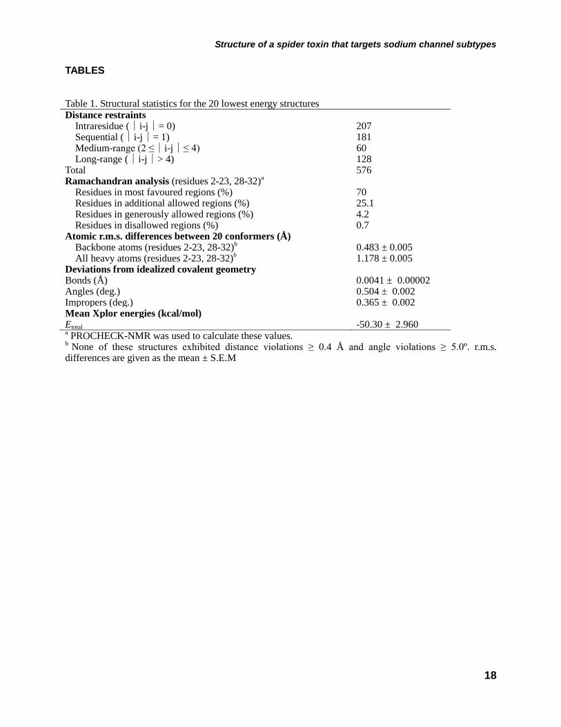
Structure of a spider toxin that targets sodium channel subtypes
18
TABLES Table 1. Structural statistics for the 20 lowest energy structures Distance restraints Intraresidue (i-j= 0) 207 Sequential (i-j= 1) 181 Medium-range (2 ≤i-j≤ 4) 60 Long-range (i-j> 4) 128 Total 576 Ramachandran analysis (residues 2-23, 28-32)a Residues in most favoured regions (%) 70 Residues in additional allowed regions (%) 25.1 Residues in generously allowed regions (%) 4.2 Residues in disallowed regions (%) 0.7 Atomic r.m.s. differences between 20 conformers (Å) Backbone atoms (residues 2-23, 28-32)b 0.483 ± 0.005 All heavy atoms (residues 2-23, 28-32)b 1.178 ± 0.005 Deviations from idealized covalent geometry Bonds (Å) 0.0041 ± 0.00002 Angles (deg.) 0.504 ± 0.002 Impropers (deg.) 0.365 ± 0.002 Mean Xplor energies (kcal/mol) Etotal -50.30 ± 2.960 a PROCHECK-NMR was used to calculate these values. b None of these structures exhibited distance violations ≥ 0.4 Å and angle violations ≥ 5.0º. r.m.s. differences are given as the mean ± S.E.M
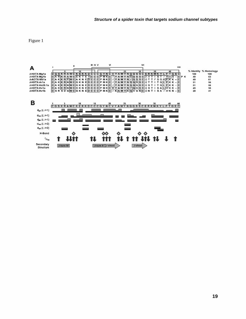
Structure of a spider toxin that targets sodium channel subtypes
19
Figure 1
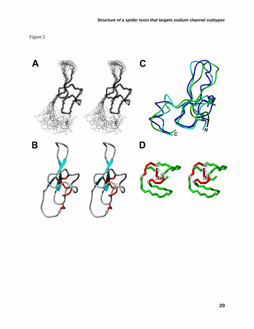
Structure of a spider toxin that targets sodium channel subtypes
20
Figure 2
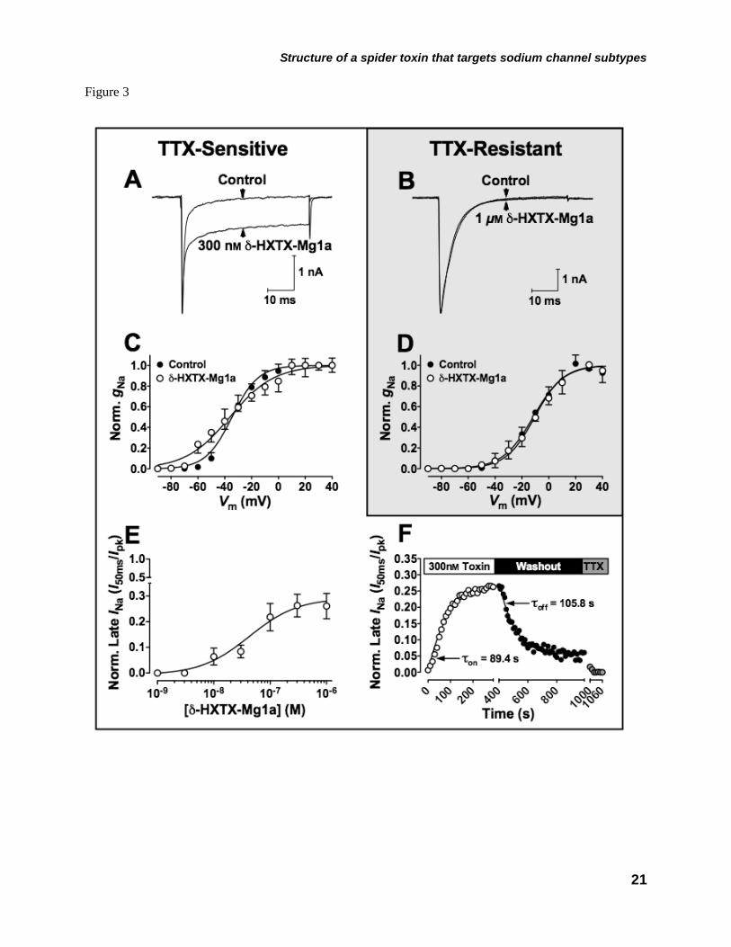
Structure of a spider toxin that targets sodium channel subtypes
21
Figure 3
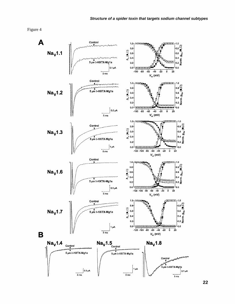
Structure of a spider toxin that targets sodium channel subtypes
22
Figure 4
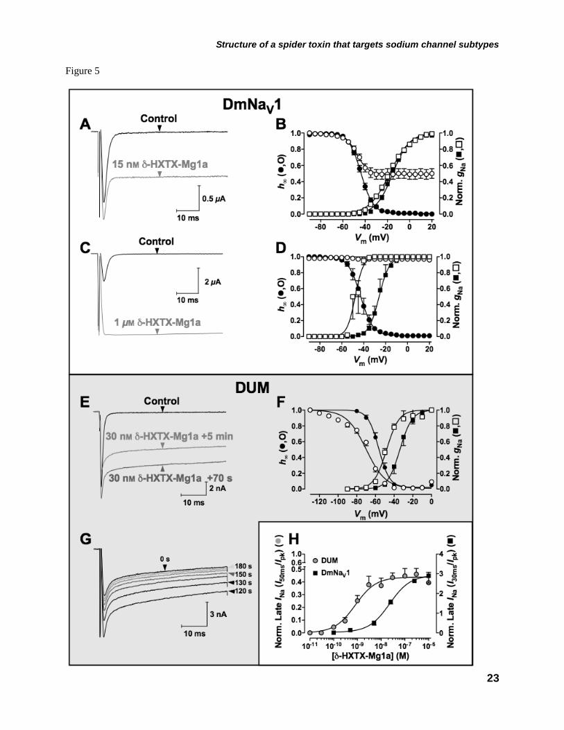
Structure of a spider toxin that targets sodium channel subtypes
23
Figure 5
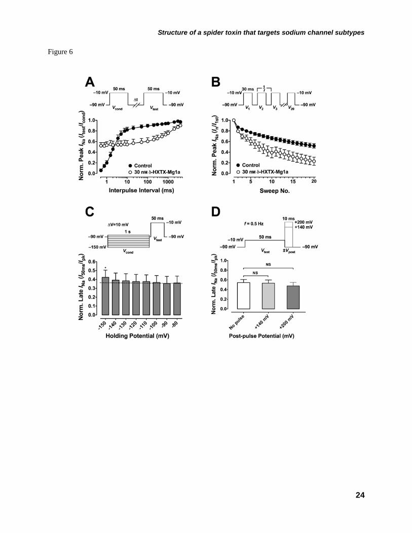
Structure of a spider toxin that targets sodium channel subtypes
24
Figure 6
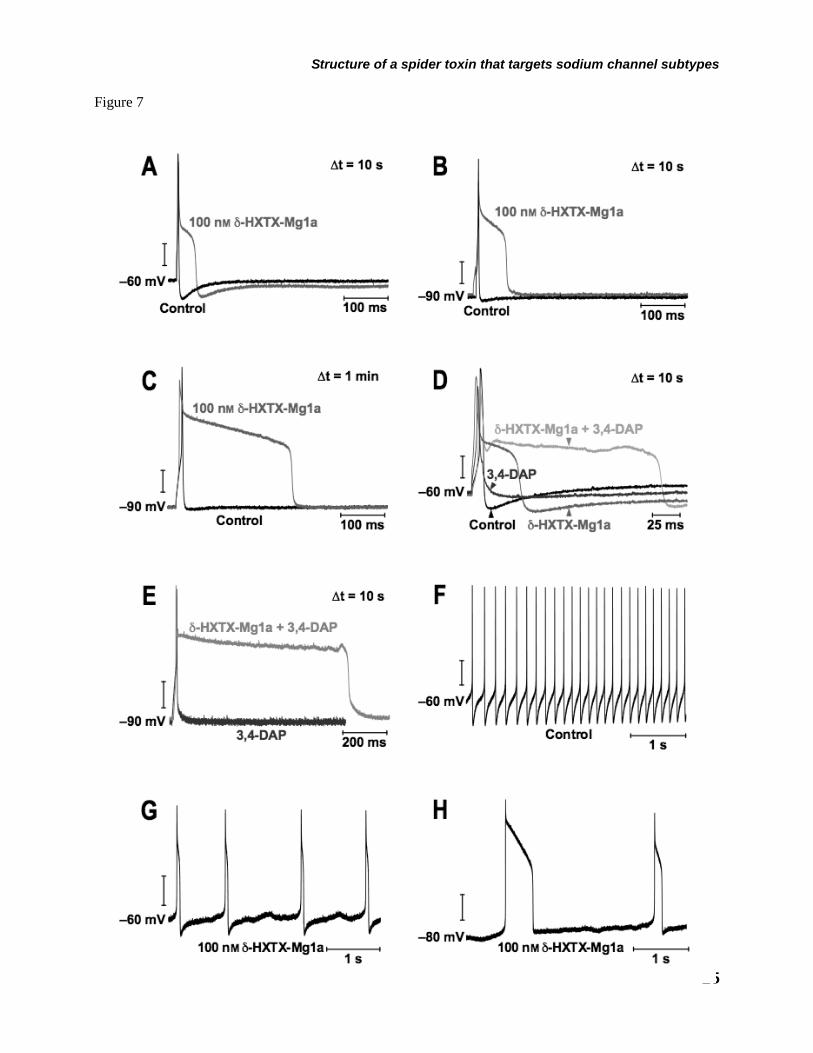
Structure of a spider toxin that targets sodium channel subtypes
25
Figure 7
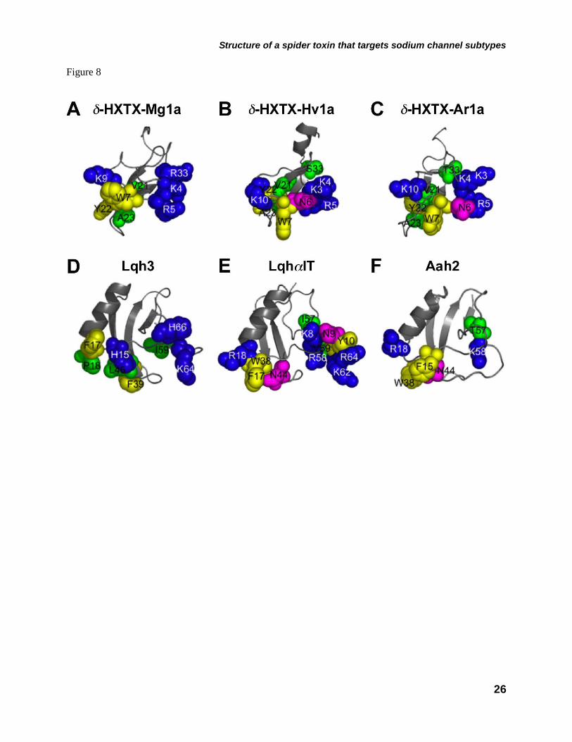
Structure of a spider toxin that targets sodium channel subtypes
26
Figure 8


![clamp Electronic Supplementary Information · Electronic Supplementary Information. Synthesis of compounds. Synthesis of [{(Dipp. Nacnac)Mg(C. 4. H. 3. N. 2)} 2] (2) To a solution](https://static.fdocument.org/doc/165x107/5f07b9817e708231d41e6c69/clamp-electronic-supplementary-electronic-supplementary-information-synthesis-of.jpg)