Surgical Anatomy of the Liver : Ηepatectomies - Dimitris P. Korkolis
67
Surgical Anatomy of the Surgical Anatomy of the Liver - Liver - Η Η epatectomies epatectomies Dimitris P. Korkolis Surgeon Saint Savvas Anticancer - Oncological Hospital of Athens
-
Upload
dimitris-p-korkolis -
Category
Health & Medicine
-
view
142 -
download
1
Transcript of Surgical Anatomy of the Liver : Ηepatectomies - Dimitris P. Korkolis

Surgical Anatomy of the Liver - Surgical Anatomy of the Liver - ΗΗepatectomiesepatectomies
Dimitris P. Korkolis
SurgeonSaint Savvas Anticancer - Oncological Hospital of Athens
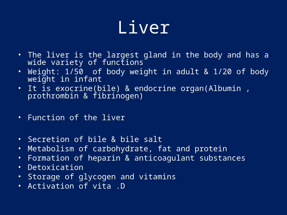
Liver• The liver is the largest gland in the body and has a wide variety of
functions• Weight: 1/50 of body weight in adult & 1/20 of body weight in infant• It is exocrine(bile) & endocrine organ(Albumin , prothrombin &
fibrinogen)
• Function of the liver
• Secretion of bile & bile salt• Metabolism of carbohydrate, fat and protein• Formation of heparin & anticoagulant substances• Detoxication• Storage of glycogen and vitamins• Activation of vita .D

EmbryologyEmbryology
44thth Week of Development Week of Development– Projection from ventral wall of Projection from ventral wall of
MidgutMidgut Cranial Bud Cranial Bud Liver Liver Caudal Bud Caudal Bud Gallbladder, extrahepatic Gallbladder, extrahepatic
biliary treebiliary tree Ventral PancreasVentral Pancreas
EmbryologyEmbryology
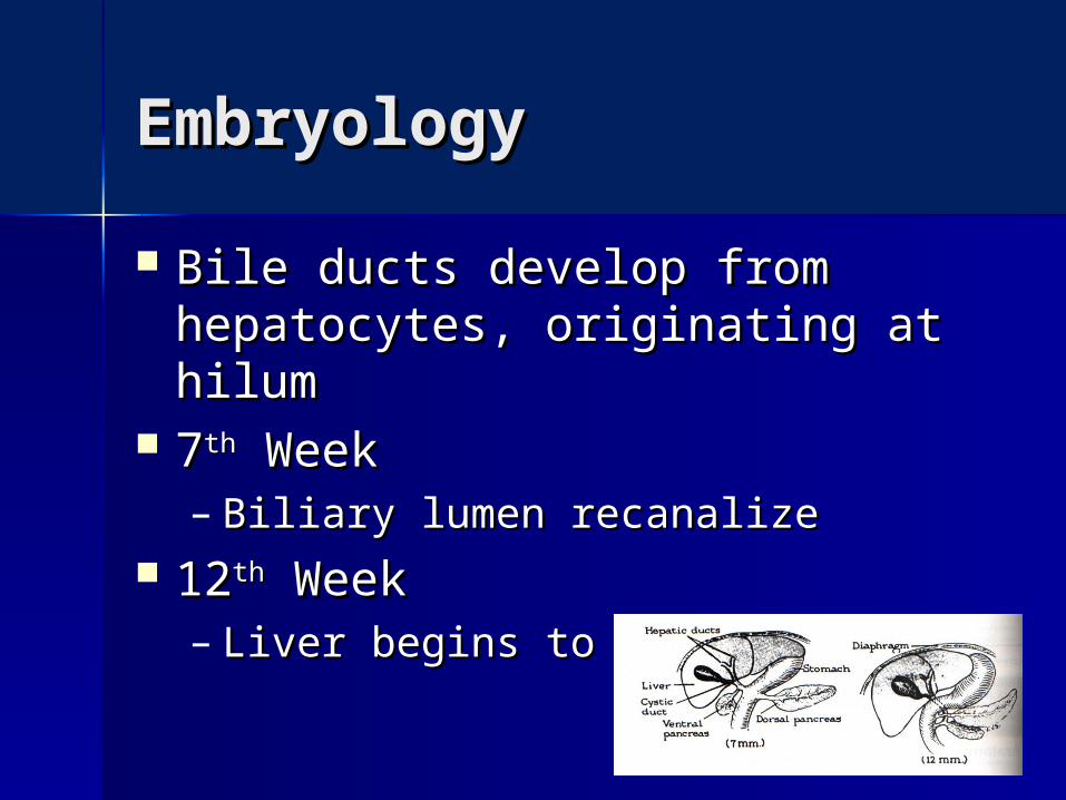
Bile ducts develop from Bile ducts develop from hepatocytes, originating at hilumhepatocytes, originating at hilum
77thth Week Week– Biliary lumen recanalize Biliary lumen recanalize
1212thth Week Week– Liver begins to secrete bileLiver begins to secrete bile

Developmental Developmental AnomaliesAnomalies Agenesis rareAgenesis rare
– Left lobe agenesis reportedLeft lobe agenesis reported Reidel’s LobeReidel’s Lobe
– Long tongue of liver extending Long tongue of liver extending inferiorly from right lobeinferiorly from right lobe
– Usually asymptomatic, may be Usually asymptomatic, may be associated with colonic or pyloric associated with colonic or pyloric obstructionobstruction
Heterotopic liver tissueHeterotopic liver tissue– Gallbladder, pancreas, adrenals, Gallbladder, pancreas, adrenals,
omphaloceleomphalocele
AnatomyAnatomy
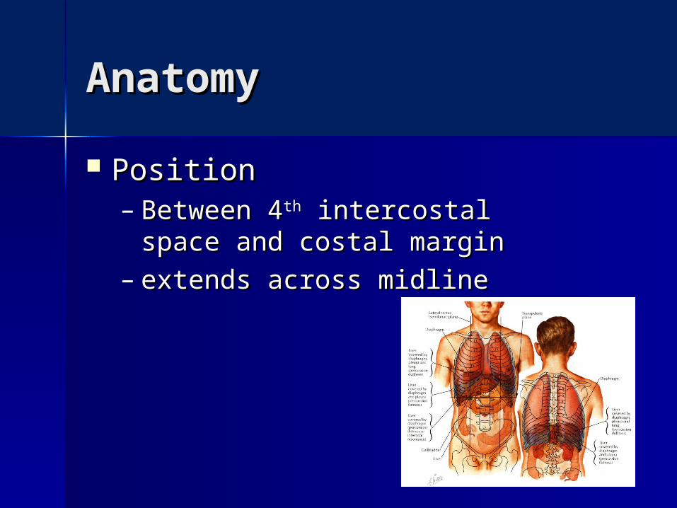
PositionPosition– Between 4Between 4thth intercostal intercostal
space and costal marginspace and costal margin– extends across midlineextends across midline
AnatomyAnatomy
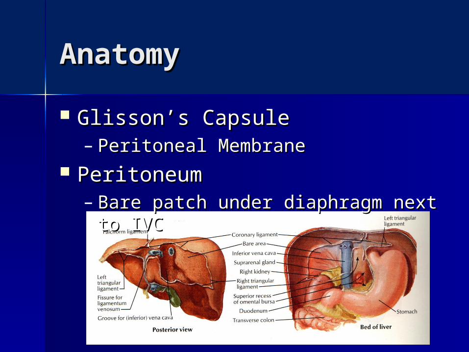
Glisson’s CapsuleGlisson’s Capsule– Peritoneal MembranePeritoneal Membrane
PeritoneumPeritoneum– Bare patch under diaphragm next to Bare patch under diaphragm next to
IVCIVC
LigamentsLigaments
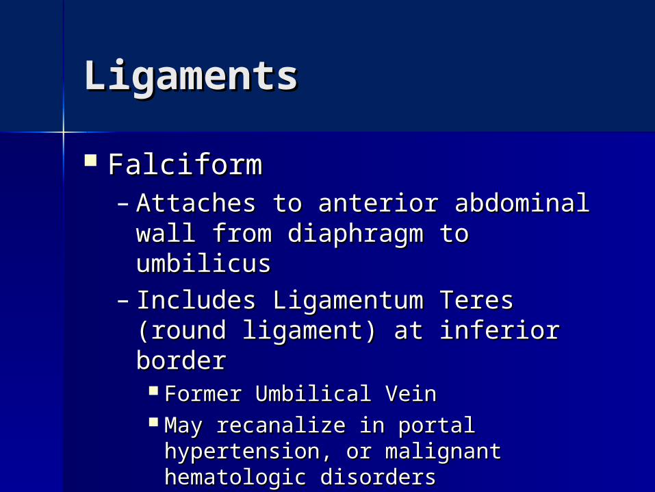
FalciformFalciform– Attaches to anterior abdominal wall Attaches to anterior abdominal wall
from diaphragm to umbilicusfrom diaphragm to umbilicus– Includes Ligamentum Teres (round Includes Ligamentum Teres (round
ligament) at inferior borderligament) at inferior border Former Umbilical VeinFormer Umbilical Vein May recanalize in portal hypertension, May recanalize in portal hypertension,
or malignant hematologic disordersor malignant hematologic disorders

LigamentsLigaments
Right and Left CoronaryRight and Left Coronary– Connect diaphragm to liverConnect diaphragm to liver– Lateral aspects become Triangular Lateral aspects become Triangular
LigamentsLigaments

LigamentsLigaments
GastrohepaticGastrohepatic– Anterior layer of lesser omentumAnterior layer of lesser omentum– Continuous with Left Triangular Continuous with Left Triangular
LigamentLigament HepatoduodenalHepatoduodenal
– Anterior border of Foramen of Anterior border of Foramen of WinslowWinslow
– Contains Portal TriadContains Portal Triad

The Ligamentum Venosum
-Fibrous band that is the remains of the ductus venosus
-Ligament of Aranthius
- It is attached to the left branch of the portal vein and ascends in a fissure on the visceral surface of the liver to be attached above to the inferior vena cava
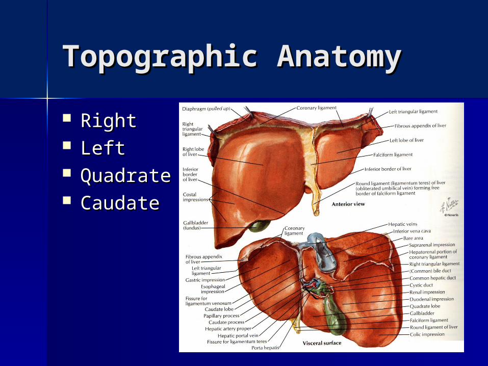
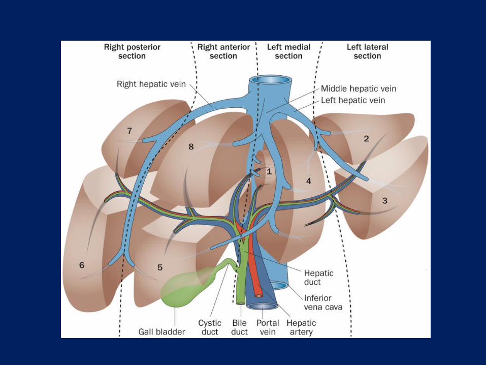
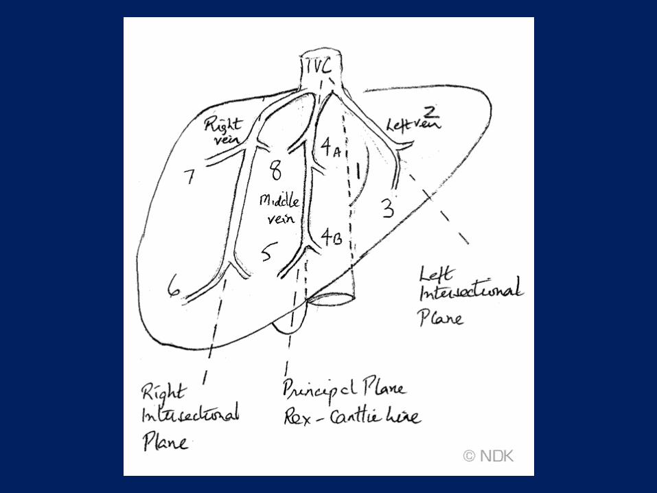
Topographic AnatomyTopographic Anatomy
RightRight LeftLeft QuadrateQuadrate CaudateCaudate
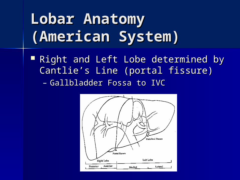
Lobar Anatomy Lobar Anatomy (American System)(American System) Right and Left Lobe determined by Right and Left Lobe determined by
Cantlie’s Line (portal fissure)Cantlie’s Line (portal fissure)– Gallbladder Fossa to IVCGallbladder Fossa to IVC
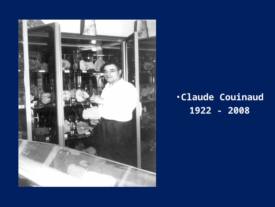
•Claude Couinaud 1922 - 2008

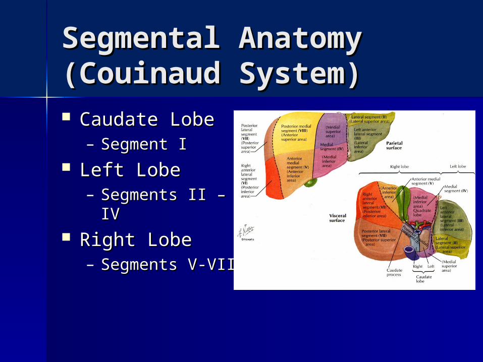
Segmental Anatomy Segmental Anatomy (Couinaud System)(Couinaud System) Caudate LobeCaudate Lobe
– Segment ISegment I Left LobeLeft Lobe
– Segments II – IV Segments II – IV Right LobeRight Lobe
– Segments V-VIIISegments V-VIII
Segmental Anatomy Segmental Anatomy (Couinaud System)(Couinaud System) Caudate LobeCaudate Lobe
– Segment ISegment I Left LobeLeft Lobe
– Segments II – IV Segments II – IV Right LobeRight Lobe
– Segments V-VIIISegments V-VIII
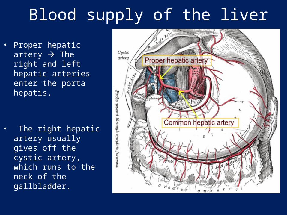
Blood supply of the liver
• Proper hepatic artery The right and left hepatic arteries enter the porta hepatis.
• The right hepatic artery usually gives off the cystic artery, which runs to the neck of the gallbladder.
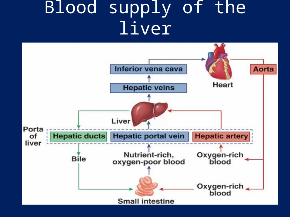
Blood supply of the liver
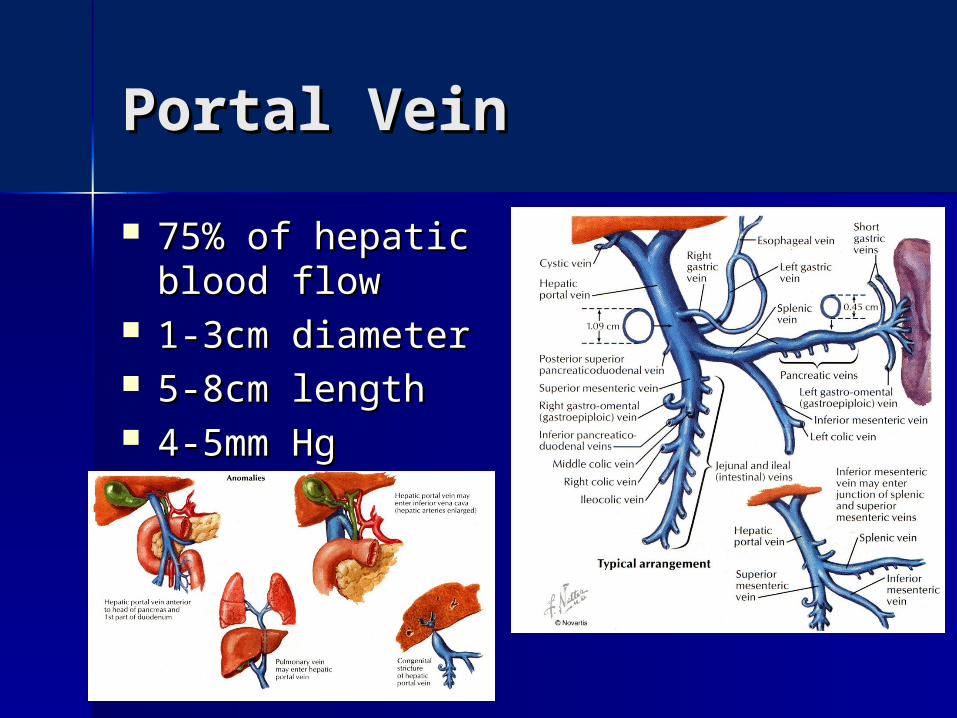
Portal VeinPortal Vein
75% of hepatic 75% of hepatic blood flowblood flow
1-3cm diameter1-3cm diameter 5-8cm length5-8cm length 4-5mm Hg4-5mm Hg

Portal VeinPortal Vein
Laminar Blood flowLaminar Blood flow– Affects distribution of amebic abscesses Affects distribution of amebic abscesses
and tumor metastasesand tumor metastases

Portal-Systemic Portal-Systemic communicationcommunication Submucosal veins of distal esophagus Submucosal veins of distal esophagus
and proximal stomachand proximal stomach Umbilical and periumbilical veinsUmbilical and periumbilical veins
– Caput MedusaeCaput Medusae– Cruveilhier-Baumgarten bruit Cruveilhier-Baumgarten bruit
Tributaries of inferior mesenteric veinsTributaries of inferior mesenteric veins– Superior hemmorhoidalsSuperior hemmorhoidals
Retroperitoneal communicationRetroperitoneal communication– Adrenal veinsAdrenal veins
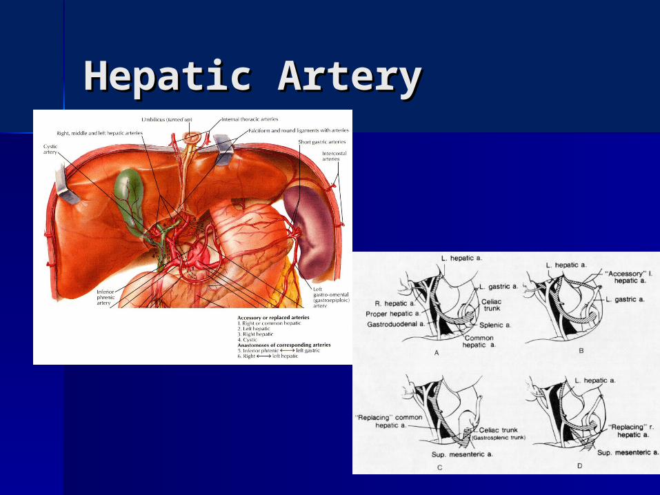
Hepatic ArteryHepatic Artery
Hepatic ArteryHepatic Artery
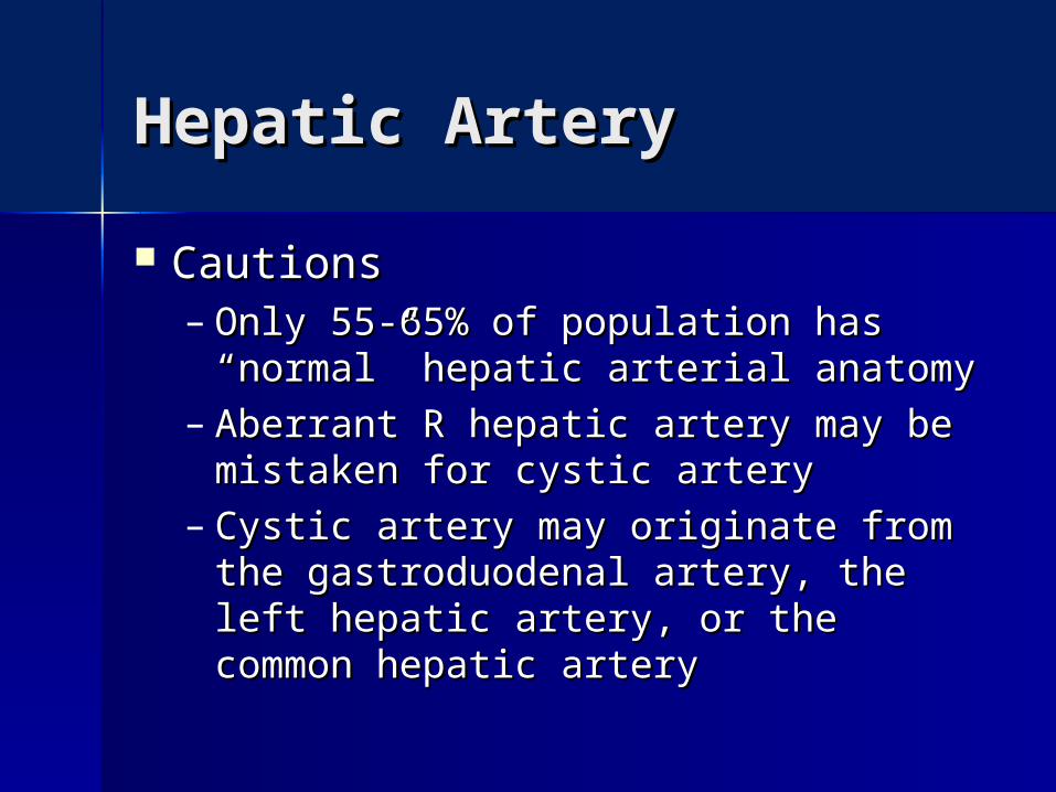
CautionsCautions– Only 55-65% of population has Only 55-65% of population has
“normal” hepatic arterial anatomy“normal” hepatic arterial anatomy– Aberrant R hepatic artery may be Aberrant R hepatic artery may be
mistaken for cystic arterymistaken for cystic artery– Cystic artery may originate from the Cystic artery may originate from the
gastroduodenal artery, the left gastroduodenal artery, the left hepatic artery, or the common hepatic artery, or the common hepatic arteryhepatic artery
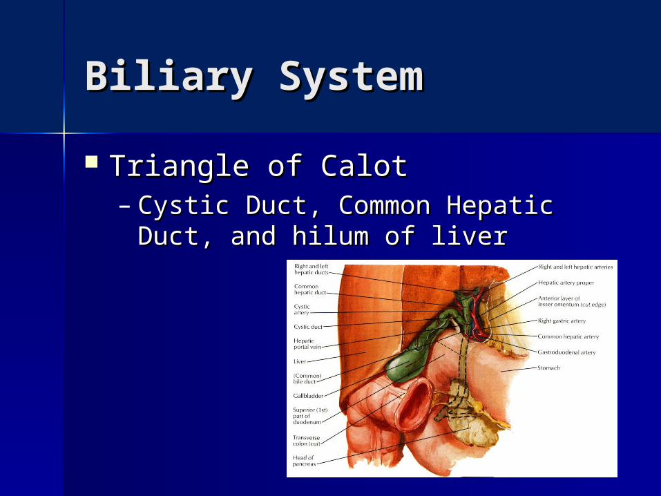
Biliary SystemBiliary System
Triangle of CalotTriangle of Calot– Cystic Duct, Common Hepatic Duct, Cystic Duct, Common Hepatic Duct,
and hilum of liverand hilum of liver
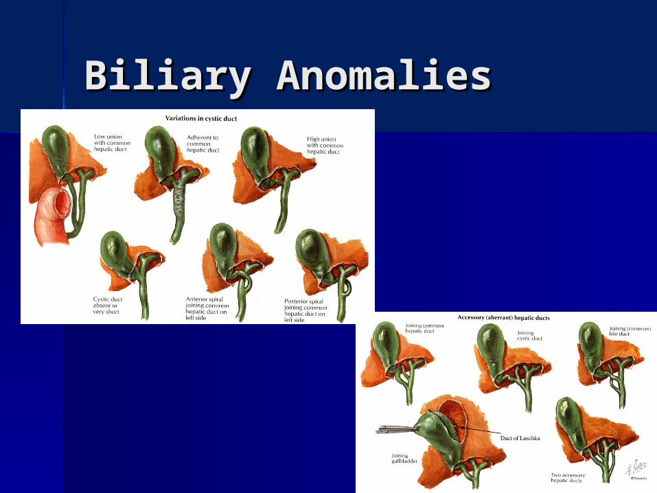
Biliary AnomaliesBiliary Anomalies

Hepatic VeinsHepatic Veins
Short extrahepatic Short extrahepatic segmentsegment– RightRight– MiddleMiddle
Usually joins leftUsually joins left
– LeftLeft Direct communication to IVC from Direct communication to IVC from
Segment I (caudate)Segment I (caudate)
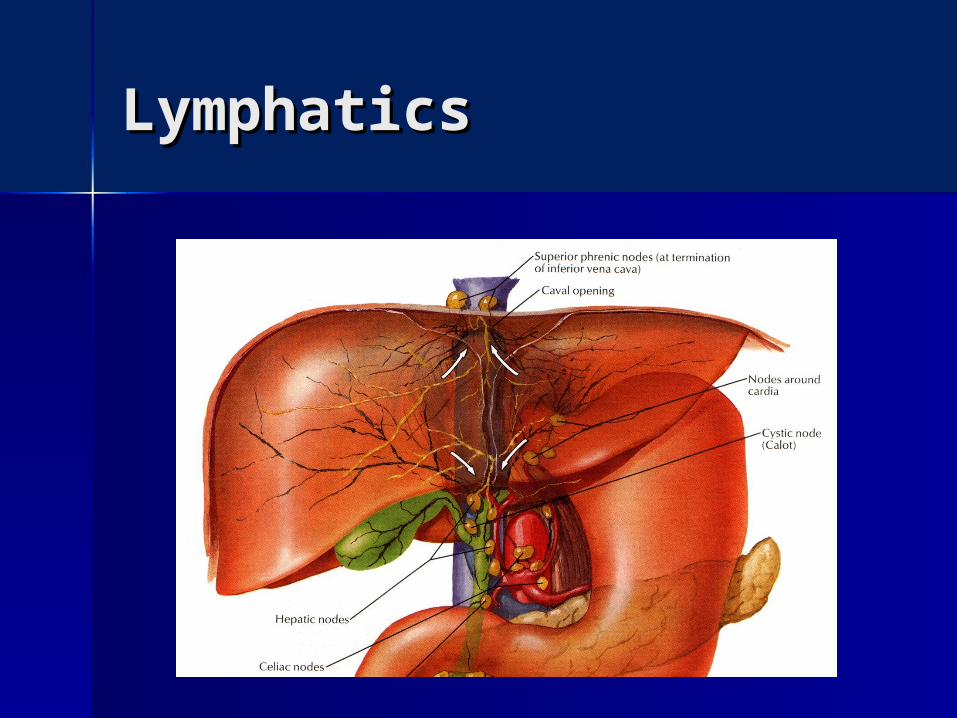
LymphaticsLymphatics
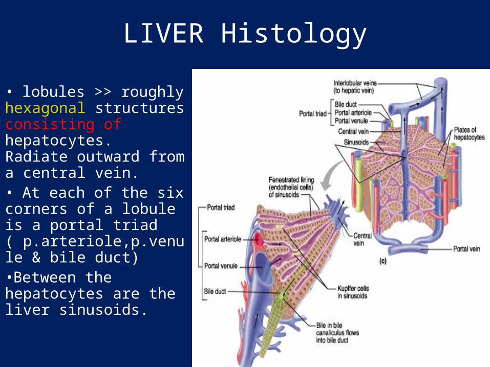
LIVER Histology
• lobules >> roughly hexagonal structures consisting of hepatocytes. Radiate outward from a central vein. • At each of the six corners of a lobule is a portal triad ( p.arteriole,p.venule & bile duct) •Between the hepatocytes are the liver sinusoids.
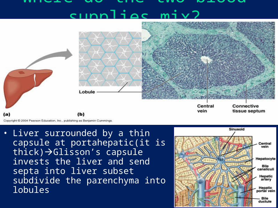
Where do the two blood supplies mix?
• Liver surrounded by a thin capsule at portahepatic(it is thick)Glisson’s capsule invests the liver and send septa into liver subset subdivide the parenchyma into lobules
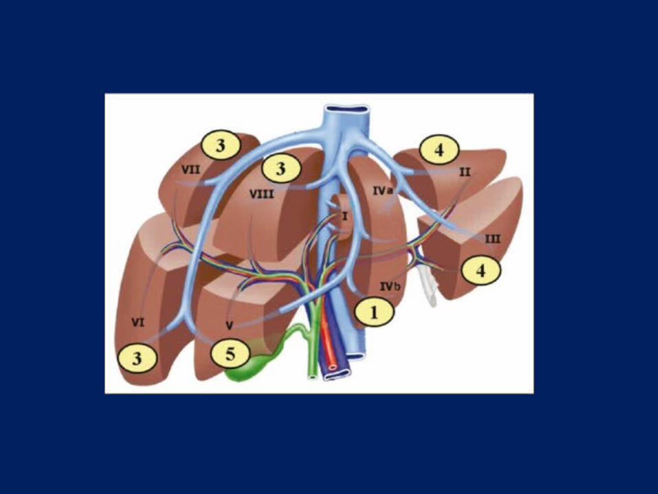
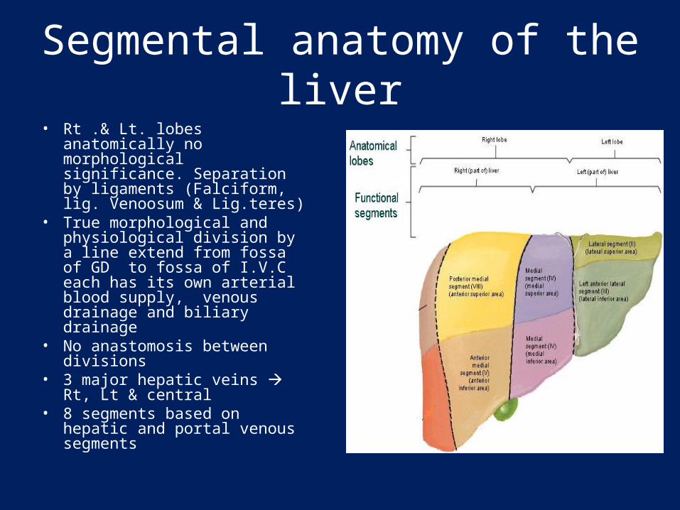
Segmental anatomy of the liver• Rt .& Lt. lobes anatomically no
morphological significance. Separation by ligaments (Falciform, lig. Venoosum & Lig.teres)
• True morphological and physiological division by a line extend from fossa of GD to fossa of I.V.C each has its own arterial blood supply, venous drainage and biliary drainage
• No anastomosis between divisions
• 3 major hepatic veins Rt, Lt & central
• 8 segments based on hepatic and portal venous segments
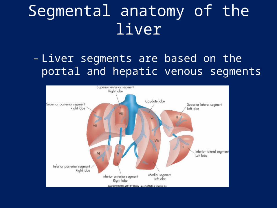
Segmental anatomy of the liver
– Liver segments are based on the portal and hepatic venous segments
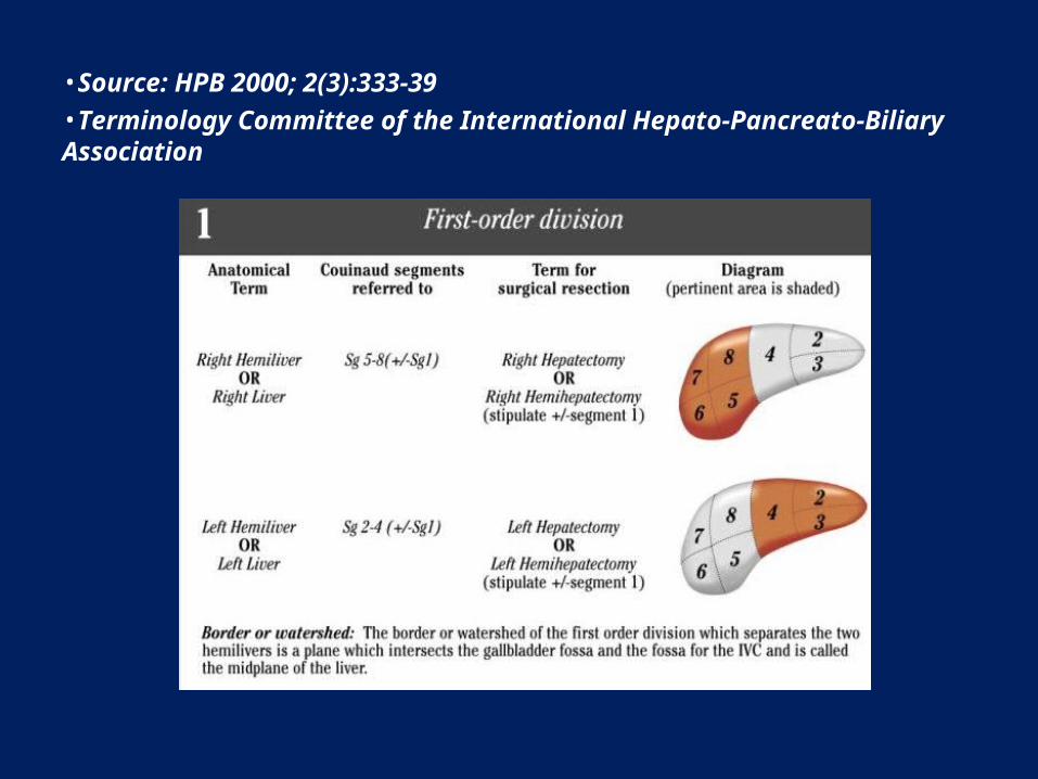
•Source: HPB 2000; 2(3):333-39•Terminology Committee of the International Hepato-Pancreato-Biliary Association