Supporting Information Experimetal details … Information Experimetal details Chemicals: McCoy’s...
4
Supporting Information Experimetal details Chemicals: McCoy’s 5α medium and fetal bovine serum (FBS) were purchased from Invitrogen, Carlsbad, CA, USA. 5-dimethylthiazol-2-yl-2,5-diphenyl tetrazolium bromide (MTT), Triton X-100, o-phenylenediamine (o-PDA) and were supplied by Sigma. N,N- dimethylformamide (DMF) was supplied by MBI, USA. Dimethyl sulfoxide (DMSO) was supplied by J. T. Baker, USA. Loading and Release Measurement of Cisplatin into/from HPB: For loading of cisplatin, 5 mg of HPB nanoparticles was added into 3 mL of cisplatin aqueous solution (2 mg/mL) with stirring in dark. After 24 hours, the HPB nanoparticles were separated via centrifuge. The concentration of cisplatin in the supernatant was determined by mixing the supernatant with o-phenylenediamine solution in DMF (1.2 mg/mL) at a volume ratio of 1:1. The mixture was heated at 100 °C for 10 min, then the absorbance at 704 nm was measure by Jusco V-570 UV/VIS/NIR spectrophotometer. For release of cisplatin, the cisplatin-loaded HPB nanoparticles (5 mg) were added into PBS buffer (pH 7.4, 5 mL) at 37 °C without stirring. The release profile of cisplatin from cisplatin-loaded HPB nanoparticles was measured by UV/VIS spectrophotometer. Cell culture: Human bladder carcinoma T24 cells were purchased from the Bioresource Collection and Research Center (BCRC, Hsinchu, Taiwan). The T24 cell line was cultured as monolayer with McCoy’s 5α medium supplemented with 10% fetal bovine serum in an incubator with humidified atmosphere of 95% air and 5% CO 2 at 37°C. Under these conditions, the plating efficiency was 90% and the doubling time was 19 hours. Single cell suspensions were obtained by trypsinization of monolayer cultures. Cell viability: T-24 cells were seeded in 96-well culture plates at 1×10 5 cells/well. After incubation overnight, the cells were treated with varying concentrations of HPB nanoparticles (0, 50, 100, 250, 500, 750 and 1000 μg/mL) and cisplatin-loaded HPB (0, 50, 100, 150, and 200 μg/mL) for 24 hours. Then, the cells were treated with 0.5 mg/mL MTT for 1 hour and the formazan crystals produced were homogenized in 100 μL of DMSO. Optical densities (OD) were read at 570 nm and 690 nm using a microplate reader (Molecular Devices, USA). The absorbance was recorded at 570 nm with a reference at 690 nm. The cells incubated in culture medium alone or 0.01% Triton X-100 served as a negative control or cell death positive control for cell viability. The effect of HPB NPs on cell viability was assessed by the relative percentage of viable cells that was normalized to that of the negative control, which Electronic Supplementary Material (ESI) for Chemical Communications This journal is © The Royal Society of Chemistry 2012
Transcript of Supporting Information Experimetal details … Information Experimetal details Chemicals: McCoy’s...
Supporting Information
Experimetal details
Chemicals: McCoy’s 5α medium and fetal bovine serum (FBS) were purchased from
Invitrogen, Carlsbad, CA, USA. 5-dimethylthiazol-2-yl-2,5-diphenyl tetrazolium bromide
(MTT), Triton X-100, o-phenylenediamine (o-PDA) and were supplied by Sigma. N,N-
dimethylformamide (DMF) was supplied by MBI, USA. Dimethyl sulfoxide (DMSO) was
supplied by J. T. Baker, USA.
Loading and Release Measurement of Cisplatin into/from HPB: For loading of cisplatin, 5
mg of HPB nanoparticles was added into 3 mL of cisplatin aqueous solution (2 mg/mL) with
stirring in dark. After 24 hours, the HPB nanoparticles were separated via centrifuge. The
concentration of cisplatin in the supernatant was determined by mixing the supernatant with
o-phenylenediamine solution in DMF (1.2 mg/mL) at a volume ratio of 1:1. The mixture was
heated at 100 °C for 10 min, then the absorbance at 704 nm was measure by Jusco V-570
UV/VIS/NIR spectrophotometer. For release of cisplatin, the cisplatin-loaded HPB
nanoparticles (5 mg) were added into PBS buffer (pH 7.4, 5 mL) at 37 °C without stirring.
The release profile of cisplatin from cisplatin-loaded HPB nanoparticles was measured by
UV/VIS spectrophotometer.
Cell culture: Human bladder carcinoma T24 cells were purchased from the Bioresource
Collection and Research Center (BCRC, Hsinchu, Taiwan). The T24 cell line was cultured as
monolayer with McCoy’s 5α medium supplemented with 10% fetal bovine serum in an
incubator with humidified atmosphere of 95% air and 5% CO2 at 37°C. Under these
conditions, the plating efficiency was 90% and the doubling time was 19 hours. Single cell
suspensions were obtained by trypsinization of monolayer cultures.
Cell viability: T-24 cells were seeded in 96-well culture plates at 1×10⁵ cells/well. After
incubation overnight, the cells were treated with varying concentrations of HPB nanoparticles
(0, 50, 100, 250, 500, 750 and 1000 μg/mL) and cisplatin-loaded HPB (0, 50, 100, 150, and
200 μg/mL) for 24 hours. Then, the cells were treated with 0.5 mg/mL MTT for 1 hour and
the formazan crystals produced were homogenized in 100 μL of DMSO. Optical densities
(OD) were read at 570 nm and 690 nm using a microplate reader (Molecular Devices, USA).
The absorbance was recorded at 570 nm with a reference at 690 nm. The cells incubated in
culture medium alone or 0.01% Triton X-100 served as a negative control or cell death
positive control for cell viability. The effect of HPB NPs on cell viability was assessed by the
relative percentage of viable cells that was normalized to that of the negative control, which
Electronic Supplementary Material (ESI) for Chemical CommunicationsThis journal is © The Royal Society of Chemistry 2012
were arbitrarily assigned 100% viability.
Confocal Microscopy: T-24 cells were seeded at a density of 1 x 10⁵ cells per well in a 4-
well culture plate with Lab-Tek chambered coverglasses at the bottom of each well. After
incubation overnight, the T-24 cells attached to the plate, and the MEM medium was replaced
with different concentrations of drug-loaded nanoparticles in serum-free MEM medium (0.5
mL/well). The cell-plated coverglasses were then washed with PBS solution three times. A
DAPI PBS buffer (2.80E-5 M, 1 mL) was then added to stain the cell nuclei for 30-60 minutes.
After being washed with PBS solution, DAPI-stained coverglasses were examined to check
the cellular localization of particles using a confocal fluorescence microscopy system (Leica
TCS SP5 II) with a 63x oil immersion objective lens.
Electronic Supplementary Material (ESI) for Chemical CommunicationsThis journal is © The Royal Society of Chemistry 2012
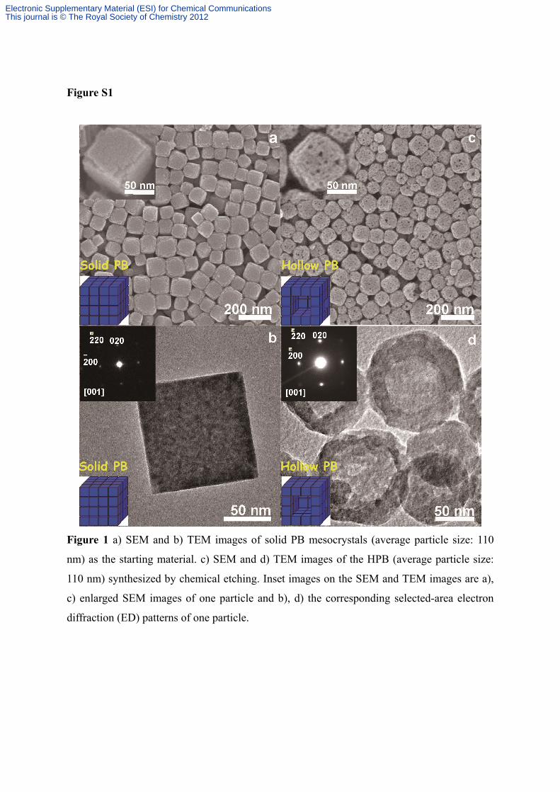
Figure S1
Figure 1 a) SEM and b) TEM images of solid PB mesocrystals (average particle size: 110
nm) as the starting material. c) SEM and d) TEM images of the HPB (average particle size:
110 nm) synthesized by chemical etching. Inset images on the SEM and TEM images are a),
c) enlarged SEM images of one particle and b), d) the corresponding selected-area electron
diffraction (ED) patterns of one particle.
Electronic Supplementary Material (ESI) for Chemical CommunicationsThis journal is © The Royal Society of Chemistry 2012
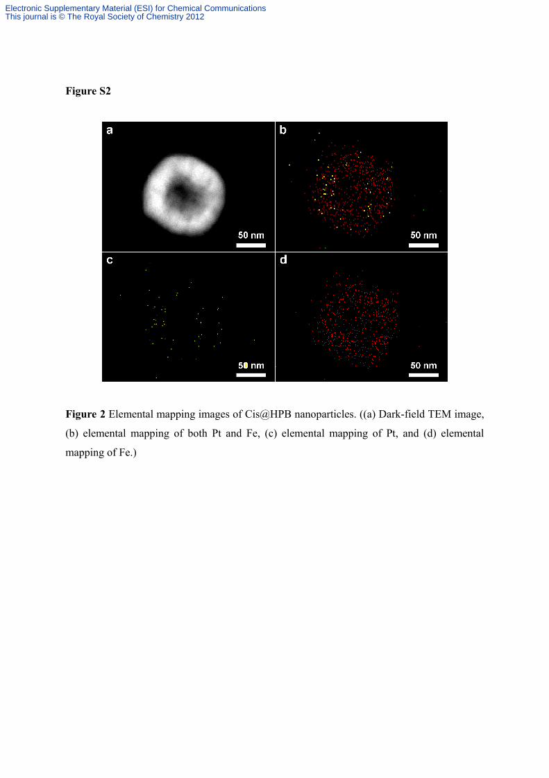
Figure S2
Figure 2 Elemental mapping images of Cis@HPB nanoparticles. ((a) Dark-field TEM image,
(b) elemental mapping of both Pt and Fe, (c) elemental mapping of Pt, and (d) elemental
mapping of Fe.)
Electronic Supplementary Material (ESI) for Chemical CommunicationsThis journal is © The Royal Society of Chemistry 2012