Supernatants of Escherichia coli K-12 Wildtype Strain and ...
12
Undergraduate Journal of Experimental Microbiology and Immunology (UJEMI) Vol. 24:1-12 Copyright © September 2019, M&I UBC September 2019 Vol. 24:1-12 Undergraduate Research Article https://jemi.microbiology.ubc.ca/ 1 Supernatants of Escherichia coli K-12 Wildtype Strain and ΔOmpC Mutant Strains do not Confer Resistance to T4 Bacteriophage Infection of K-12 WT cells Harvir Grewal, Zhong (Zack) Dang, Yu Sen (Sam) Zhai, JaeHyung (Duke) Sheen Department of Microbiology and Immunology, University of British Columbia, Vancouver, British Columbia, Canada SUMMARY T4 bacteriophage has been well established in its ability to infect Escherichia coli K-12 strains, particularly strain BW25113, through an OmpC-dependent pathway with outer membrane porin C (OmpC) and lipopolysaccharide (LPS) as receptors. Previous studies have suggested that E. coli may have a protective mechanism against phage infection that involves the production and release of OmpC-containing outer membrane vesicles (OMVs) into the culture supernatant as decoys to neutralize phage particles. In this study, we investigated if there exists a difference in the ability of T4 to infect the wildtype (WT) K-12 strain BW25113 in presence of OmpC-containing BW25113 supernatant compared to K-12 ΔompC mutant strain JW2203-1 supernatant. We hypothesized that incubating BW25113 with WT supernatant would confer an observable level of resistance to T4 infection, whilst incubation of BW25113 with ΔompC mutant supernatant would yield negligible protective effects. To test our hypothesis, we performed several growth curve assays in which BW25113 was grown in either WT or ΔompC mutant supernatant, infected with T4 bacteriophage upon inoculation and grown for up to 8 hours. Contrary to our hypothesis, the experimental results demonstrated that there is no difference in T4-mediated cell lysis between BW25113 incubation with WT versus ΔompC mutant supernatant. INTRODUCTION he continued decline in the effectiveness of antibiotics due to widespread antibiotic resistance has initiated searches for alternatives. Many researchers are revisiting phage therapy, the use of bacteria-specific viruses to treat bacterial infections. While this therapy was discovered in the early 1900s, progress in research largely stopped due to the discovery of antibiotics (1, 2). With phage therapy becoming a promising potential alternative to various antibiotic-treatments in the near future, a better understanding of phage-host interactions is required. Bacteriophage T4 is a double-stranded DNA phage that infects Escherichia coli (3). It is one of seven E. coli-specific phages (T1-T7), with the main host for T4 being E. coli K- 12 (3, 4). T4 belongs to the Myoviridae family, a group of viruses characterized by the presence of a contractile tail on each virion (4, 5). The contractile tail consists of the baseplate complex, central tube, contractile sheath, and terminator complex (6). The first step in the infection cycle is adsorption. Adsorption involves the recognition of host receptor molecules by T4 long tail fibers (LTFs) and subsequent irreversible binding of short tail fibers (STFs) to the host cell (4). LTFs and STFs are components of the baseplate, with irreversible binding of STFs triggering a conformational change in the baseplate resulting in the contraction of the contractile sheath surrounding the central tube (5). This drives the rigid central tube through the cell envelope with the help of a lysozyme domain, allowing for the ejection of viral genomic DNA into the host cytoplasm (5-7). Upon entry into the cell, T4 undergoes a lytic cycle, ultimately resulting in cell lysis and release of infectious viral progeny that can go on to infect other cells. T4 is able to adsorb to E. coli in an OmpC-dependent or OmpC-independent manner, depending on the strain (8). OmpC is one of the several general diffusion porins expressed by Gram-negative bacteria (9). Both OmpC and lipopolysaccharide (LPS) are receptors required for OmpC-dependent adsorption (8). LPS is an outer membrane component of Gram negative bacteria consisting of 3 regions termed lipid A, inner core, and outer core. T Published Online: 6 September 2019 Citation: Grewal H, Dang Z, Zhai YS, Sheen J. 2019. Supernatants of Escherichia coli K-12 Wildtype Strain and ΔOmpC Mutant Strains do not Confer Resistance to T4 Bacteriophage Infection of K-12 WT cells. UJEMI 24:1-12 Editor: Julia Huggins, University of British Columbia Copyright: © 2019 Undergraduate Journal of Experimental Microbiology and Immunology. All Rights Reserved. Address correspondence to: https://jemi.microbiology.ubc.ca/
Transcript of Supernatants of Escherichia coli K-12 Wildtype Strain and ...
Undergraduate Journal of Experimental Microbiology and Immunology (UJEMI) Vol. 24:1-12 Copyright © September 2019, M&I UBC
September 2019 Vol. 24:1-12 Undergraduate Research Article https://jemi.microbiology.ubc.ca/
1
Supernatants of Escherichia coli K-12 Wildtype Strain and ΔOmpC Mutant Strains do not Confer Resistance to T4 Bacteriophage Infection of K-12 WT cells Harvir Grewal, Zhong (Zack) Dang, Yu Sen (Sam) Zhai, JaeHyung (Duke) Sheen Department of Microbiology and Immunology, University of British Columbia, Vancouver, British Columbia, Canada SUMMARY T4 bacteriophage has been well established in its ability to infect Escherichia coli K-12 strains, particularly strain BW25113, through an OmpC-dependent pathway with outer membrane porin C (OmpC) and lipopolysaccharide (LPS) as receptors. Previous studies have suggested that E. coli may have a protective mechanism against phage infection that involves the production and release of OmpC-containing outer membrane vesicles (OMVs) into the culture supernatant as decoys to neutralize phage particles. In this study, we investigated if there exists a difference in the ability of T4 to infect the wildtype (WT) K-12 strain BW25113 in presence of OmpC-containing BW25113 supernatant compared to K-12 ΔompC mutant strain JW2203-1 supernatant. We hypothesized that incubating BW25113 with WT supernatant would confer an observable level of resistance to T4 infection, whilst incubation of BW25113 with ΔompC mutant supernatant would yield negligible protective effects. To test our hypothesis, we performed several growth curve assays in which BW25113 was grown in either WT or ΔompC mutant supernatant, infected with T4 bacteriophage upon inoculation and grown for up to 8 hours. Contrary to our hypothesis, the experimental results demonstrated that there is no difference in T4-mediated cell lysis between BW25113 incubation with WT versus ΔompC mutant supernatant.
INTRODUCTION
he continued decline in the effectiveness of antibiotics due to widespread antibiotic resistance has initiated searches for alternatives. Many researchers are revisiting
phage therapy, the use of bacteria-specific viruses to treat bacterial infections. While this therapy was discovered in the early 1900s, progress in research largely stopped due to the discovery of antibiotics (1, 2). With phage therapy becoming a promising potential alternative to various antibiotic-treatments in the near future, a better understanding of phage-host interactions is required.
Bacteriophage T4 is a double-stranded DNA phage that infects Escherichia coli (3). It is one of seven E. coli-specific phages (T1-T7), with the main host for T4 being E. coli K-12 (3, 4). T4 belongs to the Myoviridae family, a group of viruses characterized by the presence of a contractile tail on each virion (4, 5). The contractile tail consists of the baseplate complex, central tube, contractile sheath, and terminator complex (6). The first step in the infection cycle is adsorption. Adsorption involves the recognition of host receptor molecules by T4 long tail fibers (LTFs) and subsequent irreversible binding of short tail fibers (STFs) to the host cell (4). LTFs and STFs are components of the baseplate, with irreversible binding of STFs triggering a conformational change in the baseplate resulting in the contraction of the contractile sheath surrounding the central tube (5). This drives the rigid central tube through the cell envelope with the help of a lysozyme domain, allowing for the ejection of viral genomic DNA into the host cytoplasm (5-7). Upon entry into the cell, T4 undergoes a lytic cycle, ultimately resulting in cell lysis and release of infectious viral progeny that can go on to infect other cells.
T4 is able to adsorb to E. coli in an OmpC-dependent or OmpC-independent manner, depending on the strain (8). OmpC is one of the several general diffusion porins expressed by Gram-negative bacteria (9). Both OmpC and lipopolysaccharide (LPS) are receptors required for OmpC-dependent adsorption (8). LPS is an outer membrane component of Gram negative bacteria consisting of 3 regions termed lipid A, inner core, and outer core.
T
Published Online: 6 September 2019
Citation: Grewal H, Dang Z, Zhai YS, Sheen J. 2019. Supernatants of Escherichia coli K-12 Wildtype Strain and ΔOmpC Mutant Strains do not Confer Resistance to T4 Bacteriophage Infection of K-12 WT cells. UJEMI 24:1-12
Editor: Julia Huggins, University of British Columbia
Copyright: © 2019 Undergraduate Journal of Experimental Microbiology and Immunology. All Rights Reserved.
Address correspondence to: https://jemi.microbiology.ubc.ca/
UJEMI Grewal et al.
September 2019 Volume 24: 1-12 Undergraduate Research Article https://jemi.microbiology.ubc.ca/
2
For OmpC-dependent adsorption, T4 does not exhibit specificity for the outer core sugar sequence while an intact inner core is necessary for efficient adsorption (8). When OmpC is absent, T4 can only undergo adsorption under the condition that the LPS is truncated, having only one or two terminal glucose residues without a branch (8). Therefore, the requirement of OmpC for adsorption is dependent on the LPS structure. In E. coli K-12, the receptors used by T4 for infection include both OmpC and LPS as K-12 LPS is not truncated (3, 8). K-12 LPS also lacks the postulated T4 adsorption inhibitor molecule, O-antigen, consisting of repetitive units of oligosaccharides attached to the extracellular side of the outer core (8, 10, 11).
In a recent study by Manning et al., it was discovered that isolated and purified OMVs found in bacterial culture supernatant can confer resistance to antibacterial agents, including T4 bacteriophage (12). OMVs are small, spherical buds of the bacterial outer membrane (OM) that are constitutively produced by Gram-negative bacteria such as E. coli during normal growth (9, 14). In the presence of T4, growing E. coli has been found to dramatically increase OM production and release (10). Manning et al. found that T4 can irreversibly adsorb to these membrane-derived components and become inactivated (12). Co-incubation of T4 with purified OMVs resulted in a reduction of active phage, measured as a reduction in plaque forming units (PFUs) when the T4-OMV co-incubation was plated with E. coli cells (12). T4-OMV complexes were also visualized using electron microscopy (12). Hence, OMVs act as decoys that reduce the amount of active T4 phage particles able to infect E. coli by neutralizing phage particles in the surrounding solution.
While the requirement of OmpC as a co-receptor for T4 infection of E. coli K-12 strains and the neutralization of T4 by a membrane-derived factor have been well characterized, the cellular components involved in the interactions between T4 and the membrane-derived factor have yet to be investigated (8, 12). The released envelope factor has a protein composition that is representative of the OM protein composition, and includes both LPS and OmpC molecules (13, 14). Taken together, the neutralization of T4 particles by OMVs may involve OmpC that is present as a component of the OMV structure.
In this study, we investigated whether there was a difference in the ability of T4 to infect cells of a WT K-12 strain in the presence of supernatant from the WT K-12 strain BW25113 or supernatant from the ΔompC mutant K-12 strain JW2203-1. We hypothesized that WT supernatant, in which OmpC is a component of the OMVs, would confer a level of resistance against T4 infection for WT BW25113 cells by neutralizing active T4 particles when compared to the ΔompC mutant supernatant lacking OmpC in the membrane-derived factor. Differences in susceptibility of the WT strain and ΔompC mutant strain to T4 infection were assessed by a double-overlay plaque assay and growth curve assay. A modified growth curve assay was also designed to test the hypothesis of differential infection of WT cells by T4 in the presence of WT or ΔompC mutant supernatant.
UJEMI Grewal et al.
September 2019 Volume 24: 1-12 Undergraduate Research Article https://jemi.microbiology.ubc.ca/
3
METHODS AND MATERIALS
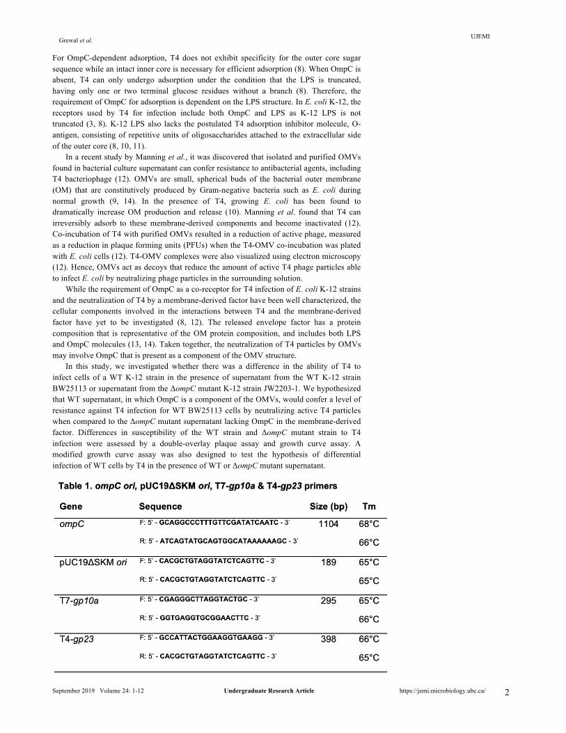
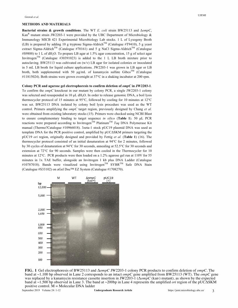
Bacterial strains & growth conditions. The WT E. coli strain BW25113 and ΔompC, KanR mutant strain JW2203-1 were provided by the UBC Department of Microbiology & Immunology MICB 421 Experimental Microbiology Lab stocks. 1 L of Lysogeny Broth (LB) is prepared by adding 10 g tryptone Sigma-AldrichTM (Catalogue #T9410), 5 g yeast extract Sigma-AldrichTM (Catalogue #70161) and 5 g NaCl Sigma-AldrichTM (Catalogue #S9888) to 1 L of dH2O. To prepare LB agar at 1.5% agar concentration, 15 g of select agar InvitrogenTM (Catalogue #30391023) is added to the 1 L LB broth mixture prior to autoclaving. BW25113 was cultivated on (w/v) LB agar for isolated colonies or inoculated in 5 mL LB broth for liquid culture applications. JW2203-1 was grown in LB agar or LB broth, both supplemented with 50 µg/mL of kanamycin sulfate GibcoTM (Catalogue #11815024). Both strains were grown overnight at 37°C in a shaking incubator at 200 rpm. Colony PCR and agarose gel electrophoresis to confirm deletion of ompC in JW2203-1. To confirm the ompC knockout in our mutant by colony PCR, a single JW2203-1 colony was selected and resuspended in 10 µL dH2O. In order to release genomic DNA, a boil lysis thermocycler protocol of 15 minutes at 95°C, followed by cooling for 10 minutes at 12°C was set. BW25113 DNA isolated by colony boil lysis procedure was used as the WT control. Primers amplifying the ompC target region, previously designed by Chang et al. were obtained from existing laboratory stocks (15). Primers were checked using NCBI Blast to ensure complementary binding to target sequence in silico (Table 1). 50 µL PCR reactions were prepared according to InvitrogenTM PlatinumTM Taq DNA Polymerase Kit manual (Thermo?Catalogue #10966018). 3zeta-1 stock pUC19 plasmid DNA was used as template DNA for the PCR positive control, amplified by pUCΔSKM primers targeting the pUC19 ori region, originally designed and provided by Fettig et al. (Table 1) (16). The thermocycler protocol consisted of an initial denaturation at 94°C for 2 minutes, followed by 30 cycles of denaturation at 94°C for 30 seconds, annealing at 52.5°C for 30 seconds and extension at 72°C for 90 seconds. Samples were then cooled in the Thermocycler for 10 minutes at 12°C. PCR products were then loaded on a 1.2% agarose gel run at 110V for 55 minutes in 1x TAE buffer, alongside an Invitrogen 1 kb plus DNA Ladder (Catalogue #10787018). Bands were visualized using InvitrogenTM SYBRTM Safe DNA Stain (Catalogue #S33102) on aGel Doc™ EZ System (Catalogue #1708270).
FIG. 1 Gel electrophoresis of BW25113 and ΔompC JW2203-1 colony PCR products to confirm deletion of ompC. The band at ~1,100 bp observed in Lane 2 corresponds to an intact ompC gene amplified from BW25113 (WT). The ompC gene was replaced by a kanamycin resistance cassette insertion in JW2203-1 (ΔompC (kanR) mutant), as shown by the expected band at ~1,500 bp observed in Lane 3. The band at ~200bp in Lane 4 represents the amplified ori region of the pUCΔSKM positive control. M = Molecular DNA ladder
UJEMI Grewal et al.
September 2019 Volume 24: 1-12 Undergraduate Research Article https://jemi.microbiology.ubc.ca/
4
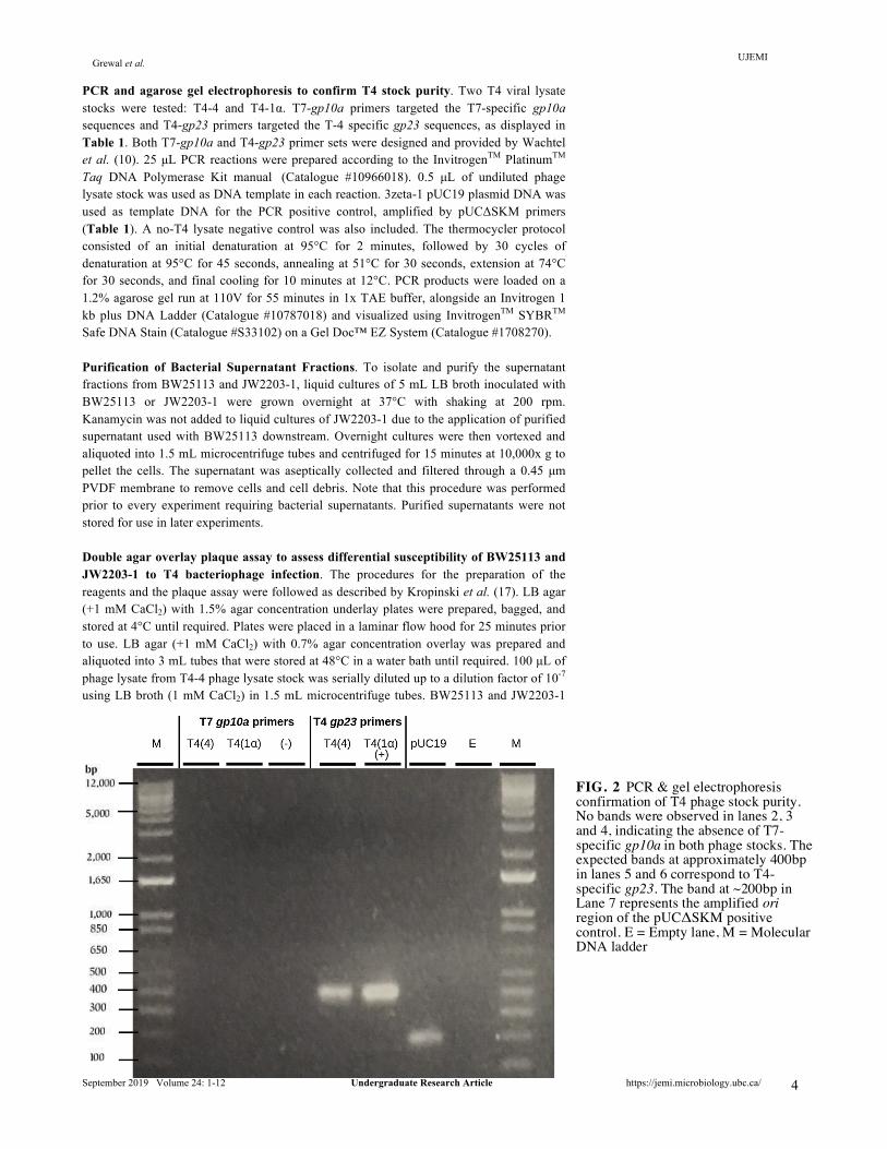
PCR and agarose gel electrophoresis to confirm T4 stock purity. Two T4 viral lysate stocks were tested: T4-4 and T4-1α. T7-gp10a primers targeted the T7-specific gp10a sequences and T4-gp23 primers targeted the T-4 specific gp23 sequences, as displayed in Table 1. Both T7-gp10a and T4-gp23 primer sets were designed and provided by Wachtel et al. (10). 25 µL PCR reactions were prepared according to the InvitrogenTM PlatinumTM Taq DNA Polymerase Kit manual (Catalogue #10966018). 0.5 µL of undiluted phage lysate stock was used as DNA template in each reaction. 3zeta-1 pUC19 plasmid DNA was used as template DNA for the PCR positive control, amplified by pUCΔSKM primers (Table 1). A no-T4 lysate negative control was also included. The thermocycler protocol consisted of an initial denaturation at 95°C for 2 minutes, followed by 30 cycles of denaturation at 95°C for 45 seconds, annealing at 51°C for 30 seconds, extension at 74°C for 30 seconds, and final cooling for 10 minutes at 12°C. PCR products were loaded on a 1.2% agarose gel run at 110V for 55 minutes in 1x TAE buffer, alongside an Invitrogen 1 kb plus DNA Ladder (Catalogue #10787018) and visualized using InvitrogenTM SYBRTM Safe DNA Stain (Catalogue #S33102) on a Gel Doc™ EZ System (Catalogue #1708270). Purification of Bacterial Supernatant Fractions. To isolate and purify the supernatant fractions from BW25113 and JW2203-1, liquid cultures of 5 mL LB broth inoculated with BW25113 or JW2203-1 were grown overnight at 37°C with shaking at 200 rpm. Kanamycin was not added to liquid cultures of JW2203-1 due to the application of purified supernatant used with BW25113 downstream. Overnight cultures were then vortexed and aliquoted into 1.5 mL microcentrifuge tubes and centrifuged for 15 minutes at 10,000x g to pellet the cells. The supernatant was aseptically collected and filtered through a 0.45 µm PVDF membrane to remove cells and cell debris. Note that this procedure was performed prior to every experiment requiring bacterial supernatants. Purified supernatants were not stored for use in later experiments. Double agar overlay plaque assay to assess differential susceptibility of BW25113 and JW2203-1 to T4 bacteriophage infection. The procedures for the preparation of the reagents and the plaque assay were followed as described by Kropinski et al. (17). LB agar (+1 mM CaCl2) with 1.5% agar concentration underlay plates were prepared, bagged, and stored at 4°C until required. Plates were placed in a laminar flow hood for 25 minutes prior to use. LB agar (+1 mM CaCl2) with 0.7% agar concentration overlay was prepared and aliquoted into 3 mL tubes that were stored at 48°C in a water bath until required. 100 µL of phage lysate from T4-4 phage lysate stock was serially diluted up to a dilution factor of 10-7 using LB broth (1 mM CaCl2) in 1.5 mL microcentrifuge tubes. BW25113 and JW2203-1
FIG. 2 PCR & gel electrophoresis confirmation of T4 phage stock purity. No bands were observed in lanes 2, 3 and 4, indicating the absence of T7-specific gp10a in both phage stocks. The expected bands at approximately 400bp in lanes 5 and 6 correspond to T4-specific gp23. The band at ~200bp in Lane 7 represents the amplified ori region of the pUCΔSKM positive control. E = Empty lane, M = Molecular DNA ladder
UJEMI Grewal et al.
September 2019 Volume 24: 1-12 Undergraduate Research Article https://jemi.microbiology.ubc.ca/
5
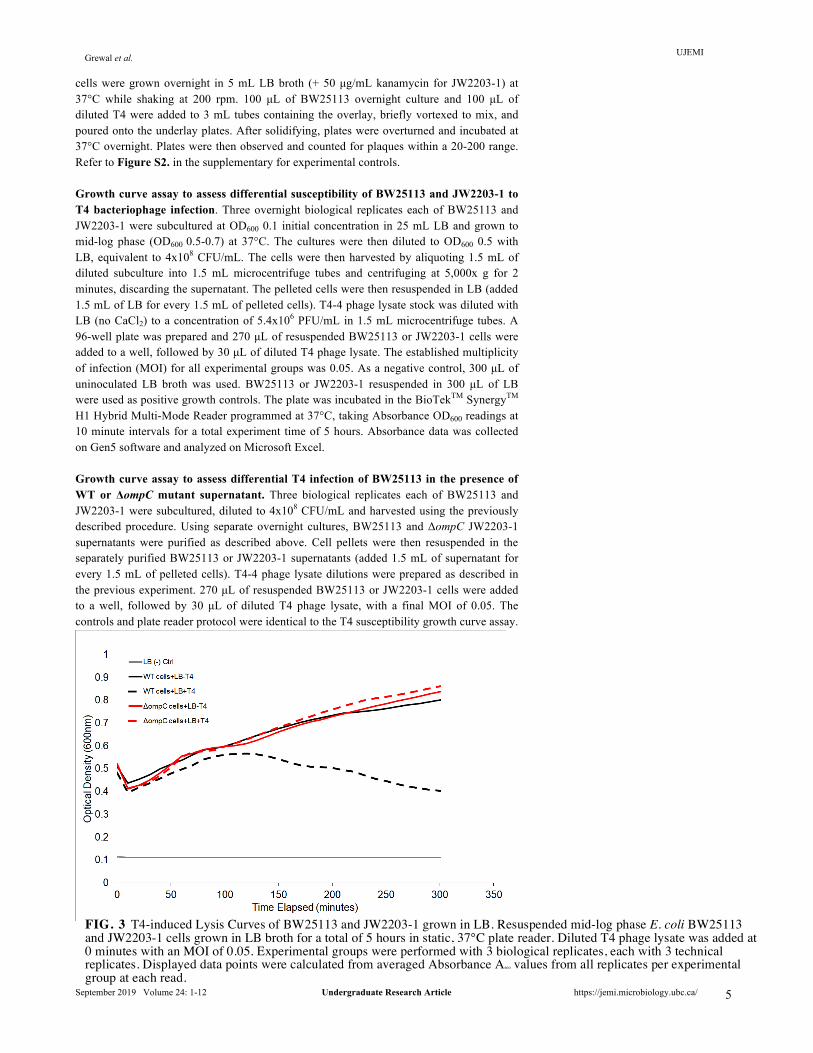
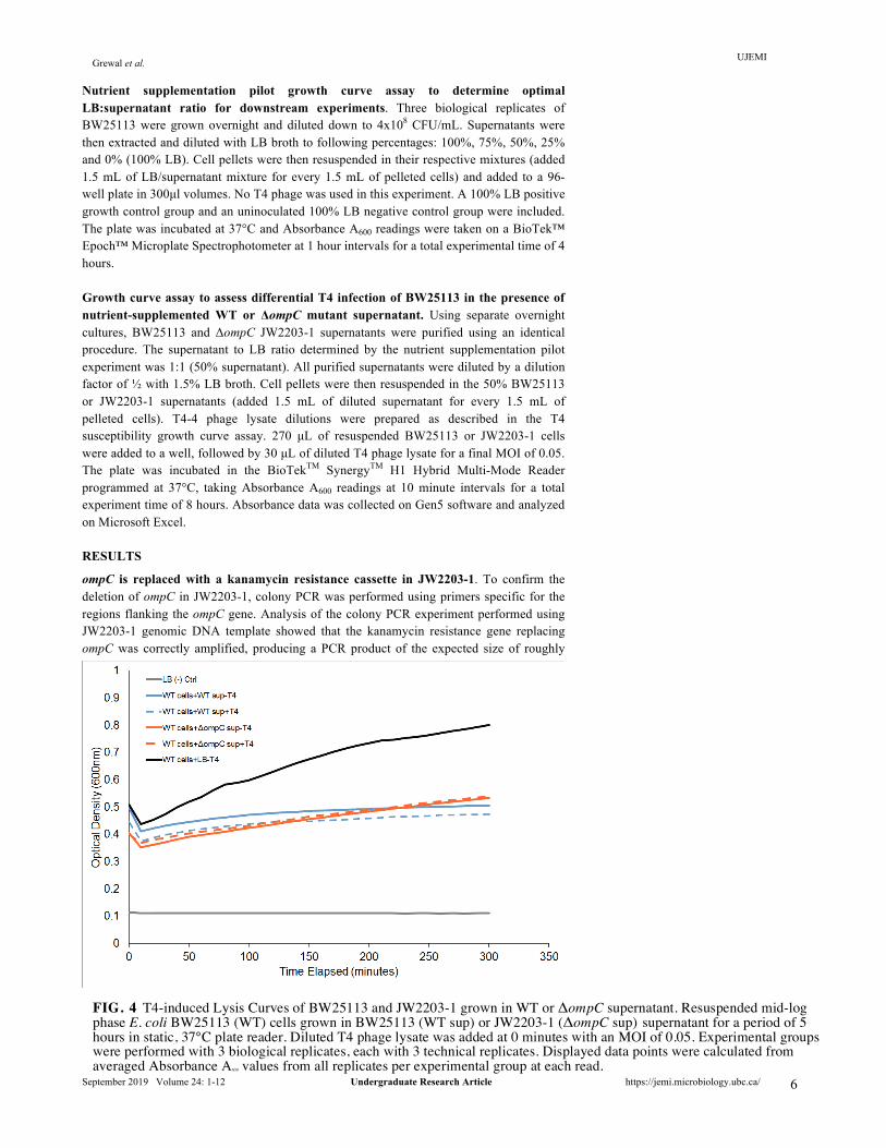
cells were grown overnight in 5 mL LB broth (+ 50 µg/mL kanamycin for JW2203-1) at 37°C while shaking at 200 rpm. 100 µL of BW25113 overnight culture and 100 µL of diluted T4 were added to 3 mL tubes containing the overlay, briefly vortexed to mix, and poured onto the underlay plates. After solidifying, plates were overturned and incubated at 37°C overnight. Plates were then observed and counted for plaques within a 20-200 range. Refer to Figure S2. in the supplementary for experimental controls. Growth curve assay to assess differential susceptibility of BW25113 and JW2203-1 to T4 bacteriophage infection. Three overnight biological replicates each of BW25113 and JW2203-1 were subcultured at OD600 0.1 initial concentration in 25 mL LB and grown to mid-log phase (OD600 0.5-0.7) at 37°C. The cultures were then diluted to OD600 0.5 with LB, equivalent to 4x10⁸ CFU/mL. The cells were then harvested by aliquoting 1.5 mL of diluted subculture into 1.5 mL microcentrifuge tubes and centrifuging at 5,000x g for 2 minutes, discarding the supernatant. The pelleted cells were then resuspended in LB (added 1.5 mL of LB for every 1.5 mL of pelleted cells). T4-4 phage lysate stock was diluted with LB (no CaCl2) to a concentration of 5.4x10⁶ PFU/mL in 1.5 mL microcentrifuge tubes. A 96-well plate was prepared and 270 µL of resuspended BW25113 or JW2203-1 cells were added to a well, followed by 30 µL of diluted T4 phage lysate. The established multiplicity of infection (MOI) for all experimental groups was 0.05. As a negative control, 300 µL of uninoculated LB broth was used. BW25113 or JW2203-1 resuspended in 300 µL of LB were used as positive growth controls. The plate was incubated in the BioTekTM SynergyTM H1 Hybrid Multi-Mode Reader programmed at 37°C, taking Absorbance OD600 readings at 10 minute intervals for a total experiment time of 5 hours. Absorbance data was collected on Gen5 software and analyzed on Microsoft Excel. Growth curve assay to assess differential T4 infection of BW25113 in the presence of WT or ΔompC mutant supernatant. Three biological replicates each of BW25113 and JW2203-1 were subcultured, diluted to 4x10⁸ CFU/mL and harvested using the previously described procedure. Using separate overnight cultures, BW25113 and ΔompC JW2203-1 supernatants were purified as described above. Cell pellets were then resuspended in the separately purified BW25113 or JW2203-1 supernatants (added 1.5 mL of supernatant for every 1.5 mL of pelleted cells). T4-4 phage lysate dilutions were prepared as described in the previous experiment. 270 µL of resuspended BW25113 or JW2203-1 cells were added to a well, followed by 30 µL of diluted T4 phage lysate, with a final MOI of 0.05. The controls and plate reader protocol were identical to the T4 susceptibility growth curve assay.
FIG. 3 T4-induced Lysis Curves of BW25113 and JW2203-1 grown in LB. Resuspended mid-log phase E. coli BW25113 and JW2203-1 cells grown in LB broth for a total of 5 hours in static, 37°C plate reader. Diluted T4 phage lysate was added at 0 minutes with an MOI of 0.05. Experimental groups were performed with 3 biological replicates, each with 3 technical replicates. Displayed data points were calculated from averaged Absorbance A600 values from all replicates per experimental group at each read.
UJEMI Grewal et al.
September 2019 Volume 24: 1-12 Undergraduate Research Article https://jemi.microbiology.ubc.ca/
6
Nutrient supplementation pilot growth curve assay to determine optimal LB:supernatant ratio for downstream experiments. Three biological replicates of BW25113 were grown overnight and diluted down to 4x10⁸ CFU/mL. Supernatants were then extracted and diluted with LB broth to following percentages: 100%, 75%, 50%, 25% and 0% (100% LB). Cell pellets were then resuspended in their respective mixtures (added 1.5 mL of LB/supernatant mixture for every 1.5 mL of pelleted cells) and added to a 96-well plate in 300µl volumes. No T4 phage was used in this experiment. A 100% LB positive growth control group and an uninoculated 100% LB negative control group were included. The plate was incubated at 37°C and Absorbance A600 readings were taken on a BioTek™ Epoch™ Microplate Spectrophotometer at 1 hour intervals for a total experimental time of 4 hours. Growth curve assay to assess differential T4 infection of BW25113 in the presence of nutrient-supplemented WT or ΔompC mutant supernatant. Using separate overnight cultures, BW25113 and ΔompC JW2203-1 supernatants were purified using an identical procedure. The supernatant to LB ratio determined by the nutrient supplementation pilot experiment was 1:1 (50% supernatant). All purified supernatants were diluted by a dilution factor of ½ with 1.5% LB broth. Cell pellets were then resuspended in the 50% BW25113 or JW2203-1 supernatants (added 1.5 mL of diluted supernatant for every 1.5 mL of pelleted cells). T4-4 phage lysate dilutions were prepared as described in the T4 susceptibility growth curve assay. 270 µL of resuspended BW25113 or JW2203-1 cells were added to a well, followed by 30 µL of diluted T4 phage lysate for a final MOI of 0.05. The plate was incubated in the BioTekTM SynergyTM H1 Hybrid Multi-Mode Reader programmed at 37°C, taking Absorbance A600 readings at 10 minute intervals for a total experiment time of 8 hours. Absorbance data was collected on Gen5 software and analyzed on Microsoft Excel. RESULTS
ompC is replaced with a kanamycin resistance cassette in JW2203-1. To confirm the deletion of ompC in JW2203-1, colony PCR was performed using primers specific for the regions flanking the ompC gene. Analysis of the colony PCR experiment performed using JW2203-1 genomic DNA template showed that the kanamycin resistance gene replacing ompC was correctly amplified, producing a PCR product of the expected size of roughly
FIG. 4 T4-induced Lysis Curves of BW25113 and JW2203-1 grown in WT or ΔompC supernatant. Resuspended mid-log phase E. coli BW25113 (WT) cells grown in BW25113 (WT sup) or JW2203-1 (ΔompC sup) supernatant for a period of 5 hours in static, 37°C plate reader. Diluted T4 phage lysate was added at 0 minutes with an MOI of 0.05. Experimental groups were performed with 3 biological replicates, each with 3 technical replicates. Displayed data points were calculated from averaged Absorbance A600 values from all replicates per experimental group at each read.
UJEMI Grewal et al.
September 2019 Volume 24: 1-12 Undergraduate Research Article https://jemi.microbiology.ubc.ca/
7
1,500 bp, as displayed in Figure 1. As a positive control, we also verified that ompC is present in the BW25113 (WT) strain. Results of the colony PCR using BW25113 genomic DNA template showed that a band of the expected 1,100 bp size was produced Figure 1. Thus, we verified that ompC was replaced by a kanamycin resistance cassette in the ΔompC mutant JW2203-1. T4-4 and T4-1α bacteriophage contain T4 and are not contaminated with T7 bacteriophage. To confirm the purity of stock T4-4 bacteriophage prior to use in downstream experiments, PCR was performed on both phage stocks to test for T7 phage contamination. Note that T4-4 phage lysate stock was used for all experiments. Gel electrophoresis of the PCR products in Figure 2. show that no bands corresponding to T7-specific gp10a (295 bp) were observed in both bacteriophage stocks. Additionally, amplification of T4-specific gp23 occurred as expected, producing the corresponding bands of approximately 398 bp in size. Infection of E. coli K-12 strain BW25113 by T4 is OmpC-dependent, while E. coli K-12 strain JW2203-1 is protected from T4 infection due to ompC deletion. To determine whether BW25113 and JW2203-1 differ in their susceptibility to T4 bacteriophage infection, a double agar overlay plaque overlay assay and growth curve assay were performed.. Positive growth controls with only BW25113 or JW2203-1 cells added to the overlay exhibited confluent growth after overnight incubation at 37°C, confirming cell viability in the assay and the absence of phage contamination in the reagents (Supplementary Fig. S2). No growth was observed on the overlay-only negative growth control. Small plaques of uniform shape and size were observed for the condition of BW25113 infected with T4 (Supplementary Fig. S1). To verify the viral titre of the T4-4 phage stock, the phage lysate concentration was calculated from averaged PFU counts at the 10-7 dilution factor. The final undiluted phage lysate concentration was calculated to be 7.15x10⁹ PFU/mL, as expected. As predicted, no plaques were observed in plates containing JW2203-1 infected with T4 bacteriophage.
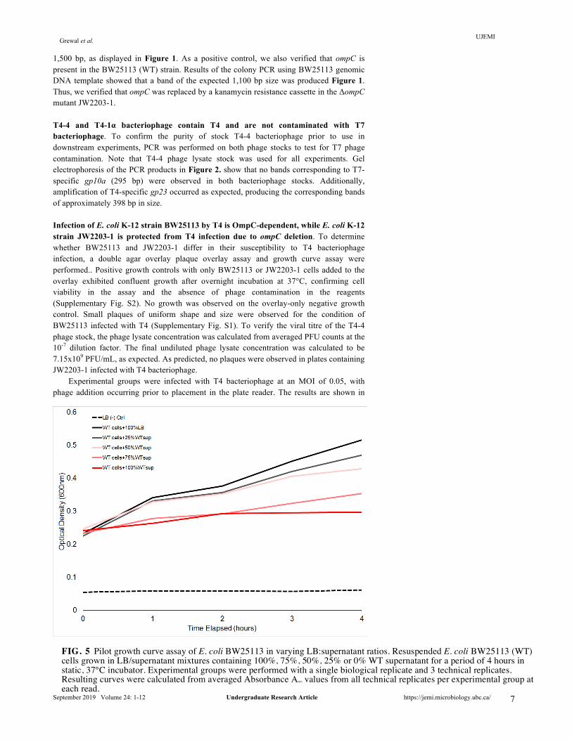
Experimental groups were infected with T4 bacteriophage at an MOI of 0.05, with phage addition occurring prior to placement in the plate reader. The results are shown in
FIG. 5 Pilot growth curve assay of E. coli BW25113 in varying LB:supernatant ratios. Resuspended E. coli BW25113 (WT) cells grown in LB/supernatant mixtures containing 100%, 75%, 50%, 25% or 0% WT supernatant for a period of 4 hours in static, 37°C incubator. Experimental groups were performed with a single biological replicate and 3 technical replicates. Resulting curves were calculated from averaged Absorbance A600 values from all technical replicates per experimental group at each read.
UJEMI Grewal et al.
September 2019 Volume 24: 1-12 Undergraduate Research Article https://jemi.microbiology.ubc.ca/
8
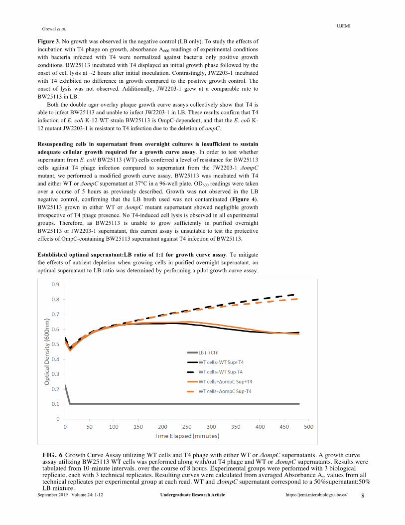
Figure 3. No growth was observed in the negative control (LB only). To study the effects of incubation with T4 phage on growth, absorbance A600 readings of experimental conditions with bacteria infected with T4 were normalized against bacteria only positive growth conditions. BW25113 incubated with T4 displayed an initial growth phase followed by the onset of cell lysis at ~2 hours after initial inoculation. Contrastingly, JW2203-1 incubated with T4 exhibited no difference in growth compared to the positive growth control. The onset of lysis was not observed. Additionally, JW2203-1 grew at a comparable rate to BW25113 in LB.
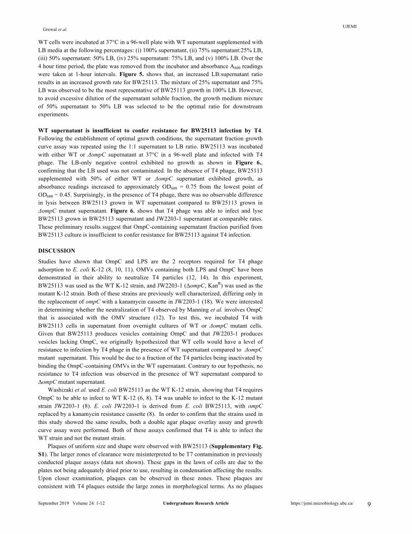
Both the double agar overlay plaque growth curve assays collectively show that T4 is able to infect BW25113 and unable to infect JW2203-1 in LB. These results confirm that T4 infection of E. coli K-12 WT strain BW25113 is OmpC-dependent, and that the E. coli K-12 mutant JW2203-1 is resistant to T4 infection due to the deletion of ompC. Resuspending cells in supernatant from overnight cultures is insufficient to sustain adequate cellular growth required for a growth curve assay. In order to test whether supernatant from E. coli BW25113 (WT) cells conferred a level of resistance for BW25113 cells against T4 phage infection compared to supernatant from the JW2203-1 ΔompC mutant, we performed a modified growth curve assay. BW25113 was incubated with T4 and either WT or ΔompC supernatant at 37°C in a 96-well plate. OD600 readings were taken over a course of 5 hours as previously described. Growth was not observed in the LB negative control, confirming that the LB broth used was not contaminated (Figure 4). BW25113 grown in either WT or ΔompC mutant supernatant showed negligible growth irrespective of T4 phage presence. No T4-induced cell lysis is observed in all experimental groups. Therefore, as BW25113 is unable to grow sufficiently in purified overnight BW25113 or JW2203-1 supernatant, this current assay is unsuitable to test the protective effects of OmpC-containing BW25113 supernatant against T4 infection of BW25113. Established optimal supernatant:LB ratio of 1:1 for growth curve assay. To mitigate the effects of nutrient depletion when growing cells in purified overnight supernatant, an optimal supernatant to LB ratio was determined by performing a pilot growth curve assay.
FIG. 6 Growth Curve Assay utilizing WT cells and T4 phage with either WT or ΔompC supernatants. A growth curve assay utilizing BW25113 WT cells was performed along with/out T4 phage and WT or ΔompC supernatants. Results were tabulated from 10-minute intervals, over the course of 8 hours. Experimental groups were performed with 3 biological replicate, each with 3 technical replicates. Resulting curves were calculated from averaged Absorbance A600 values from all technical replicates per experimental group at each read. WT and ΔompC supernatant correspond to a 50%supernatant:50% LB mixture.
UJEMI Grewal et al.
September 2019 Volume 24: 1-12 Undergraduate Research Article https://jemi.microbiology.ubc.ca/
9
WT cells were incubated at 37°C in a 96-well plate with WT supernatant supplemented with LB media at the following percentages: (i) 100% supernatant, (ii) 75% supernatant:25% LB, (iii) 50% supernatant: 50% LB, (iv) 25% supernatant: 75% LB, and (v) 100% LB. Over the 4 hour time period, the plate was removed from the incubator and absorbance A600 readings were taken at 1-hour intervals. Figure 5. shows that, an increased LB:supernatant ratio results in an increased growth rate for BW25113. The mixture of 25% supernatant and 75% LB was observed to be the most representative of BW25113 growth in 100% LB. However, to avoid excessive dilution of the supernatant soluble fraction, the growth medium mixture of 50% supernatant to 50% LB was selected to be the optimal ratio for downstream experiments. WT supernatant is insufficient to confer resistance for BW25113 infection by T4. Following the establishment of optimal growth conditions, the supernatant fraction growth curve assay was repeated using the 1:1 supernatant to LB ratio. BW25113 was incubated with either WT or ΔompC supernatant at 37°C in a 96-well plate and infected with T4 phage. The LB-only negative control exhibited no growth as shown in Figure 6., confirming that the LB used was not contaminated. In the absence of T4 phage, BW25113 supplemented with 50% of either WT or ΔompC supernatant exhibited growth, as absorbance readings increased to approximately OD600 = 0.75 from the lowest point of OD600 = 0.45. Surprisingly, in the presence of T4 phage, there was no observable difference in lysis between BW25113 grown in WT supernatant compared to BW25113 grown in ΔompC mutant supernatant. Figure 6. shows that T4 phage was able to infect and lyse BW25113 grown in BW25113 supernatant and JW2203-1 supernatant at comparable rates. These preliminary results suggest that OmpC-containing supernatant fraction purified from BW25113 culture is insufficient to confer resistance for BW25113 against T4 infection.
DISCUSSION
Studies have shown that OmpC and LPS are the 2 receptors required for T4 phage adsorption to E. coli K-12 (8, 10, 11). OMVs containing both LPS and OmpC have been demonstrated in their ability to neutralize T4 particles (12, 14). In this experiment, BW25113 was used as the WT K-12 strain, and JW2203-1 (ΔompC, KanR) was used as the mutant K-12 strain. Both of these strains are previously well characterized, differing only in the replacement of ompC with a kanamycin cassette in JW2203-1 (18). We were interested in determining whether the neutralization of T4 observed by Manning et al. involves OmpC that is associated with the OMV structure (12). To test this, we incubated T4 with BW25113 cells in supernatant from overnight cultures of WT or ΔompC mutant cells. Given that BW25113 produces vesicles containing OmpC and that JW2203-1 produces vesicles lacking OmpC, we originally hypothesized that WT cells would have a level of resistance to infection by T4 phage in the presence of WT supernatant compared to ΔompC mutant supernatant. This would be due to a fraction of the T4 particles being inactivated by binding the OmpC-containing OMVs in the WT supernatant. Contrary to our hypothesis, no resistance to T4 infection was observed in the presence of WT supernatant compared to ΔompC mutant supernatant.
Washizaki et al. used E. coli BW25113 as the WT K-12 strain, showing that T4 requires OmpC to be able to infect to WT K-12 (6, 8). T4 was unable to infect to the K-12 mutant strain JW2203-1 (8). E. coli JW2203-1 is derived from E. coli BW25113, with ompC replaced by a kanamycin resistance cassette (8). In order to confirm that the strains used in this study showed the same results, both a double agar plaque overlay assay and growth curve assay were performed. Both of these assays confirmed that T4 is able to infect the WT strain and not the mutant strain.
Plaques of uniform size and shape were observed with BW25113 (Supplementary Fig. S1). The larger zones of clearance were misinterpreted to be T7 contamination in previously conducted plaque assays (data not shown). These gaps in the lawn of cells are due to the plates not being adequately dried prior to use, resulting in condensation affecting the results. Upon closer examination, plaques can be observed in these zones. These plaques are consistent with T4 plaques outside the large zones in morphological terms. As no plaques
UJEMI Grewal et al.
September 2019 Volume 24: 1-12 Undergraduate Research Article https://jemi.microbiology.ubc.ca/
10
were observed for the mutant, our results were consistent with those of Washizaki et al. (8). We confirmed the requirement of OmpC for T4 to infect E. coli K-12 strain BW25113 (8).
A growth curve assay also supported the plaque assay results. As shown in Figure 3., the onset of lysis was observed for BW25113 incubated with T4 compared to BW25113 incubated without T4. JW2203-1 incubated with and without T4 showed results similar to BW25113 incubated without T4, with no lysis observed. T4 was added prior to start of the incubation period and the first OD600 reading at Time = 0 minutes. The MOI of 0.05 used was effective in producing an observable effect without inducing lysis too abruptly and drastically, which would have prevented an effective comparison of the experimental conditions tested for the supernatant experiments. As such, the same MOI and methods were used to prepare the cells for the remaining growth curve assays.
Upon establishing that BW25113 is lysed by T4 phage via an OmpC-dependent pathway, we focused on attempting to optimize our growth curve assay for testing the effects of the different supernatants on T4 infection of BW25113. The preliminary supernatant experiment showed insufficient growth when growing BW25113 in only the supernatant extracted from either WT or mutants cells (Fig. 4). For each growth condition, there were 3 biological replicates, with 3 technical replicates for each biological replicate. The condition with WT cells infected with T4 in 1.5% LB exhibited normal growth as expected (Fig. 4). As such, the lack of adequate growth was not due to issues with the viability of the subcultured cells or growth conditions in the plate reader. A likely explanation is that the purified overnight supernatant was nutrient-depleted. E. coli cells must be in exponential phase for efficient infection by T4 (19). We attempted to mitigate this issue by supplementing the supernatants with LB broth at the following supernatant concentrations: 0%, 25%, 50%, and 75% (Fig. 5). The supernatant to LB ratios of either 50%:50% or 25%:75% displayed growth most similar to the condition with WT cells growing in 100% LB. In order to avoid excessive dilution of the supernatant, which would dilute the soluble factors of interest, a 1:1 supernatant to LB ratio was decided to be used in future experiments.
A subsequent growth curve assay using the 1:1 supernatant-LB mixture was performed. BW25113 cells were incubated with T4 and either WT or ΔompC mutant supernatant, diluted by a factor of ½ with LB. Results showed that incubation of BW25113 with OmpC-containing WT supernatant did not confer an observable difference in the level of resistance to T4 infection when in comparison to incubation with ΔompC mutant supernatant (Fig. 6).
A possible explanation for this result may be the low concentration of OMVs in the supernatant due to insufficient growth time. BW25113 and JW2203-1 cells were grown overnight, with the supernatants extracted and filtered the next day. This may not have been adequate time for the production and release of sufficient amounts of protective OMVs into solution. While Manning et al. used a similar procedure that involved the growth of cells overnight, the filtered supernatant was centrifuged a second time at 38,400x g for 3 hours and the resulting pellet was resuspended and filtered (12) in order to isolate OMVs. This step was not performed in our procedure due to resource limitations. It is also hypothesized that OmpC protein may not be present in high enough concentrations in the WT OMVs to confer noticeable resistance. Compounded with low OMV concentrations, a higher concentration of OMVs is required to observe a significant effect on T4 phage infection. Manning et al. incubated T4 phage with purified OMVs prior to adding the T4-OMV mixture to cells, whereas in our procedure, T4 was added directly to the cell-supernatant mixture. In the case of insufficient OMV production, T4 virions are more likely to come into contact with OmpC expressed on BW25113, rather than on OMVs. The use of concentrated OMVs may be key in conferring resistance against T4 phage infection in WT cells observed by Manning et al. (12). Due to the lack of sufficient amounts of these soluble factors, the hypothesized WT decoy mechanism to reduce the amount of T4 particles able to bind and lyse the cells may be limited. Lastly, the dilution of the supernatant fraction with LB may have diluted the OMVs below a threshold concentration to produce an observable effect, assuming that a sufficient amount of OMVs were produced by the overnight cultures.
Aside from an insufficient growth time period, the growth conditions may also have been suboptimal for the production of sufficient amounts OMVs. OMVs appear to protect
UJEMI Grewal et al.
September 2019 Volume 24: 1-12 Undergraduate Research Article https://jemi.microbiology.ubc.ca/
11
Gram-negative bacteria from membrane-active antibiotics (12, 20). Increased vesiculation has been observed in enterotoxigenic E. coli in response to polymyxin exposure (12). Conclusions In conclusion, our results confirmed that T4 infection of E. coli K-12 strain BW25113 is OmpC-dependent, and that JW2203-1 is resistant to T4 infection due to the lack of OmpC on its surface. Filtered supernatant from overnight WT cultures did not confer resistance against T4 infection of WT cells compared to supernatant from ΔompC mutants. While OMVs were not purified and quantified, the observed lack of a difference in susceptibility may have been due to an insufficient amount of OMVs present in the filtrates. An increased focus towards OMV isolation and quantification may be necessary. Future Directions To continue investigating the potential protective effects of OMV-associated OmpC against T4 phage infection, several additional quantitative and qualitative experiments can be performed. To address the limited nutrient availability and dilution of the supernatant fraction in our growth curve assay, a modified assay using concentrated LB broth could be used to increase the growth rate while reducing the total supernatant dilution factor. More significantly, future work could involve purifying and quantifying OMVs from BW25113 and JW2203-1 supernatant, as described by Manning et al. (12). Growth curve assays consisting of BW25113 incubated in LB supplemented with purified OMVs could more accurately determine the influence of OmpC against T4 phage infection in a dose-dependent method (12). Additionally, using purified OMVs, Western blots can be performed to quantify and compare OmpC protein expression between BW25113 and JW2203-1 on OMVs. Exposure to a membrane-active antibiotic to induce greater vesiculation is a viable option for future studies. Thus, hypervesiculating mutants such as E. coli AD600 △yieM (MK1248) could be used in future growth curve assays in order to increase the production of OmpC-containing OMVs (12). BW25113 could also be converted into a hypervesiculating mutant and compared with a similarly altered hypervesiculating JW2203-1 mutant. Alternatively, the effects of abiotic and biotic stressors on bacterial cells could be investigated in conjunction with increased OMV production or ompC expression. ACKNOWLEDGEMENTS
We are grateful for the support, funding and laboratory facilities provided by the Department of Microbiology & Immunology at the University of British Columbia. We give our special thanks to Dr. David Oliver and Aaron Liu for their endless support and mentorship throughout the project. We are also grateful to our laboratory colleagues of team 1-alpha and team 2-gamma for supplying us with the T4 phage lysate stocks and the Wesbrook Media Room staff for providing all the equipment and laboratory supplies. CONTRIBUTIONS
This project was the collaborative effort of HG, ZD, SZ, and DS, with each author being involved in every step of the process in both experimental and writing tasks. Each author were delegated a portion of each assignment, but every author made revisions and edits to produce the final draft of each assignments
REFERENCES
1. Bragg R, van der Westhuizen W, Lee JY, Coetsee E, Boucher C. 2014. Bacteriophages as potential treatment option for antibiotic resistant bacteria. Adv Exp Med Biol 807:97-110.
2. Lin DM, Koskella B, Lin HC. 2017. Phage therapy: An alternative to antibiotics in the age of multi-drug resistance. World journal of gastrointestinal pharmacology and therapeutics 8:162-173.
3. Bertozzi Silva J, Storms Z, Sauvageau D. 2016. Host receptors for bacteriophage adsorption. FEMS Microbiology Letters 363.
4. Yap ML, Rossmann MG. 2014. Structure and function of bacteriophage T4. Future microbiology 9:1319-1327.
5. Yap ML, Klose T, Arisaka F, Speir JA, Veesler D, Fokine A, Rossmann MG. 2016. Role of bacteriophage T4 baseplate in regulating assembly and infection. Proceedings of the National Academy of Sciences 113:2654-2659.
UJEMI Grewal et al.
September 2019 Volume 24: 1-12 Undergraduate Research Article https://jemi.microbiology.ubc.ca/
12
6. Leiman PG, Shneider MM. 2012. Contractile Tail Machines of Bacteriophages, p 93-114. In Rossmann MG, Rao VB (ed), Viral Molecular Machines doi:10.1007/978-1-4614-0980-9_5. Springer US, Boston, MA.
7. Aksyuk AA, Leiman PG, Kurochkina LP, Shneider MM, Kostyuchenko VA, Mesyanzhinov VV, Rossmann MG. 2009. The tail sheath structure of bacteriophage T4: a molecular machine for infecting bacteria. The EMBO journal 28:821-829.
8. Washizaki A, Yonesaki T, Otsuka Y. 2016. Characterization of the interactions between Escherichia coli receptors, LPS and OmpC, and bacteriophage T4 long tail fibers. MicrobiologyOpen 5:1003-1015.
9. Fernandez L, Hancock RE. 2012. Adaptive and mutational resistance: role of porins and efflux pumps in drug resistance. Clin Microbiol Rev 25:661-81.
10. Amir Wachtel AG, Zach Sagorin, Emmanuel Etti. 11/17/2017 O16 Serotype O Antigen Expression in Escherichia coli K-12 May Confer Resistance Against T4 Bacteriophage Infection by Preventing Adsorption. The Journal of Experimental Microbiology and Immunology (JEMI) 3.
11. Currie CG, Poxton IR. 1999. The lipopolysaccharide core type of Escherichia coli O157:H7 and other non-O157 verotoxin-producing E. coli. FEMS Immunology & Medical Microbiology 24:57-62.
12. Manning AJ, Kuehn MJ. 2011. Contribution of bacterial outer membrane vesicles to innate bacterial defense. BMC microbiology 11:258-258.
13. Loeb MR, Kilner J. 1978. Release of a special fraction of the outer membrane from both growing and phage T4-infected Escherichia coli B. Biochimica et Biophysica Acta (BBA) - Biomembranes 514:117-127.
14. Schwechheimer C, Kuehn MJ. 2015. Outer-membrane vesicles from Gram-negative bacteria: biogenesis and functions. Nat Rev Microbiol 13:605-19.
15. Chuan Chuan Chang CWY, Regina Onal, and Yue Zhang. 09/09/2018 Genetic characterization and investigation of kanamycin susceptibility of ompC and ompF single gene deletion mutants of Escherichia coli K-12. The Journal of Experimental Microbiology and Immunology (JEMI) 22.
16. Naomi Fettig AR, Nicole Mar, Angela Sunario. 09/09/2018. Growth phase of host Escherichia coli MG1655 influences T4 and T7 bacteriophage replication patterns during coinfection. The Journal of Experimental Microbiology and Immunology (JEMI) 22.
17. Kropinski AM, Mazzocco A, Waddell TE, Lingohr E, Johnson RP. 2009. Enumeration of Bacteriophages by Double Agar Overlay Plaque Assay, p 69-76. In Clokie MRJ, Kropinski AM (ed), Bacteriophages: Methods and Protocols, Volume 1: Isolation, Characterization, and Interactions doi:10.1007/978-1-60327-164-6_7. Humana Press, Totowa, NJ.
18. Baba T, Ara T, Hasegawa M, Takai Y, Okumura Y, Baba M, Datsenko KA, Tomita M, Wanner BL, Mori H. 2006. Construction of Escherichia coli K-12 in-frame, single-gene knockout mutants: the Keio collection. Molecular systems biology 2:2006.0008-2006.0008.
19. Bryan D, El-Shibiny A, Hobbs Z, Porter J, Kutter EM. 2016. Bacteriophage T4 Infection of Stationary Phase E. coli: Life after Log from a Phage Perspective. Frontiers in microbiology 7:1391-1391.
20. Kulkarni HM, Nagaraj R, Jagannadham MV. 2015. Protective role of E. coli outer membrane vesicles against antibiotics. Microbiological Research 181:1-7.