Structure, organization, and functions of cellulose ... · PDF file3 Braz. J. Plant Physiol.,...
13
Braz. J. Plant Physiol., 19(1):1-13, 2007 REVIEW Annually, plants produce about 180 billion tons of cellulose making it the largest reservoir of organic carbon on Earth. Cellulose is a linear homopolymer of β(1-4)-linked glucose residues. The coordinated synthesis of glucose chains is orchestrated by specific plasma membrane-bound cellulose synthase complexes (CelS). The CelS is postulated to be composed of approximately 36 cellulose synthase (CESA) subunits. The CelS synthesizes 36 glucose chains in close proximity before they are further organized into microfibrils that are further associated with other cell wall polymers. The 36 glucose chains in a microfibril are stabilized by intra- and inter-hydrogen bonding which confer great stability on microfibrils. Several elementary microfibrils come together to form macrofibrils. Many CESA isoforms appear to be involved in the cellulose biosynthetic process and at least three types of CESA isoforms appear to be necessary for the functional organization of CelS in higher plants. Key words: cellulose biosynthesis, cellulose synthase (CESA), cellulose synthase complex (CelS), microfibril Estrutura, organização e funções dos complexos da sintase da celulose em plantas superiores: Anualmente, as plantas produzem aproximadamente 180 bilhões de toneladas de celulose, sendo o maior reservatório de carbono orgânico no planeta. A celulose é um homopolímero linear composto por resíduos de glicose unidos por meio de ligações β(1-4). A síntese coordenada das cadeias de glicose é orquestrada por complexos específicos ligados à membrana plasmática (CelS). Postula-se que o CelS é composto por aproximadamente 36 subunidades da sintase da celulose (CESA). Cada CelS sintetiza 36 cadeias de glicose dispostas lado a lado antes de serem organizadas em microfibrilas, que são, poste- riormente, associadas com outros polímeros da parede celular. As 36 cadeias de glicose presentes em uma microfibrila são estabilizadas por pontes de hidrogênio intra e inter-cadeias, conferindo grande estabilidade às microfibrilas. As microfibrilas elementares são dispostas lado a lado, permitindo a formação das macrofibrilas. Várias isoformas da CESA podem estar envolvidas no processo de biossíntese de celulose e, no mínimo, três tipos de isoformas da CESA podem ser necessárias para a organização funcional de cada CelS em plantas superiores. Palavras-chave: biossíntese de celulose, complexo da sintase da celulose (CelS), microfibrilas, sintase da celulose (CESA) Structure, organization, and functions of cellulose synthase complexes in higher plants Reginaldo A. Festucci-Buselli 1* , Wagner C. Otoni 1 and Chandrashekhar P. Joshi 2 1 Departamento de Biologia Vegetal, Laboratório de Cultura de Tecidos, BIOAGRO, Universidade Federal de Viçosa, Viçosa 36570-000, MG, Brazil. 2 Biotechnology Research Center, School of Forest Resources and Environmental Science, Michigan Technological University, Houghton, Michigan 49931, USA. *Corresponding author: [email protected]. Phone: 55-31-3899-2930, Fax: 55-31-3899-2580 Received: 30 January 2007; Returned for revision: 26 April 2007; Accepted: 26 May 2007 INTRODUCTION Cellulose is an outstanding commodity due to its abundance and distinctive structural properties. For example, its tension resistance is comparable to that of steel (Eckardt, 2003). Even though cellulose has great commercial value for the pulp, paper, and textile as well as chemical industries which use it to produce commercially important polymers, cellulose biosynthesis in trees is still not well understood. Due to the economical significance of tree cellulose for forest product industries, we have
Transcript of Structure, organization, and functions of cellulose ... · PDF file3 Braz. J. Plant Physiol.,...
Braz. J. Plant Physiol., 19(1):1-13, 2007
REVIEW
Annually, plants produce about 180 billion tons of cellulose making it the largest reservoir of organic carbon on Earth.Cellulose is a linear homopolymer of β(1-4)-linked glucose residues. The coordinated synthesis of glucose chains isorchestrated by specific plasma membrane-bound cellulose synthase complexes (CelS). The CelS is postulated to becomposed of approximately 36 cellulose synthase (CESA) subunits. The CelS synthesizes 36 glucose chains in closeproximity before they are further organized into microfibrils that are further associated with other cell wall polymers. The36 glucose chains in a microfibril are stabilized by intra- and inter-hydrogen bonding which confer great stability onmicrofibrils. Several elementary microfibrils come together to form macrofibrils. Many CESA isoforms appear to beinvolved in the cellulose biosynthetic process and at least three types of CESA isoforms appear to be necessary for thefunctional organization of CelS in higher plants.Key words: cellulose biosynthesis, cellulose synthase (CESA), cellulose synthase complex (CelS), microfibril
Estrutura, organização e funções dos complexos da sintase da celulose em plantas superiores: Anualmente, as plantasproduzem aproximadamente 180 bilhões de toneladas de celulose, sendo o maior reservatório de carbono orgânico noplaneta. A celulose é um homopolímero linear composto por resíduos de glicose unidos por meio de ligações β(1-4). Asíntese coordenada das cadeias de glicose é orquestrada por complexos específicos ligados à membrana plasmática(CelS). Postula-se que o CelS é composto por aproximadamente 36 subunidades da sintase da celulose (CESA). CadaCelS sintetiza 36 cadeias de glicose dispostas lado a lado antes de serem organizadas em microfibrilas, que são, poste-riormente, associadas com outros polímeros da parede celular. As 36 cadeias de glicose presentes em uma microfibrilasão estabilizadas por pontes de hidrogênio intra e inter-cadeias, conferindo grande estabilidade às microfibrilas. Asmicrofibrilas elementares são dispostas lado a lado, permitindo a formação das macrofibrilas. Várias isoformas da CESApodem estar envolvidas no processo de biossíntese de celulose e, no mínimo, três tipos de isoformas da CESA podemser necessárias para a organização funcional de cada CelS em plantas superiores.Palavras-chave: biossíntese de celulose, complexo da sintase da celulose (CelS), microfibrilas, sintase da celulose(CESA)
Structure, organization, and functions of cellulose synthasecomplexes in higher plants
Reginaldo A. Festucci-Buselli1*, Wagner C. Otoni1 and Chandrashekhar P. Joshi2
1Departamento de Biologia Vegetal, Laboratório de Cultura de Tecidos, BIOAGRO, Universidade Federal de Viçosa,Viçosa 36570-000, MG, Brazil. 2Biotechnology Research Center, School of Forest Resources and EnvironmentalScience, Michigan Technological University, Houghton, Michigan 49931, USA. *Corresponding author:[email protected]. Phone: 55-31-3899-2930, Fax: 55-31-3899-2580
Received: 30 January 2007; Returned for revision: 26 April 2007; Accepted: 26 May 2007
INTRODUCTION
Cellulose is an outstanding commodity due to itsabundance and distinctive structural properties. Forexample, its tension resistance is comparable to that ofsteel (Eckardt, 2003). Even though cellulose has great
commercial value for the pulp, paper, and textile as well as
chemical industries which use it to produce commercially
important polymers, cellulose biosynthesis in trees is still
not well understood. Due to the economical significance
of tree cellulose for forest product industries, we have
2
Braz. J. Plant Physiol., 19(1):1-13, 2007
R.A. FESTUCCI-BUSELLI et al.
focused our attention in this review on cellulosebiosynthesis in trees. Most of the recent findings
concerning the molecular mechanism of cellulose
biosynthesis in higher plants resulted from research in
model herbaceous plants and fiber crops and have been
reviewed recently (Somerville, 2006).
Cellulose is synthesized by cellulose synthase
enzymes (CESAs) and is regarded as a major sink for
atmospheric carbon in plants because it is the main
component of the plant cell wall (Delmer and Haigler,
2002). Many plant cell walls of commercial importance are
made up of three layers: middle lamellae, primary cell wall,
and secondary cell wall. The secondary cell wall is further
subdivided into three sub-layers called S1, S2, and S3. All
the layers present in the cell wall have two phases:
microfibrilar and matrix (Brett and Waldron, 1990). The
microfibrilar phase, a crystalline phase, is composed of
microfibrils of cellulose and the matrix phase, a non-
crystall ine phase, is composed of a variety of
polysaccharides (pectins and hemicelluloses), proteins,
and phenolic compounds (lignin, ferulic acid, coumaric
acid, and others) (Brett and Waldron, 1990).
There is a definite distinction between chemical
composition of the primary and secondary cell walls. The
most remarkable one is concerning the quantities of
cellulose and lignin. The amount of lignin in secondary
cell walls of trees like Populus trichocarpa (poplar) is 19-
21%, whereas in the primary cell wall lignin is absent
(Mellerowicz et al., 2001). The content of cellulose,
expressed in dry weight, in the primary cell wall of poplars
is 20-30%, whereas in the secondary cell wall it is 40-50%
(Mellerowicz et al., 2001). For instance, some clones of
Eucalyptus grandis cultivated in Brazil have a cellulose
content of 43.9 – 49.7% in the secondary cell wall (Gomide
et al., 2005).
In some special cases, when the developing
angiosperm wood is under tension stress, a gelatinous
layer (G layer) is formed instead of the S2 or S3 layer
(Timell, 1969; Mellerowicz et al., 2001; Pilate et al., 2004).
The G layer is composed almost exclusively of highly
crystalline axially oriented microfibrils of cellulose. An
increase of 10-20% in the cellulose content and a
significant decrease in lignin content has been observed
in tension wood in response to the deposition of the G
layer. Thus tension wood may represent an excellent
system to study processes involved in cellulose
biosynthesis (Wu et al., 2000; Pilate et al., 2004). For
example, in Populus tremuloides (aspen), thecoexpression of three CesAs , namely, PtrCesA1 ,PtrCesA2, and PtrCesA3, suggests that these threeCesAs may be important for the biosynthesis of highlycrystalline cellulose present in tension wood fibers(Bhandari et al., 2006). It has been reported thatKORRIGAN (KOR), a type of cellulase, is involved incellulose biosynthesis allowing proper formation ofmicrofibrils of cellulose I (Delmer and Haigler, 2002;Molhoj et al., 2002). The aspen Kor gene (PtrKor) hasbeen isolated and its expression pattern has beenanalyzed in aspen. Similar to three aspen CESAs, a highamount of PtrKOR protein was detected in developingxylem as well as on the upper side of the bent aspen stemin response to tension stress. In contrast, a very lowamount of PtrKOR protein was detected on the oppositeside of the bent stem experiencing compression stress(Bhandari et al., 2006).
In this review, we will discuss the process of cellulosebiosynthesis. In addition, structural characteristics ofcellulose and CESAs, as well as the structural,organizational, and functional features of CESAcomplexes (CelS) will be discussed.
MOLECULAR AND SUPRAMOLECULARFEATURES OF CELLULOSE
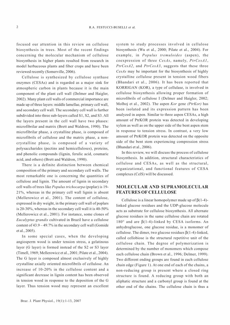
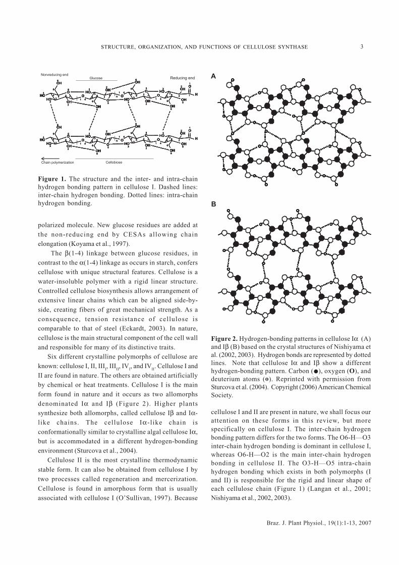
Cellulose is a linear homopolymer made up of β(1-4)-linked glucose residues and the UDP-glucose moleculeacts as substrate for cellulose biosynthesis. All alternateglucose residues in the same cellulose chain are rotated180° and are β(1-4)-linked by CESA isoforms. Ananhydroglucose, one glucose residue, is a monomer ofcellulose. The dimer, two glucose residues β(1-4)-linked,called cellobiose is the structural repetitive unit of thecellulose chain. The degree of polymerization isdetermined by the number of monomers which composeeach cellulose chain (Brown et al., 1996; Delmer, 1999).Two different ending groups are found in each cellulosechain edge (Figure 1). At one end of each of the chains, anon-reducing group is present where a closed ringstructure is found. A reducing group with both analiphatic structure and a carbonyl group is found at theother end of the chains. The cellulose chain is thus a
3
Braz. J. Plant Physiol., 19(1):1-13, 2007
STRUCTURE, ORGANIZATION, AND FUNCTIONS OF CELLULOSE SYNTHASE
Figure 1. The structure and the inter- and intra-chainhydrogen bonding pattern in cellulose I. Dashed lines:inter-chain hydrogen bonding. Dotted lines: intra-chainhydrogen bonding.
polarized molecule. New glucose residues are added atthe non-reducing end by CESAs allowing chainelongation (Koyama et al., 1997).
The β(1-4) linkage between glucose residues, incontrast to the α(1-4) linkage as occurs in starch, conferscellulose with unique structural features. Cellulose is awater-insoluble polymer with a rigid linear structure.Controlled cellulose biosynthesis allows arrangement ofextensive linear chains which can be aligned side-by-side, creating fibers of great mechanical strength. As aconsequence, tension resistance of cellulose iscomparable to that of steel (Eckardt, 2003). In nature,cellulose is the main structural component of the cell walland responsible for many of its distinctive traits.
Six different crystalline polymorphs of cellulose areknown: cellulose I, II, III
I, III
II, IV
I, and IV
II. Cellulose I and
II are found in nature. The others are obtained artificiallyby chemical or heat treatments. Cellulose I is the mainform found in nature and it occurs as two allomorphsdenominated Iα and Iβ (Figure 2). Higher plantssynthesize both allomorphs, called cellulose Iβ and Iα-l ike chains. The cellulose Iα-l ike chain isconformationally similar to crystalline algal cellulose Iα,but is accommodated in a different hydrogen-bondingenvironment (Sturcova et al., 2004).
Cellulose II is the most crystalline thermodynamic
stable form. It can also be obtained from cellulose I bytwo processes called regeneration and mercerization.
Cellulose is found in amorphous form that is usuallyassociated with cellulose I (O’Sullivan, 1997). Because
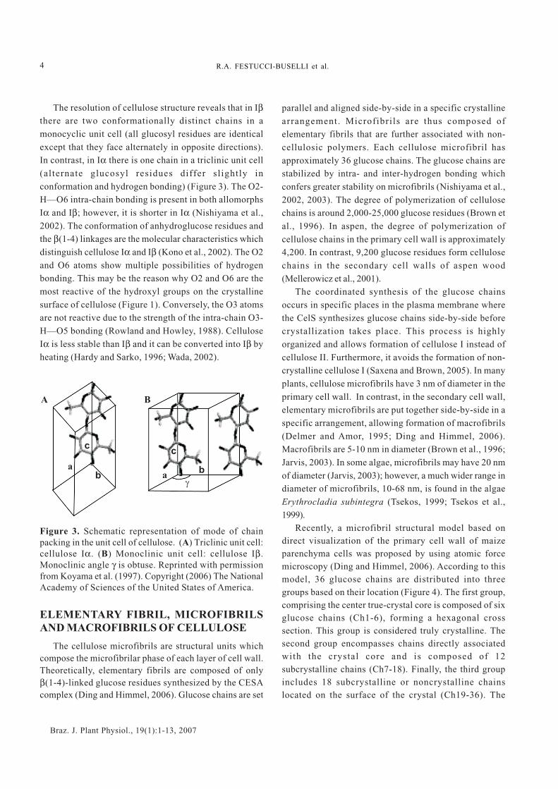
Figure 2. Hydrogen-bonding patterns in cellulose Iα (A)and Iβ (B) based on the crystal structures of Nishiyama etal. (2002, 2003). Hydrogen bonds are represented by dottedlines. Note that cellulose Iα and Iβ show a differenthydrogen-bonding pattern. Carbon ( ), oxygen (O), anddeuterium atoms (o). Reprinted with permission fromSturcova et al. (2004). Copyright (2006) American ChemicalSociety.
OH OH
OH OHO
O
O
O
O O O
OH
OH
OH
OH
OH
HHOHO
HO
HO
HO
12
3
45
6
6
4
3
2
1 4
5
124
53
6
6
5
23
1 4
1 4
6
6
5
12
3
45
32
6
6
5
5
1
1
2
32
3
4
OH OH
OH OHO
O
O
O
O O O
OH
OH
OH
OH
OH
HHOHO
HO
HO
HO
12
3
45
6
6
4
3
2
1 4
5
124
53
6
6
5
23
1 4
1 4
6
6
5
12
3
45
32
6
6
5
5
1
1
2
32
3
4
Glucose
n
n
CellobioseChain polymerization
Reducing endNon-reducing end
OH OH
OH OHO
O
O
O
O O O
OH
OH
OH
OH
OH
HHOHO
HO
HO
HO
12
3
45
6
6
4
3
2
1 4
5
124
53
6
6
5
23
1 4
1 4
6
6
5
12
3
45
32
6
6
5
5
1
1
2
32
3
4
OH OH
OH OHO
O
O
O
O O O
OH
OH
OH
OH
OH
HHOHO
HO
HO
HO
12
3
45
6
6
4
3
2
1 4
5
124
53
6
6
5
23
1 4
1 4
6
6
5
12
3
45
32
6
6
5
5
1
1
2
32
3
4
OH OH
OH OHO
O
O
O
O O O
OH
OH
OH
OH
OH
HHOHO
HO
HO
HO
12
3
45
6
6
4
3
2
1 4
5
124
53
6
6
5
23
1 4
1 4
6
6
5
12
3
45
32
6
6
5
5
1
1
2
32
3
4
OH OH
OH OHO
O
O
O
O O O
OH
OH
OH
OH
OH
HHOHO
HO
HO
HO
12
3
45
6
6
4
3
2
1 4
5
124
53
6
6
5
23
1 4
1 4
6
6
5
12
3
45
32
6
6
5
5
1
1
2
32
3
4
OH OH
OH OHO
O
O
O
O O O
OH
OH
OH
OH
OH
HHOHO
HO
HO
HO
12
3
45
6
6
4
3
2
1 4
5
124
53
6
6
5
23
1 4
1 4
6
6
5
12
3
45
32
6
6
5
5
1
1
2
32
3
4
OH OH
OH OHO
O
O
O
O O O
OH
OH
OH
OH
OH
HHOHO
HO
HO
HO
12
3
45
6
6
4
3
2
1 4
5
124
53
6
6
5
23
1 4
1 4
6
6
5
12
3
45
32
6
6
5
5
1
1
2
32
3
4
OH OH
OH OHO
O
O
O
O O O
OH
OH
OH
OH
OH
HHOHO
HO
HO
HO
12
3
45
6
6
4
3
2
1 4
5
124
53
6
6
5
23
1 4
1 4
6
6
5
12
3
45
32
6
6
5
5
1
1
2
32
3
4
OH OH
OH OHO
O
O
O
O O O
OH
OH
OH
OH
OH
HHOHO
HO
HO
HO
12
3
45
6
6
4
3
2
1 4
5
124
53
6
6
5
23
1 4
1 4
6
6
5
12
3
45
32
6
6
5
5
1
1
2
32
3
4
n
n
-
cellulose I and II are present in nature, we shall focus ourattention on these forms in this review, but morespecifically on cellulose I. The inter-chain hydrogenbonding pattern differs for the two forms. The O6-H—O3inter-chain hydrogen bonding is dominant in cellulose I,whereas O6-H—O2 is the main inter-chain hydrogenbonding in cellulose II. The O3-H—O5 intra-chainhydrogen bonding which exists in both polymorphs (Iand II) is responsible for the rigid and linear shape ofeach cellulose chain (Figure 1) (Langan et al., 2001;Nishiyama et al., 2002, 2003).
°
°°
°
°°
°
°
° °
°°
°
°°
°
°°
°
°
° °
°°
°
° °
°
°°
°
°
°°
°°
°
° °
°
°°
°
°
°°
°°
B
°
°°
°
°°
°
°
° °
°°
°
°°
°
°°
°
°
° °
°°
°
°°
°
°°
°
°
° °
°°°
°°
°
°°
°
°
° °
°°°
°°
°
°°
°
°
° °
°°
°
°°
°
°°
°
°
° °
°°°
°°
°
°°
°
°
° °
°°°
°°
°
°°
°
°
° °
°°
°
° °
°
°°
°
°
°°
°°
°
° °
°
°°
°
°
°°
°°
°
° °
°
°°
°
°
°°
°°°
° °
°
°°
°
°
°°
°°°
° °
°
°°
°
°
°°
°°
°
° °
°
°°
°
°
°°
°°°
° °
°
°°
°
°
°°
°°°
° °
°
°°
°
°
°°
°°
A
4
Braz. J. Plant Physiol., 19(1):1-13, 2007
R.A. FESTUCCI-BUSELLI et al.
The resolution of cellulose structure reveals that in Iβthere are two conformationally distinct chains in amonocyclic unit cell (all glucosyl residues are identicalexcept that they face alternately in opposite directions).In contrast, in Iα there is one chain in a triclinic unit cell(alternate glucosyl residues differ slightly inconformation and hydrogen bonding) (Figure 3). The O2-H—O6 intra-chain bonding is present in both allomorphsIα and Iβ; however, it is shorter in Iα (Nishiyama et al.,2002). The conformation of anhydroglucose residues andthe β(1-4) linkages are the molecular characteristics whichdistinguish cellulose Iα and Iβ (Kono et al., 2002). The O2and O6 atoms show multiple possibilities of hydrogenbonding. This may be the reason why O2 and O6 are themost reactive of the hydroxyl groups on the crystallinesurface of cellulose (Figure 1). Conversely, the O3 atomsare not reactive due to the strength of the intra-chain O3-H—O5 bonding (Rowland and Howley, 1988). CelluloseIα is less stable than Iβ and it can be converted into Iβ byheating (Hardy and Sarko, 1996; Wada, 2002).
Figure 3. Schematic representation of mode of chainpacking in the unit cell of cellulose. (A) Triclinic unit cell:cellulose Iα. (B) Monoclinic unit cell: cellulose Iβ.Monoclinic angle γ is obtuse. Reprinted with permissionfrom Koyama et al. (1997). Copyright (2006) The NationalAcademy of Sciences of the United States of America.
ELEMENTARY FIBRIL, MICROFIBRILSAND MACROFIBRILS OF CELLULOSE
The cellulose microfibrils are structural units whichcompose the microfibrilar phase of each layer of cell wall.Theoretically, elementary fibrils are composed of onlyβ(1-4)-linked glucose residues synthesized by the CESAcomplex (Ding and Himmel, 2006). Glucose chains are set
parallel and aligned side-by-side in a specific crystallinearrangement. Microfibrils are thus composed ofelementary fibrils that are further associated with non-cellulosic polymers. Each cellulose microfibril hasapproximately 36 glucose chains. The glucose chains arestabilized by intra- and inter-hydrogen bonding whichconfers greater stability on microfibrils (Nishiyama et al.,2002, 2003). The degree of polymerization of cellulosechains is around 2,000-25,000 glucose residues (Brown etal., 1996). In aspen, the degree of polymerization ofcellulose chains in the primary cell wall is approximately4,200. In contrast, 9,200 glucose residues form cellulosechains in the secondary cell walls of aspen wood(Mellerowicz et al., 2001).
The coordinated synthesis of the glucose chainsoccurs in specific places in the plasma membrane wherethe CelS synthesizes glucose chains side-by-side beforecrystallization takes place. This process is highlyorganized and allows formation of cellulose I instead ofcellulose II. Furthermore, it avoids the formation of non-crystalline cellulose I (Saxena and Brown, 2005). In manyplants, cellulose microfibrils have 3 nm of diameter in theprimary cell wall. In contrast, in the secondary cell wall,elementary microfibrils are put together side-by-side in aspecific arrangement, allowing formation of macrofibrils(Delmer and Amor, 1995; Ding and Himmel, 2006).Macrofibrils are 5-10 nm in diameter (Brown et al., 1996;Jarvis, 2003). In some algae, microfibrils may have 20 nmof diameter (Jarvis, 2003); however, a much wider range indiameter of microfibrils, 10-68 nm, is found in the algaeErythrocladia subintegra (Tsekos, 1999; Tsekos et al.,1999).
Recently, a microfibril structural model based ondirect visualization of the primary cell wall of maizeparenchyma cells was proposed by using atomic forcemicroscopy (Ding and Himmel, 2006). According to thismodel, 36 glucose chains are distributed into threegroups based on their location (Figure 4). The first group,comprising the center true-crystal core is composed of sixglucose chains (Ch1-6), forming a hexagonal crosssection. This group is considered truly crystalline. Thesecond group encompasses chains directly associatedwith the crystal core and is composed of 12subcrystalline chains (Ch7-18). Finally, the third groupincludes 18 subcrystalline or noncrystalline chainslocated on the surface of the crystal (Ch19-36). The
A B
a
b
c
a
c
b
�

5
Braz. J. Plant Physiol., 19(1):1-13, 2007
STRUCTURE, ORGANIZATION, AND FUNCTIONS OF CELLULOSE SYNTHASE
Figure 4. Cross-section of the 36-chain elementary fibrilshowing number of chains. Chains are numbered from Ch1to Ch36 and categorized into three groups: group-C1 (Ch1-6) contains six true crystalline chains, group-C2 (Ch7-18)contains twelve subcrystalline chains, and group-C3(Ch19-36) contains 18 subcrystalline or noncrystallinechains. Reprinted with permission from Ding and Himmel(2006). Copyright (2006) American Chemical Society.
groups 2 and 3 form protection and transition phasesbetween the crystal core and later-deposited non-crystalline polymers. It is postulated that this model isstructurally similar to the cellulose Iβ model (Figures 2Band 3B).
CELLULOSE SYNTHASE COMPLEX(CelS)
The CelS, usually called the terminal complex orrosette, is proposed to be composed of six subunitswhich are arranged in hexagonal symmetry. It is 25-30 nmin diameter (Mueller and Brown, 1980; Delmer, 1999). Eachsubunit of CelS has six CESA isoforms which produce sixglucan chains (Brown and Saxena, 2000). The CelS is thustheoretically composed of 36 CESAs. At least three typesof CESA isoforms, called α
1, α
2, and β, may be necessary
for the spontaneous arrangement of CESAs in each CelS(Doblin et al., 2002; Ding and Himmel, 2006). Consistentwith this hypothesis, different types of CesA isoformshave been found to be coexpressed in Arabidopsis
thaliana: AtCesA4, AtCesA7, and AtCesA8 (Taylor et al.,2003); barley: HvCesA1, HvCesA2, and HvCesA6 (Burtonet al., 2004); poplar: PtrCesA1, PtrCesA2, and PtrCesA3
(Joshi et al., 2004; Bhandari et al., 2006); and rice:OsCesA4, OsCesA7, and OsCesA9 (Tanaka et al., 2003).However, expression of several CesA isoforms in the same
cell does not necessarily indicate that they interact andare part of the same CelS. According to the expressionpattern and functional redundancy, three situations arepossible: (i) different isoforms are expressed in differentcells; (ii) different isoforms are expressed in the same cell,but they are functionally redundant; and (iii) differentisoforms are expressed in the same cell, but they areessential and are not redundant (Perrin, 2001).
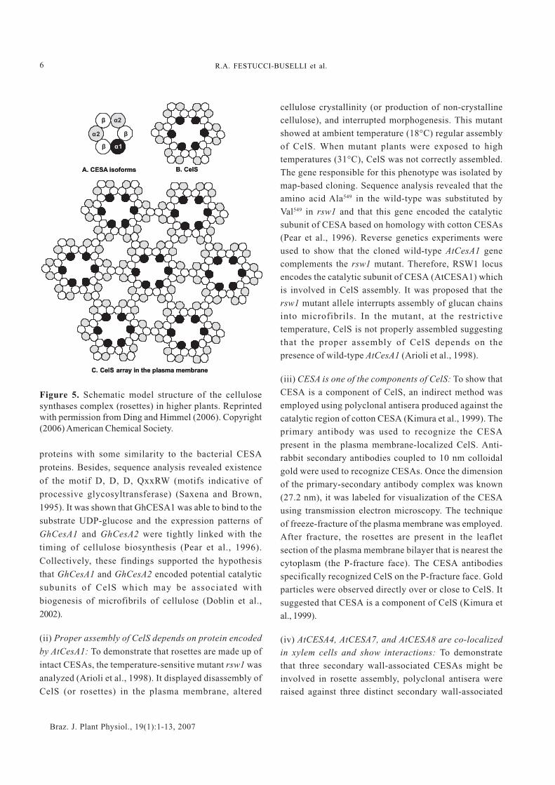
Several models have been proposed to explaininteractions among CESA isoforms, allowing properformation of CelS (Scheible et al., 2001; Doblin et al., 2002;Joshi, 2003a). These models are based on the hypothesisthat one elementary fibril is composed of 36 glucosechains, which are synthesized by 36 CESA isoformsarranged in a planar configuration in the plasmamembrane (Delmer, 1999; Kimura et al., 1999; Scheible etal., 2001). The number of CESA isoforms and the type ofinteractions among them are the distinctive features ofeach model. In the model proposed by Ding and Himmel(2006), 36 CESA isoforms are assembled into rosetteslocated in the plasma membrane (Figure 5). Each CelS hassix identical subunits composed of six CESA isoforms:
one α1, two α
2, and three β (Figure 5A). Three types of
protein-protein interactions (β-β, α1-β, and α
2-β) may be
involved in the spontaneous assembly of CelS (Figure
5B). Several CelS arranged in the plasma membrane may
form a honeycomb array (Figure 5C). The honeycomb
array structure could synthesize a great number of
elementary fibrils to form a macrofibril.
CelS IS COMPOSED OF CESA ISOFORMSTHAT ARE ENCODED BY CesA GENES
Several experiments were successfully conducted to
suggest that CelS are made up of multiple CESA isoforms
that are encoded by distinct CesA genes.
(i) CelS (rosette) is associated with biogenesis of
microfibrils of cellulose: The discovery of the cotton
CesA genes encoding potential catalytic subunits of CelS
revealed the involvement of CelS in biosynthesis of
microfibrils of cellulose. The identification of potential
components of the plant CelS complex was achieved by a
molecular approach. Two cDNA clones GhCESA1 and
GhCESA2 were identified in cotton fiber enriched in
cellulose (Pear et al., 1996). These cDNAs encoded
Ch35 Ch36 Ch19
Ch28 Ch27 Ch26
Ch34 Ch18 Ch7 Ch20
Ch33 Ch17 Ch1 Ch8
Ch32 Ch16 Ch6 Ch2
Ch31 Ch15 Ch5 Ch3
Ch30 Ch14 Ch4 Ch11
Ch29 Ch13 Ch12 Ch25
Ch21
Ch9 Ch22
Ch10
Ch24
Ch23
Ch35 Ch36 Ch19
Ch28 Ch27 Ch26
Ch34 Ch18 Ch7 Ch20
Ch33 Ch17 Ch1 Ch8
Ch32 Ch16 Ch6 Ch2
Ch31 Ch15 Ch5 Ch3
Ch30 Ch14 Ch4 Ch11
Ch29 Ch13 Ch12 Ch25
Ch21
Ch9 Ch22
Ch10
Ch24
Ch23
6
Braz. J. Plant Physiol., 19(1):1-13, 2007
R.A. FESTUCCI-BUSELLI et al.
Figure 5. Schematic model structure of the cellulosesynthases complex (rosettes) in higher plants. Reprintedwith permission from Ding and Himmel (2006). Copyright(2006) American Chemical Society.
proteins with some similarity to the bacterial CESA
proteins. Besides, sequence analysis revealed existence
of the motif D, D, D, QxxRW (motifs indicative of
processive glycosyltransferase) (Saxena and Brown,
1995). It was shown that GhCESA1 was able to bind to the
substrate UDP-glucose and the expression patterns of
GhCesA1 and GhCesA2 were tightly linked with the
timing of cellulose biosynthesis (Pear et al., 1996).
Collectively, these findings supported the hypothesis
that GhCesA1 and GhCesA2 encoded potential catalytic
subunits of CelS which may be associated with
biogenesis of microfibrils of cellulose (Doblin et al.,
2002).
(ii) Proper assembly of CelS depends on protein encoded
by AtCesA1: To demonstrate that rosettes are made up of
intact CESAs, the temperature-sensitive mutant rsw1 was
analyzed (Arioli et al., 1998). It displayed disassembly of
CelS (or rosettes) in the plasma membrane, altered
cellulose crystallinity (or production of non-crystallinecellulose), and interrupted morphogenesis. This mutantshowed at ambient temperature (18°C) regular assemblyof CelS. When mutant plants were exposed to hightemperatures (31°C), CelS was not correctly assembled.The gene responsible for this phenotype was isolated bymap-based cloning. Sequence analysis revealed that theamino acid Ala549 in the wild-type was substituted byVal549 in rsw1 and that this gene encoded the catalyticsubunit of CESA based on homology with cotton CESAs(Pear et al., 1996). Reverse genetics experiments wereused to show that the cloned wild-type AtCesA1 genecomplements the rsw1 mutant. Therefore, RSW1 locusencodes the catalytic subunit of CESA (AtCESA1) whichis involved in CelS assembly. It was proposed that thersw1 mutant allele interrupts assembly of glucan chainsinto microfibrils. In the mutant, at the restrictivetemperature, CelS is not properly assembled suggestingthat the proper assembly of CelS depends on thepresence of wild-type AtCesA1 (Arioli et al., 1998).
(iii) CESA is one of the components of CelS: To show that
CESA is a component of CelS, an indirect method wasemployed using polyclonal antisera produced against the
catalytic region of cotton CESA (Kimura et al., 1999). Theprimary antibody was used to recognize the CESA
present in the plasma membrane-localized CelS. Anti-rabbit secondary antibodies coupled to 10 nm colloidal
gold were used to recognize CESAs. Once the dimensionof the primary-secondary antibody complex was known
(27.2 nm), it was labeled for visualization of the CESAusing transmission electron microscopy. The technique
of freeze-fracture of the plasma membrane was employed.After fracture, the rosettes are present in the leaflet
section of the plasma membrane bilayer that is nearest thecytoplasm (the P-fracture face). The CESA antibodies
specifically recognized CelS on the P-fracture face. Goldparticles were observed directly over or close to CelS. It
suggested that CESA is a component of CelS (Kimura etal., 1999).
(iv) AtCESA4, AtCESA7, and AtCESA8 are co-localizedin xylem cells and show interactions: To demonstratethat three secondary wall-associated CESAs might beinvolved in rosette assembly, polyclonal antisera wereraised against three distinct secondary wall-associated
β
β
β
α2
α2
α1
A. CESA isoforms B. CelS
C. CelS array in the plasma membrane
β
β
β
α2
α2
α1
β
β
β
α2
α2
α1
A. isoforms B. CelS
C. CelS array in the plasma membrane
CESA

7
Braz. J. Plant Physiol., 19(1):1-13, 2007
STRUCTURE, ORGANIZATION, AND FUNCTIONS OF CELLULOSE SYNTHASE
Arabidopsis CESAs, namely, AtCESA4, AtCESA7, andAtCESA8 (Taylor et al., 2003). Tissue prints of the stemsections were hybridized with AtCESA4, AtCESA7, andAtCESA8 antibodies, indicating that the labeling in thexylem and interfascicular region in tissue prints wascaused by specific recognition of AtCESA4, AtCESA7,and AtCESA8 proteins. It suggested that these genes areco-expressed in the same cells although no cellulardetails were visible. The next step was to verify if there isactual interaction between them. To accomplish this, ahistidine tag was fused at the amino terminal of AtCESA4,AtCESA7, and AtCESA8 proteins. Such a tag allowspurification of tagged protein on a Ni column andverification of interacting proteins with tagged proteincan be done using Western blot provided antibodies forthe interacting proteins are available. Mutant plants weretransformed with the respective constructs carrying thewild-type AtCesA gene and the proteins were extractedfrom the transgenic stems. Successful complementationof the mutant with such a construct suggested that His-tag did not interfere with the biological function of CESA.The purification of AtCESA4, AtCESA7, and AtCESA8and interacting proteins was performed by usingimmobilized metal affinity chromatography. Theseproteins were resolved by polyacrylamide gelelectrophoresis and transferred to a nitrocellulosemembrane and probed with AtCESA4, AtCESA7, orAtCESA8 antibodies. According to the data, there arepositive interactions among them and consequently theycan be part of the same complex (Taylor et al., 2003). Taken together, these findings support that CelS is atleast composed of diverse CESA isoforms encoded bydistinct CesA genes. Read and Bacic (2002) have furthersuggested that there are other non-CESA components ofrosettes but so far no other proteins have been proven tobe part of the rosettes.
ASSEMBLY OF CelS AT THE PLASMAMEMBRANE AND BIOSYNTHESIS OFCELLULOSE
The cellulose is deposited on the external surface ofeach plant cell. It cannot be synthesized inside the cellsbecause it is a water-insoluble polymer with a rigid linearstructure. To accomplish this, the CelS needs to beorganized throughout the plasma membrane, forming a
channel through which UDP-glucose (a soluble molecule)
can pass from the cytoplasm, and be further converted
into cellulose (an insoluble polymer) by the action of
CESA enzymes. Therefore, it is crucial that CelS is
accurately assembled in the plasma membrane.
It has been postulated that two phases of assembly of
the CESAs are required (Saxena and Brown, 2005). In the
first phase, probably within the cytoplasmatic domain of
CelS, three different homodimers could be folded, forming
a linear array with six particles. A distinctive CESA may be
present in each dimer. In the second phase, apparently in
the endoplasmic reticulum and the Golgi apparatus, the
linear arrays may be arranged in a rosette with a six-fold
symmetry. The assembled CelS may then be transported
to the plasma membrane for its activation and subsequent
cellulose microfibril synthesis (Haigler and Brown, 1986).
Two steps may be necessary for cellulose crystallization.
The first one could be formation of monomolecular
glucan sheets. Glucan sheets may be folded side-by-side
by van der Waals forces inside of CelS rows. In the
second step, six separate glucan chain sheets could pass
through the CelS orifice and may then be hydrogen-
bonded into the crystalline cellulose I microfibril.In plants, besides CESAs, at least P-sucrose synthase
(P-SUSY), which is associated with the plasma membrane,and membrane-bound endo-1,4-β-D-glucanase (KOR) areinvolved in cellulose biosynthesis (Read and Bacic,2002). The formation of CelS takes place with thecoordinate expression of CesAs. Even though firmevidence is not available, it is hypothesized that once theCelS is properly folded and assembled in the plasmamembrane, P-SUSY might be converting sucrose intoUDP-glucose and fructose. The released UDP is recycled,becoming available again to SUSY for formation of UDP-glucose (Haigler et al., 2001). The UDP-glucose moleculesare then β(1-4)-linked by CESAs. The polymerization ofglucose chains, assembling, processing, and formation ofmicrofibrils of cellulose I seem to be a highlysynchronized process catalyzed by CelS. During theconversion of glucose chains into the microfibril ofcellulose I, it has been suggested that monitoring andediting of the microfibril of cellulose I takes placeinvolving KOR, which is associated with the plasmamembrane (Delmer and Haigler, 2002; Molhoj et al., 2002).It has been shown that the deposition of celluloseinvolves regulated intracellular traffic of KOR1 (Robert etal., 2005).
8
Braz. J. Plant Physiol., 19(1):1-13, 2007
R.A. FESTUCCI-BUSELLI et al.
STRUCTURAL FEATURES OF CESAPROTEINS
The genes encoding CESAs were first discovered in
Acetobacter xylinum (Saxena et al., 1990; Wong et al.,
1990). After six years, the first plant CesAs, GhCesA1 and
GhCesA2 were identified in cotton (Pear et al., 1996).
Since then, CesAs have been identified in several species.
Arabidopsis thaliana has at least ten CesAs which are
denominated AtCesA1-10 (Richmond and Somerville,
2000), following the proposed nomenclature for the genes
involved in cellulose synthesis (Delmer, 1999). Nine CesA
genes were identified in maize (Holland et al., 2000). In
aspen , seven CesA members were extensively
characterized (Joshi et al., 2004) and the presence of an
additional two members was suggested (Liang and Joshi,
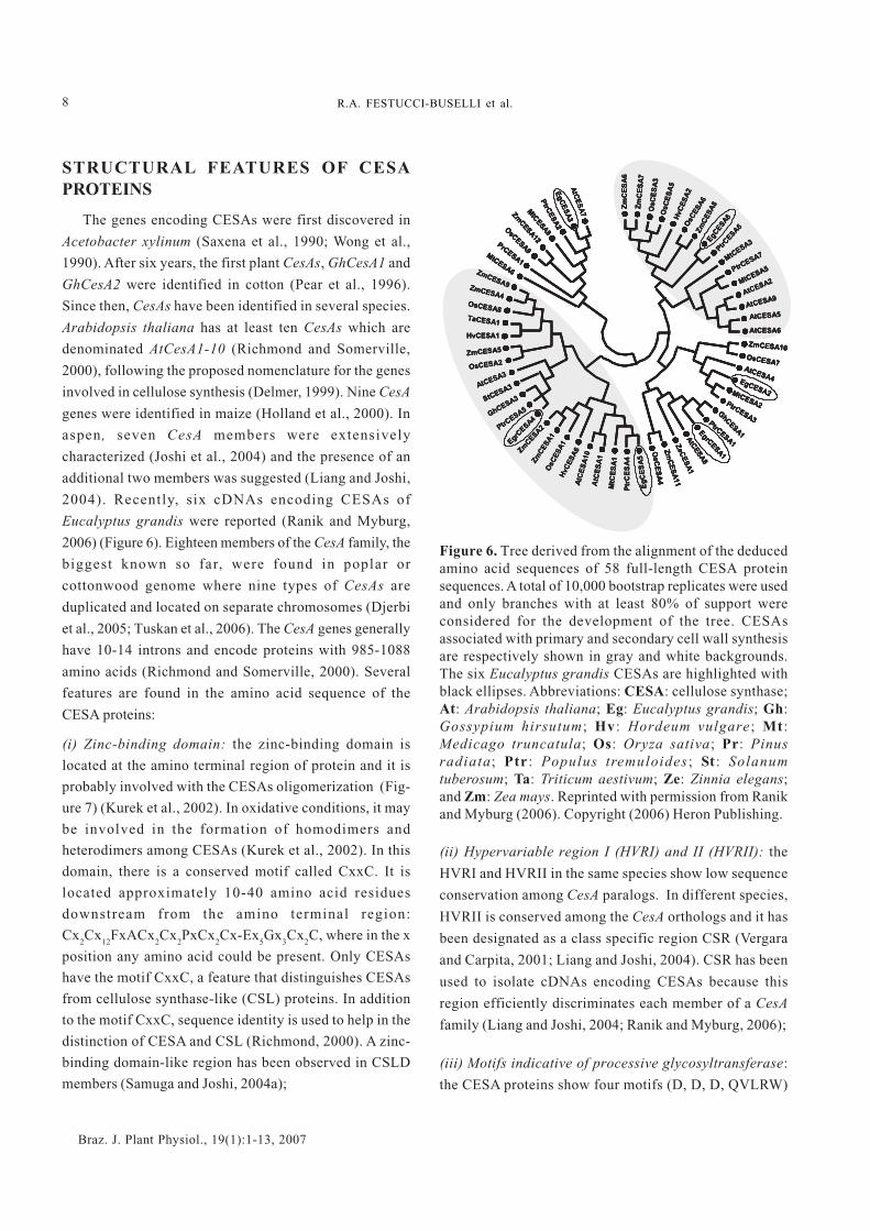
2004). Recently, six cDNAs encoding CESAs of
Eucalyptus grandis were reported (Ranik and Myburg,
2006) (Figure 6). Eighteen members of the CesA family, the
biggest known so far, were found in poplar or
cottonwood genome where nine types of CesAs are
duplicated and located on separate chromosomes (Djerbi
et al., 2005; Tuskan et al., 2006). The CesA genes generally
have 10-14 introns and encode proteins with 985-1088
amino acids (Richmond and Somerville, 2000). Several
features are found in the amino acid sequence of the
CESA proteins:
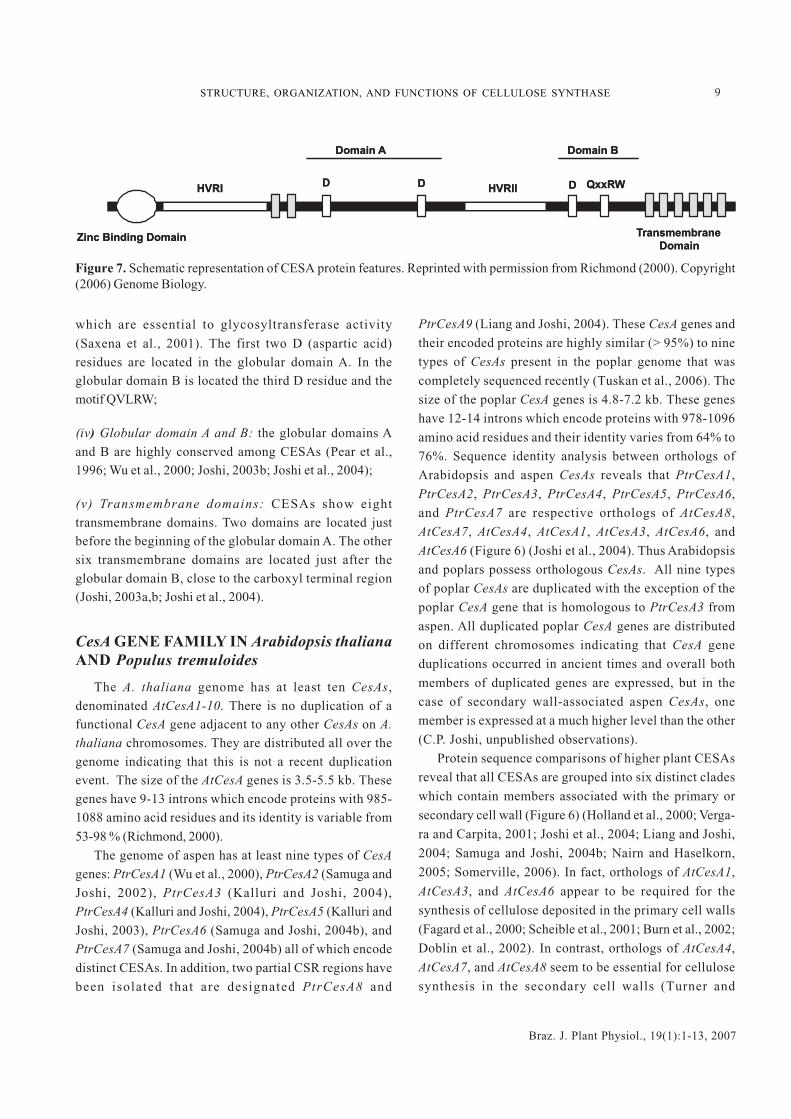
(i) Zinc-binding domain: the zinc-binding domain is
located at the amino terminal region of protein and it is
probably involved with the CESAs oligomerization (Fig-
ure 7) (Kurek et al., 2002). In oxidative conditions, it may
be involved in the formation of homodimers and
heterodimers among CESAs (Kurek et al., 2002). In this
domain, there is a conserved motif called CxxC. It is
located approximately 10-40 amino acid residues
downstream from the amino terminal region:
Cx2Cx
12FxACx
2Cx
2PxCx
2Cx-Ex
5Gx
3Cx
2C, where in the x
position any amino acid could be present. Only CESAs
have the motif CxxC, a feature that distinguishes CESAs
from cellulose synthase-like (CSL) proteins. In addition
to the motif CxxC, sequence identity is used to help in the
distinction of CESA and CSL (Richmond, 2000). A zinc-
binding domain-like region has been observed in CSLD
members (Samuga and Joshi, 2004a);
Figure 6. Tree derived from the alignment of the deducedamino acid sequences of 58 full-length CESA proteinsequences. A total of 10,000 bootstrap replicates were usedand only branches with at least 80% of support wereconsidered for the development of the tree. CESAsassociated with primary and secondary cell wall synthesisare respectively shown in gray and white backgrounds.The six Eucalyptus grandis CESAs are highlighted withblack ellipses. Abbreviations: CESA: cellulose synthase;At: Arabidopsis thaliana; Eg: Eucalyptus grandis; Gh:Gossypium hirsutum ; Hv : Hordeum vulgare ; Mt :Medicago truncatula; Os: Oryza sativa; Pr: Pinusradiata; Ptr : Populus tremuloides ; St : Solanumtuberosum; Ta: Triticum aestivum; Ze: Zinnia elegans;and Zm: Zea mays. Reprinted with permission from Ranikand Myburg (2006). Copyright (2006) Heron Publishing.
(ii) Hypervariable region I (HVRI) and II (HVRII): the
HVRI and HVRII in the same species show low sequence
conservation among CesA paralogs. In different species,
HVRII is conserved among the CesA orthologs and it has
been designated as a class specific region CSR (Vergara
and Carpita, 2001; Liang and Joshi, 2004). CSR has been
used to isolate cDNAs encoding CESAs because this
region efficiently discriminates each member of a CesA
family (Liang and Joshi, 2004; Ranik and Myburg, 2006);
(iii) Motifs indicative of processive glycosyltransferase:
the CESA proteins show four motifs (D, D, D, QVLRW)
AtC
ES
A7
Eg
CE
SA
3
PtrC
ES
A2
MtC
ESA8
ZmCESA12
OsC
ESA9
PrCESA1M
tCESA6
EgrC
ESA4
ZmCESA9ZmCESA4OsCESA8
TaCESA1
HvCESA1
ZmCESA5
OsCESA2
AtCESA3
StCESA3
GhCESA3
PtrCESA5
ZmCESA2
Zm
CES
A1
OsC
ES
A1
HvC
ES
A6
AtC
ES
A10
AtC
ES
A1
MtC
ES
A1
Ptr
CE
SA
4
Eg
CE
SA
5
ZmCESA10
OsCESA7AtCESA4EgCESA2MtCESA2PtrCESA3
GhC
ESA1
PtrCESA1
EgrC
ESA1
AtC
ES
A8
ZeC
ES
A1
Zm
CE
SA
11
OsC
ES
A4
Zm
CE
SA
6
Zm
CE
SA
7
OsC
ES
A3
OsC
ES
A5
HvC
ES
A2
OsC
ES
A6
Zm
CE
SA
8EgCESA6
PtrCESA6
MtC
ESA3
PtrCESA7
MtCESA5
AtCESA2
AtCESA9
AtCESA5
AtCESA6
AtC
ES
A7
Eg
CE
SA
3
PtrC
ES
A2
MtC
ESA8
ZmCESA12
OsC
ESA9
PrCESA1M
tCESA6
AtC
ES
A7
Eg
CE
SA
3
PtrC
ES
A2
MtC
ESA8
ZmCESA12
OsC
ESA9
PrCESA1M
tCESA6
EgrC
ESA4
ZmCESA9ZmCESA4OsCESA8
TaCESA1
HvCESA1
ZmCESA5
OsCESA2
AtCESA3
StCESA3
GhCESA3
PtrCESA5
EgrC
ESA4
ZmCESA9ZmCESA4OsCESA8
TaCESA1
HvCESA1
ZmCESA5
OsCESA2
AtCESA3
StCESA3
GhCESA3
PtrCESA5
ZmCESA2
Zm
CES
A1
OsC
ES
A1
HvC
ES
A6
AtC
ES
A10
AtC
ES
A1
MtC
ES
A1
Ptr
CE
SA
4
Eg
CE
SA
5
ZmCESA2
Zm
CES
A1
OsC
ES
A1
HvC
ES
A6
AtC
ES
A10
AtC
ES
A1
MtC
ES
A1
Ptr
CE
SA
4
Eg
CE
SA
5
ZmCESA10
OsCESA7AtCESA4EgCESA2MtCESA2PtrCESA3
GhC
ESA1
PtrCESA1
EgrC
ESA1
AtC
ES
A8
ZeC
ES
A1
Zm
CE
SA
11
OsC
ES
A4
ZmCESA10
OsCESA7AtCESA4EgCESA2MtCESA2PtrCESA3
GhC
ESA1
PtrCESA1
EgrC
ESA1
AtC
ES
A8
ZeC
ES
A1
Zm
CE
SA
11
OsC
ES
A4
Zm
CE
SA
6
Zm
CE
SA
7
OsC
ES
A3
OsC
ES
A5
HvC
ES
A2
OsC
ES
A6
Zm
CE
SA
8EgCESA6
PtrCESA6
MtC
ESA3
PtrCESA7
MtCESA5
AtCESA2
AtCESA9
AtCESA5
AtCESA6
Zm
CE
SA
6
Zm
CE
SA
7
OsC
ES
A3
OsC
ES
A5
HvC
ES
A2
OsC
ES
A6
Zm
CE
SA
8EgCESA6
PtrCESA6
MtC
ESA3
PtrCESA7
MtCESA5
AtCESA2
AtCESA9
AtCESA5
AtCESA6
9
Braz. J. Plant Physiol., 19(1):1-13, 2007
STRUCTURE, ORGANIZATION, AND FUNCTIONS OF CELLULOSE SYNTHASE
Figure 7. Schematic representation of CESA protein features. Reprinted with permission from Richmond (2000). Copyright(2006) Genome Biology.
which are essential to glycosyltransferase activity
(Saxena et al., 2001). The first two D (aspartic acid)residues are located in the globular domain A. In theglobular domain B is located the third D residue and themotif QVLRW;
(iv) Globular domain A and B: the globular domains Aand B are highly conserved among CESAs (Pear et al.,1996; Wu et al., 2000; Joshi, 2003b; Joshi et al., 2004);
(v) Transmembrane domains: CESAs show eighttransmembrane domains. Two domains are located justbefore the beginning of the globular domain A. The othersix transmembrane domains are located just after theglobular domain B, close to the carboxyl terminal region(Joshi, 2003a,b; Joshi et al., 2004).
CesA GENE FAMILY IN Arabidopsis thalianaAND Populus tremuloides
The A. thaliana genome has at least ten CesAs,denominated AtCesA1-10. There is no duplication of afunctional CesA gene adjacent to any other CesAs on A.
thaliana chromosomes. They are distributed all over thegenome indicating that this is not a recent duplicationevent. The size of the AtCesA genes is 3.5-5.5 kb. Thesegenes have 9-13 introns which encode proteins with 985-1088 amino acid residues and its identity is variable from
53-98 % (Richmond, 2000).
The genome of aspen has at least nine types of CesA
genes: PtrCesA1 (Wu et al., 2000), PtrCesA2 (Samuga and
Joshi, 2002), PtrCesA3 (Kalluri and Joshi, 2004),PtrCesA4 (Kalluri and Joshi, 2004), PtrCesA5 (Kalluri and
Joshi, 2003), PtrCesA6 (Samuga and Joshi, 2004b), andPtrCesA7 (Samuga and Joshi, 2004b) all of which encode
distinct CESAs. In addition, two partial CSR regions have
been isolated that are designated PtrCesA8 and
PtrCesA9 (Liang and Joshi, 2004). These CesA genes and
their encoded proteins are highly similar (> 95%) to nine
types of CesAs present in the poplar genome that was
completely sequenced recently (Tuskan et al., 2006). The
size of the poplar CesA genes is 4.8-7.2 kb. These genes
have 12-14 introns which encode proteins with 978-1096
amino acid residues and their identity varies from 64% to
76%. Sequence identity analysis between orthologs of
Arabidopsis and aspen CesAs reveals that PtrCesA1,
PtrCesA2, PtrCesA3, PtrCesA4, PtrCesA5, PtrCesA6,
and PtrCesA7 are respective orthologs of AtCesA8,
AtCesA7, AtCesA4, AtCesA1, AtCesA3, AtCesA6, and
AtCesA6 (Figure 6) (Joshi et al., 2004). Thus Arabidopsis
and poplars possess orthologous CesAs. All nine types
of poplar CesAs are duplicated with the exception of the
poplar CesA gene that is homologous to PtrCesA3 from
aspen. All duplicated poplar CesA genes are distributed
on different chromosomes indicating that CesA gene
duplications occurred in ancient times and overall both
members of duplicated genes are expressed, but in the
case of secondary wall-associated aspen CesAs, one
member is expressed at a much higher level than the other
(C.P. Joshi, unpublished observations).
Protein sequence comparisons of higher plant CESAs
reveal that all CESAs are grouped into six distinct clades
which contain members associated with the primary or
secondary cell wall (Figure 6) (Holland et al., 2000; Verga-
ra and Carpita, 2001; Joshi et al., 2004; Liang and Joshi,
2004; Samuga and Joshi, 2004b; Nairn and Haselkorn,
2005; Somerville, 2006). In fact, orthologs of AtCesA1,
AtCesA3, and AtCesA6 appear to be required for the
synthesis of cellulose deposited in the primary cell walls
(Fagard et al., 2000; Scheible et al., 2001; Burn et al., 2002;
Doblin et al., 2002). In contrast, orthologs of AtCesA4,
AtCesA7, and AtCesA8 seem to be essential for cellulose
synthesis in the secondary cell walls (Turner and
D D D QxxRWHVRIIHVRI
Zinc Binding Domain Transmembrane
Domain
Domain A Domain B
D D D QxxRWHVRIIHVRI
Zinc Binding Domain Transmembrane
Domain
Domain A Domain B
1 0
Braz. J. Plant Physiol., 19(1):1-13, 2007
R.A. FESTUCCI-BUSELLI et al.
Somerville, 1997; Taylor et al., 1999, 2000, 2003; Zhong et
al., 2003). The latter three CESA isoforms are specific to
secondary cell wall formation and they actually
physically interact with each other so it is highly
probable that they are located in the same CelS as
discussed previously (Taylor et al., 2003).
In the same way, PtrCesA1, PtrCesA2, and PtrCesA3
are involved with cellulose deposition in the secondary
cell wall and PtrCesA4, PtrCesA5, PtrCesA6, and
PtrCesA7 may be involved with cellulose deposition in
the primary cell wall (Joshi et al., 2004; Kalluri and Joshi,
2004; Samuga and Joshi, 2004b). The gene AtCesA8
present in the lew2-1 mutant genome was further
characterized and its disruption increases osmotic and
drought stress tolerance, indicating that cellulose
synthesis may be involved in stress tolerance (Chen et
al., 2005), but the cause and consequence relationship
needs to be delineated.
THE MOST IMPORTANT UNSOLVEDTOPICS IN CELLULOSE BIOSYNTHESIS
Although some remarkable progress has been made
during the past decade towards unraveling the process of
cellulose biosynthesis, a number of unanswered
questions still remain. In higher plants, do two distinct
types of complexes of CESAs and other associated
enzymes synthesize cellulose Iα and cellulose Iβ? How is
cellulose synthesized and deposited during primary and
secondary cell wall formation? How is each glucose
residue inverted 180° with respect to its neighboring
residues? Are monosaccharides or disaccharides
substrates for CESAs? How are glucose chains aligned
side-by-side to generate a microfibril of cellulose? How
many proteins are necessary to fully assemble the CelS
and synthesize cellulose? What is the exact function of
each of these proteins? How are CESAs arranged in the
subunits of CelS? Why are there so many CesAs in
plants? Do the transmembrane helices of the CESA
proteins form a pore in the plasma membrane through
which the growing glucan chain can pass? Are CesAs
regulated at both transcriptional and post-transcriptional
phases? Would it be possible to produce wood with highcontents of better cellulose and still maintain thenecessary physical and mechanical properties of wood?
Would it be possible to reconstitute functional CelS invitro that is capable of synthesizing cellulose I? Whatrole(s) do other proteins (such as KOR) play that havebeen shown to affect cellulose biosynthesis inArabidopsis mutants? Will overexpression of SUSY orother enzymes involved in photosynthetic carbonfixation improve cellulose biosynthesis? Which proteinscontrol economically important cellulose traits such asmicrofibril angle, DP, and crystallinity?
In conclusion, there are still a large number ofquestions that need to be answered to fully understandthe process of cellulose biosynthesis in plants that hasdirect connection with utilization of cellulose foragricultural and forest product manufacturing. Thecharacterization of regulatory regions of CesAs genesand their transcription factors might provide insightsabout regulatory processes involved in the specificexpression patterns of CesAs genes and consequently
cellulose production.
Acknowledgments: The authors would like toacknowledge the doctoral scholarship awarded to RAFBby FAPEMIG (Minas Gerais State Research Foundation,Brazil). CPJ wishes to thank the National ScienceFoundation (award # IBN-0236492), Consortium for PlantBiotechnology Research (award # G012026-249), and theUnited States Department of Agriculture (award # 2005-35103-15256) for supporting research in his laboratory.We would like to express our gratitude to Drs Shi-YouDing, Mike Jarvis, Todd Richmond, Martin Ranik, andBernard Henrissat for permission to use adaptedversions of the Figures shown in this review. RAFBwould like to thank Professor Raimundo Santos Barrosfor his suggestions and criticisms of the first drafts of
this manuscript.
REFERENCES
Arioli T, Peng L, Betzner AS, Burn J, Wittke W, Herth W,Camilleri C, Höfte H, Plazinski J, Birch R, Cork A, GloverJ, Redmond J, Williamson RE (1998) Molecular analysisof cellulose biosynthesis in Arabidopsis. Science279:717-720.
Bhandari S, Fujino T, Thammanagowda S, Zhang D, Xu F,Joshi CP (2006) Xylem-specific and tension stress-responsive coexpression of KORRIGAN endoglucanaseand three secondary wall-associated cellulose synthasegenes in aspen trees. Planta 224:828-837.
1 1
Braz. J. Plant Physiol., 19(1):1-13, 2007
STRUCTURE, ORGANIZATION, AND FUNCTIONS OF CELLULOSE SYNTHASE
Caracterização tecnológica, para produção de celulose,da nova geração de clones de Eucalyptus do Brasil.Rev. Árv. 29:129-137.
Haigler CH, Brown Jr RM (1986) Transport of rosettesfrom the Golgi apparatus to the plasma membrane inisolated mesophyll cells of Zinnia elegans duringdifferentiation to tracheary elements in suspensionculture. Protoplasma 134:111-120.
Haigler CH, Ivanova-Datcheva M, Hogan PS, Salnikov VV,Hwang S, Martin K, Delmer DP (2001) Carbonpartitioning to cellulose synthesis. Plant Mol. Biol.47:29-51.
Hardy BJ, Sarko A (1996) Molecular dynamics simulationsand diffraction-based analysis of the native cellulosefiber: structural modeling of the I-α and I-β phases andtheir interconversion. Polymer 37:1833-1839.
Holland N, Holland D, Helentjaris T, Dhugga KS,Xoconostle-Cazares B, Delmer DP (2000) A comparativeanalysis of the plant cellulose synthase (CesA) genefamily. Plant Physiol. 123:1313-1323.
Jarvis M. 2003. Cellulose stacks up. Nature 426:611-612.Joshi CP (2003a) Xylem-specific and tension stress
responsive expression of cellulose synthase genes fromaspen trees. Appl. Biochem. Biotechnol. 105:17-26.
Joshi CP (2003b) Molecular biology of cellulosebiosynthesis in plants. In: Pandalai S (ed), RecentResearch Development in Plant Molecular Biology, vol.1, pp.19-38. Research Signpost Press, Kerala.
Joshi CP, Bhandari S, Ranjan P, Kalluri UC, Liang X, FujinoT, Samuga A (2004) Genomics of cellulose biosynthesisin poplars. New Phytol. 164:53-61.
Kalluri UC, Joshi CP (2003) Isolation and characterizationof a new, full-length cellulose synthase cDNA,PtrCESA5 from developing xylem of aspen trees. J. Exp.Bot. 54:2187-2188.
Kalluri UC, Joshi CP (2004) Differential expression patternsof two cellulose synthase genes are associated withprimary and secondary cell wall development in aspentrees. Planta 220:47-55.
Kimura S, Laosinchai W, Itoh T, Cui X, Linder CR, BrownJr RM (1999) Immunogold labeling of rosette terminalcellulose-synthesizing complexes in the vascular plantVigna angularis. Plant Cell 11:2075-2085.
Kono H, Yunoki S, Shikano T, Fujiwara M, Erata T, KawaiM (2002) CP/MAS 13C NMR study of cellulose andcellulose derivatives. 1. Complete assignment of theCP/MAS 13C NMR spectrum of the native cellulose. J.Am. Chem. Soc. 124:7506-7511.
Koyama M, Helbert W, Imai T, Sugiyama J, Henrissat B(1997) Parallel-up structure evidences the molecular
Brett C, Waldron K (1990) Physiology and biochemistryof plant cell walls. In: Black M, Chapman J (eds), Topicsin Plant Physiology, vol. 2, pp.4-57. Unwin Hyman,London.
Brown Jr RM, Saxena IM, Kudlicka K (1996) Cellulosebiosynthesis in higher plants. Trends Plant Sci. 1:149-156.
Brown Jr RM, Saxena IM (2000) Cellulose biosynthesis: amodel for understanding the assembly of biopolymers.Plant Physiol. Biochem. 38:57-67.
Burn JE, Hocart CH, Birch RJ, Cork AC, Williamson RE(2002) Functional analysis of the cellulose synthasegenes CesA1, CesA2, and CesA3 in Arabidopsis. PlantPhysiol. 129:797-807.
Burton RA, Shirley NJ, King BJ, Harvey AJ, Fincher GB(2004) The CesA gene family of barley. Quantitativeanalysis of transcripts reveals two groups ofcoexpressed genes. Plant Physiol. 134:224-236.
Chen Z, Hong X, Zhang H, Wang Y, Li X, Zhu JK, Gong Z(2005) Disruption of the cellulose synthase gene,AtCesA8/IRX1, enhances drought and osmotic stresstolerance in Arabidopsis. Plant J. 43:273-283.
Delmer DP, Amor Y (1995) Cellulose biosynthesis. PlantCell 7:987-1000.
Delmer DP (1999) Cellulose biosynthesis: exciting timesfor a difficult field of study. Annu. Rev. Plant Physiol.Plant Mol. Biol. 50:245-276.
Delmer DP, Haigler CH (2002) The regulation of metabolicflux to cellulose, a major sink for carbon in plants. Metab.Eng. 4:22-28.
Ding SY, Himmel ME (2006) The maize primary cell wallmicrofibril: a new model derived from direct visualization.J. Agric. Food Chem. 54:597-606.
Djerbi S, Lindskog M, Arvestad L, Sterky F, Teeri TT (2005)The genome sequence of Black Cottonwood (Populustrichocarpa) reveals 18 conserved cellulose synthase(CesA) genes. Planta 221:739-746.
Doblin MS, Kurek I, Jacob-Wilk D, Delmer DP (2002)Cellulose biosynthesis in plants: from genes to rosettes.Plant Cell Physiol. 43:1407-1420.
Eckardt NA (2003) Cellulose synthesis takes the CesA train.Plant Cell 15:1685-1687.
Fagard M, Desnos T, Desprez T, Goubet F, Refrégier G,Mouille G, McCann M, Rayon C, Vernhettes S, Höfte H(2000) PROCUSTE1 encodes a cellulose synthaserequired for normal cell elongation specifically in rootsand dark-grown hypocotyls of Arabidopsis. Plant Cell12:2409-2423.
Gomide JL, Colodette JL, de Oliveira RC, Silva CM (2005)
1 2
Braz. J. Plant Physiol., 19(1):1-13, 2007
R.A. FESTUCCI-BUSELLI et al.
directionality during biosynthesis of bacterial cellulose.Proc. Natl. Acad. Sci. USA 94:9091-9095.
Kurek I, Kawagoe Y, Jacob-Wilk D, Doblin M, Delmer D(2002) Dimerization of cotton fiber cellulose synthasecatalytic subunits occurs via oxidation of the zinc-binding domains. Proc. Natl. Acad. Sci. USA 99:11109-11114.
Langan P, Nishiyama Y, Chanzy H (2001) X-ray structureof mercerized cellulose II at 1 Å resolution.Biomacromolecules 2:410-416.
Liang X, Joshi CP (2004) Molecular cloning of ten distincthypervariable regions from cellulose synthase genesuperfamily in aspen trees. Tree Physiol. 24:543-550.
Mellerowicz EJ, Baucher M, Sundberg B, Boerjan W (2001)Unraveling cell wall formation in the woody dicot stem.Plant Mol. Biol. 47:239-274.
Molhoj M, Pagant S, Hofte H (2002) Towardsunderstanding the role of membrane bound endo-β-glucanases in cellulose biosynthesis. Plant Cell Physiol.43:1399-1406.
Mueller SC, Brown Jr RM (1980) Evidence for anintramembrane component associated with a cellulosemicrofibril synthesizing complex in higher plants. J. CellBiol. 84:315-326.
Nairn CJ, Haselkorn T (2005) Three loblolly pine CesAgenes expressed in developing xylem are orthologousto secondary cell wall CesA genes of angiosperms. NewPhytol. 166:907-915.
Nishiyama Y, Langan P, Chanzy H (2002) Crystal structureand hydrogen-bonding system in cellulose Iβ fromsynchrotron X-ray and neutron fiber diffraction. J. Am.Chem. Soc. 124:9074-9082.
Nishiyama Y, Sugiyama J, Chanzy H, Langan P (2003)Crystal structure and hydrogen bonding system incellulose I alpha from synchrotron X-ray and neutronfiber diffraction. J. Am. Chem. Soc. 125:14300-14306.
O’Sullivan AC (1997) Cellulose: the structure slowlyunravels. Cellulose 4:173-207.
Pear JR, Kawagoe Y, Schreckengost WE, Delmer DP, StalkerDM (1996) Higher plants contain homologs of thebacterial CelA genes encoding the catalytic sub-unitof cellulose synthase. Proc. Natl. Acad. Sci. USA93:12637-12642.
Perrin RM (2001) Cellulose: how many cellulose synthasesto make a plant? Curr. Biol. 11:213-216.
Pilate G, Déjardin A, Laurans F, Leplé JC (2004) Tensionwood as a model for functional genomics of woodformation. New Phytol. 164:63-72.
Ranik M, Myburg AA (2006) Six new cellulose synthasegenes from Eucalyptus are associated with primary and
secondary cell wall biosynthesis. Tree Physiol. 26:545-556.
Read SM, Bacic T (2002) Prime time for cellulose. Science295:59-60.
Richmond TA (2000) Higher plant cellulose synthases.Genome Biol. 1:3001.1-3001.6.
Richmond TA, Somerville CR (2000) The cellulosesynthase superfamily. Plant Physiol. 124:495-498.
Robert S, Bichet A, Grandjean O, Kierzkowski D, Satiat-Jeunemaιtre B, Pelletier S, Hauser MT, Hofte H,Vernhettes S (2005) An Arabidopsis endo-1,4-β-D-glucanase involved in cellulose synthesis undergoesregulated intracellular cycling. Plant Cell 17:3378-3389.
Rowland SP, Howley PS (1988) Hydrogen bonding onaccessible surfaces of cellulose from various sourcesand relationship to order within crystalline regions. J.Polym. Sci., Part A: Polym. Chem. 26:1769-1778.
Samuga A, Joshi CP (2002) A new cellulose synthase gene(PtrCesA2) from aspen xylem is orthologous toArabidopsis AtCesA7 (irx3) gene associated withsecondary cell wall synthesis. Gene 296:37-44.
Samuga A, Joshi CP (2004a) Cloning and characterizationof cellulose synthase-like gene, PtrCSLD2 fromdeveloping xylem of aspen trees. Physiol. Plant. 120:641-651.
Samuga A, Joshi CP (2004b) Expression patterns of twoprimary cell wall-related cellulose synthase cDNAs,PtrCESA6 and PtrCESA7 from aspen. Gene 334:71-80.
Saxena IM, Lin FC, Brown Jr RM (1990) Cloning andsequencing of the cellulose synthase catalytic sub-unitgene of Acetobacter xylinum. Plant Mol. Biol. 15:673-683.
Saxena IM, Brown Jr RM (1995) Identification of a secondcellulose synthase gene (acsAII) in Acetobacterxylinum. J. Bacteriol. 177:5276-5283.
Saxena IM, Brown Jr RM, Dandekar T (2001) Structure–function characterization of cellulose synthase:relationship to other glycosyltransferases.Phytochemistry 57:1135-1148.
Saxena IM, Brown Jr RM (2005) Cellulose biosynthesis:current views and evolving concepts. Ann. Bot. 96:9-21.
Scheible WR, Eshed R, Richmond T, Delmer D, SomervilleC (2001) Modifications of cellulose synthase conferresistance to isoxaben and thiazolidinone herbicides inArabidopsis ixr1 mutants. Proc. Natl. Acad. Sci. USA98:10079-10084.
Somerville CR (2006) Cellulose synthesis in higher plants.Annu. Rev. Cell Dev. Biol. 22:53-78.
Sturcova A, His I, Apperley DC, Sugiyama J, Jarvis MC(2004) Structural details of crystalline cellulose fromhigher plants. Biomacromolecules 5:1333-1339.
1 3
Braz. J. Plant Physiol., 19(1):1-13, 2007
STRUCTURE, ORGANIZATION, AND FUNCTIONS OF CELLULOSE SYNTHASE
Tanaka K, Murata K, Yamazaki M, Onosato K, Miyao A,Hirochika H (2003) Three distinct rice cellulose synthasecatalytic subunit genes required for cellulose synthesisin the secondary wall. Plant Physiol. 133:73-83.
Taylor NG, Laurie S, Turner SR (2000) Multiple cellulosesynthase catalytic subunits are required for cellulosesynthesis in Arabidopsis. Plant Cell 12:2529-2540.
Taylor NG, Scheible WR, Cutler S, Somerville CR, TurnerSR (1999) The irregular xylem3 locus of Arabidopsisencodes a cellulose synthase required for secondarycell wall synthesis. Plant Cell 11:769-780.
Taylor NG, Howells RM, Huttly AK, Vickers K, Turner SR(2003) Interactions among three distinct CESA proteinsessential for cellulose synthesis. Proc. Natl. Acad. Sci.USA 100:1450-1455.
Timell TE (1969) The chemical composition of tensionwood. Svensk Papperstidning 72:173-181.
Tsekos I (1999) The sites of cellulose synthesis in algae:diversity and evolution of cellulose-synthesizingenzyme complexes. J. Phycol. 35:635-655.
Tsekos I, Orolagas N, Herth W (1999) Cellulose microfibrilassembly and orientation in some bangiophyte redalgae: relationship between synthesizing terminalcomplexes and microfibril structure, shape, anddimensions. Phycologia 38:217-224.
Turner SR, Somerville CR (1997) Collapsed xylem phenotype
of Arabidopsis identifies mutants deficient in cellulosedeposition in the secondary cell wall. Plant Cell 9:689-701.
Tuskan GA et al. (2006) The genome of western blackcottonwood, Populus trichocarpa (Torr. & Gray exBrayshaw). Science 313:1596-1604.
Vergara CE, Carpita NC (2001) β-D-Glycan synthases andthe CesA gene family: lessons to be learned from themixed-linkage (1 3),(1�4)β-D-glucan synthase. PlantMol. Biol. 47:145-160.
Wada MJ (2002) Lateral thermal expansion of cellulose Iβand IIII polymorphs. J. Polym. Sci., Part B: Polym. Phys.40:1095-1102.
Wong HC, Fear AL, Calhoon RD, Eichinger GH, Mayer R,Amikam D, Benziman M, Gelfand DH, Meade JH,Emerick AW, Bruner R, Ben-Bassat A, Tal R (1990)Genetic organization of the cellulose synthase operonin Acetobacter xylinum. Proc. Natl. Acad. Sci. USA87:8130-8134.
Wu L, Joshi CP, Chiang VL (2000) A xylem-specificcellulose synthase gene from aspen (Populustremuloides) is responsive to mechanical stress. PlantJ. 22:495-502.
Zhong R, Morrison WH, Freshour GD, Hahn MG, Ye ZH(2003) Expression of a mutant form of cellulose synthaseAtCesA7 causes dominant negative effect on cellulosebiosynthesis. Plant Physiol. 132:786-795.
�