Structural Basis for the Enantiospecificities of R- and S ...
41
1 Structural Basis for the Enantiospecificities of R- and S-Specific Phenoxypropionate/α-Ketoglutarate Dioxygenases Tina A. Müller, 1 Maria I. Zavodszky, 2 Michael Feig, 2,3 Leslie A. Kuhn, 2 and Robert P. Hausinger 1,2 1 Department of Microbiology & Molecular Genetics, 2 Department of Biochemistry & Molecular Biology, and 3 Department of Chemistry, Michigan State University, East Lansing, Michigan 48824-4320 Reprint requests to: Robert P. Hausinger, Department of Microbiology and Molecular Genetics, 6193 Biomedical Physical Sciences; Tel.: 517-355-6463 ext. 1610; Fax: 517-353-8957; E-mail: [email protected] Running title: Dichlorprop hydroxylase enantiospecificity 43 pages, 5 pages supplementary material, 2 schemes, 3 figures, 2 tables Supplementary material includes coordinate files in PDB format of the most favored docked models of RdpA and SdpA (RdpA.pdb and SdpA.pdb) and a word document containing one table and three figures with their legends (RdpA-SdpA.doc). March 16, 2006
Transcript of Structural Basis for the Enantiospecificities of R- and S ...
1
Structural Basis for the Enantiospecificities of R- and S-Specific
Phenoxypropionate/α-Ketoglutarate Dioxygenases
Tina A. Müller,1 Maria I. Zavodszky,2 Michael Feig,2,3 Leslie A. Kuhn,2 and Robert P.
Hausinger1,2
1Department of Microbiology & Molecular Genetics, 2Department of Biochemistry & Molecular
Biology, and 3Department of Chemistry, Michigan State University, East Lansing, Michigan
48824-4320
Reprint requests to: Robert P. Hausinger, Department of Microbiology and Molecular
Genetics, 6193 Biomedical Physical Sciences; Tel.: 517-355-6463 ext. 1610; Fax: 517-353-8957;
E-mail: [email protected]
Running title: Dichlorprop hydroxylase enantiospecificity
43 pages, 5 pages supplementary material, 2 schemes, 3 figures, 2 tables
Supplementary material includes coordinate files in PDB format of the most favored docked
models of RdpA and SdpA (RdpA.pdb and SdpA.pdb) and a word document containing one table
and three figures with their legends (RdpA-SdpA.doc).
March 16, 2006
2
Abstract
(R)- and (S)-dichlorprop/α-ketoglutarate dioxygenases (RdpA and SdpA) catalyze the oxidative
cleavage of 2-(2,4-dichlorophenoxy)propanoic acid (dichlorprop) and 2-(4-chloro-2-methyl-
phenoxy)propanoic acid (mecoprop) to form pyruvate plus the corresponding phenol concurrent
with the conversion of α-ketoglutarate (αKG) to succinate plus CO2. RdpA and SdpA are strictly
enantiospecific, converting only the (R) or the (S) enantiomer, respectively. Homology models
were generated for both enzymes on the basis of the structure of the related enzyme TauD (PDB
code 1OS7). Docking was used to predict the orientation of the appropriate mecoprop
enantiomer in each protein and the predictions were tested by characterizing the activities of site-
directed variants of the enzymes. Mutant proteins changed at residues predicted to interact with
(R)- or (S)-mecoprop exhibited significantly reduced activity, often accompanied by increased
Km values, consistent with roles for these residues in substrate binding. Four of the designed
SdpA variants were (slightly) active with (R)-mecoprop. The results of the kinetic investigations
are consistent with the identification of key interactions in the structural models and demonstrate
that enantiospecificity is coordinated by the interactions of a number of residues in RdpA and
SdpA. Most significantly, residues Phe171 in RdpA and Glu69 in SdpA apparently act by
hindering the binding of the wrong enantiomer more than the correct one, as judged by the
observed decreases in Km when these side chains are replaced by Ala.
Keywords: dioxygenase; enantiospecificity; mecoprop; site-directed mutagenesis; structural
modeling; docking
3
Abbreviations: 2,4-D, 2,4-dichlorophenoxyacetic acid; AlkB, alkylation-damaged DNA
repair enzyme; AtsK, alkyl sulfatase; ANS, anthocyanidin synthase; CarC, carbapenam synthase;
CAS, clavaminate synthase; CSD, Cambridge Structural Database; DAOCS,
deacetoxycephalosporin C synthase; dichlorprop, 2-(2,4-dichlorophenoxy)propanoic acid; FIH,
factor inhibiting hypoxia-inducible factor; αKG, α-ketoglutarate; mecoprop, 2-(4-chloro-2-methyl-
phenoxy)propanoic acid; NTA, nitrilotriacetic acid; PAHX, phytanoyl-coenzyme A 2-hydroxylase;
PDB, Protein Data Bank; RdpA, (R)-specific dichlorprop/αKG dioxygenase; SdpA, (S)-specific
dichlorprop/αKG dioxygenase; TfdA, 2,4-D/αKG dioxygenase; TauD, taurine/αKG dioxygenase;
TR, tropinone reductase; SDS-PAGE, sodium dodecyl sulfate-polyacrylamide gel electrophoresis.
4
Phenoxyalkanoic acids are systemic and post-emergence inhibitors of broadleaf weeds and are
among the most widely applied herbicides in the world (Worthing and Hance 1991; Ahrens
1994; Donaldson et al. 2002). These synthetic auxins (Åberg 1973; Loos 1975; Ahrens 1994)
include 2,4-dichlorophenoxyacetic acid (2,4-D) along with the chiral representatives 2-(2,4-
dichlorophenoxy)propanoic acid (dichlorprop) and 2-(4-chloro-2-methyl-phenoxy)propanoic
acid (mecoprop), of which only the (R)-enantiomers are herbicidally active (Matell 1953).
Microorganisms able to degrade these phenoxyalkanoic acid herbicides have been isolated from
different environments, and their degradative pathways have been elucidated (Zipper et al. 1996;
Hausinger et al. 1997; Tett et al. 1997; Müller et al. 1999; Müller et al. 2001). For example, the
first step in 2,4-D metabolism is side chain hydroxylation to form an unstable intermediate that
decomposes by elimination of the phenol derivative (Scheme 1).
Scheme 1
The 2,4-D hydroxylase (TfdA) from Cupriavidus necator (formerly Ralstonia eutropha)
JMP134(pJP4) has been intensively studied and shown to require FeII as a cofactor and α-
ketoglutarate (αKG) as a co-substrate (Fukumori and Hausinger 1993b; Fukumori and Hausinger
1993a; Saari and Hausinger 1998; Hegg et al. 1999; Hogan et al. 2000; Dunning Hotopp and
Hausinger 2002). For mecoprop or dichlorprop metabolism, best studied in the soil bacterium
Sphingomonas herbicidovorans MH, the enantiomers are separately transported into the cell by
distinct uptake systems (Nickel et al. 1997), and enantiomer-specific (R)- and (S)-
dichlorprop/αKG dioxygenases (RdpA and SdpA) catalyze the initial degradation steps as
illustrated in Scheme 2 (Nickel et al. 1997; Müller 2004; Müller et al. 2004b). RdpA and SdpA
share 30% amino acid sequence identity to each other and 30% and 37% identity, respectively, to
TfdA, with no significant gaps in alignment quality, indicating they are all close structural
5
homologs (Sander and Schneider 1991). The substituted phenol products released from these
FeII/αKG-dependent dioxygenases are subsequently converted to the corresponding catechols
and further metabolized by the modified ortho-cleavage pathway.
Scheme 2
The herbicide-degrading dioxygenases belong to a large family of mononuclear, non-
heme FeII enzymes that catalyze a broad array of reactions (reviewed in Hausinger 2004; Clifton
et al. 2006) including hydroxylations, epoxidations, desaturations, ring formation, ring
expansion, and, as only recently discovered, chlorinations (Vaillancourt et al. 2005a;
Vaillancourt et al. 2005b). Crystal structures have been elucidated for several family members
including taurine/αKG dioxygenase (TauD) (Elkins et al. 2002; O'Brien et al. 2003), alkyl
sulfatase (AtsK) (Müller et al. 2004a; Müller et al. 2005), clavaminate synthase (CAS) (Zhang et
al. 2000), deacetoxycephalosporin C synthase (DAOCS) (Valegård et al. 1998), anthocyanidin
synthase (ANS) (Wilmouth et al. 2002), carbapenam synthase (CarC) (Clifton et al. 2003),
proline 3-hydroxylase (Clifton et al. 2001), the factor inhibiting hypoxia-inducible factor (FIH)
(Dann et al. 2002; Elkins et al. 2003), phytanoyl-coenzyme A 2-hydroxylase (PAHX)
(McDonough et al. 2005), and the DNA repair enzyme AlkB (Yu et al. 2006). The structures
reveal a common β-jelly roll or double-stranded β-helix fold containing a metal ion-binding
motif: His1-X-Asp/Glu-Xn-His2 (where n varies from 40-153 residues). Three water molecules
occupy the remaining metal ligand positions in the resting enzyme. Two water molecules are
displaced upon binding of the cosubstrate, with the αKG C-2 keto group coordinating opposite
the carboxylate side chain and the αKG C-1 carboxyl group binding opposite either His1 (TauD,
AtsK, CAS, FIH) or opposite His2 (DAOCS, ANS, CarC, PAHX, and AlkB), with a nearby Arg
residue (located 15-22 residues beyond His2 in the sequence) providing additional stabilization to
6
the C-1 carboxylate in the cases of TauD, AtsK, CAS, CarC, and AlkB. Another Arg residue
(located in the sequence about 10 residues beyond His2) is positioned to form an ion pair with the
C-5 carboxylate of αKG in all structures except FIH, where a Lys located elsewhere in the
sequence provides stabilization. Unlike other FeII sites, the αKG-bound metallocenters exhibit a
characteristic metal-to-ligand charge-transfer transition (Pavel et al. 1998; Hegg et al. 1999; Ryle
et al. 1999; Trewick et al. 2002) conferring a lilac color to this state of the enzymes. The primary
substrate (e.g., taurine in the case of TauD) does not bind to the metal center, but the
aforementioned crystallographic studies and additional spectroscopic evidence (Ho et al. 2001;
Zhou et al. 2001) indicates that substrate binding leads to the loss of the final water molecule,
thus creating a site for binding of oxygen. In the case of TauD, oxidative decarboxylation of
αKG has been shown to produce an FeIV-oxo intermediate species that inserts oxygen into the
unactivated C-H bond (Price et al. 2003a; Price et al. 2003b; Proshlyakov et al. 2004; Riggs-
Gelasco et al. 2004; Grzyska et al. 2005; Price et al. 2005).
Sequence alignments highlight several potential key residues of the (R)- and (S)-
dichlorprop/αKG dioxygenases from S. herbicidovorans MH (Müller 2004). The His1-X-
Asp/Glu-Xn-His2 motif of RdpA is comprised of residues His111, Asp113, and His270, while
that of SdpA involves His102, Asp104, and His257. Fifteen residues beyond His2 are residues
predicted to interact with the C-1 carboxylate of αKG, Arg285 and His272, respectively.
Furthermore, Arg281 and Arg268 of the two proteins are predicted to form salt bridges to the
αKG C-5 carboxylate, with additional interactions involving Thr138 of RdpA and Thr129 of
SdpA. In contrast to these conserved residues, essentially nothing is known about the
phenoxypropanoic acid binding sites of these proteins, especially with regard to the structural
basis of enantiospecificity.
7
Here, we describe the construction of homology models of SdpA and RdpA from S.
herbicidovorans MH and the use of docking to identify residues likely to be involved in
herbicide binding. Previous homology models have led to successes in elucidating or designing
specificity-conferring interactions in ligands. For instance, homology modeling of a cercarial
(human parasite) elastase led to the development of an effective elastase inhibitor (Cohen et al.
1991) and to understanding the specificity determinants for ligands binding to a parasite tRNA
synthetase versus its human homolog (Sukuru et al. 2006). Here, we test by site-directed
mutagenesis and kinetic analysis the residues predicted to be involved in substrate binding,
enantiospecificity, or catalysis. The activity experiments are consistent with the key residues
identified by modeling being involved in substrate binding. We provide additional evidence that
several amino acids are responsible for the enantiospecificity of RdpA and SdpA, demonstrate
that the active site of SdpA is less specific than RdpA for its substrate, and discuss the structural
implications of these results.
8
Results
SdpA and RdpA Homology Models
RdpA and SdpA were aligned with TauD (Figure S1, Supplementary Material), and homology
models were created as described in Materials and Methods (Figure S2, Supplementary Material)
using the TauD structure as a structural template (O'Brien et al. 2003). The two
phenoxypropionate-degrading proteins are predicted to contain jelly roll or double-stranded β-
helix folds comprised of eight β-strands with connecting loops, as is typical of this enzyme
family (Hausinger 2004; Clifton et al. 2006). The homology models contain FeII-binding sites
(His111, Asp113, and His270 in RdpA or His102, Asp104, and His257 in SdpA), as expected
from former sequence alignments with other FeII/αKG-dependent dioxygenases (Müller 2004).
The high degree of active-site sequence identity and strong orientation of key side chains by
interactions with the FeII are supportive of the active site being the most conserved and
structurally accurate part of the RdpA and SdpA models. Detailed analysis of favored αKG
binding motifs in other members of this enzyme family indicate the iron is chelated by αKG with
its keto group positioned opposite Asp113 of RdpA or Asp104 of SdpA and its C-1 carboxylate
located so as to interact with His111 and His102, respectively. The positively charged residues
Arg285 in RdpA and His272 in SdpA are well positioned to provide additional stabilization of
the αKG C-1 carboxylate, and in each protein the C-5 carboxylate of αKG forms a salt bridge
with Arg268 and Arg281, respectively. Whereas the RdpA structure represents only one subunit
of the predicted trimeric protein, SdpA is suggested to be monomeric, based on gel filtration
experiments (Müller 2004).
9
Docking of Substrates into the RdpA and SdpA Structures
The natural substrates (R)- and (S)-mecoprop were docked into the active sites of RdpA and
SdpA to gain insight into the basis of enzyme enantiospecificity. First, the co-substrate αKG was
modeled into the active sites of RdpA and SdpA using two distinct conformations, as found in
crystal structures of FeII/αKG-dependent dioxygenases (Clifton et al. 2006). The flat
conformation has the five-membered ring formed by the metal chelate co-planar with the C-5
carboxylate, whereas the twist conformation has the two planes forming a 90 ° angle. The
resulting four models were energy-minimized and used as targets for substrate docking with the
program SLIDE (Zavodszky et al. 2002), with the assumption that the substrate carbon
undergoing hydroxylation would be located approximately at the same position relative to the
iron center as the key carbon atom of taurine in TauD (Elkins et al. 2002; O'Brien et al. 2003).
The mecoprop docking interactions with RdpA and SdpA were analyzed in detail, and one model
of each protein was selected based on the most favorable interactions between enzyme and
substrate (see below). These models are illustrated in Figure 1 with the corresponding plots of
mecoprop interactions shown in Figure 2.
Figures 1 and 2
Binding of (R)-mecoprop to RdpA
The substrate (R)-mecoprop consists of a hydrophobic phenoxy ring and a polar propanoic acid,
with both components needing to be accommodated and bound by the active site. The αKG
10
conformation leading to the most favorable interactions has αKG in the twist conformation and
positions the phenoxy ring of (R)-mecoprop as illustrated in Figure 1A (with the corresponding
interactions plotted in Figure 2A). The mecoprop carboxylate interacts with the amide nitrogen
of Ser114, the hydroxyl group of Tyr221, and a guanidino nitrogen of Arg285. The Tyr221
hydroxyl group also is predicted to lie near (3.5 Å) the substrate ether oxygen atom and could
play a role in directing enantiospecificity. Residues lining the hydrophobic substrate-binding
pocket include Val80, Leu83, Ile106, Gly107, and Phe171 (Figures 1A and 2A), with Val80 and
Leu83 being well positioned to interact with the propanoic acid methyl group. The terminal
carbon atom (CZ) of the Phe171 side chain is 4.1 Å from the phenoxy group of (R)-mecoprop;
since LigPlot has a 4.0 Å threshold for hydrophobic interactions, this interaction is missed in
Figure 2A.
To directly test the importance of potential substrate-binding residues of RdpA identified
by the homology modeling and substrate docking procedures, variant forms of the enzymes were
created by site-directed mutagenesis. To eliminate the bulky and polar Tyr221 and Arg285
residues, Y221A and R285A mutants were generated. In the presumed “hydrophobic pocket”,
Val80, Leu83, Ile106 and Phe171 each were changed to alanine to reduce hydrophobic
interactions and thereby decrease the binding affinity of (R)-mecoprop to the active site.
Replacing Gly107, positioned near and coplanar with the phenoxy ring, with a bulkier
hydrophobic residue is expected to hinder substrate binding, so this residue was mutated to Ile
and Asn in the double mutants I106G/G107I and I106G/G107N, respectively.
The specific activities of the RdpA variants were tested using (R)-mecoprop and the
racemic mixture (Table 1). The (S) enantiomer is not sold commercially and was available in
very limited supply, so the RdpA variants were not tested with this compound. When the RdpA
11
variants were examined using 4 mM (R)-mecoprop, the V80A and F171A variants exhibited
approximately 60% of wild-type enzyme activity, I106A had about 30% of that activity and
L83A, Y221A, R285A were ~10% active. The activities of the double mutants I106G/G107I and
I106G/G107N were further reduced compared to that of I106A, consistent with a bulkier side
chain at position 107 presenting steric hindrance to substrate binding in a reactive conformation.
The RdpA variants retaining at least 10% of wild-type enzyme activity were subjected to
more detailed kinetic characterization (Table 2). The maximal concentration of substrates that
could be tested was 4 mM due to solubility limitations; therefore, Km values higher than 800 µM
are only approximations and possess large errors. The Km values of V80A and R285A RdpA
variants were at least 5-fold increased over that of the wild-type enzyme, whereas that of the
I106A RdpA variant was more than 20-fold greater, supporting the described docking orientation
of the model. The effect on the R285A variant can be understood in terms of decreased
interaction with the substrate carboxylate, while the changes observed for the V80A and I106A
variants are likely to arise from loss of hydrophobic interactions. The F171A protein had a three-
fold lower Km, indicating either that this residue does not interact specifically with the phenoxy
ring of (R)-mecoprop or that F171 is actually slightly hindering (R)-mecoprop binding. The
calculated kcat of all mutant proteins was similar to the wild-type value with the exception of the
R285A mutant enzyme. Arg285 is postulated to interact with both the mecoprop carboxylate and
the C-1 carboxylate of αKG, so it could directly influence catalysis.
Binding of (S)-mecoprop to SdpA
12
The most favorable orientation of (S)-mecoprop was obtained with αKG in the twist
conformation in SdpA and (S)-mecoprop bound as illustrated in Figure 1B (with the
corresponding ligand interactions shown in Figure 2B). In this model, the substrate carboxylate
interacts with the amide nitrogen of Ser105, the hydroxyl group of Tyr107, and the guanidino
nitrogens of Arg274. Additional active site residues near the polar carboxylate include His272
and His208. Also of interest, the substrate ether oxygen atom is predicted to lie within 3.4 Å of
the two carboxyl oxygens of Glu69. If protonated or bridged by the proton of a bound water
molecule, the Glu69 carboxyl group could confer specificity to the (S) enantiomer by making a
hydrogen bond with the ether oxygen (a prediction not borne out by experimental results, vide
infra). Residues predicted to be in contact with the hydrophobic ring of (S)-mecoprop include
Ala71, Ala72, Leu82, Val84, Gly97, Asn98, Gln162 and Arg207 (with the latter four residues
depicted in Figure 1B). Pi-cation interactions between positively charged side chains like Arg207
and aromatic side groups like that in mecoprop can contribute very favorably to ligand binding
(Mitchell et al. 1994).
To experimentally test the importance of several of these residues for the binding of (S)-
mecoprop in the active site of SdpA, a series of mutant proteins was constructed. Glu69, His208,
His272 and Arg274 each were changed to alanine to eliminate the bulky and charged side chains
proposed to interact with the substrate carboxylate. The extended side chain of Arg207 was
eliminated in the R207A SdpA variant, and Gln162 was substituted with the corresponding,
hydrophobic residue found in RdpA to generate the Q162F variant.
The specific activities of the SdpA variants were tested using the (S) enantiomer, the
racemic mixture, and the (R) enantiomer of mecoprop (Table 1). The activity of all SdpA
mutants was strongly reduced. The most active SdpA variants were the E69A and R207A
13
proteins, while all other variants exhibited less than 5% of the wild-type enzyme activity. No
activity was detected in the R274A sample. E69A, R207A, and H208A were subjected to
detailed kinetic characterization (Table 2). When analyzed with (S)-mecoprop, the R207A and
H208A samples had 15- and 18-fold increases, respectively, in their Km values compared to the
wild-type enzyme. The H208A result is compatible with an ionic interaction between the
mecoprop carboxylate group and His208. A similar interaction might be possible with the highly
flexible Arg207 residue or this side chain may participate in a π-cation interaction with the
aromatic group of the substrate, in either case accounting for the large Km change in the R207A
variant. By contrast to these results, the E69A variant exhibited a lower Km than the native SdpA,
arguing against the ether oxygen having a significant role in substrate binding. With (S)-
mecoprop, the kcat was reduced four-fold for the E69A and R207A SdpA mutants compared to
wild-type enzyme, and even greater reductions were noted for the H208A variant.
Residues determining enantiospecificity in RdpA and SdpA
A direct comparison of the active sites in the RdpA and SdpA models is shown in Figure 3. The
different substrate enantiomers are predicted to bind with remarkably similar geometries in the
two predicted structures, with the carboxyl groups and aromatic rings nearly overlapping but at
distinct angles. RdpA residues Leu83, Gln93, Ile95 (omitted for clarity), Ile106, Phe171, and
Arg285 (shown in Figure 1A) could hinder binding of (S)-mecoprop; the corresponding residues
in SdpA are Ala72, Leu82, Val84, Gly97, Gln162, and His272, respectively. Similarly, SdpA
residues Glu69, Asn98, Ser161, His208 and Arg207 could restrict binding of (R)-mecoprop;
Val80, Gly107, Val170, Tyr221, and Val220 occupy these positions in RdpA. Ala72, Gly97,
14
and Gln162 are predicted to allow access by the (S) enantiomer. In general, side-chain
interactions suggest that SdpA is less specific than RdpA for its substrate. This finding is
compatible with activity measurements (Table 1), such as the activity with 2 mM (S)-mecoprop
versus that with 4 mM of the racemate, that show inhibition of SdpA activity by (R)-mecoprop.
However, RdpA remains completely active towards the (R) enantiomer in the presence of (S)-
mecoprop (Müller 2004).
Figure 3
Site-directed variants of RdpA and SdpA, created and purified as described earlier, were
used to test a subset of the above-mentioned residues for their importance in controlling
enantioselectivity. Single substitutions included the L83A, Q93A, I106A, F171A, F171Q, and
R285A variants of RdpA along with the E69A, Q162F, R207A, R207V, and H208A variants of
SdpA. In addition, to test the possibility that Ile106/Gly107 (large residue/small residue) in
RdpA versus Gly97/Asn98 (small residue/large residue) in SdpA confers some measure of
enantiospecificity to the enzymes, the I106G/G107I and I106G/G107N double variants of RdpA
along with G97N/N98G and G97I/N98G double variants of SdpA were generated. Each enzyme
was tested for activity using its enantiomer, the racemate, and, in case of the SdpA mutants, with
(R)-mecoprop, too (Tables 1 and 2).
Among the RdpA variants were several that show evidence of inhibition by the (S)
enantiomer. For example, the Q93A variant retains 60% of wild-type enzyme activity, but this
dropped to 35% when the racemate was provided, suggesting that Gln93 prevents the incorrect
enantiomer from binding to and inhibiting the enzyme. Significant decreases in activity also were
apparent when comparing rates using the racemic mixture of mecoprop versus the (R)-
enantiomer with the Y221A and R285A variants; these results suggest Tyr221 and Arg285
15
contribute to enantiospecificity. These variant proteins exhibited ~5% to ~15% of the wild-type
enzyme activity. Less dramatic differences were observed using the I106A, F171A, F171Q, and
I106G/G107N variants, but in each case the activity was reduced in the presence of the incorrect
enantiomer. The I106A, F171A, F171Q, and R285A proteins were subjected to detailed kinetic
analysis with (R)-mecoprop (Table 2). With the exception of the R285A variant (which exhibited
~20% of the wild-type enzyme kcat), the proteins retained over 70% of the wild-type enzyme kcat.
The I106A and R285A variants exhibited 23- and 4-fold increases in Km, the F171Q form
exhibited a Km similar to the wild-type enzyme, and the F171A mutant had a three-fold lower
Km. The latter result is consistent with Phe171 hindering binding of the correct (R) enantiomer
while helping to exclude the (S) enantiomer from the RdpA active site, so that the expanded
active site in the F171A variant more readily binds its substrate.
Significantly, four SdpA variants (E69A, R207A, R207V, and G97I/N98G) showed
slight activity with the opposite enantiomer, (R)-mecoprop (Table 1). These results suggest that
Glu69, Arg207, and Gly97/Asn98 contribute to determining the enantiomeric specificity, as
predicted by docking. They are compatible both with the postulated interaction of Glu69 with the
(S)-mecoprop ether oxygen and with steric constraints on (R)-mecoprop binding imposed by
Glu69, Arg207, and Asn98. Kinetic analyses of the E69A variant (Table 2) argue against a
positive, stabilizing interaction between Glu69 and the substrate ether atom, because the Km of
this variant is less than that of the wild-type enzyme. These results are most consistent with
Glu69 discriminating against binding by the opposite enantiomer while slightly hindering
binding of the correct enantiomer. The apparent Km of wild-type SdpA was approximately
doubled when examined using the racemic mixture, as expected from earlier work (Müller
2004). Similar results were obtained for the E69A variant, while the Km was essentially
16
unchanged for the R207A protein when comparing the single enantiomer versus the racemate.
The R207A Km results are consistent with both substrate enantiomers binding to the active site
with similar affinities. This point was directly demonstrated by assaying the R207A enzyme with
(R)-mecoprop, yielding a Km of 4.0 ± 1.3 mM and a kcat of ~0.16 min-1. For comparison, the Km
of E69A SdpA for (R)-mecoprop was determined to be 5.4 ± 1.7 mM with a kcat of 65 ± 15 min-1.
The kcat was further reduced for the E69A and H208A variants when using the racemic mixture,
consistent with binding and inhibition by the (R) enantiomer. The low activities of the H208A
and G97I/N98G proteins precluded a similar analysis.
17
Discussion
On the basis of the modeled RdpA and SdpA structures, we identified several amino acid
residues that are likely to interact with (R)- and (S)-mecoprop, including several that are likely to
contribute to the enantiospecificities of the two enzymes. The structural modeling performed
here incorporated small-scale conformational changes in optimizing the protein-ligand complex
through flexible docking using SLIDE and energy minimization of the structures. However,
many enzymes, including selected representatives of the FeII/αKG dioxygenases (Clifton et al.
2006), are known to undergo larger scale conformational changes upon ligand binding. The
problem then becomes one of predicting protein conformational change as well as the orientation
of ligand binding, a prodigious task for which no generally proven method exists. Success has
been shown on sub-problems for some complexes, e.g., predicting relative degrees of flexibility
within a protein and how this flexibility redistributes upon complex formation (Gohlke et al.
2004), predicting large-scale conformational change between two protein states (Tama et al.
2004), evaluating conformational ensembles of the ligand and protein structures that are
consistent with binding (Zavodszky et al. 2004), and predicting ligand binding by docking into a
series of experimentally known enzyme conformations that approximate larger scale protein
flexibility (Lorber and Shoichet 1998). Molecular dynamics simulation of the protein-ligand
complex is another option. However, it is generally difficult to access substantially different
ligand orientations and protein conformations in molecular dynamics simulations. In our case,
the situation is further complicated given the trimeric state of RdpA and the possible influence of
subunit interfaces on active-site conformations. Therefore, the accuracy of homology modeling
and prediction of (R)- and (S)-mecoprop interactions with the RdpA and SdpA enzymes may be
18
limited by not anticipating medium to large-scale protein conformational change. The predictive
value of this modeling - identifying residues likely to be responsible for recognizing the (R)-
versus (S)- enantiomer of mecoprop - can be assessed by consistency with the binding effects of
mutations of these residues. Site-specific variants of RdpA and SdpA were created, their
activities measured, and, in selected cases, detailed kinetic experiments were performed. The
results confirm the residues involved in substrate or co-substrate binding and provide useful
insights into the structural basis of enantiomeric specificity.
Basis of the Substrate-Binding Specificity of RdpA and SdpA
(R)- and (S)-mecoprop docked to the predicted structures of RdpA and SdpA, respectively,
indicate that the mecoprop carboxylates and phenoxy rings are similarly positioned in the two
proteins, with the substrate methyl group oriented in opposite configurations. The activity and
kinetic data for R285A and Y221A variants of RdpA are compatible with the (R)-mecoprop
carboxylate interacting with both the guanidino group of Arg285 and the hydroxyl group of
Tyr221, where Tyr221 also forms a potential hydrogen bond to the ether oxygen atom. Similarly,
activity and kinetic measurements of the R274A and H208A proteins are consistent with SdpA
using the corresponding residues Arg274 and His208 to bind the carboxylate of (S)-mecoprop.
Comparable Arg residues are conserved in many FeII/αKG-dependent dioxygenases, where they
play a dual role of binding the primary substrate and the C1-carboxyl group of αKG (Valegård et
al. 1998; Elkins et al. 2002; O'Brien et al. 2003; Hausinger 2004; Müller et al. 2004a). Such a
dual role is predicted for Arg285 of RdpA, whereas Arg274 and His272 split these functions in
SdpA.
19
Examination of the mutant forms of RdpA and SdpA also provided insights into the
specificity for the distinct mecoprop enantiomers. For RdpA variants, Gln93, Ile106, and Phe171
are proposed to sterically exclude binding by the incorrect enantiomer, whereas the Q93A variant
yields results consistent with inhibition by (S)-mecoprop. Similarly, the results with the SdpA
mutants are consistent with Glu69, Asn98, and Arg207 and His208 acting to prevent binding of
the (R) enantiomer. In particular, the E69A, R207A, and G97I/N98G variants exhibited activity
with the incorrect substrate, (R)-mecoprop. Hydrophobic interactions are also likely to be critical
for substrate discrimination. For example, Val80 and Leu83 in RdpA are predicted to be well
positioned to interact with the propanoic acid methyl group, whereas Glu69 and Ala72 occupy
these positions in SdpA. We conclude that several residues in each protein play a role in
dictating the substrate enantiospecificity.
Comparison of the Proposed RdpA- and SdpA-Substrate Interactions with 2,4-D Binding by TfdA
RdpA and SdpA are 30% and 37% identical to TfdA, for which a homology model was
previously constructed and its 2,4-D substrate docked (Elkins et al. 2002). Predicted interactions
in theTfdA model previously were tested by site-directed mutagenesis and kinetic analyses
(Hogan et al. 2000; Dunning Hotopp and Hausinger 2002), similar to what is described here for
RdpA and SdpA. In the case of TfdA, the substrate carboxylate was suggested to interact with
Arg278, His214, and Lys71. While the TfdA Arg and His residues have direct counterparts in the
mecoprop-degrading enzymes (Arg285 and Tyr221 in RdpA; Arg274 and His208 in SdpA),
there is no indication of a carboxylate-stabilizing Lys in RdpA or SdpA. Also of interest, Lys71
and/or Lys95 of TfdA was suggested to interact with the ether oxygen atom of 2,4-D. Again, a
20
Lys does not appear to function in this manner in the mecoprop-degrading enzymes; however,
Tyr221 of RdpA is reasonably well positioned for this role.
TfdA is slightly active with (S)-dichlorprop, but not with (R)-dichlorprop (Saari et al.
1999). Based on secondary structure alignment, TfdA residues Ala109 and Asn110 correspond to
the Gly97/Asn98 (small residue/large residue) pair in SdpA, versus Ile106/Gly107 in RdpA.
Evidence described earlier is consistent with this pair of RdpA and SdpA residues contributing to
the enantiospecificity of these enzymes. The presence of (S)-dichlorprop-degrading activity in
TfdA supports this proposal. 2,4-D converting TfdAs from other strains also show slight
activities with phenoxypropanoic acids. Like TfdA from C. necator JMP134(pJP4), TfdA from
Burkholderia cepacia strain RASC cleaves the ether bond of (S)-dichlorprop (Saari et al. 1999),
whereas TfdA from Alcaligenes denitrificans exclusively converts the (R) enantiomer (Tett et al.
1997). Interestingly, PCR-derived DNA fragments reveal that these enzymes share 86% amino
acid sequence identity in the corresponding region of the proteins (Saari et al. 1999); if the entire
proteins exhibit this level of identity, the enantiospecificity is likely to be conferred by a few key
amino acid residues.
Conclusion and Relationship to Other Enantiospecific Enzymes
The stereocenter-recognition-model of Sundaresan and Abrol (Sundaresan and Abrol 2002;
2005), much like the simpler three-point interaction model (Davankov 1997), generalizes the
interactions between a chiral substrate and an enzyme or receptor by postulating that at least
three points of interaction are needed to distinguish between enantiomers with one chiral center.
For the enantiospecific binding of mecoprop to RdpA and SdpA, the active site could be
21
expected to interact with the carboxylic acid, either the ether oxygen atom or the phenoxy ring,
and the methyl group. The predicted substrate interactions and kinetic results for RdpA and its
variants are consistent with such mecoprop interactions. That is, several protein side chains are
predicted to position the carboxylate, Tyr221 likely forms a hydrogen bond to the ether oxygen,
while several residues, notably the Ile106/Gly107 pair, position the phenoxy ring, and Val80 and
Leu83 interact with the methyl group. In contrast, the SdpA model displays only two clear
interactions with (S)-mecoprop: Tyr107 and Arg274 side chains position the carboxylate and
several including the Gly97Asn98 pair position the phenoxy ring, but the methyl group exhibits
little interaction with the active site. Therefore, the active site of SdpA appears to be inherently
less enantiospecific for (S) mecoprop and more easily binds the opposite (R) enantiomer. The
observed inhibition of SdpA by the (R) enantiomer and the reactivity of certain SdpA variants
with (R)-mecoprop support this hypothesis.
In general, enantioselectivity is known to arise from both stabilization of the preferred
substrate and relative destabilization of binding by the other enantiomer (Sundaresan and Abrol
2002; 2005). For example, tropinone reductase (TR)-1 and TR-II reduce tropinone in a
stereoselective manner to produce tropine and Ψ-tropine. Tropinone is differently oriented in the
active sites of the two enzymes, with both proteins providing stabilizing interactions and TR-I
acting to prevent the inappropriate binding mode by a repulsion interaction involving the
positively-charged nitrogen of tropinone and a positively-charged histidine residue (Nakajima et
al. 1998). RdpA and SdpA are proposed to conform to this general pattern by using both positive
and negative interactions to enhance binding by the appropriate enantiomer.
22
Materials and methods
Recombinant Plasmids
Mutagenesis of rdpA and sdpA within pMec15 and pMec19 was carried out by using the
Stratagene Quickchange System (Stratagene, LaJolla, CA) and the mutagenic primers listed in
Table S1 (Supplementary Material). Each mutation was confirmed by sequence analysis (Davis
Sequencing, Davis, CA). These constructs produce “wild-type” and variant enzymes as His6-
tagged fusion proteins.
Enzyme Purification
Escherichia coli C41(DE3) (Miroux and Walker 1996) cells harboring pMec15, pMec151,
pMec152, pMec153, pMec154, pMec155, pMec156, pMec157, pMec158, pMec160, and
pMec161 were used to purify His6-tagged wild-type RdpA and its V80A, Q93A, Y221A,
R285A, I106G/G107I, I106G/G107N, L83A, I106A, F171A, and F171Q variants, respectively.
Similarly, E. coli BL21(DE3) (Novagen, Madison, WI) cells harboring pMec19, pMec191,
pMec192, pMec193, pMec194, pMec195, pMec196, pMec197, and pMec198 were used to
purify His6-tagged wild-type SdpA and its E69A, H208A, H272A, H274A, G97N/N98G,
G98I/N98G, Q162F, R207A, and R207V variants, respectively. The cells were grown with
constant shaking in Erlenmeyer flasks containing terrific broth supplemented with 200 µg per ml
of ampicillin. Starter cultures grown overnight at 37 °C were used to inoculate (0.5%, v/v) media
for growth at room temperature. Upon reaching an OD600 between 0.3–0.7, isopropyl-β-D-
thiogalactopyranoside was added to a final concentration of 1 mM and the cultures were grown
23
for an additional 16-18 hours. To overexpress wild type and variant forms of His6-tagged RdpA
in E. coli C41(DE3) cells, it was important to fill flasks to 50% of their volume with medium and
that the shaking be kept at moderate levels to minimize aeration and decrease growth rate. The
cells were harvested by centrifugation at 6,000 g for 10 min at 4 °C. To prepare cell extracts,
each 1 g of the cell pellet was resuspended in 2.5 ml of lysis buffer (50 mM NaH2PO4, 300 mM
NaCl, 10 mM imidazole, pH 8) and the cells were disrupted by sonication with cooling on ice
between pulses. The disrupted cells were centrifuged at 130,000 g for 60 min at 4 °C and the
clarified cell extracts were stored in aliquots at –80 °C. RdpA and SdpA accounted for up to 23%
and 33% of the protein in the respective soluble cell extracts.
His6-RdpA and its variants were purified by a one-step procedure carried out at room
temperature. Up to 100 mg of protein was loaded onto a Ni-bound NTA-agarose column (5 x 2.5
cm; Invitrogen, Carlsbad, CA) in lysis buffer. Unbound proteins were removed from the column
with five column volumes of wash buffer (50 mM NaH2PO4, 300 mM NaCl, 50 mM imidazole,
pH 8). His6-RdpA and its variants were released from the resin with elution buffer (50 mM
NaH2PO4, 300 mM NaCl, 200 mM imidazole, pH adjusted to 8) containing 20% glycerol to
increase stability. His6-SdpA and its mutants were purified as described for the His6-RdpA
except that no glycerol was included in the elution buffer (see representative purifications in
Figure S3, Supplementary Material). Purified proteins were stored on ice for up to 4 days.
Protein concentrations were determined by using the Bio-Rad protein assay (Bio-Rad,
Hercules, CA) with bovine serum albumin as the standard. Sodium dodecyl sulfate-
polyacrylamide gel electrophoresis (SDS-PAGE) analysis was carried out according to
established procedures (Laemmli 1970).
24
Assays
Typical assays contained 100 µM (NH4)2Fe(SO4)2, 1 mM ascorbic acid, 1 mM αKG and the
indicated amount of substrate in 100 mM imidazole buffer (pH 6.75). All assays were carried out
at 30 °C. To determine kinetic parameters for phenoxypropanoic acids, the coupled continuous
enzyme assay was used as previously described (Müller 2004). Units of activity were defined as
µmol of product produced per min per mg protein. Values of kcat were calculated using Mr
34,950 for the His6-RdpA variants and 33,460 for the His6-SdpA variants.
Structural Modeling
Homology models of RdpA and SdpA were generated on the basis of the structure of the related
taurine/αKG dioxygenase (TauD) from E. coli (O'Brien et al. 2003). With 33% and 29% identity
to RdpA and SdpA and no significant gaps in the alignment, TauD (PDB code 1OS7, chain A)
meets the criterion of at least 25% identity over at least 80 residues (Sander and Schneider 1991)
to be an accurate structural template for modeling. Multiple alignments for the dichlorprop
hydroxylase structures were obtained with the Bioinfo Meta Server (http://bioinfo.pl/Meta) and
one was selected based on optimal agreement with predicted secondary structures. Side chains of
RdpA and SdpA were reconstructed with the MMTSB Tool Set (Feig et al. 2000; Feig et al.
2004). Non-conserved loops and missing residues were added with Modeller (Fiser et al. 2000;
Fiser et al. 2002). The iron was placed into the active site according to the TauD structure and
the side chains in the predicted binding sites were adjusted using Insight II (Accelrys) to have
similar conformations to the corresponding side chains from TauD, since conserved residues in
25
catalytic sites tend to be highly structurally conserved. Catalytic sites are even more strongly
conserved in structure when they involve a metal center, which strongly orients its ligating side
chains. In particular, residues His111, Asp113, and His270 in RdpA and His102, Asp104, and
His257 in SdpA were repositioned to bind the FeII metallocenter as they do in TauD, and αKG
was positioned with its C-1 carboxylate plus C-2 carbonyl group chelating the metal and its C-5
carboxylate forming an ion pair with Arg268 or Arg281, respectively. The conformation of the
free αKG (ID code COTPAC) was obtained from the Cambridge Structural Database (CSD).
To model αKG interactions with RdpA and SdpA as accurately as possible, multiple
bound αKG conformations were retrieved from crystal structures of other αKG dependent
dioxygenases (PDB codes 1OS7, 1GY9, 1GQW, 1OII, 1OIJ, 1OIK, 1NX4, and 1NX8). This
analysis revealed two distinct conformations. The flat conformation has the five-membered ring
formed by chelating the FeII nearly co-planar with the C-5 carboxylate, whereas the twist
conformation has the two planes at a 90 ° angle. Both conformations could be accommodated by
RdpA and SdpA, and four models (RdpA and SdpA with the flat and the twist αKG
conformation) were built and energy minimized in the context of the protein structure, with an
implicit aqueous solvent (Generalized Born with solvent accessibility correction) using the
MMTSB tool set and the CHARMM22 force field (Brooks et al. 1983; Feig et al. 2004).
Docking of Substrates
The energy-minimized structures with each of the αKG conformations were used as
targets for docking with the program SLIDE (Schnecke and Kuhn 2000; Zavodszky et al. 2002),
which models the small-scale protein and ligand side-chain rotations that are ubiquitous in
26
induced fit between proteins and their ligands (Zavodszky and Kuhn 2005). The free (R)-
mecoprop conformation was obtained from CSD. Alternative low-energy conformations for both
the (R) and the (S) enantiomer, reflecting the range of conformations likely to be energetically
accessible, were generated from connectivity information using Omega v1.8.b3 (OpenEye
Scientific Software). The energy window allowed for the conformers was 20 kcal/mol, ensuring
very thorough search of the ligands’ conformations. Nine conformers each of (R)- and (S)-
mecoprop and of the CSD structure were generated, and all 27 conformers were docked into each
of the four models (RdpA with flat and twisted αKG conformations, and likewise for SdpA) with
SLIDE using the default parameter set (Schnecke and Kuhn 2000; Zavodszky et al. 2002). To
identify the most likely binding orientation, the dockings of mecoprop into RdpA and SdpA were
filtered using the following geometric criteria for catalysis: the distance between the FeII and the
C7 carbon atom of mecoprop should be ~4 Å and the C7-H4 bond should orient toward the FeII.
In addition, based on the interactions observed between enzymes of this class and their
substrates, the carboxylate group of mecoprop is expected to bind in the same region and make
similar interactions as the taurine sulfate group bound to TauD, anchoring the substrate for
catalysis. Among the (R)- mecoprop dockings satisfying these criteria, the one with the most
favorable interactions positioned the mecoprop phenoxy ring into the enlarged hydrophobic
pocket formed by the replacement of bulky Tyr73 in TauD by Leu83 in RdpA and the propanoic
acid methyl group in the hydrophobic pocket created by replacing His70 of TauD with Val80.
This comprised the favored RdpA-(R)-mecoprop model. The (S)-mecoprop dockings satisfying
the above geometric criteria were very similar to each other, and the one with optimal protein-
substrate hydrogen bonds was selected.
27
Figures 1, 3, and S2 (Supplementary Material) of RdpA and SdpA structures and active
sites were produced using the program PyMOL (DeLano Scientific LLC, South San Francisco,
CA). Ligand interactions were depicted using LIGPLOT v.4.4.2 (Wallace et al. 1995).
Electronic supplementary material
Coordinate files in PDB format containing the atomic coordinates of the modeled RdpA and
SdpA structures in complex with (R)- and (S) mecoprop and a document file containing a table,
three figures, and their legends.
Acknowledgments
These studies were supported by a fellowship for prospective researchers from the Swiss
National Science Foundation (to T.A.M.), NSF CAREER grant 0447799 (to M.F.), and National
Institutes of Health (AI53877 to L.A.K and GM063584 to R.P.H.). We thank Professor Hans-
Peter Kohler for generously providing (S)-mecoprop and OpenEye (Santa Fe, NM) for
generously providing their Omega software for our use.
28
References
Åberg, B. 1973. Plant growth regulators. XXXI. Some monochloro-monomethylphenoxyacetic
and related optically active propionic acids. Swedish J. Agric. Res. 3: 49-62.
Ahrens, W.H. 1994. Herbicide Handbook (WSSA), 7th ed. Weed Science Society of America,
Champaign.
Brooks, B.R., Bruccoleri, R.E., Olafson, B.D., States, D.J., Swaminathan, S., and Karplus, M.
1983. CHARMM: a program for macromolecular energy, minimization, and dynamics
calculations. J. Comp. Chem. 4: 187-217.
Clifton, I.J., Doan, L.X., Sleeman, M.C., Topf, M., Suzuki, H., Wilmouth, R.C., and Schofield,
C.J. 2003. Crystal structure of carbapenem synthase (CarC). J. Biol. Chem. 278: 20843-
20850.
Clifton, I.J., Hsueh, L.-C., Baldwin, J.E., Harlos, K., and Schofield, C.J. 2001. Structure of
proline 3-hydroxylase. Evolution of the family of 2-oxoglutarate dependent
dioxygenases. Eur. J. Biochem. 268: 6625-6636.
Clifton, I. J., McDonough, M. A., Ehrismann, D., Kershaw, N. J., Granatino, N., and Schofield,
C. J. 2006. Structural studies on 2-oxoglutarate oxygenases and related double-stranded
β-helix fold proteins. J. Inorg. Biochem. 100: in press.
Cohen, F.E., Gregoret, L.M., Amiri, P., Aldape, K., Raily, J., and McKerrow, J.H. 1991.
Arresting tissue invasion of a parasite by protease inhibitors chosen with the aid of
computer modeling. Biochemistry 30: 11221-11229.
Dann, C.E., III, Bruick, R.K., and Deisenhofer, J. 2002. Structure of a factor-inhibiting hypoxia-
inducible factor 1: an essential asparaginyl hydroxylase involved in the hypoxic response
pathway. Proc. Natl. Acad. Sci. USA 99: 15351-15356.
29
Davankov, V.A. 1997. The nature of chiral recognition: Is it a three-point interaction? Chirality
9: 99-102.
Donaldson, D., Kiely, T., and Grube, A. 2002. Pesticides industry sales and usage. 1998 and
1999 market estimates., pp. 1-32. U.S. Environmental Protection Agency, Washington,
D. C.
Dunning Hotopp, J.C., and Hausinger, R.P. 2002. Probing the 2,4-dichlorophenoxyacetate/α-
ketoglutarate dioxygenase substrate binding site by site-directed mutagenesis and
mechanism-based inactivation. Biochemistry 41: 9787-9794.
Elkins, J.M., Hewitson, K.S., McNeill, L.A., Seibel, J.F., Schlemminger, I., Pugh, C.W.,
Ratcliffe, P.J., and Schofield, C.J. 2003. Structure of factor-inhibiting hypoxia-inducible
factor (HIF) reveals mechanism of oxidative modification of HIF-1α. J. Biol. Chem. 278:
1802-1806.
Elkins, J.M., Ryle, M.J., Clifton, I.J., Dunning Hotopp, J.C., Lloyd, J.S., Burzlaff, N.I., Baldwin,
J.E., Hausinger, R.P., and Roach, P.L. 2002. X-ray crystal structure of Escherichia coli
taurine/α-ketoglutarate dioxygenase complexed to ferrous iron and substrates.
Biochemistry 41: 5185-5192.
Feig, M., Karanicolas, J., and Brooks, C.L., III. 2004. MMTSB tool set: enhanced sampling and
multiscale modeling methods for applications in structural biology. J. Molec. Graphics
Modeling 22: 377-395.
Feig, M., Rotkiewicz, P., Kolinski, A., Skolnick, J., and Brooks, C.L., III. 3rd. 2000. Accurate
reconstruction of all-atom protein representations from side-chain-based low-resolution
models. Proteins 41: 86-97.
30
Fiser, A., Do, R.K., and Sali, A. 2000. Modeling of loops in protein structures. Protein Sci. 9:
1753-1773.
Fiser, A., Feig, M., Brooks, C.L., 3rd, and Sali, A. 2002. Evolution and physics in comparative
protein structure modeling. Acc. Chem. Res. 35: 413-421.
Fukumori, F., and Hausinger, R.P. 1993a. Alcaligenes eutrophus JMP134 "2,4-
dichlorophenoxyacetate monooxygenase" is an α-ketoglutarate-dependent dioxygenase.
J. Bacteriol. 175: 2083-2086.
Fukumori, F., and Hausinger, R.P. 1993b. Purification and characterization of 2,4-
dichlorophenoxyacetate/α-ketoglutarate dioxygenase. J. Biol. Chem. 268: 24311-24317.
Gohlke, H., Kuhn, L. A., and Case, D. A. 2004. Change in protein flexibility upon complex
formation: analysis of Ras-Raf using molecular dynamics and a molecular framework
approach. Proteins: Struct. Funct. Bioinf. 56: 322-337.
Grzyska, P.K., Ryle, M.J., Monterosso, G.R., Liu, J., Ballou, D.P., and Hausinger, R.P. 2005.
Steady-state and transient kinetic analyses of taurine/α-ketoglutarate dioxygenase: effects
of oxygen concentration, alternative sulfonates, and active site variants on the Fe(IV)
intermediate. Biochemistry 44: 3845-3855.
Hausinger, R.P. 2004. Fe(II)/α-ketoglutarate-dependent hydroxylases and related enzymes. Crit.
Rev. Biochem. Mol. Biol. 39: 21-68.
Hausinger, R.P., Fukumori, F., Hogan, D.A., Sassanella, T.M., Kamagata, Y., Takami, H., and
Saari, R.E. 1997. Biochemistry of 2,4-D degradation: evolutionary implications. In
Microbial Diversity and Genetics of Biodegradation. (eds. K. Horikoshi, M. Fukuda, and
T. Kudo), pp. 35-51. Japan Scientific Press, Tokyo.
31
Hegg, E.L., Whiting, A.K., Saari, R.E., McCracken, J., Hausinger, R.P., and Que, L., Jr. 1999.
Herbicide-degrading α-keto acid-dependent enzyme TfdA: metal coordination
environment and mechanistic insights. Biochemistry 38: 16714-16726.
Ho, R.Y.N., Mehn, M.P., Hegg, E.L., Liu, A., Ryle, M.A., Hausinger, R.P., and Que, L., Jr.
2001. Resonance Raman studies of the iron(II)-α-keto acid chromophore in model and
enzyme complexes. J. Am. Chem. Soc. 123: 5022-5029.
Hogan, D.A., Smith, S.R., Saari, E.A., McCracken, J., and Hausinger, R.P. 2000. Site-directed
mutagenesis of 2,4-dichlorophenoxyacetic acid/α-ketoglutarate dioxygenase.
Identification of residues involved in metallocenter formation and substrate binding. J.
Biol. Chem. 275: 12400-12409.
Laemmli, U.K. 1970. Cleavage of structural proteins during the assembly of the head of
bacteriophage T4. Nature (London) 227: 680-685.
Loos, M.A. 1975. Phenoxyalkanoic acids. In Herbicides. Chemistry, Degradation and Mode of
Action, 2nd ed. (ed. D.D. Kaufmann), pp. 1-128. Marcel Dekker, Inc., New York.
Lorber, D. M. and Shoichet, B. K. 1998. Flexible ligand docking using conformational
ensembles. Protein Sci. 7: 938-950.
Matell, M. 1953. Stereochemical studies on plant growth regulators. VII. Optically active α-(2-
methyl-4-chlorophenoxy)-propionic acid and α-(2,4-dichlorophenoxy)-n-butyric acid and
their steric relations. Arkiv for Kemi 6: 365-373.
McDonough, M. A., Kavanagh, K. L., Butler, D., Searls, T., Oppermann, U., and Schofield, C. J.
2005. Structure of human phytanoyl-CoA 2-hydroxylase identifies molecular
mechanisms of Refsum disease. J. Biol. Chem. 280: 41101-41110.
32
Miroux, B., and Walker, J.E. 1996. Over-production of proteins in Escherichia coli: mutant hosts
that allow synthesis of some membrane protein and globular proteins at high levels. J.
Mol. Biol. 260: 289-298.
Mitchell, J.B.O., Nandi, L., McDonald, I.K., Thornton, J.M., and Price, S.L. 1994.
Amino/aromatic interactions in proteins: is the evidence stacked against hydrogen
bonding? J. Mol. Biol. 239: 315-331.
Müller, I., Kahnert, A., Pape, T., Sheldrick, G.M., Meyer-Klaucke, W., Dierks, T., Kertesz, M.,
and Usòn, I. 2004a. Crystal structure of the alkylsulfatase AtsK: insights into the catalytic
mechanism of the Fe(II) α-ketoglutarate-dependent dioxygenase superfamily.
Biochemistry 43: 3075-3088.
Müller, I., Stückl, C., Wakeley, J., Kertesz, M., and Usòn, I. 2005. Succinate complex crystal
structures of α-ketoglutarate-dependent dioxygenase AtsK. Steric aspects of enzyme self-
hydroxylation. J. Biol. Chem. 280: 5716-5723.
Müller, R.H., Jorks, S., Kleinsteuber, S., and Babel, W. 1999. Comamonas acidovorans strain
MC1: a new isolate capable of degrading the chiral herbicides dichlorprop and mecoprop
and the herbicides 2,4-D and MCPA. Microbiol. Res. 154: 241-246.
Müller, R.H., Kleinsteuber, S., and Babel, W. 2001. Physiological and genetic characteristics of
two bacterial strains utilizing phenoxypropionate and phenoxyacetate herbicides.
Microbiol. Res. 156: 121-131.
Müller, T.A. 2004. Metabolism of phenoxyalkanoic acid herbicides in Sphingomonas
herbicidovorans MH: Cloning and characterization of two enantiospecific α-
ketoglutarate dependent dioxygenases and degradation analysis., pp. 167. Swiss Federal
Institute of Technology Zurich, Zurich.
33
Müller, T.A., Byrde, S.M., Werlen, C., van der Meer, J.R., and Kohler, H.-P.E. 2004b. Genetic
analysis of phenoxyalkanoic acid degradation in Sphingomonas herbicidovorans MH.
Appl. Environ. Microbiol. 70: 6066-6075.
Nakajima, K., Yamashita, A., Akama, H., Nakatsu, T., Kato, H., Hashimoto, T., Oda, J., and
Yamada, Y. 1998. Crystal structures of two tropinone reductases: different reaction
stereospecificities in the same protein fold. Proc. Natl. Acad. Sci. USA 95: 4876-4881.
Nickel, K., Suter, M.J.-F., and Kohler, H.-P.E. 1997. Involvement of two α-ketoglutarate-
dependent dioxygenases in enantioselective degradation of (R)- and (S)-mecoprop by
Sphingomonas herbicidovorans MH. J. Bacteriol. 179: 6674-6679.
O'Brien, J.R., Schuller, D.J., Yang, V.S., Dillard, B.D., and Lanzilotta, W.N. 2003. Substrate-
induced conformational changes in Escherichia coli taurine/α-ketoglutarate dioxygenase
and insight into the oligomeric structure. Biochemistry 42: 5547-5554.
Pavel, E.G., Zhou, J., Busby, R.W., Gunsior, M., Townsend, C.A., and Solomon, E.I. 1998.
Circular dichroism and magnetic circular dichroism spectroscopic studies of the non-
heme ferrous active site in clavaminate synthase and its interaction with α-ketoglutarate
cosubstrate. J. Am. Chem. Soc. 120: 743-753.
Price, J.C., Barr, E.W., Glass, T.E., Krebs, C., and Bollinger, J.M., Jr. 2003a. Evidence for
hydrogen abstraction from C1 of taurine by the high-spin Fe(IV) intermediate detected
during oxygen activation by taurine:α-ketoglutarate dioxygenase (TauD). J. Am. Chem.
Soc. 125: 13008-13009.
Price, J.C., Barr, E.W., Hoffart, L.M., Krebs, C., and Bollinger, J.M., Jr. 2005. Kinetic dissection
of the catalytic mechanism of taurine:α-ketoglutarate dioxygenase (TauD) from
Escherichia coli. Biochemistry 44: 8138-8147.
34
Price, J.C., Barr, E.W., Tirupati, B., Bollinger, J.M., Jr., and Krebs, C. 2003b. The first direct
characterization of a high-valent iron intermediate in the reaction of an α-ketoglutarate-
dependent dioxygenase: a high-spin Fe(IV) complex in taurine/α-ketoglutarate
dioxygenase (TauD) from Escherichia coli. Biochemistry 42: 7497-7508.
Proshlyakov, D.A., Henshaw, T.F., Monterosso, G.R., Ryle, M.J., and Hausinger, R.P. 2004.
Direct detection of oxygen intermediates in the non-heme Fe enzyme taurine/α-
ketoglutarate dioxygenase. J. Am. Chem. Soc. 126: 1022-1023.
Riggs-Gelasco, P.J., Price, J.C., Guyer, R.B., Brehm, J.H., Barr, E.W., Bollinger, J.M., Jr., and
Krebs, C. 2004. EXAFS spectroscopic evidence for an Fe=O unit in the Fe(IV)
intermediate observed during oxygen activation by taurine:α-ketoglutarate dioxygenase.
J. Am. Chem. Soc. 126: 8108-8109.
Ryle, M.J., Padmakumar, R., and Hausinger, R.P. 1999. Stopped-flow kinetic analysis of
Escherichia coli taurine/α-ketoglutarate dioxygenase: interactions with α-ketoglutarate,
taurine, and oxygen. Biochemistry 38: 15278-15286.
Saari, R.E., and Hausinger, R.P. 1998. Ascorbic acid-dependent turnover and reactivation of 2,4-
dichlorophenoxyacetic acid/α-ketoglutarate dioxygenase using thiophenoxyacetic acid.
Biochemistry 37: 3035-3042.
Saari, R.E., Hogan, D.A., and Hausinger, R.P. 1999. Stereospecific degradation of the
phenoxypropionate herbicide dichlorprop. J. Molec. Catalysis B 6: 421-428.
Sander, C., and Schneider, R. 1991. Database of homology-derived protein structures and the
structural meaning of sequence alignment. Proteins: Struct. Funct. Genet. 9: 56-68.
Schnecke, V., and Kuhn, L.A. 2000. Virtual screening with solvation and ligand-induced
complementarity. Perspect. Drug Discov. Design 20: 171-190.
35
Sundaresan, V., and Abrol, R. 2002. Towards a general model for protein-substrate
stereoselectivity. Protein Sci. 11: 1330-1339.
Sundaresan, V., and Abrol, R. 2005. Biological chiral recognition: the substrate's perspective.
Chirality 17 Suppl: S30-39.
Sukuru, S. C. K., Crepin, T., Milev, Y., Marsh, L. C., Hill, J. B., Anderson, R. J., Morris, J. C.,
Rohatgi, A., O'Mahony, G., Grøtli, M., Danel, F., Page, M. G. P., Härtlein, M., Cusack,
S., Kron, M. A., and Kuhn, L. A. 2006. Discovering new classes of Brugia malayi
asparaginyl tRNA synthetase inhibitors and relating specificity to conformational change,
J. Comput. Aided Molec. Des., in press.
Tama, F., Miyashita, O., and Brooks, C. L., III 2004. Flexible multi-scale fitting of atomic
structures into low-resolution electron density maps with elastic network normal mode
analysis. J. Mol. Biol. 337: 985-999
Tett, V.A., Willetts, A.J., and Lappin-Scott, H.M. 1997. Biodegradation of the chlorophenoxy
herbicide (R)-(+)-mecoprop by Alcaligenes denitrificans. Biodegradation 8: 43-52.
Trewick, S.C., Henshaw, T.F., Hausinger, R.P., Lindahl, T., and Sedgwick, B. 2002. Oxidative
demethylation by Escherichia coli AlkB directly reverts DNA base damage. Nature 419:
174-178.
Vaillancourt, F.H., Yeh, E., Vosburg, D.A., O'Connor, S.E., and Walsh, C.T. 2005a. Cryptic
chlorination by a non-haem iron enzyme during cyclopropyl amino acid biosynthesis.
Nature 436: 1191-1194.
Vaillancourt, F.H., Yin, J., and Walsh, C.T. 2005b. SyrB2 in syringomycin E biosynthesis is a
nonheme FeII α-ketoglutarate- and O2-dependent halogenase. Proc. Natl. Acad. Sci. USA
102: 10111-10116.
36
Valegård, K., Terwisscha van Scheltinga, A.C., Lloyd, M.D., Hara, T., Ramaswamy, S.,
Perrakis, A., Thompson, A., Lee, W.-J., Baldwin, J.E., Schofield, C.J., et al. 1998.
Structure of a cephalosporin synthase. Nature (London) 394: 805-809.
Wallace, A.C., Laskowski, R.A., and Thornton, J.M. 1995. LIGPLOT: a program to generate
schematic diagrams of protein-ligand interactions. Protein Eng 8: 127-134.
Wilmouth, R.C., Turnbull, J.J., Welford, R.W.D., Clifton, I.J., Prescott, A.G., and Schofield, C.J.
2002. Structure and mechanism of anthocyanidin synthase from Arabidopsis thaliana.
Structure 10: 93-103.
Worthing, C.R., and Hance, R.J. 1991. The Pesticide Manual - A World Compendium, 9th ed.
The British Crop Council, Farnham, Great Britain.
Yu, B., Edstrom, W. C., Benach, J., Hamuro, Y., Weber, P. C., Gibney, B. R., and Hunt, J. F.
2006. Crystal structures of catalytic complexes of the oxidative DNA/RNA repair
enzyme AlkB. Nature 439: 879-884.
Zavodszky, M.I., and Kuhn, L.A. 2005. Side-chain flexibility in protein-ligand binding: the
minimal rotation hypothesis. Protein Sci. 14: 1104-1114.
Zavodszky, M. I., Lei, M., Day, A. R., Thorpe, M. F., and Kuhn, L. A. 2004. Modeling
correlated main-chain motions in proteins for flexible molecular recognition. Proteins:
Struct. Funct. Bioinf. 57: 243-261.
Zavodszky, M.I., Sanschagrin, P.C., Korde, R.S., and Kuhn, L.A. 2002. Distilling the essential
features of a protein surface for improving protein-ligand docking, scoring, and virtual
screening. J. Comput. Aided Molec. Des. 16: 883-902.
37
Zhang, Z., Ren, J., Stammers, D.K., Baldwin, J.E., Harlos, K., and Schofield, C.J. 2000.
Structural origins of the selectivity of the trifunctional oxygenase clavaminic acid
synthase. Nature Struct. Biol. 7: 127-133.
Zhou, J., Kelly, W.L., Bachmann, B.O., Gunsior, M., Townsend, C.A., and Solomon, E.I. 2001.
Spectroscopic studies of substrate interactions with clavaminate synthase 2, a
multifunctional α-KG-dependent non-heme iron enzyme: correlation with mechanisms
and reactivities. J. Am. Chem. Soc. 123: 7388-7398.
Zipper, C., Nickel, K., Angst, W., and Kohler, H.-P.E. 1996. Complete microbial degradation of
both enantiomers of the chiral herbicide mecoprop [(RS)-2-(4-chloro-2-
methylphenoxy)propionic acid] in an enantioselective manner by Sphingomonas
herbicidovorans sp. nov. Appl. Environ. Microbiol. 62: 4318-4322.
38
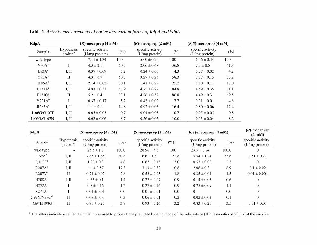
Table 1. Activity measurements of native and variant forms of RdpA and SdpA RdpA (R)-mecoprop (4 mM) (R)-mecoprop (2 mM) (R,S)-mecoprop (4 mM)
Sample Hypothesis probeda
specific activity
(U/mg protein) (%) specific activity (U/mg protein) (%) specific activity
(U/mg protein) (%)
wild type -- 7.11 ± 1.34 100 5.60 ± 0.26 100 6.46 ± 0.44 100 V80Ab I 4.3 ± 2.1 60.5 2.06 ± 0.48 36.8 2.7 ± 0.5 41.8 L83Ac I, II 0.37 ± 0.09 5.2 0.24 ± 0.06 4.3 0.27 ± 0.02 4.2 Q93Ab II 4.3 ± 0.7 60.5 3.27 ± 0.23 58.3 2.27 ± 0.15 35.2 I106Ac I, II 2.14 ± 0.025 30.1 1.41 ± 0.29 25.2 1.10 ± 0.11 17.0 F171Ac I, II 4.83 ± 0.31 67.9 4.75 ± 0.22 84.8 4.59 ± 0.35 71.1 F171Qc II 5.2 ± 0.4 73.1 4.86 ± 0.52 86.8 4.49 ± 0.31 69.5 Y221Ab I 0.37 ± 0.17 5.2 0.43 ± 0.02 7.7 0.31 ± 0.01 4.8 R285Ac I, II 1.1 ± 0.1 14.8 0.92 ± 0.06 16.4 0.80 ± 0.06 12.4
I106G/G107Id I, II 0.05 ± 0.03 0.7 0.04 ± 0.03 0.7 0.05 ± 0.05 0.8 I106G/G107Nd I, II 0.62 ± 0.06 8.7 0.56 ± 0.05 10.0 0.53 ± 0.04 8.2
SdpA (S)-mecoprop (4 mM) (S)-mecoprop (2 mM) (R,S)-mecoprop (4 mM) (R)-mecoprop
(4 mM)
Sample Hypothesis probeda
specific activity
(U/mg protein) (%) specific activity
(U/mg protein) (%) specific activity
(U/mg protein) (%) specific activity
(U/mg protein) wild type -- 25.5 ± 1.7 100.0 28.96 ± 3.6 100 23.5 ± 0.74 100.0 0
E69Ad I, II 7.85 ± 1.65 30.8 6.6 ± 1.3 22.8 5.54 ± 1.24 23.6 0.51 ± 0.22 Q162Fe I, II 1.22 ± 0.3 4.8 0.87 ± 0.15 3.0 0.53 ± 0.08 2.3 0 R207Ae I, II 4.4 ± 0.57 17.3 3.13 ± 0.52 10.8 2.08 ± 0.3 8.9 0.1 ± 0.02 R207Ve II 0.71 ± 0.07 2.8 0.52 ± 0.05 1.8 0.35 ± 0.04 1.5 0.01 ± 0.004 H208Ad I, II 0.35 ± 0.1 1.4 0.27 ± 0.07 0.9 0.14 ± 0.05 0.6 0 H272Ad I 0.3 ± 0.16 1.2 0.27 ± 0.16 0.9 0.25 ± 0.09 1.1 0 R274Ad I 0.01 ± 0.01 0.0 0.01 ± 0.01 0.0 0 0.0 0
G97N/N98Gd II 0.07 ± 0.03 0.3 0.06 ± 0.01 0.2 0.02 ± 0.03 0.1 0 G97I/N98Gd II 0.96 ± 0.27 3.8 0.93 ± 0.26 3.2 0.83 ± 0.26 3.5 0.01 ± 0.01
a The letters indicate whether the mutant was used to probe (I) the predicted binding mode of the substrate or (II) the enantiospecificity of the enzyme.
39
b Values were determined by measuring activities of samples isolated from three independent cell cultures. c Values were determined by triplicate measurements of a single sample. d Values were determined by measuring activities of samples isolated from two independent cell cultures. e Values were determined by triplicate (for (R,S)-mecoprop) or duplicate (for (S)- and (R)-mecoprop) measurements of a single sample.
40
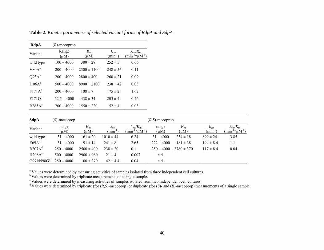
Table 2. Kinetic parameters of selected variant forms of RdpA and SdpA RdpA (R)-mecoprop
Variant Range (µM)
Km (µM)
kcat (min-1)
kcat/Km (min-1*µM-1)
wild type 100 – 4000 380 ± 28 252 ± 5 0.66
V80Aa 200 – 4000 2300 ± 1100 248 ± 56 0.11
Q93Aa 200 – 4000 2800 ± 400 260 ± 21 0.09
I106Ab 500 – 4000 8900 ± 2100 238 ± 42 0.03
F171Ab 200 – 4000 108 ± 7 175 ± 2 1.62
F171Qb 62.5 – 4000 438 ± 34 203 ± 4 0.46
R285Aa 200 – 4000 1550 ± 220 52 ± 4 0.03
SdpA (S)-mecoprop (R,S)-mecoprop
Variant range (µM)
Km (µM)
kcat (min-1)
kcat/Km (min-1*µM-1)
range (µM)
Km (µM)
kcat (min-1)
kcat/Km (min-1*µM-1)
wild type 31 – 4000 161 ± 20 1010 ± 44 6.24 31 – 4000 234 ± 18 899 ± 24 3.85 E69Ac 31 – 4000 91 ± 14 241 ± 8 2.65 222 – 4000 181 ± 38 194 ± 8.4 1.1 R207Ad 250 – 4000 2500 ± 400 238 ± 20 0.1 250 – 4000 2780 ± 370 117 ± 8.4 0.04 H208Ac 500 – 4000 2900 ± 960 21 ± 4 0.007 n.d. G97I/N98Gc 250 – 4000 1100 ± 270 42 ± 4.4 0.04 n.d. a Values were determined by measuring activities of samples isolated from three independent cell cultures. b Values were determined by triplicate measurements of a single sample. c Values were determined by measuring activities of samples isolated from two independent cell cultures. d Values were determined by triplicate (for (R,S)-mecoprop) or duplicate (for (S)- and (R)-mecoprop) measurements of a single sample.
41
Figure 1. Stereoviews of the most favorable models of the active sites of RdpA (A) and SdpA
(B) with bound substrates. Shown are selected protein residues that are predicted to interact with
the substrate. For clarity reasons, the FeII ligands are not depicted. (R)- and (S)-mecoprop are
indicated in light blue and turquoise, respectively, and αKG in yellow.
Figure 2. LIGPLOT diagrams of the active site models of RdpA (A) and SdpA (B). Shown are
the most important interactions between the substrate and the active site according to the model.
Figure 3. Stereoviews of the superimposition of the substrate-bound active site models of RdpA
and SdpA. Shown are selected residues predicted to be involved in dictating enantiospecificity.
Amino acid residues of RdpA are depicted in pink, residues of SdpA in green. (R)-mecoprop is
shown in purple and (S)-mecoprop in turquoise. (A) View with the focus on the propanoic acid
moiety and its binding to the active sites. (B) Same as in (A), but rotated to focus on the phenoxy
ring and its interaction with the amino acid residues Ile106/Gly107 in RdpA and Gly97/Asn98 in
SdpA.