Spatial analysis of key signaling proteins by high-content ... · 8 (Packard ViewPlateTM), and...
40
1 Spatial analysis of key signaling proteins by high-content solid-phase cytometry in Hep3B cells treated with an inhibitor of Cdc25 dual-specificity phosphatases. Andreas Vogt, Takahito Adachi, Alexander P. Ducruet, Jon Chesebrough, Kaoru Nemoto, Brian I. Carr, and John S. Lazo * Departments of Pharmacology (A.V., A.P.D., J.C., K.N., J.S.L.), and Surgery (T.A., B.I.C.), University of Pittsburgh, Pittsburgh , Pennsylvania 15261 *Address for Correspondence: John S. Lazo, Department of Pharmacology, Biomedical Science Tower E-1340, University of Pittsburgh, Pittsburgh, PA 15261; Telephone: 412- 648-9319; Fax: 412-648-2229; Email: [email protected] Running Title: Spatial analysis of signaling kinases Keywords: dual-specificity phosphatase, Cdc25, mitogen-activated protein kinase, cancer, high-content screening, spatial analysis, chemical complementation Copyright 2001 by The American Society for Biochemistry and Molecular Biology, Inc. JBC Papers in Press. Published on March 26, 2001 as Manuscript M100078200 by guest on May 30, 2018 http://www.jbc.org/ Downloaded from
Transcript of Spatial analysis of key signaling proteins by high-content ... · 8 (Packard ViewPlateTM), and...
1
Spatial analysis of key signaling proteins by high-content solid-phase cytometry in
Hep3B cells treated with an inhibitor of Cdc25 dual-specificity phosphatases.
Andreas Vogt, Takahito Adachi, Alexander P. Ducruet, Jon Chesebrough, Kaoru
Nemoto, Brian I. Carr, and John S. Lazo*
Departments of Pharmacology (A.V., A.P.D., J.C., K.N., J.S.L.), and Surgery (T.A.,
B.I.C.), University of Pittsburgh, Pittsburgh , Pennsylvania 15261
*Address for Correspondence: John S. Lazo, Department of Pharmacology, Biomedical
Science Tower E-1340, University of Pittsburgh, Pittsburgh, PA 15261; Telephone: 412-
648-9319; Fax: 412-648-2229; Email: [email protected]
Running Title: Spatial analysis of signaling kinases
Keywords: dual-specificity phosphatase, Cdc25, mitogen-activated protein kinase,
cancer, high-content screening, spatial analysis, chemical complementation
Copyright 2001 by The American Society for Biochemistry and Molecular Biology, Inc.
JBC Papers in Press. Published on March 26, 2001 as Manuscript M100078200 by guest on M
ay 30, 2018http://w
ww
.jbc.org/D
ownloaded from
2
Summary
Protein phosphorylation frequently results in the subcellular redistribution of key
signaling molecules, and this spatial change is critical for their activity. Here we have
probed the effects of a Cdc25 inhibitor, 2-(2-mercaptoethanol)-3-methyl-1,4-
naphthoquinone, or Compound 5, on the spatial regulation and activation kinetics of
tyrosine phosphorylation-dependent signaling events using two methods: i) high-content,
fluorescence-based, automated solid phase cytometry and ii) a novel cellular assay for
Cdc25A activity in intact cells. Immunofluorescence studies demonstrated that
Compound 5 produced a concentration-dependent nuclear accumulation of phospho-Erk
and phospho-p38, but not NFkB. Immunoblot analysis confirmed Erk phosphorylation
and nuclear accumulation, and in vitro kinase assays showed that Compound 5-activated
Erk was competent to phosphorylate its physiological substrate, the transcription factor
Elk-1. Pretreatment of cells with the MEK inhibitor U-0126 prevented the induction by
Compound 5 of nuclear phospho-Erk, but not phospho-p38 accumulation, and protected
cells from the antiproliferative effects of Compound 5. Overexpression of Cdc25A in
whole cells caused dephosphorylation of Erk that was reversed by Compound 5. The
data show that an inhibitor of Cdc25 increases Erk phosphorylation and nuclear
accumulation and support the hypothesis that Cdc25A regulates Erk phosphorylation
status.
by guest on May 30, 2018
http://ww
w.jbc.org/
Dow
nloaded from
3
Introduction
Approximately one third of mammalian proteins are thought to be posttranslationally
modified by phosphorylation (1). The human genome contains hundreds of protein
kinases (2), but the reversibility of the phosphorylation process suggests that
phosphatases also play a major role in the regulation of protein phosphorylation. While
the roles and cellular functions of kinases have been extensively studied, protein
phosphatases have received much less attention. A long held view has been that
phosphatases merely serve to reverse the actions of protein kinases. More recently has it
been recognized that phosphatases may be as numerous and as tightly regulated as
protein kinases, with as widely varying substrate specificities and signaling functions (3).
The preference of certain phosphatases for one phosphorylated hydroxy amino acid over
others has resulted in the current classification of phosphatases as serine/threonine
specific (STPase)1, tyrosine-specific (PTPases), and dual-specific (DSPases)
phosphatases. While several highly potent and selective inhibitors of STPases have been
isolated from natural sources, selective PTPase or DSPase inhibitors are still rare.
Protein kinases and phosphatases are part of a complex signaling network of tightly
regulated dynamic processes. The nature and details of network organization are just
beginning to be unveiled, but their abundance, diversity, and substrate specificity alone
cannot explain how these molecules function to regulate complex biochemical pathways.
An emerging concept in signaling specificity is the subcellular location at which
signaling events occur (4). Most protein movements within the cell are consistent with a
random diffusion process. However, it is now being recognized that spreading as well as
by guest on May 30, 2018
http://ww
w.jbc.org/
Dow
nloaded from
4
restriction of signaling events to certain regions of the cell is driven by the availability of
sites for protein-protein and protein-second messenger interactions (5). Phosphorylation
of many key signaling molecules causes a subcellular redistribution that is critical for
biological activity (6-8).
Despite the potential importance that spatial regulation might have in signal transduction
and the considerable information that can be derived from localization studies, a lack of
readily available, quantitative analytical tools to assess the subcellular localization of
multiple signal transduction molecules has impeded progress in this area. Current
fluorescent imaging techniques have low-throughput and are not well suited for the
dissection of how complex signaling networks are coordinated. In this report, we have
used a novel, automated, fluorescence-based, multiparametric, solid-phase cytometer, the
Cellomics ArrayScan II (9) to rapidly quantitate the effects of a synthetic K vitamin
analog, Compound 5, on the spatial regulation of a subset of key signal transduction
molecules.
Compound 5 was discovered to be a potent inhibitor of hepatoma cell growth in a small
targeted library of synthetic K Vitamin analogs (10, 11). It has antiphosphatase activity
that is thought to contribute to its antiproliferative activity. Most notably, it is one of the
most potent in vitro inhibitors of the Cdc25 phosphatase family of dual-specificity
phosphatases reported to date (12). In vitro, Compound 5 is approximately 10- and 100-
fold more potent against Cdc25 compared with the prototype DSPase VHR, or protein
tyrosine phosphatase 1B (PTP1B), respectively (12). Its ability to cause a dual cell cycle
by guest on May 30, 2018
http://ww
w.jbc.org/
Dow
nloaded from
5
arrest in G1 and G2 phase as well as increased phosphorylation of the Cdc25 substrates,
Cdc2 (Cdk1), Cdk2, and Cdk4, are consistent with Cdc25 phosphatase inhibition by
Compound 5 (12). Recent work from our laboratories has also demonstrated that
Compound 5 causes increased tyrosine phosphorylation on a number of proteins,
including the epidermal growth factor receptor (EGFR) and extracellular signal-regulated
kinase (Erk) in hepatocytes (13) and MCF-7 cells (14), but it is unknown how Compound
5 enhanced Erk phosphorylation or whether Compound 5 treatment changed Erk
subcellular localization. A possible link between the mitogenic signal transduction and
Cdc25A has been described by Galaktionov et al (15), who reported that Cdc25A
associates with Raf-1, a key upstream activator of Erk, in mammalian cells and frog
oocytes. More recently, evidence for a possible functional involvement of Cdc25A in
the Erk pathway was presented by Xia et al. (16), who reported that co-expression of
Cdc25A, together with Raf-1, prevented Raf-1 activation in response to PDGF in
NIH3T3 cells. Nonetheless, no direct evidence for Cdc25A involvement in Erk
phosphorylation or activity has been reported.
In the current study, we demonstrated that Cdc25A expression could reduce Erk
phosphorylation, and described a novel cell-based assay revealing that Compound 5
directly interfered with Cdc25A function on Erk phosphorylation. Using quantitative,
fluorescence-based, solid phase cytometry, we documented that Erk
hyperphosphorylation by Compound 5 resulted in increased nuclear accumulation of
kinase-active phospho-Erk. Thus, an inhibitor of Cdc25 increased Erk phosphorylation,
by guest on May 30, 2018
http://ww
w.jbc.org/
Dow
nloaded from
6
which further supported the hypothesis that Cdc25A regulates Erk phosphorylation
status.
by guest on May 30, 2018
http://ww
w.jbc.org/
Dow
nloaded from
7
Experimental Procedures
Reagents. Compound 5 , 2-(2-mercaptoethanol)-3-methyl-1,4-naphthoquinone, has been
described previously (10). Human recombinant interleukin 1-α (IL-1α) was from R&D
systems (Minneapolis, MN). Mouse monoclonal anti-phospho-Erk antibody (E10) and
the MEK inhibitor U-0126 were from New England Biolabs (Beverly, MA). Mouse
monoclonal Erk 2 antibody was from Upstate Biotechnology (UBI, Lake Placid, NY).
Primary antibodies for phospho-p38 and the p65 subunit of NFkB were components of a
commercially available assay kit (Cellomics, Pittsburgh, PA). Anti-Oct-1 antibody was
from Santa Cruz Biotechnology (Santa Cruz, CA), and anti-HSP90 antibody was from
BD Transduction Laboratories (San Diego, CA). Secondary antibodies were Alexa-
Fluor 488 conjugated goat anti-mouse (p-Erk), goat anti-rabbit (p38, p-JNK), or donkey
anti-goat (NFkB) IgG (Molecular Probes, Eugene, OR).
Cell culture. Cells were maintained in Dulbecco’s Minimum Essential Medium
(DMEM) containing 10% fetal bovine serum (FBS, HyClone, Logan, UT), and 1%
penicillin-streptomycin (Life Technologies, Inc., Rockville, MD) in a humidified
atmosphere of 5% CO2 at 37oC. HeLa, PC-3, DU-145, and NIH 3T3 cells were from
ATCC. Rat-1 fibroblasts were obtained from Dr. Guillermo Romero, University of
Pittsburgh. Hep3B human hepatoma cells have been characterized previously (17).
Indirect immunofluorescence. Hep3B, HeLa, PC-3, DU-145, Rat-1. or NIH 3T3 cells
(4,000 cells per well) were plated in the wells of a collagen-coated 96 well darkwell plate
by guest on May 30, 2018
http://ww
w.jbc.org/
Dow
nloaded from
8
(Packard ViewPlateTM), and allowed to attach overnight. Cells were treated for the times
indicated with Compound 5 or IL-1α, fixed with 3.7% formaldehyde in PBS and
permeabilized with PBS/Triton X-100. Cells were stained with antibodies against
phospho-Erk, phospho-p38, phospho-JNK, or the 65 kDa subunit of NFkB, and washed
with PBS/Tween20. Nuclei were stained with Hoechst 33342 fluorescent dye, and
immunoreactive cells were visualized by AlexaFluor 488 secondary antibodies
(Molecular Probes, Eugene, OR) using an XF100 filter set at excitation/emission
wavelengths of 494/519 nm (Alexa 488), and 350/461 nm (Hoechst), respectively. Plates
were analyzed by automated image analysis on the ArrayScan II system (Cellomics,
Pittsburgh, PA) using the previously described cytoplasm-to-nuclear translocation
algorithm (18). Control experiments omitting primary antibodies were performed each
time to assess the amount of non-specific background staining.
Cell Fractionation and Western Blotting. Cytosolic and nuclear fractions were
prepared using a slightly modified procedure as published by Schreiber et al. (19).
Hep3B cells were plated in 100 mm tissue culture dishes, exposed to 10 µM Compound 5
for the indicated periods of time, and harvested by centrifugation. Cell pellets were
resuspended in 200 µl of hypotonic buffer (10 mM HEPES, pH 7.9, 1.5 mM MgCl2, 10
mM KCl, 0.2 mM PMSF, 0.5 mM DTT, and 0.5% Nonidet-P-40), incubated on ice for 10
min, disrupted by repeated aspiration through a 20-gauge needle, and centrifuged at 2,500
x g for 15 min. The supernatant was collected as cytosolic extract. Nuclear pellets were
resuspended in nuclear extraction buffer (20 mM HEPES, pH 7.9, 10% glycerol, 1.5 mM
MgCl2, 400 mM KCl, 0.2 mM EDTA, 0.2 mM PMSF, and 0.5 mM DTT), incubated on
by guest on May 30, 2018
http://ww
w.jbc.org/
Dow
nloaded from
9
ice for 1 h, and centrifuged at 13,000 x g to collect the nuclear fraction. Solubilized
proteins were resolved by 10% SDS-PAGE and transferred to polyvinylidene difluoride
membranes (NEN, Boston, MA). Membranes were probed with anti-phospho-Erk, anti-
Oct-1, and anti HSP-90 antibodies. Positive antibody reactions were visualized using
peroxidase-conjugated secondary antibodies (Jackson ImmunoResearch, West Grove,
PA) and an enhanced chemiluminescence detection system (Renaissance, NEN, Boston,
MA) according to manufacturer’s instructions.
Erk activity assay. Erk activity in cytosolic and nuclear fractions was determined using
a non-radioactive immunoprecipitation kit (Cell Signaling Technologies, Beverly, MA).
Briefly, 200 µg of nuclear or cytosolic proteins were incubated with 15 µl of agarose-
conjugated anti-phospho-Erk antibody, and incubated overnight at 4oC with gentle
rocking. Immunoprecipitates were pelleted and washed twice with kinase buffer (25 mM
Tris, pH 7.5, 5 mM β-glycerophosphate, 2 mM DTT, 0.1 mM Na3VO4, and 10 mM
MgCl2). Pellets were resuspended in 50 µl of kinase buffer supplemented with 200 µM
ATP and 2 µg of Elk-1 GST fusion protein, and incubated for 30 min at 30oC.
Immunoprecipitates were boiled in SDS-PAGE sample buffer and analyzed by Western
blot using an anti-phospho-Elk-1 antibody.
Growth inhibition assay. The antiproliferative activity of Compound 5 in combination
with the MEK inhibitor, U-0126, was measured by a previously described assay based on
fluorimetric quantitation of total cellular DNA content using the fluorochrome Hoechst
33258 (20). Briefly, cells were grown in 96 well microplates and treated every day with
by guest on May 30, 2018
http://ww
w.jbc.org/
Dow
nloaded from
10
for three days with various concentrations of Compound 5 in the presence or absence of
the MEK inhibitor, U-0126 (5 µM). Cells were lysed by repeated freeze-thawing and
cellular DNA was quantitated as described (20).
Cell transfections. Mammalian expression plasmids encoding full length wild-type and
catalytically inactive C430S mutant Cdc25A in a pcDNA3 vector were generously
provided by Dr. Thomas Roberts, Dana Farber Cancer Institute (16). Transfections were
carried out by the Lipofectamine method as per manufacturer’s instructions (Life
Technologies, Rockville, MD). Briefly, HeLa cells (100,000 per well) were plated in the
wells of a six well plate and transfected with 0.5 µg of cDNA in OPTI-MEM transfection
medium using Lipofectamine PlusTM reagent (Life Technologies, Rockville, MD).
Three hours after transfection, the medium was replaced with complete growth medium
and the cells were allowed to recover for 48 h. Cells were treated with 0-20 µM
Compound 5 for 30 min and protein lysates were prepared and analyzed by SDS-PAGE
and Western Blot analysis for phospho-Erk and Erk 2 levels as described above. For
quantitation of protein expression levels, X-ray films were scanned on a Molecular
Dynamics personal SI densitometer and analyzed using the ImageQuant software
package (Ver. 4.1, Molecular Dynamics, Sunnyvale, CA).
by guest on May 30, 2018
http://ww
w.jbc.org/
Dow
nloaded from
11
Results
A fluorescence-based high-content assay for phospho-Erk nuclear accumulation.
Compound 5 was previously found to induce the prolonged phosphorylation of tyrosines
on a number of signaling proteins in the Erk cascade, including Erk1 and Erk2 (13, 14).
We first asked whether this increase in tyrosine phosphorylation was associated with a
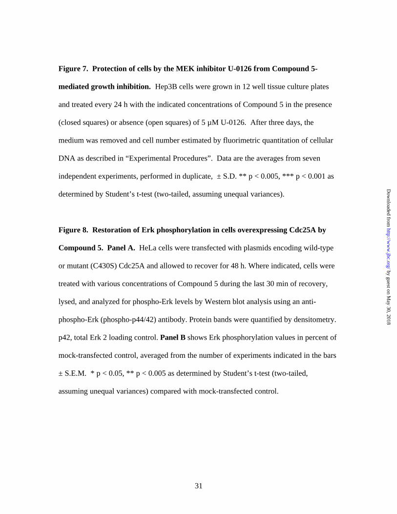
change in phospho-Erk nuclear accumulation. Hep3B cells were either incubated with
vehicle (DMSO) (Fig. 1A-C) or Compound 5 (Figure 1D-F) for 30 min and
immunostained with antibodies against a dually phosphorylated (Thr202/Tyr204) form of
Erk (Fig. 1B,C,E,F). Nuclei were visualized by Hoechst 33342 staining (Fig. 1A and D).
Figure 1B shows that vehicle-treated cells had very low levels of phospho-Erk, most of
which was diffusely distributed in the cytoplasm. Treatment of cells with Compound 5
resulted in a substantial increase in total phospho-Erk, with prominent nuclear
accumulation (Fig. 1E). Overlay images (Fig. 1C and F) illustrate the quantitation of
cytoplasmic and nuclear phospho-Erk levels. Fluorescence-labeled cells were analyzed
in two separate channels by the ArrayScan II and the cytoplasm-to-nuclear distribution
determined by a previously described algorithm (18). Hoechst 33342 staining (Fig. 1A
and D) defined the nuclear area. Phospho-Erk fluorescence intensity within this nuclear
area was referred to as “cytonuclear intensity”. To assess the amount of fluorescently
labeled phospho-Erk in the cytoplasm, a set of concentric rings spaced by two pixels was
placed around the nuclear boundary. Phospho-Erk fluorescence intensity within the ring
area was referred to as “cytoring intensity”. Both cytonuclear and cytoring intensities
were normalized to the total cytonuclear or cytoring area, and are expressed as average
by guest on May 30, 2018
http://ww
w.jbc.org/
Dow
nloaded from
12
intensity per pixel. All cytoplasmic-to-nuclear difference values were calculated by
subtracting the average cytoring intensity per pixel from the average cytonuclear intensity
per pixel. Thus, an increase in the cytonuclear difference value is indicative of Erk
activation through phosphorylation, translocation, or both.
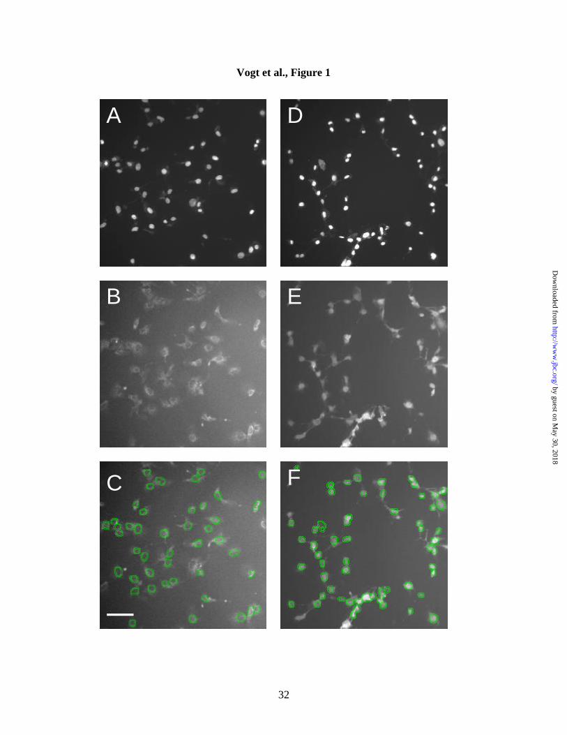
Induction of phospho-Erk and phospho-p38, but not NFkB, by Compound 5. We
next examined whether Compound 5 caused selective nuclear Erk accumulation by
comparing its effects to those of other signaling events, which have also been reported to
be activated in a tyrosine phosphorylation-dependent manner, and are thought to mediate
stress responses. Cells were treated for 30 min with either 10 µM Compound 5 or 25
ng/ml interleukin-1 alpha (IL-1α), immunostained with phospho-Erk, phospho-p38,
phospho-JNK, or p65 NFkB antibodies, respectively and analyzed for differences in
cytoplasmic-to-nuclear fluorescence intensity. A total of 100 cells were imaged in each
well. Figure 2 shows that Compound 5 lead to a dramatic increase in nuclear
accumulation of phospho-Erk and phospho-p38, but had only a moderate effect on
phospho-JNK, and did not affect the nuclear accumulation of NFkB. IL-1α, in contrast,
activated all three stress-response mediators (p38, JNK, and NFkB), but not Erk. Thus,
the activity profile of Compound 5 was distinct from that of the cytokine IL-1α,
suggesting that Compound 5 was not a general stress-inducing agent.
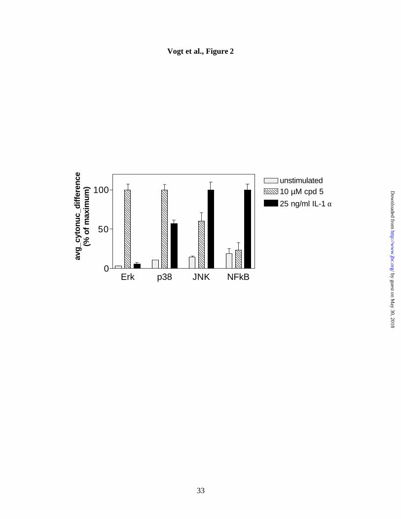
Kinetics of Erk and p38 activation by Compound 5. Experiments with the stress
inducer and phosphatase inhibitor, sodium arsenite, previously demonstrated that p38 and
Erk were activated with different kinetics in a variety of cell lines (21). These authors
by guest on May 30, 2018
http://ww
w.jbc.org/
Dow
nloaded from
13
also reported that Erk activation was abrogated by dominant negative forms of p38 and
the p38 specific kinase inhibitor, SB-203580, suggesting an involvement of p38 in Erk
activation. We thus examined concentration-dependence and kinetics of phospho-Erk
and phospho-p38 activation in Hep3B cells. Figure 3 shows that maximal stimulation of
both Erk and p38 was obtained at 10 µM Compound 5. Moreover, continuous exposure
to 10 µM Compound 5 caused a progressively greater activation and nuclear
accumulation with similar temporal characteristics (Figure 3B). We have also found that
the p38 inhibitor SB-203580 did not inhibit phospho-Erk nuclear accumulation (data not
shown). These results suggest that Compound 5 acted differently than the nonspecific
tyrosine phosphatase inhibitor sodium arsenite.
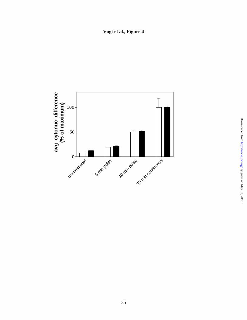
Irreversibility of Compound 5 action. Compound 5 is a sulfhydryl-arylating agent and
its sustained antiphosphatase activity has been ascribed to covalent modification of
critical cysteine residues on dual-specific and tyrosine phosphatases (17). To test
whether its effects were irreversible, we treated cells with Compound 5 for 5 or 10 min,
followed by washout, and compared the magnitude of phospho-Erk and phospho-p38
accumulation to that obtained after a 30 min continuous exposure. Figure 4 shows that
short pulses of Compound 5 resulted in substantial activation of both Erk and p38,
consistent with a rapid and persistent inhibition of cellular phosphatases after compound
removal.
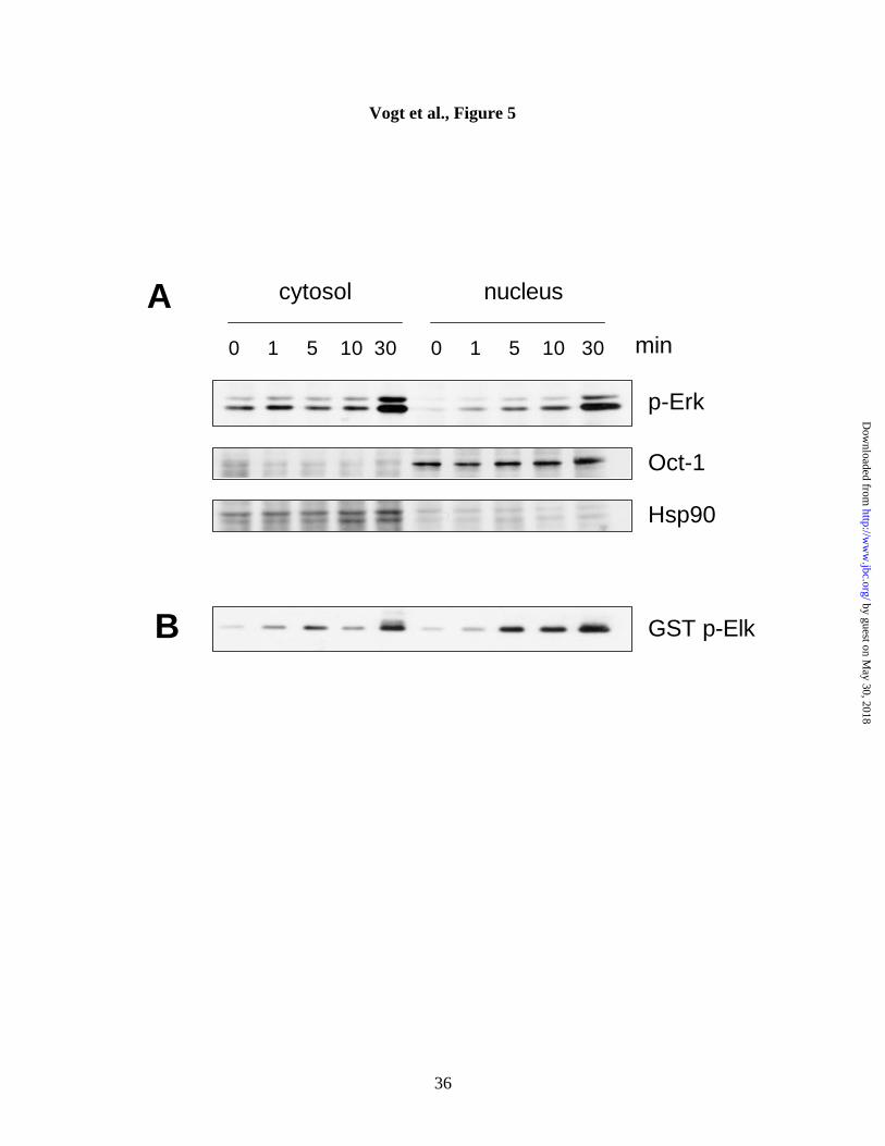
Biochemical analysis confirms phospho-Erk nuclear accumulation. We next
validated the results from the automated fluorescence-based analysis by conventional
by guest on May 30, 2018
http://ww
w.jbc.org/
Dow
nloaded from
14
biochemical methods. Cells were treated with 10 µM Compound 5 for the indicated
times, lysed, separated into cytosolic and nuclear fractions, and analyzed by Western blot
using a phospho-Erk antibody (Figure 5A). Untreated cells had almost no nuclear
phospho-Erk, consistent with the whole cell images in Figure 1B. Within minutes,
Compound 5 caused a time-dependent and sustained increase in nuclear phospho-Erk
accumulation. In contrast, cytosolic phospho-Erk levels in control cells were higher than
those in the nucleus and increased only after a longer exposure to Compound 5 (30 min,
Figure 5A). The results from the immunoblot analysis thus confirmed those from the less
arduous solid phase cytometry studies.
Phosphorylated Erk from Compound 5 treated cells is activated and phosphorylates
Elk-1. We then used the identical lysates from Compound 5 treated cells to investigate
whether the observed Erk phosphorylation resulted in an increase in Erk kinase activity.
It is thought that upon phosphorylation by MEK1 and MEK2 in the cytosol, a fraction of
Erk translocates to the nucleus, where it phosphorylates and activates transcription
factors, such as c-fos, c-jun, and Elk-1 (22). To investigate whether phosphorylated Erk
was functional in Compound 5-treated cells, we examined its ability to phosphorylate the
transcription factor, Elk-1. Phospho-Erk was immunoprecipitated from Compound 5
treated and untreated cells and immunoprecipitates were subjected to an in vitro kinase
assay using recombinant GST-Elk-1 fusion protein as a substrate. Assay mixtures were
separated on SDS-PAGE and immunoblotted with an anti-phospho-Elk-1 antibody.
Figure 5B shows that nuclear phospho-Erk had kinase activity and that its kinetics of
activation correlated well with its phosphorylation status. Compound 5-induced nuclear
by guest on May 30, 2018
http://ww
w.jbc.org/
Dow
nloaded from
15
phospho-Erk was thus functional and able to phosphorylate its physiological substrate,
Elk-1.
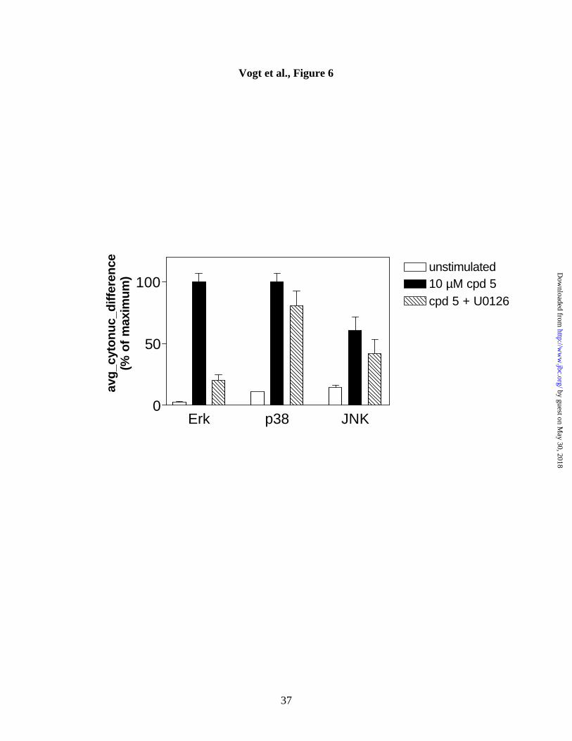
MEK inhibition and nuclear translocation of phospho-Erk, phospho-p38 or
phospho-JNK. We next investigated possible consequences of Erk or p38 activation by
Compound 5. We first examined whether inhibition of MEK, the direct upstream
activating kinase for Erk, would reduce phospho-Erk nuclear accumulation. Cells were
pretreated with the MEK1/MEK2 inhibitor U-0126 (23) for 45 min, stimulated with
Compound 5 for an additional 30 min in the presence of the inhibitor, and analyzed on
the ArrayScan II for nuclear accumulation of phospho-Erk, phospho-p38, and phospho-
JNK. Consistent with results from Figure 2, Compound 5 caused a robust increase in
nuclear phospho-Erk and phospho-p38, but had only a partial effect on phospho-JNK
(Figure 6). Inclusion of 10 µM U-0126 caused almost complete inhibition of Compound
5-induced Erk activation, but, as expected, had little or no effect on p38 or JNK
activation. These data suggest that MEK inhibition is sufficient to inhibit phospho-Erk
nuclear accumulation by Compound 5.
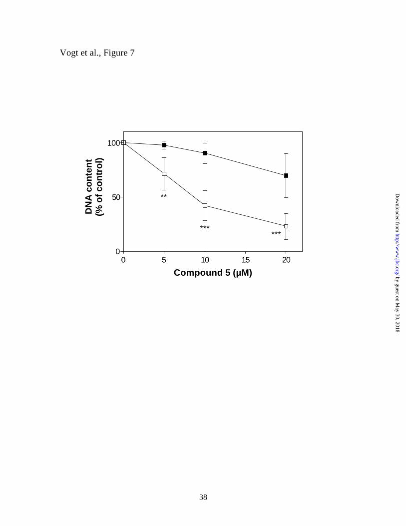
The MEK inhibitor U-0126 protects cells from the antiproliferative effects of
Compound 5. To determine whether the activation of Erk or p38 played a role in
mediating the antiproliferative activity of Compound 5, cells were incubated with the
indicated concentrations of Compound 5, either in the presence or absence of 5 µM U-
0126 for 72h. Cells were harvested, stained with Hoechst 33258 and cellular DNA was
quantified by fluorimetry as previously described (17). Figure 7 shows that inclusion of
by guest on May 30, 2018
http://ww
w.jbc.org/
Dow
nloaded from
16
the MEK inhibitor significantly reduced Compound 5-mediated cell growth inhibition.
This strongly suggests that activation of the Erk pathway is the major determinant in the
antiproliferative effects of Compound 5. In contrast, p38 activation, which has been
implicated in cell death in many cell types, did not appear to mediate growth inhibition of
Hep3B cells by Compound 5 since in the presence of U-0126, cell growth continued
despite high levels of nuclear phospho-p38 but depressed levels of phospho-Erk (see
Figure 6).
The effects of Compound 5 on nuclear phospho-Erk accumulation are cell type
dependent. To determine whether the observed accumulation of phospho-Erk was
specific for Hep3B cells, we examined the ability of Compound 5 to induce nuclear
phospho-Erk accumulation in a variety of mammalian cell lines using the ArrayScan II.
We found Compound 5-induced nuclear phospho-Erk accumulation was not unique to
Hep3B cells but that the magnitude of response varied with cell type. Cell lines fell into
three categories based on the magnitude of phospho-Erk induction. Strong responders
were NIH 3T3, Rat-1, and Hep3B cells, which showed up to 24- , 75 -, and 57-fold
increases over control cells, respectively, in nuclear phospho-Erk levels 30 min after
exposure to 10 µM Compound 5 (data not shown). DU-145 and PC-3 prostate cancer
cells were less responsive (2-3 fold increase with 30 min exposure to 10 µM Compound
5), and HeLa cells did not respond to Compound 5 with enhanced nuclear phospho-Erk
accumulation at concentrations up to 30 µM (data not shown). Thus, the induction of
phospho-Erk nuclear accumulation by Compound 5 was not limited to Hep3B cells, but
instead constituted a more generalized phenomenon.
by guest on May 30, 2018
http://ww
w.jbc.org/
Dow
nloaded from
17
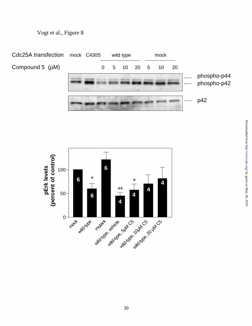
Compound 5 restores phospho-Erk levels after Cdc25A overexpression. Because in
vitro studies had shown that Compound 5 was most effective against the Cdc25 family of
DSPases (12), we investigated whether the effects of a brief treatment with Compound 5
on phospho-Erk nuclear accumulation could be attributed to Cdc25A inhibition. Previous
reports have revealed that the tyrosine phosphorylation status and activity of Raf-1,
which is an upstream activator of Erk, is controlled by Cdc25A (16). Thus, we
hypothesized that ectopic expression of Cdc25A might reduce Erk phosphorylation and
provide a novel assay system to examine the acute actions of Compound 5 against
intracellular Cdc25A. We selected HeLa cells as a model because in the absence of
ectopic Cdc25A no nuclear phospho-Erk accumulation was seen with Compound 5 in
these cells, possibly due to low endogenous Cdc25A activity. We predicted that this
model would, therefore, have the lowest background and that any effect seen with a small
molecule could be assigned to an action on the ectopically expressed Cdc25A. As
illustrated in Figure 8A, ectopic Cdc25A expression reduced Erk phosphorylation by
50% (p<0.05, Figure 8B). This reduction in Erk phosphorylation absolutely required the
intrinsic phosphatase activity of Cdc25A because a catalytically inactive Cdc25A
(C430S) did not reduce Erk phosphorylation in these cells. We then asked whether
Compound 5 was able to restore Erk phosphorylation after ectopic expression of wild-
type Cdc25A. Cells transiently transfected with wild-type Cdc25A were allowed to
recover for 48 h and, during the last 30 min of recovery, treated with vehicle or
increasing concentrations of Compound 5. Figure 8B shows that Compound 5 gradually
restored Erk phosphorylation to mock/control levels. Consistent with the inherent
by guest on May 30, 2018
http://ww
w.jbc.org/
Dow
nloaded from
18
unresponsiveness of HeLa cells to Compound 5 (see above), the levels of Erk
phosphorylation in untransfected cells were not markedly changed upon Compound 5
treatment (Figure 8A). Furthermore, phospho-Erk levels in Cdc25A-expressing,
Compound 5-treated cells never exceeded those in untransfected cells (Figure 8B). These
results support the hypothesis that Compound 5 interfered with Cdc25A-mediated
dephosphorylation of Erk.
by guest on May 30, 2018
http://ww
w.jbc.org/
Dow
nloaded from
19
Discussion
PTPases and DSPases play a major role in receptor-mediated signal transduction events.
For example, the kinase activities of growth factor receptors are regulated by tyrosine
autophosphorylation. Signals initiated at the cell surface are transmitted by a series of
cytoplasmic kinases that sequentially phosphorylate each other, eventually leading to
activation of members of the MAPK superfamily, namely Erk, p38, and SAPK/JNK, by
dual phosphorylation on tyrosine and threonine residues. PTPases dephosphorylate
tyrosines and thus inactivate growth factor receptors, whereas the signal at the level of
MAPK is attenuated by dephosphorylation of the MAPKs on both tyrosine and threonine
by specific MKPs. Recently, the cell cycle phosphatase Cdc25A has been proposed to
regulate tyrosine phosphorylation and activity of Raf-1, a key element in the Erk
signaling cascade (16).
In contrast to their upstream activating kinases, tyrosine / threonine phosphorylated
MAPKs translocate to the nucleus where they phosphorylate and activate their respective
protein targets, which include several transcription factors. Very few studies have
addressed spatial aspects of phosphorylation-dependent signaling events and to our
knowledge, none have investigated small molecules that might perturb the subcellular
localization of key signal transducers in the context of tyrosine phosphorylation. Here we
have used a high-content, cell-based assay to evaluate the temporal and spatial dynamics
of three key parallel signaling molecules in response to Compound 5, a synthetic K
vitamin analog with in vitro antiphosphatase activity (12) and antiproliferative activity in
a variety of cell lines (10). Using this novel methodology, we found that Compound 5
by guest on May 30, 2018
http://ww
w.jbc.org/
Dow
nloaded from
20
caused rapid and irreversible nuclear accumulation of phospho-Erk and phospho-p38.
The observed activation of Erk by a compound known to cause growth inhibition (11-12,
17) is somewhat surprising since brief activation of Erk is often associated with
mitogenesis and survival. In contrast, JNK and p38 are thought to be mediators of stress
responses and apoptosis (24). We considered the possibility that p38 activation might be
a factor in the antiproliferative activity of Compound 5. In the presence of the MEK1
inhibitor U-0126, however, which inhibits Erk activation, Hep3B cells grew despite
having high levels of phospho-p38. In contrast, we found that pretreatment of cells with
U-0126 not only prevented Compound 5-induced Erk phosphorylation, but also protected
cells from the growth inhibitory effects of Compound 5. These results strongly support
our previous suggestion that prolonged activation of the Erk pathway is causally involved
in the growth inhibitory effects of Compound 5 (13, 17) . Moreover, our conclusion is in
agreement with a growing body of data documenting an involvement of Erk in growth
inhibition in neuronal cells (25), NIH3T3 cells (26), and MCF-7 cells (14). In addition,
there is increasing evidence that p38 does not appear to exclusively mediate cytotoxicity,
but can be cytoprotective under certain conditions (27-29).
The inability of Compound 5 to induce NFkB and, to a lesser extent, JNK, suggests
specificity and that it is not a general stress-inducing stimulus. Both NFkB and JNK are
activated by a variety of extracellular stimuli, such as oxidative stress or inflammatory
cytokines. In addition, the broad PTPase inhibitors vanadate and pervanadate have been
found to induce NFkB (30, 31), providing further support for a unique and more specific
action associated with Compound 5. We recently demonstrated that Compound 5
by guest on May 30, 2018
http://ww
w.jbc.org/
Dow
nloaded from
21
selectively inhibited members of the DSPase family with median inhibitory values of 4
µM for Cdc25B2 and Cdc25A, while it was 10-fold less active against VHR, a prototype
MKP, and 100-fold less active against PTP1B (12). Furthermore, Compound 5 caused a
cell cycle arrest in both G1 and G2, which correlated with enhanced phosphorylation of
the Cdc25 substrates Cdk1, Cdk2, and Cdk4, respectively (12). We suggested that the
growth inhibitory properties of Compound 5 might be due to inhibition of the Cdc25
family, but in large part due to a lack of appropriate assays, there has been no direct
evidence that Compound 5 inhibits Cdc25 phosphatases in the cell. To investigate
whether Cdc25A could affect Erk phosphorylation status and be inhibited within cells by
Compound 5, we devised a chemical complementation strategy based on earlier
observations that Cdc25A associated with the Raf-1 oncoprotein (15). Functional
evidence that Cdc25A regulates Raf-1 activity was obtained by Xia et al. (16), who
showed that overexpression of Raf-1 together with wild-type Cdc25A reduced PDGF-
mediated Raf-1 tyrosine phosphorylation in NIH 3T3 cells. Raf-1 is one of the most
important upstream activators of the Erk cascade (32). We thus hypothesized that
Cdc25A overexpression would result in decreased Erk phosphorylation and that an
inhibitor of Cdc25A would restore Erk phosphorylation to normal levels, by chemically
complementing the loss-of-function phenotype caused by Cdc25A overexpression. To
simplify the analysis, we chose HeLa cells, which did not respond to Compound 5 with
increased nuclear phospho-Erk accumulation. By treating Cdc25A-overexpressing cells
with concentrations of Compound 5 that did not cause Erk hyperphosphorylation under
normal growth conditions, we were able to demonstrate that Compound 5 specifically
inhibited the effects of the overexpressed Cdc25A protein on Erk phosphorylation. Thus,
by guest on May 30, 2018
http://ww
w.jbc.org/
Dow
nloaded from
22
we have obtained, for the first time, evidence that Cdc25A regulates endogenous Erk
phosphorylation status in whole cells, and that Compound 5 affected Cdc25A function in
the cell.
Although the concentrations of Compound 5 required for inhibition of the MKP VHR in
vitro are an order of magnitude higher than those for Cdc25A inhibition, it is possible that
inhibition of MKPs by Compound 5 also contributes to Erk and p38 activation. A
number of cytosolic and nuclear MKPs, which have overlapping substrate specificities,
have been described. For example, the Erk isoforms are selectively inhibited by MKP-3,
whereas M3/6 selectively dephosphorylates JNK (33). MKP-1 and 2 preferentially
dephosphorylate JNK, but also have some activity toward p38 (34, 35). More recently, a
p38 specific phosphatase, MKP-5, has been reported (36). The prototype DSPase VHR,
which seems to reside in the nucleus, dephosphorylates Erk (37), but its effect on other
kinases has not been examined. The fact that Compound 5 only partially activated JNK
suggests that it may have some selectivity. At this time, we do not have any information
about whether Compound 5 has any specificity for the different MKPs, but this
information should become available as we expanding our chemical complementation
strategy to probe for cell-active inhibitors of MKPs.
In summary, using the ArrayScan II, we were able to quickly and quantitatively probe
selective activation of tyrosine phosphorylation-dependent signal transduction events by
a small molecule dual-specificity phosphatase inhibitor in intact cells. By performing
fluorescence-based spatial analysis in a high-throughput compatible format, we
by guest on May 30, 2018
http://ww
w.jbc.org/
Dow
nloaded from
23
demonstrated that this inhibitor selectively activated dual-specificity phosphatase-
dependent cellular events. Subsequent analyses using both genetic and pharmacological
tools identified activation of the Erk pathway as the dominant component mediating
Compound 5’s antiproliferative activity, and provided direct evidence that it could
interfere with Cdc25A function in the cell. We propose that the combination of high-
content, cell-based analyses coupled with a chemical complementation approach will be a
powerful technique to identify cell active inhibitors of a variety of cellular targets.
by guest on May 30, 2018
http://ww
w.jbc.org/
Dow
nloaded from
24
References
1. Zolnierowicz, S. and Bollen, M. (2000) EMBO J. 19, 483-488
2. Venter, J. C. et al. (2001) Science 291, 1304-1351
3. Tonks, N. K. and Neel, B. G. (1996) Cell 87, 365-368
4. Pawson, T. and Scott, J. D. (1997) Science 278, 2075-2080
5. Teruel, M. N. and Meyer, T. (2000) Cell 103, 181-184
6. Brunet, A., Roux, D., Lenormand, P., Dowd, S., Keyse, S., and Pouyssegur, J.
(1999) EMBO J 18, 664-674
7. Dalal, S. N., Schweitzer, C. M., Gan, J., and DeCaprio, J. A. (1999) Mol. Cell. Biol.
19, 4465-4479
8. Liu, F., Rothblum-Oviatt, C., Ryan, C. E., and Piwnica-Worms, H. (1999) Mol.
Cell. Biol. 19, 5113-5123
9. Giuliano, K. A., DeBiasio, R. L., Dunlay, T., Gough, A., Volosky, J. M., Zock, J.,
Pavlakis, G. N., and Taylor, D. L. (1997) Journal of Biomolecular Screening 2,
249-259
10. Nishikawa, Y., Carr, B. I., Wang, M., Kar, S., Finn, F., Dowd, P., Zheng, Z. B.,
Kerns, J., and Naganathan, S. (1995) J. Biol. Chem. 270, 28304-28310
11. Kerns, J., Naganathan, S., Dowd, P., Finn, F. M., and Carr, B. I. (1995) Bioorg
Chem 23, 101-108
12. Tamura, K., Southwick, E. C., Kerns, J., Rosi, K., Carr, B. I., Wilcox, C., and Lazo,
J. S. (2000) Cancer Res. 60, 1317-1325
13. Wang, Z., Wang, M., and Carr, B. I. (2000) J. Cell Physiol. 183, 338-346
by guest on May 30, 2018
http://ww
w.jbc.org/
Dow
nloaded from
25
14. Kar, S. and Carr, B. I. (2000) J. Cell Physiol. 185, 386-393
15. Galaktionov, K., Jessus, C., and Beach, D. (1995) Genes Dev. 9, 1046-1058
16. Xia, K., Lee, R. S., Narsimhan, R. P., Mukhopadhyay, N. K., Neel, B. G., and
Roberts, T. M. (1999) Mol. Cell. Biol. 19, 4819-4824
17. Nishikawa, Y., Wang, Z., Kerns, J., Wilcox, C. S., and Carr, B. I. (1999) J. Biol.
Chem. 274, 34803-34810
18. Ding, G. J., Fischer, P. A., Boltz, R. C., Schmidt, J. A., Colaianne, J. J., Gough, A.,
Rubin, R. A., and Miller, D. K. (1998) J. Biol. Chem. 273, 28897-28905
19. Schreiber, E., Matthias, P., Muller, M. M., and Schaffner, W. (1989) Nucleic Acids
Res. 17, 6419
20. Rago, R., Mitchen, J., and Wilding, G. (1990) Anal. Biochem. 191, 31-34
21. Ludwig, S., Hoffmeyer, A., Goebeler, M., Kilian, K., Hafner, H., Neufeld, B., Han,
J., and Rapp, U. R. (1998) J. Biol. Chem. 273, 1917-1922
22. Chen, R. H., Sarnecki, C., and Blenis, J. (1992) Mol. Cell. Biol. 12, 915-927
23. Favata, M. F., Horiuchi, K. Y., Manos, E. J., Daulerio, A. J., Stradley, D. A.,
Feeser, W. S., Van Dyk, D. E., Pitts, W. J., Earl, R. A., Hobbs, F., Copeland, R. A.,
Magolda, R. L., Scherle, P. A., and Trzaskos, J. M. (1998) J. Biol. Chem. 273,
18623-18632
24. Xia, Z., Dickens, M., Raingeaud, J., Davis, R. J., and Greenberg, M. E. (1995)
Science 270, 1326-1331
25. Stanciu, M., Wang, Y., Kentor, R., Burke, N., Watkins, S., Kress, G., Reynolds, I.,
Klann, E., Angiolieri, M. R., Johnson, J. W., and DeFranco, D. B. (2000) J. Biol.
Chem. 275, 12200-12206
by guest on May 30, 2018
http://ww
w.jbc.org/
Dow
nloaded from
26
26. Pumiglia, K. M. and Decker, S. J. (1997) Proc. Natl. Acad. Sci. U S A 94, 448-452
27. Ivanov, V. N. and Ronai, Z. (2000) Oncogene 19, 3003-3012
28. Liu, R. Y., Fan, C., Liu, G., Olashaw, N. E., and Zuckerman, K. S. (2000) J. Biol.
Chem. 275, 21086-21093
29. Maher, P. (1999) J. Biol. Chem. 274, 17491-17498
30. Imbert, V., Rupec, R. A., Livolsi, A., Pahl, H. L., Traenckner, E. B., Mueller-
Dieckmann, C., Farahifar, D., Rossi, B., Auberger, P., Baeuerle, P. A., and Peyron,
J. F. (1996) Cell 86, 787-798
31. Chen, F., Demers, L. M., Vallyathan, V., Ding, M., Lu, Y., Castranova, V., and Shi,
X. (1999) J. Biol. Chem. 274, 20307-20312
32. Cobb, M. H. and Goldsmith, E. J. (1995) J. Biol. Chem. 270, 14843-14846
33. Muda, M., Theodosiou, A., Rodrigues, N., Boschert, U., Camps, M., Gillieron, C.,
Davies, K., Ashworth, A., and Arkinstall, S. (1996) J. Biol. Chem. 271, 27205-
27208
34. Chu, Y., Solski, P. A., Khosravi-Far, R., Der, C. J., and Kelly, K. (1996) J. Biol.
Chem. 271, 6497-6501
35. Hirsch, D. D. and Stork, P. J. (1997) J. Biol. Chem. 272, 4568-4575
36. Tanoue, T., Moriguchi, T., and Nishida, E. (1999) J. Biol. Chem. 274, 19949-19956
37. Todd, J. L., Tanner, K. G., and Denu, J. M. (1999) J. Biol. Chem. 274, 13271-13280
by guest on May 30, 2018
http://ww
w.jbc.org/
Dow
nloaded from
27
Footnotes
1Abbreviations
DSPase, dual-specificity phosphatase; Erk, Extracellular signal-regulated kinase; Hsp90,
heat shock protein 90; IL-1α, Interleukin-1 alpha; JNK, c-jun terminal kinase; MAPK,
mitogen-activated protein kinase; MKP, mitogen-activated protein kinase phosphatase;
NFkB, nuclear factor kappa B; PTPase, protein tyrosine phosphatase; SDS-PAGE,
sodium polyacrylamide gel electrophoresis; STPase, serine/threonine phosphatase; VHR,
VH-1-related phosphatase.
Acknowledgments
We thank Donald B. DeFranco, University of Pittsburgh, and Kenneth A. Giuliano,
Cellomics, Inc., for helpful discussions and critical review of this manuscript, and
Meifung Wang for excellent technical assistance. This work was supported by NIH
grants CA 78039, CA 52995, and CA 82723, the Fiske Drug Discovery Fund, and a seed
grant by The Pittsburgh Tissue Engineering Initiative.
by guest on May 30, 2018
http://ww
w.jbc.org/
Dow
nloaded from
28
Figure Legends
Figure 1. Quantitation of phospho-Erk nuclear accumulation in Compound 5-
treated Hep3B cells by the nuclear to cytoplasmic translocation algorithm.
Untreated (A-C) and Compound 5-treated (D-F) Hep3B cells were stained with Hoechst
33342 fluorescent dye (A and D) or with an anti-phospho-Erk antibody followed by a
fluorescently tagged secondary antibody (B, C, E, F). Images were acquired in two
separate channels on an ArrayScan II system, and analyzed for both nuclear and
cytoplasmic phospho-Erk expression. Nuclear masks were generated from Hoechst
33342-stained nuclei, and analysis parameters adjusted to exclude irregularly shaped or
sized nuclei as well as aggregate cells. For determination of cytoplasmic intensity, the
nuclear boundary was eroded by two pixels, and fitted with two concentric circles placed
around the nuclear mask (panels C and F). Cytonuclear differences were calculated by
subtracting the average cytosolic fluorescence pixel intensity from the average nuclear
fluorescence pixel intensity. Bar = 55 µm.
Figure 2. Selective activation of Erk and p38, but not NFkB by Compound 5.
Hep3B cells (4,000) were plated in each of the 96 wells of a darkwell plate, treated with
Compound 5 or vehicle, and stained with anti-phospho-Erk, phospho-p38, phospho-JNK,
and p65NFkB antibodies. A minimum of 100 cells per well were analyzed with the
previously described (18) nuclear to cytoplasm translocation algorithm on the ArrayScan
II (Cellomics, Pittsburgh, PA). Cytoplasm-to-nuclear difference values were calculated
by guest on May 30, 2018
http://ww
w.jbc.org/
Dow
nloaded from
29
as described in the legend to Figure 1 and normalized to the maximum signal obtained
(10 µM Compound 5 for Erk and p38, and 25 ng/ml IL-1α for JNK and NFkB). Data
shown are the averages from quadruplicate wells ± S.D. and are from a single experiment
that has been repeated at least two times with identical results.
Figure 3. Kinetics and concentration-dependence of Compound 5-induced phospho-
Erk and phospho-p38 nuclear accumulation. Hep3B cells were incubated for 30 min
with increasing concentrations of Compound 5 (A), or for the indicated amounts of time
with 10 µM Compound 5 (B). Average cytonuclear differences were obtained by
quantitation of phospho-Erk or phospho-p38 staining. Data are the averages ± S.E.M.
from quadruplicate wells, with approximately 100 cells being scored in each well.
Figure 4. Pulse-treatment with Compound 5 and partial activation of Erk and p38.
Cells were treated for various lengths of time with Compound 5 followed by compound
removal and incubation in fresh medium. After a total of 30 min, cells were washed,
fixed and stained with anti-phospho-Erk (open bars) or anti-phospho-p38 (solid bars)
antibodies. Numeric values for phospho-Erk and phospho-p38 nuclear accumulation
were obtained as described in the legend to Figure 1, and data were normalized to the
maximum signal obtained (30 min of continuous exposure to Compound 5). Data are the
averages ± S.E.M. from quadruplicate wells. Similar results were obtained in a second
independent experiment.
by guest on May 30, 2018
http://ww
w.jbc.org/
Dow
nloaded from
30
Figure 5. Confirmation of Compound 5-mediated phospho-Erk activation by
immunoblot analysis and in vitro kinase assay. Hep3B cells were grown to
subconfluency in 100 mm dishes, treated for the indicated lengths of time with
Compound 5 (10 µM), and harvested. (A) Nuclear and cytoplasmic fractions of treated
and untreated Hep3B cells were separated on SDS-PAGE and immunoblotted with an
anti-phospho-Erk antibody (p-Erk). Equal protein loading and the quality of the cellular
separation procedure were demonstrated by reprobing the identical blots with anti-Oct-1
(nuclear marker) or anti-Hsp-90 (cytosolic marker) antibodies. (B) Proteins from nuclear
and cytosolic fractions were immunoprecipitated with anti-phospho-Erk antibody-agarose
conjugate, and the immunoprecipitates subjected to an in vitro kinase assay using
recombinant GST-Elk-1 fusion protein. Reaction mixtures were separated on SDS-
PAGE and immunoblotted with an anti-phospho-Elk-1 antibody. Data shown are
representative of three experiments giving similar results.
Figure 6. Inhibition of phospho-Erk, but not phospho-p38 or phospho-JNK nuclear
translocation by a MEK inhibitor. Hep3B cells were pretreated with 10 µM U-0126
for 45 min, and subsequently with vehicle (open bars), 10 µM Compound 5 (closed bars)
or a mixture of 10 µM Compound 5 and 10 µM U-0126 (hatched bars). After 30 min,
cells were fixed and stained with anti-phospho-Erk, anti-phospho-p38, or anti-phospho-
JNK antibodies. Data shown are the averages, normalized to the maximum signal
obtained, from quadruplicate wells ± S.D. The conditions for maximum stimulation
were: 10 µM compound for Erk and p38; 25 ng/ml IL-1α for JNK and NFkB. Results
are from a single representative experiment that has been repeated at least two times.
by guest on May 30, 2018
http://ww
w.jbc.org/
Dow
nloaded from
31
Figure 7. Protection of cells by the MEK inhibitor U-0126 from Compound 5-
mediated growth inhibition. Hep3B cells were grown in 12 well tissue culture plates
and treated every 24 h with the indicated concentrations of Compound 5 in the presence
(closed squares) or absence (open squares) of 5 µM U-0126. After three days, the
medium was removed and cell number estimated by fluorimetric quantitation of cellular
DNA as described in “Experimental Procedures”. Data are the averages from seven
independent experiments, performed in duplicate, ± S.D. ** p < 0.005, *** p < 0.001 as
determined by Student’s t-test (two-tailed, assuming unequal variances).
Figure 8. Restoration of Erk phosphorylation in cells overexpressing Cdc25A by
Compound 5. Panel A. HeLa cells were transfected with plasmids encoding wild-type
or mutant (C430S) Cdc25A and allowed to recover for 48 h. Where indicated, cells were
treated with various concentrations of Compound 5 during the last 30 min of recovery,
lysed, and analyzed for phospho-Erk levels by Western blot analysis using an anti-
phospho-Erk (phospho-p44/42) antibody. Protein bands were quantified by densitometry.
p42, total Erk 2 loading control. Panel B shows Erk phosphorylation values in percent of
mock-transfected control, averaged from the number of experiments indicated in the bars
± S.E.M. * p < 0.05, ** p < 0.005 as determined by Student’s t-test (two-tailed,
assuming unequal variances) compared with mock-transfected control.
by guest on May 30, 2018
http://ww
w.jbc.org/
Dow
nloaded from
32
Vogt et al., Figure 1
A
E
D
B
C F
by guest on May 30, 2018
http://ww
w.jbc.org/
Dow
nloaded from
33
Vogt et al., Figure 2
Erk p38 JNK NFkB0
50
100unstimulated10 µM cpd 5
25 ng/ml IL-1 α
avg
_cyt
on
uc_
dif
fere
nce
(% o
f m
axim
um
)
by guest on May 30, 2018
http://ww
w.jbc.org/
Dow
nloaded from
34
Vogt et al., Figure 3
0 5 10 15 200
50
100
phospho-Erkphospho-p38
Compound 5 (µM)
avg
_cyt
on
uc_
dif
f(%
of
max
imu
m)
0 10 20 300
50
100 phospho-p38
phospho-Erk
time (min)
avg
_cyt
on
uc_
dif
f(%
of
max
imu
m)
by guest on May 30, 2018
http://ww
w.jbc.org/
Dow
nloaded from
35
Vogt et al., Figure 4
unsti
mula
ted
5 m
in pu
lse
10 m
in pu
lse
30 m
in co
ntinu
ous
0
50
100
avg
_cyt
on
uc_
dif
fere
nce
(% o
f m
axim
um
)
by guest on May 30, 2018
http://ww
w.jbc.org/
Dow
nloaded from
36
Vogt et al., Figure 5
GST p-Elk
cytosol nucleusA
B
p-Erk
Oct-1
Hsp90
0 1 5 10 30 min0 1 5 10 30
by guest on May 30, 2018
http://ww
w.jbc.org/
Dow
nloaded from
37
Vogt et al., Figure 6
Erk p38 JNK0
50
100unstimulated10 µM cpd 5
cpd 5 + U0126
avg
_cyt
on
uc_
dif
fere
nce
(% o
f m
axim
um
)
by guest on May 30, 2018
http://ww
w.jbc.org/
Dow
nloaded from
38
Vogt et al., Figure 7
0 5 10 15 200
50
100
**
******
Compound 5 (µM)
DN
A c
on
ten
t(%
of
co
ntr
ol)
by guest on May 30, 2018
http://ww
w.jbc.org/
Dow
nloaded from
39
Vogt et al., Figure 8
mock
wild-ty
pe
mutant
wild-ty
pe, v
ehicl
e
wild-ty
pe, 5
µM C
5
wild-ty
pe, 1
0µM C
5
wild-ty
pe, 2
0 µM C
50
50
100
* ***
6
6
6
44
44
pE
rk l
evel
s(p
erc
en
t o
f co
ntr
ol)
p42Erk2
phospho-p42phospho-p44
Cdc25A transfection mock C430S wild type mock
Compound 5 (µM) 0 5 10 20 5 10 20
by guest on May 30, 2018
http://ww
w.jbc.org/
Dow
nloaded from
Brian I. Carr and John S. LazoAndreas Vogt, Takahito Adachi, Alexander P. Ducruet, Jon Chesebrough, Kaoru Nemoto,
Hep3B cells treated with an inhibitor of Cdc25 dual-specificity phosphatasesSpatial analysis of key signaling proteins by high-content solid-phase cytometry in
published online March 26, 2001J. Biol. Chem.
10.1074/jbc.M100078200Access the most updated version of this article at doi:
Alerts:
When a correction for this article is posted•
When this article is cited•
to choose from all of JBC's e-mail alertsClick here
by guest on May 30, 2018
http://ww
w.jbc.org/
Dow
nloaded from