SHORT REPORT Open Access Role of Serine140 in the mode of ... · SHORT REPORT Open Access Role of...
10
SHORT REPORT Open Access Role of Serine140 in the mode of action of Mycobacterium tuberculosis β-ketoacyl-ACP Reductase (MabA) Leonardo A Rosado 1,2,4 , Rafael Andrade Caceres 2,3 , Walter Filgueira de Azevedo Jr 3,4 , Luiz A Basso 1,4* and Diógenes S Santos 1,4* Abstract Background: Tuberculosis (TB) still remains one of the most deadly infectious diseases in the world. Mycobacterium tuberculosis β-ketoacyl-ACP Reductase (MabA) is a member of the fatty acid elongation system type II, providing precursors of mycolic acids that are essential to the bacterial cell growth and survival. MabA has been shown to be essential for M. tuberculosis survival and to play a role in intracellular signal transduction of bacilli. Findings: Here we describe site-directed mutagenesis, recombinant protein expression and purification, steady-state kinetics, fluorescence spectroscopy, and molecular modeling for S140T and S140A mutant MabA enzymes. No enzyme activity could be detected for S140T and S140A. Although the S140T protein showed impaired NADPH binding, the S140A mutant could bind to NADPH. Computational predictions for NADPH binding affinity to WT, S140T and S140A MabA proteins were consistent with fluorescence spectroscopy data. Conclusions: The results suggest that the main role of the S140 side chain of MabA is in catalysis. The S140 side chain appears to also play an indirect role in NADPH binding. Interestingly, NADPH titrations curves shifted from sigmoidal for WT to hyperbolic for S140A, suggesting that the S140 residue may play a role in displacing the pre-existing equilibrium between two forms of MabA in solution. The results here reported provide a better understanding of the mode of action of MabA that should be useful to guide the rational (function-based) design of inhibitors of MabA enzyme activity which, hopefully, could be used as lead compounds with anti-TB action. Keywords: Mycobacterium tuberculosis, β-Ketoacyl-ACP Reductase, MabA, Site-directed mutagenesis, Enzyme activity, Fluorescence spectroscopy, Molecular modeling Findings Background Tuberculosis (TB) remains the leading cause of mortality due to a bacterial pathogen, Mycobacterium tuberculosis [1]. In 2008, there were an estimated 8.9-9.9 million inci- dent cases of TB, killing two million people annually [2]. In addition, there were an estimated 0.5 million cases of multi-drug resistant TB (MDR-TB), which is defined as strains resistant to, at least, isoniazid and rifampicin [2]. The emergence of extensively drug-resistant (XDR-TB) TB cases, defined as cases in persons with TB whose iso- lates are MDR that are also resistant to a fluoroquino- lone and, at least, one second-line injectable agent (amikacin, kanamycin and/or capreomycin) [2,3], its widespread distribution [4], and unprecedented fatality rate [5], raise the prospect of virtually incurable and deadly TB worldwide. Recently, a total new strain was identified as resistant to all first and second line of anti- TB drugs tested [6,7]. There is thus is an urgent need for the development of new antimycobacterial agents. * Correspondence: [email protected]; [email protected] 1 Centro de Pesquisas em Biologia Molecular e Funcional (CPBMF), Instituto Nacional de Ciência e Tecnologia em Tuberculose (INCT-TB), Pontifícia Universidade Católica do Rio Grande do Sul (PUCRS), Av. Ipiranga 6681, Porto Alegre, RS 90619-900, Brazil 4 Programa de Pós-Graduação em Biologia Celular e Molecular, PUCRS, Av. Ipiranga 6681 – Tecnopuc – Prédio 92A, ZIP CODE 90619-900, Porto Alegre, RS, Brazil Full list of author information is available at the end of the article © 2012 Rosado et al.; licensee BioMed Central Ltd. This is an Open Access article distributed under the terms of the Creative Commons Attribution License (http://creativecommons.org/licenses/by/2.0), which permits unrestricted use, distribution, and reproduction in any medium, provided the original work is properly cited. Rosado et al. BMC Research Notes 2012, 5:526 http://www.biomedcentral.com/1756-0500/5/526
Transcript of SHORT REPORT Open Access Role of Serine140 in the mode of ... · SHORT REPORT Open Access Role of...
Rosado et al. BMC Research Notes 2012, 5:526http://www.biomedcentral.com/1756-0500/5/526
SHORT REPORT Open Access
Role of Serine140 in the mode of action ofMycobacterium tuberculosis β-ketoacyl-ACPReductase (MabA)Leonardo A Rosado1,2,4, Rafael Andrade Caceres2,3, Walter Filgueira de Azevedo Jr3,4, Luiz A Basso1,4*
and Diógenes S Santos1,4*
Abstract
Background: Tuberculosis (TB) still remains one of the most deadly infectious diseases in the world. Mycobacteriumtuberculosis β-ketoacyl-ACP Reductase (MabA) is a member of the fatty acid elongation system type II, providingprecursors of mycolic acids that are essential to the bacterial cell growth and survival. MabA has been shown to beessential for M. tuberculosis survival and to play a role in intracellular signal transduction of bacilli.
Findings: Here we describe site-directed mutagenesis, recombinant protein expression and purification,steady-state kinetics, fluorescence spectroscopy, and molecular modeling for S140T and S140A mutant MabAenzymes. No enzyme activity could be detected for S140T and S140A. Although the S140T protein showedimpaired NADPH binding, the S140A mutant could bind to NADPH. Computational predictions for NADPH bindingaffinity to WT, S140T and S140A MabA proteins were consistent with fluorescence spectroscopy data.
Conclusions: The results suggest that the main role of the S140 side chain of MabA is in catalysis. The S140 sidechain appears to also play an indirect role in NADPH binding. Interestingly, NADPH titrations curves shifted fromsigmoidal for WT to hyperbolic for S140A, suggesting that the S140 residue may play a role in displacing thepre-existing equilibrium between two forms of MabA in solution. The results here reported provide a betterunderstanding of the mode of action of MabA that should be useful to guide the rational (function-based)design of inhibitors of MabA enzyme activity which, hopefully, could be used as lead compounds withanti-TB action.
Keywords: Mycobacterium tuberculosis, β-Ketoacyl-ACP Reductase, MabA, Site-directed mutagenesis, Enzymeactivity, Fluorescence spectroscopy, Molecular modeling
FindingsBackgroundTuberculosis (TB) remains the leading cause of mortalitydue to a bacterial pathogen, Mycobacterium tuberculosis[1]. In 2008, there were an estimated 8.9-9.9 million inci-dent cases of TB, killing two million people annually [2].In addition, there were an estimated 0.5 million cases of
* Correspondence: [email protected]; [email protected] de Pesquisas em Biologia Molecular e Funcional (CPBMF), InstitutoNacional de Ciência e Tecnologia em Tuberculose (INCT-TB), PontifíciaUniversidade Católica do Rio Grande do Sul (PUCRS), Av. Ipiranga 6681, PortoAlegre, RS 90619-900, Brazil4Programa de Pós-Graduação em Biologia Celular e Molecular, PUCRS, Av.Ipiranga 6681 – Tecnopuc – Prédio 92A, ZIP CODE 90619-900, Porto Alegre,RS, BrazilFull list of author information is available at the end of the article
© 2012 Rosado et al.; licensee BioMed CentralCommons Attribution License (http://creativecreproduction in any medium, provided the or
multi-drug resistant TB (MDR-TB), which is defined asstrains resistant to, at least, isoniazid and rifampicin [2].The emergence of extensively drug-resistant (XDR-TB)TB cases, defined as cases in persons with TB whose iso-lates are MDR that are also resistant to a fluoroquino-lone and, at least, one second-line injectable agent(amikacin, kanamycin and/or capreomycin) [2,3], itswidespread distribution [4], and unprecedented fatalityrate [5], raise the prospect of virtually incurable anddeadly TB worldwide. Recently, a total new strain wasidentified as resistant to all first and second line of anti-TB drugs tested [6,7]. There is thus is an urgent needfor the development of new antimycobacterial agents.
Ltd. This is an Open Access article distributed under the terms of the Creativeommons.org/licenses/by/2.0), which permits unrestricted use, distribution, andiginal work is properly cited.
Rosado et al. BMC Research Notes 2012, 5:526 Page 2 of 10http://www.biomedcentral.com/1756-0500/5/526
The type II fatty acid biosynthesis system (FAS-II) ispresent in bacteria, plants and organisms of the phylumApicomplexa, but generally considered to be absentfrom mammals. M. tuberculosis mabA(fabG1)–encodedβ-ketoacyl-ACP Reductase (MabA) is a member ofFAS-II system, which elongates acyl fatty acid precursorsyielding the long carbon chain of the meromycolatebranch of mycolic acids, the hallmark of mycobacteria[8]. It has been shown that MabA is essential forM. tuberculosis survival [9]. Post-translational phosphor-ylation of MabA by Ser/Thr protein kinase activity hasbeen shown to negatively regulate mycolic acid biosyn-thesis [10]. The dehydratases of M. tuberculosis FAS-II(HadAB and HadBC) and methyltransferases have re-cently been shown to be part of a mycolic acid biosyn-thesis interactome, which is involved in coordinatingelongation and chemical modification of mycolic acids[11]. For enzyme-targeted drug programs, however,mechanistic analysis should always be a top priority be-cause effective enzyme inhibitors take advantage of en-zyme chemistry to achieve inhibition [12]. As MabArepresents a possible target for anti-tubercular agent de-velopment, the work here described was undertaken.MabA belongs to the family of short-chain dehydro-
genases/Reductase (SDR) [13], displaying a preferencefor NADPH over NADH, and higher specificity for longβ-ketoacyl chains [14]. Crystal structure of MabArevealed a conserved Rossmann fold and the Ser-Tyr-Lyscatalytic triad conserved among SDR members [15]. Theconserved catalytic triad of MabA corresponds to thefollowing signature: S140(X)12Y153(X)3 K157. The cata-lytic triads of SDR proteins catalyze abstraction of theproton from the substrate grouping H-C-O-H, althoughthey also catalyze reduction of C = C and C = N doublebonds, and mediate dehydratase, as well as sulfotransfer-ase, isomerase, and decarboxylation reactions [16]. Thedependence of MabA initial velocity on pH values hasidentified a single enzyme group with a pKa value of 9.6that abolishes activity upon deprotonation, which hasbeen proposed to likely be either Tyr or Lys componentof the Ser-Tyr-Lys triad [17]. However, it has been sug-gested that the Ser138 of the catalytic triad plays a cata-lytic role in Drosophila alcohol dehydrogenase, an SDRprotein [18]. On the other hand, the Ser residues of thecatalytic triad of SDR proteins have been suggested toplay a minor role, if any, in catalysis [19]. Analysis ofMabA crystal structure in the apo form showed a 90° ro-tation of Tyr153 phenol ring that was proposed to beinduced by the hydroxyl group of “catalytic” Ser140 [15].In this particular arrangement, S140 is placed into theposition occupied by the nicotinamide ribose of NADPin the holo-form [15]. However, there has been no re-port on the role, if any, of S140 in the mode of bindingand/or catalysis of MabA.
Side-directed mutagenesis, recombinant protein expressionand purificationTo evaluate the role of S140 residue in MabA, S140T andS140A mutants were produced by site-directed mutagen-esis. The singly mutated genes corresponding to S140Tand S140A were generated using the Quick Change Site-Directed Mutagenesis Kit (Stratagene) according to themanufacturer’s instructions and pET23a(+):mabA as thetemplate. The mutant genes were sequenced in their en-tirety to ensure that no unexpected mutations occurred.The recombinant plasmid was transformed into Escheri-chia coli BL21(DE3) cells (Novagen) and grown in Luria-Bertani medium 50 g mL-1 carbenicillin, at 37°C to avalue of 0.4 for absorbance at 600 nm, and induced bythe addition of isopropyl-1-thio-β-D-galactopyranoside(IPTG) to a final concentration of 0.1 mM. Cells wereallowed to grow for an additional 4 h and harvested bycentrifugation at 20,800 g for 30 min. Soluble S140T andS140A mutant MabA proteins were purified to homo-geneity as described elsewhere [17,20]. Samples of thepurification steps were analyzed by SDS-PAGE [21] andprotein content by the Bradford’s method [22].
Steady-state kinetics measurementsActivity assays of homogeneous S140T and S140A mutantenzymes were carried out under steady-state conditions at25°C and 100 mM HEPES, pH 7.0, measuring decrease inabsorbance at 370 nm (E = 2,320 M-1 cm-1) upon oxida-tion of NADPH (1.5 – 6 mM) in the presence ofacetoacetyl-CoA (AcAcCoA: 2 – 8 mM). These concentra-tions are well above the KM values for NADPH (26 μM)and AcAcCoA (165 μM) [17]. In addition, increasing mu-tant enzyme concentrations (0.48 μM ≤ S140T ≤ 3.45 μM;0.36 μM ≤ S140A ≤ 2.6 μM) were tested to try to detectβ-ketoacyl Reductase activity, if any. The S140T andS140A mutants had no detectable activity under theseexperimental conditions.
Fluorescence spectroscopyTo ascertain whether these mutations affected catalysisor substrate binding, enhancement in nucleotide fluores-cence upon NADPH binding to MabA was employed toevaluate the overall dissociation constant for binarycomplex formation at equilibrium (Kd), as previouslydescribed [20]. Fluorescence titration of NADPH bind-ing to either S140T or S140A mutants at equilibriumwas carried out at 25°C by making micro liter additionsof 10 mM NADPH to 2 mL of 4 μM mutant protein in100 mM Hepes, pH 7.0, keeping the dilution to a max-imum of 0.75%. Nucleotide fluorescence excitation andemission wavelengths were, respectively, 360 and520 nm (maximum λem = 460 nm). The slits for excita-tion and emission were, respectively, 1.5 and 5 nm. Con-trol measurements were performed under the same
Rosado et al. BMC Research Notes 2012, 5:526 Page 3 of 10http://www.biomedcentral.com/1756-0500/5/526
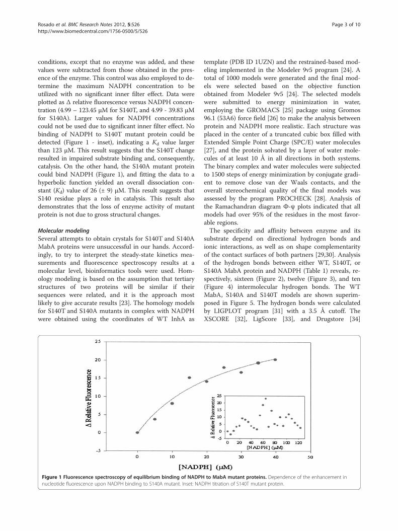
conditions, except that no enzyme was added, and thesevalues were subtracted from those obtained in the pres-ence of the enzyme. This control was also employed to de-termine the maximum NADPH concentration to beutilized with no significant inner filter effect. Data wereplotted as Δ relative fluorescence versus NADPH concen-tration (4.99 – 123.45 μM for S140T, and 4.99 - 39.83 μMfor S140A). Larger values for NADPH concentrationscould not be used due to significant inner filter effect. Nobinding of NADPH to S140T mutant protein could bedetected (Figure 1 - inset), indicating a Kd value largerthan 123 μM. This result suggests that the S140T changeresulted in impaired substrate binding and, consequently,catalysis. On the other hand, the S140A mutant proteincould bind NADPH (Figure 1), and fitting the data to ahyperbolic function yielded an overall dissociation con-stant (Kd) value of 26 (± 9) μM. This result suggests thatS140 residue plays a role in catalysis. This result alsodemonstrates that the loss of enzyme activity of mutantprotein is not due to gross structural changes.
Molecular modelingSeveral attempts to obtain crystals for S140T and S140AMabA proteins were unsuccessful in our hands. Accord-ingly, to try to interpret the steady-state kinetics mea-surements and fluorescence spectroscopy results at amolecular level, bioinformatics tools were used. Hom-ology modeling is based on the assumption that tertiarystructures of two proteins will be similar if theirsequences were related, and it is the approach mostlikely to give accurate results [23]. The homology modelsfor S140T and S140A mutants in complex with NADPHwere obtained using the coordinates of WT InhA as
Figure 1 Fluorescence spectroscopy of equilibrium binding of NADPHnucleotide fluorescence upon NADPH binding to S140A mutant. Inset: NAD
template (PDB ID 1UZN) and the restrained-based mod-eling implemented in the Modeler 9v5 program [24]. Atotal of 1000 models were generated and the final mod-els were selected based on the objective functionobtained from Modeler 9v5 [24]. The selected modelswere submitted to energy minimization in water,employing the GROMACS [25] package using Gromos96.1 (53A6) force field [26] to make the analysis betweenprotein and NADPH more realistic. Each structure wasplaced in the center of a truncated cubic box filled withExtended Simple Point Charge (SPC/E) water molecules[27], and the protein solvated by a layer of water mole-cules of at least 10 Å in all directions in both systems.The binary complex and water molecules were subjectedto 1500 steps of energy minimization by conjugate gradi-ent to remove close van der Waals contacts, and theoverall stereochemical quality of the final models wasassessed by the program PROCHECK [28]. Analysis ofthe Ramachandran diagram Φ-ψ plots indicated that allmodels had over 95% of the residues in the most favor-able regions.The specificity and affinity between enzyme and its
substrate depend on directional hydrogen bonds andionic interactions, as well as on shape complementarityof the contact surfaces of both partners [29,30]. Analysisof the hydrogen bonds between either WT, S140T, orS140A MabA protein and NADPH (Table 1) reveals, re-spectively, sixteen (Figure 2), twelve (Figure 3), and ten(Figure 4) intermolecular hydrogen bonds. The WTMabA, S140A and S140T models are shown superim-posed in Figure 5. The hydrogen bonds were calculatedby LIGPLOT program [31] with a 3.5 Å cutoff. TheXSCORE [32], LigScore [33], and Drugstore [34]
to MabA mutant proteins. Dependence of the enhancement inPH titration of S140T mutant protein.
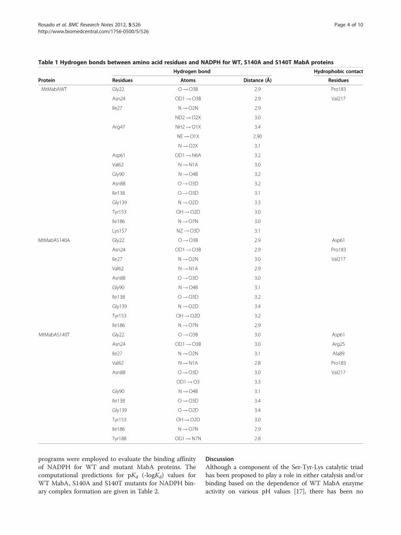
Table 1 Hydrogen bonds between amino acid residues and NADPH for WT, S140A and S140T MabA proteins
Hydrogen bond Hydrophobic contact
Protein Residues Atoms Distance (Å) Residues
MtMabAWT Gly22 O!O3B 2.9 Pro183
Asn24 OD1!O3B 2.9 Val217
Ile27 N!O2N 2.9
ND2!O2X 3.0
Arg47 NH2!O1X 3.4
NE!O1X 2.90
N!O2X 3.1
Asp61 OD1!N6A 3.2
Val62 N!N1A 3.0
Gly90 N!O4B 3.2
Asn88 O!O3D 3.2
Ile138 O!O3D 3.1
Gly139 N!O2D 3.3
Tyr153 OH!O2D 3.0
Ile186 N!O7N 3.0
Lys157 NZ!O3D 3.1
MtMabAS140A Gly22 O!O3B 2.9 Asp61
Asn24 OD1!O3B 2.9 Pro183
Ile27 N!O2N 3.0 Val217
Val62 N!N1A 2.9
Asn88 O!O3D 3.0
Gly90 N!O4B 3.1
Ile138 O!O3D 3.2
Gly139 N!O2D 3.4
Tyr153 OH!O2D 3.2
Ile186 N!O7N 2.9
MtMabAS140T Gly22 O!O3B 3.0 Asp61
Asn24 OD1!O3B 3.0 Arg25
Ile27 N!O2N 3.1 Ala89
Val62 N!N1A 2.8 Pro183
Asn88 O!O3D 3.0 Val217
OD1!O3 3.3
Gly90 N!O4B 3.1
Ile138 O!O3D 3.4
Gly139 O!O2D 3.4
Tyr153 OH!O2D 3.0
Ile186 N!O7N 2.9
Tyr188 OG1!N7N 2.8
Rosado et al. BMC Research Notes 2012, 5:526 Page 4 of 10http://www.biomedcentral.com/1756-0500/5/526
programs were employed to evaluate the binding affinityof NADPH for WT and mutant MabA proteins. Thecomputational predictions for pKd (-logKd) values forWT MabA, S140A and S140T mutants for NADPH bin-ary complex formation are given in Table 2.
DiscussionAlthough a component of the Ser-Tyr-Lys catalytic triadhas been proposed to play a role in either catalysis and/orbinding based on the dependence of WT MabA enzymeactivity on various pH values [17], there has been no
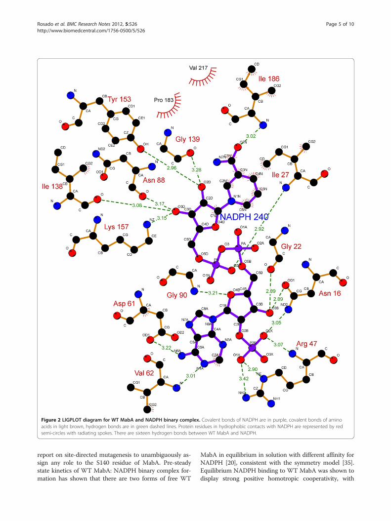
Figure 2 LIGPLOT diagram for WT MabA and NADPH binary complex. Covalent bonds of NADPH are in purple, covalent bonds of aminoacids in light brown, hydrogen bonds are in green dashed lines. Protein residues in hydrophobic contacts with NADPH are represented by redsemi-circles with radiating spokes. There are sixteen hydrogen bonds between WT MabA and NADPH.
Rosado et al. BMC Research Notes 2012, 5:526 Page 5 of 10http://www.biomedcentral.com/1756-0500/5/526
report on site-directed mutagenesis to unambiguously as-sign any role to the S140 residue of MabA. Pre-steadystate kinetics of WT MabA: NADPH binary complex for-mation has shown that there are two forms of free WT
MabA in equilibrium in solution with different affinity forNADPH [20], consistent with the symmetry model [35].Equilibrium NADPH binding to WT MabA was shown todisplay strong positive homotropic cooperativity, with
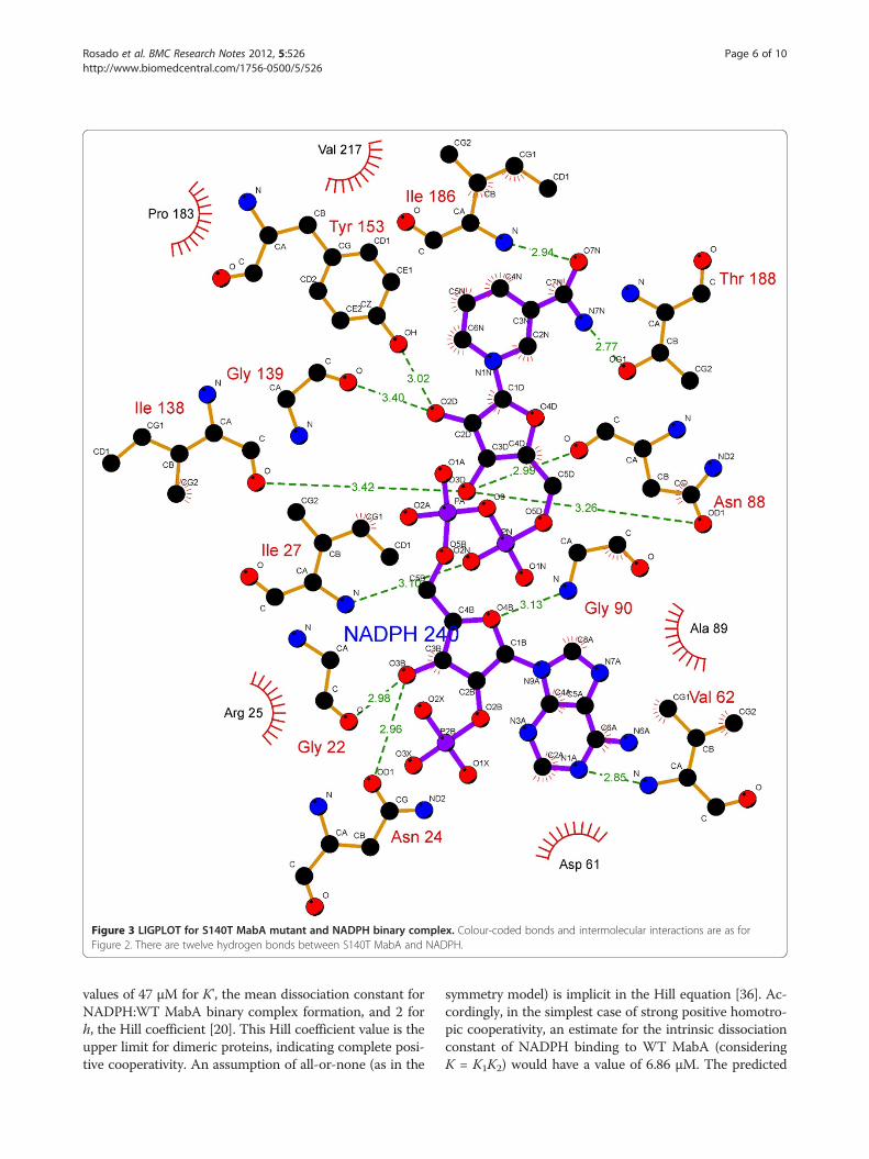
Figure 3 LIGPLOT for S140T MabA mutant and NADPH binary complex. Colour-coded bonds and intermolecular interactions are as forFigure 2. There are twelve hydrogen bonds between S140T MabA and NADPH.
Rosado et al. BMC Research Notes 2012, 5:526 Page 6 of 10http://www.biomedcentral.com/1756-0500/5/526
values of 47 μM for K', the mean dissociation constant forNADPH:WT MabA binary complex formation, and 2 forh, the Hill coefficient [20]. This Hill coefficient value is theupper limit for dimeric proteins, indicating complete posi-tive cooperativity. An assumption of all-or-none (as in the
symmetry model) is implicit in the Hill equation [36]. Ac-cordingly, in the simplest case of strong positive homotro-pic cooperativity, an estimate for the intrinsic dissociationconstant of NADPH binding to WT MabA (consideringK = K1K2) would have a value of 6.86 μM. The predicted
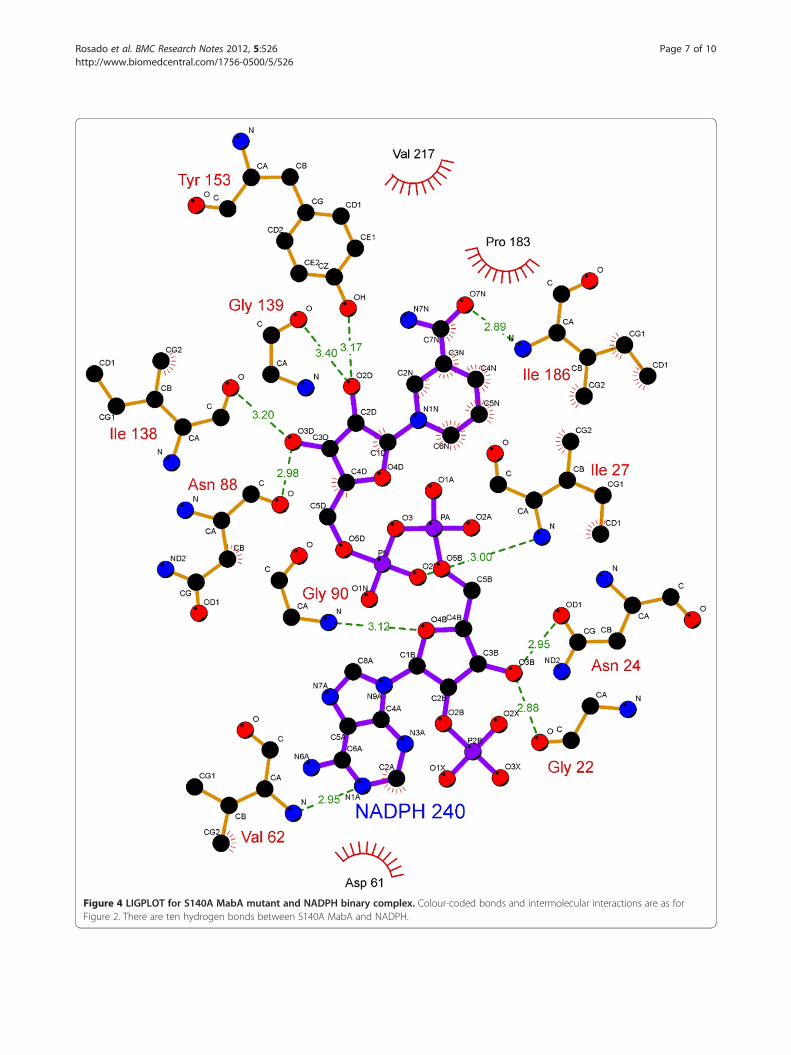
Figure 4 LIGPLOT for S140A MabA mutant and NADPH binary complex. Colour-coded bonds and intermolecular interactions are as forFigure 2. There are ten hydrogen bonds between S140A MabA and NADPH.
Rosado et al. BMC Research Notes 2012, 5:526 Page 7 of 10http://www.biomedcentral.com/1756-0500/5/526
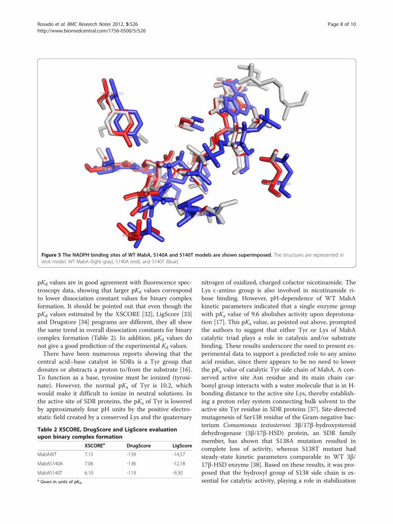
Figure 5 The NADPH binding sites of WT MabA, S140A and S140T models are shown superimposed. The structures are represented instick model. WT MabA (light gray), S140A (red), and S140T (blue).
Rosado et al. BMC Research Notes 2012, 5:526 Page 8 of 10http://www.biomedcentral.com/1756-0500/5/526
pKd values are in good agreement with fluorescence spec-troscopy data, showing that larger pKd values correspondto lower dissociation constant values for binary complexformation. It should be pointed out that even though thepKd values estimated by the XSCORE [32], LigScore [33]and Drugstore [34] programs are different, they all showthe same trend in overall dissociation constants for binarycomplex formation (Table 2). In addition, pKd values donot give a good prediction of the experimental Kd values.There have been numerous reports showing that the
central acid–base catalyst in SDRs is a Tyr group thatdonates or abstracts a proton to/from the substrate [16].To function as a base, tyrosine must be ionized (tyrosi-nate). However, the normal pKa of Tyr is 10.2, whichwould make it difficult to ionize in neutral solutions. Inthe active site of SDR proteins, the pKa of Tyr is loweredby approximately four pH units by the positive electro-static field created by a conserved Lys and the quaternary
Table 2 XSCORE, DrugScore and LigScore evaluationupon binary complex formation
XSCOREa DrugScore LigScore
MabAWT 7.15 -139 -14,57
MabAS140A 7.06 -136 -12,18
MabAS140T 6.10 -119 -9,30a Given in units of pKd.
nitrogen of oxidized, charged cofactor nicotinamide. TheLys E-amino group is also involved in nicotinamide ri-bose binding. However, pH-dependence of WT MabAkinetic parameters indicated that a single enzyme groupwith pKa value of 9.6 abolishes activity upon deprotona-tion [17]. This pKa value, as pointed out above, promptedthe authors to suggest that either Tyr or Lys of MabAcatalytic triad plays a role in catalysis and/or substratebinding. These results underscore the need to present ex-perimental data to support a predicted role to any aminoacid residue, since there appears to be no need to lowerthe pKa value of catalytic Tyr side chain of MabA. A con-served active site Asn residue and its main chain car-bonyl group interacts with a water molecule that is in H-bonding distance to the active site Lys, thereby establish-ing a proton relay system connecting bulk solvent to theactive site Tyr residue in SDR proteins [37]. Site-directedmutagenesis of Ser138 residue of the Gram-negative bac-terium Comamonas testosteroni 3β/17β-hydroxysteroiddehydrogenase (3β/17β-HSD) protein, an SDR familymember, has shown that S138A mutation resulted incomplete loss of activity, whereas S138T mutant hadsteady-state kinetic parameters comparable to WT 3β/17β-HSD enzyme [38]. Based on these results, it was pro-posed that the hydroxyl group of S138 side chain is es-sential for catalytic activity, playing a role in stabilization
Rosado et al. BMC Research Notes 2012, 5:526 Page 9 of 10http://www.biomedcentral.com/1756-0500/5/526
and/or polarization of carbonyl substrate and hydrogenbonding to the hydroxyl group of the conserved Tyr [38].These authors have also pointed out that “it is not clearwhy in all SDR sequences thus far characterized Thr isconspicuously absent at this position, since our in vitroreplacement of Ser138 with Thr yielded an active proteinwith identical catalytic constants”. However, here weshow that the MabA S140T mutant has impairedNADPH binding and no enzyme activity could bedetected, suggesting that, contrary to 3β/17β-HSD, Thrcannot replace Ser140 in MabA. It is tempting to suggestthat impaired NADPH binding of S140T mutant as com-pared to WT and S140A MabA proteins is due to thebulkier side chain of threonine, which is borne out by theweaker intermolecular contact between NADPH and en-zyme binding site (Figure 5). Although the S140A mutantis able to bind NADPH, this mutation results in inactiveMabA enzyme. Accordingly, these results point to S140residue of MabA as a residue whose main role appears tobe in catalysis, in agreement with S138 of the catalytictriad of Drosophila alcohol dehydrogenase, also an SDRprotein [18]. Notwithstanding, the S140 side chain mayalso play an indirect role in NADPH binding as theS140T showed impaired dinucleotide binding, in agree-ment with both the MabA: NADPH (Figures 2, 3, 4 and5) and MabA:cofactor: substrate molecular models [39].Interestingly, a conspicuous consequence of replacingS140 with threonine (Figure 3) or alanine (Figure 4) is onLys157 which is no longer within the 3.5 Å cutoff forhydrogen bonding distance. Accordingly, the S140 resi-due appears to anchor the Lys157 catalytic triad ofMabA. On the other hand, the S140 residue appears tohave no effect on the Tyr153 of the catalytic triad ofMabA. It has been proposed that a switch from “closed”to “open” conformation of MabA upon NADPH bindingbrings the phenol ring of catalytic Tyr153 side chain tothe active site and that Ser140 induces this rearrange-ment [38]. However, we have recently shown that thereare two forms of MabA in solution that differ in affinityfor NADPH [20], which could imply that the “open” and“closed” conformations exist in equilibrium prior toNADPH binding. Interestingly, although tetramericβ-ketoacyl Reductase from Escherichia coli [40], Plasmo-dium falciparum [41] and Brassica napus [42] displaynegative homotropic cooperativity; β-ketoacyl Reductasefrom M. tuberculosis (MabA) [20] and Staphylococcus aur-eus [43] exhibit positive homotropic cooperativity. Unfor-tunately, attempts to obtain crystals of S140T and S140AMabA mutants have been unsuccessful in our hands.
ConclusionsThe S140 side chain appears to play a role in MabA ca-talysis that is likely not lowering the pKa value ofTyr153. Interestingly, there was a shift from sigmoidal
for WT MabA [20] to hyperbolic for S140A (Figure 1)titration curve upon NADPH binding. This finding sug-gests that the S140 residue may play a role in displacingthe pre-existing equilibrium between two forms ofMabA in solution, thereby affecting enzyme activity.Nina M. Goodey and Stephen J. Benkovic have pointedout that chemical compounds that can shift the equilib-rium to protein conformers with lower substrate affinityand/or less catalytically competent must also be consid-ered in drug design [44]. Ideally though, the crystalstructure of S140A mutant protein should be obtainedto shed further light on the role of this residue in MabA.
AbbreviationsTB: Tuberculosis; MDR-TB: Multi-Drug Resistant TB; XDR-TB: Extensively Drug-Resistant; FAS: Fatty Acid biosynthesis System; MabA: Mycobacteriumtuberculosis β-ketoacyl-ACP Reductase; SDR: Short-chain Dehydrogenases/Reductase; Kd: Overall dissociation constant; 3β/17β-HSD: Comamonastestosteroni 3β/17β-hydroxysteroid dehydrogenase.
Competing interestsThe authors declare that they have no competing interests.
Authors’ contributionsConceived and designed the experiments: LAR, RAC, WFA, DSS and LAB.Performed the experiments: LAR and RAC. Analyzed the data: LAR, RAC andLAB. Contributed reagents/materials/analysis tools: WFA, LAB and DSS. Wrotethe paper: LAR, LAB and DSS. All authors read and approved the finalmanuscript.
AcknowledgmentsFinancial support for this work was provided by Millennium InitiativeProgram and National Institute of Science and Technology on Tuberculosis,MCT-CNPq, Ministry of Health-Department of Science and Technology (Brazil)to DSS and LAB LAB and DSS also acknowledge financial support awardedby FAPERGS-CNPq-PRONEX-2009. DSS (304051/1975-06), LAB (520182/99-5)and WFAJr (300851/98-7) are research career awardees from the NationalCouncil for Scientific and Technological Development of Brazil (CNPq). LARand RAC were supported by studentships from CNPq.
Author details1Centro de Pesquisas em Biologia Molecular e Funcional (CPBMF), InstitutoNacional de Ciência e Tecnologia em Tuberculose (INCT-TB), PontifíciaUniversidade Católica do Rio Grande do Sul (PUCRS), Av. Ipiranga 6681, PortoAlegre, RS 90619-900, Brazil. 2Programa de Pós-Graduação em Medicina eCiências da Saúde, PUCRS, Av. Ipiranga 6681, Porto AlegreRS 90619-900,Brazil. 3Faculdade de Biociências, Laboratório de Bioquímica Estrutural,PUCRS, Av. Ipiranga 6681, Porto Alegre, RS 90619-900, Brazil. 4Programa dePós-Graduação em Biologia Celular e Molecular, PUCRS, Av. Ipiranga 6681 –Tecnopuc – Prédio 92A, ZIP CODE 90619-900, Porto Alegre, RS, Brazil.
Received: 7 May 2012 Accepted: 14 September 2012Published: 25 September 2012
References1. Harries AD, Dye C: Tuberculosis. Ann Trop Med Parasitol 2006, 100:415–431.2. World Health Organization: Global Tuberculosis Control -Epidemiology,
Strategy, Financing. www.who.int/tb/publications/global_report/2009.3. Centers for Disease Control and Prevention (CDC): Emergence of
Mycobacterium tuberculosis with extensive resistance to second-linedrugs – worldwide, 2004-2006. MMWR Morb Mortal Wkly Rep 2006,55:301–305.
4. Dorman SE, Chaisson RE: From magic bullets back to the magicmountain: the rise of extensively drug-resistant tuberculosis. Nat Med2007, 13:295–298.
5. Singh JA, Upshur R, Padayatchi N: XDR-TB in South Africa: no time fordenial or complacency. PLoS Med 2007, 4:e50.
Rosado et al. BMC Research Notes 2012, 5:526 Page 10 of 10http://www.biomedcentral.com/1756-0500/5/526
6. Velayati AA, Farnia P, Masjedi MR, Ibrahim TA, Tabarsi P, Haroun RZ, KuanHO, Ghanavi J, Farnia P, Varahram M: Totally drug-resistant tuberculosisstrains: evidence of adaptation at the cellular level. Eur Respir J 2009,34:1202–1203.
7. Velayati AA, Masjedi MR, Farnia P, Tabarsi P, Ghanavi J, ZiaZarifi AH, HoffnerSE: Emergence of new forms of totally drug-resistant tuberculosis bacilli.Chest 2009, 136:420–425.
8. Banerjee A, Sugantino M, Sacchettini JC, Jacobs WR Jr: The mabA gene fromthe inhA operon of Mycobacterium tuberculosis encodes a 3-ketoacylreductase that fails to confer isoniazid resistance. Microbiology 1998,144:2697–2707.
9. Parish T, Roberts G, Laval F, Schaeffer M, Daffe M, Dunkan K: Functionalcomplementation of the essential gene fabG1 of Mycobacteriumtuberculosis by Mycobacterium smegmatis fabG but not Escherichia colifabG. J Bacteriol 2007, 189:3721–3728.
10. Veyron-Churlet R, Zanella-Cleon I, Cohen-Gonsaud M, Molle V, Kremer L:Phosphorylation of the Mycobacterium tuberculosis β-ketoacyl-acyl carrierprotein reductase MabA regulates mycolic acid biosynthesis. J Biol Chem2010, 285:12714–12725.
11. Cantaloube S, Veyron-Churlet R, Haddache N, Daffé M, Zerbib D: TheMycobacterium tuberculosis FAS-II dehydratases and methyltransferasesdefine the specificity of the mycolic acid elongation complexes. PLoSOne 2011, 6(12):e29564.
12. Robertson JG: Enzymes as a special class of therapeutic target: clinicaldrugs and modes of action. Curr Opin Struct Biol 2007, 17:674–679.
13. Oppermann U, Filling C, Hult M, Shafqat N, Wu X, Lindh M, Shafqat J, NordlingE, Kallberg Y, Persson B, Jörnval H: Short-chain dehydrogenases/reductases(SDR): the 2002 update. Chem Biol Interact 2003, 143–144:247–253.
14. Marrakchi H, Ducasse S, Labesse G, Montrozier H, Margeat E, Emorine L,Charpentier X, Mamadou D, Quémard A: MabA (FabG1), a Mycobacteriumtuberculosis protein involved in the long-chain fatty acid elongationsystem FAS-II. Microbiology 2002, 148:951–960.
15. Cohen-Gonsaud M, Ducasse S, Hoh F, Zerbib D, Labesse G, Quémard A:Crystal structure of MabA from Mycobacterium tuberculosis, a reductaseinvolved in long-chain fatty acid biosynthesis. J Mol Biol 2002,320:249–261.
16. Kanavagh KL, Jörnvall H, Persson B, Oppermann U: The SDR superfamily:functional and structural diversity within a family of metabolic andregulatory enzymes. Cell Mol Life Sci 2008, 65:3895–3906.
17. Silva RG, Carvalho LP, Blanchard JS, Santos DS, Basso LA: Mycobacteriumtuberculosis beta-ketoacyl-acyl carrier protein (ACP) reductase: kineticand chemical mechanisms. Biochemistry 2006, 45:13064–13073.
18. Winberg J-O, Brendskag MK, Sylte I, Lindstad RI, McKinley-McKee JS: Thecatalytic triad in Drosophila alcohol dehydrogenase: pH, temperatureand molecular modelling studies. J Mol Biol 1999, 294:601–616.
19. Persson B, Krook M, Jörnvall H: Characteristics of short-chain alcoholdehydrogenases and related enzymes. Eur J Biochem 1991, 200:537–543.
20. Silva RG, Rosado LA, Santos DS, Basso LA: Mycobacterium tuberculosis beta-ketoacyl-ACP reductase: alpha-secondary kinetic isotope effects andkinetic and equilibrium mechanisms of substrate binding. Arch BiochemBiophys 2008, 471:1–10.
21. Laemmli UK: Cleavage of structural proteins during the assembly of thehead of bacteriophage T4. Nature 1970, 227:680–685.
22. Bradford MM, McRorie RA, Williams WL: A rapid and sensitive method forthe quantification of microgram quantities of protein utilizing theprinciple of protein-dye binding. Anal Biochem 1976, 72:248–254.
23. Kroemer RT, Doughty SW, Robinson AJ, Richards WG: Prediction of thethree-dimensional structure of human interleukin-7 by homologymodeling. Protein Eng 1996, 9:493–498.
24. Sali A, Blundell TL: Comparative protein modelling by satisfaction ofspatial restraints. J Mol Biol 1993, 234:779–815.
25. van Der Spoel D, Lindahl E, Hess B, Groenhof G, Mark AE, Berendsen HJ:GROMACS: fast, flexible, and free. J Comp Chem 2005, 26:1701–1718.
26. Ooestenbrik C, Soares TA, van der Vegt NF, van Gunsteren WF: Validationof the 53A6 GROMOS force field. Eur Biophys J 2005, 34:273–284.
27. Berendsen HJC, Postma JPM, van Gunsteren WF, Hermans J: Interactionmodels for water in relation to protein hydration. In Intermolecular Forces.Edited by Pullman B. Dordrecht: Reidel D. Publishing Company;1981:331–342.
28. Laskowski RA, Macarthur MW, Moss DS, Thornton JM: PROCHECK: aprogram to check the stereochemical quality of protein structures. J ApplCryst 1993, 26:283–291.
29. Azevedo WF Jr, Mueller-Dieckmann JH, Schulze-Gahmen U, Worland PJ,Sausville E, Kim SH: Structural basis for specificity and potency of aflavonoid inhibitor of human CDK2, a cell cycle kinase. Proc Natl Acad SciUSA 1996, 93:2735–2740.
30. Azevedo WF Jr, Canduri F, Fadel V, Teodoro LG, Hial V, Gomes RA:Molecular model for the binary complex of uropepsin and pepstatin.Biochem Biophys Res Commun 2001, 287:277–281.
31. Wallace AC, Laskowski RA, Thornton JM: LIGPLOT: a program generateschematic diagrams of protein–ligand interactions. Protein Eng 1995,8:127–134.
32. Wang R, Lai L, Wang S: Further development and validation of empiricalscoring functions for structure-based binding affinity prediction. J Comput-Aided Molecular Des 2002, 16:11–26.
33. Fan H, Schneidman-Duhovny D, Irwin J, Dong GQ, Shoichet B, Sali A:Statistical Potential for Modeling and Ranking of Protein-LigandInteractions. J Chem Inf Model 2011, 51:3078–3092.
34. Velec HF, Gohlke H, Klebe G: DrugScore(CSD)-knowledge-based scoringfunction derived from small molecule crystal data with superiorrecognition rate of near-native ligand poses and better affinityprediction. J Med Chem 2005, 48:6296–6303.
35. Monod J, Wyman J, Changeux J-P: On the nature of allosteric transitions:a plausible model. J Mol Biol 1965, 12:88–118.
36. Hill AV: The combinations of haemoglobin with oxygen and with carbonmonoxide. Biochem J 1913, 7:471–480.
37. Filling C, Berndt KD, Benach J, Knapp S, Prozorovski T, Nordling E,Ladenstein R, Jörnvall H, Oppermann U: Critical residues for structure andcatalysis in short-chain dehydrogenases/reductases. J Biol Chem 2002,277:25677–25684.
38. Oppermann UCT, Filling C, Berndt KD, Persson B, Benach J, Ladenstein R,Jörnvall H: Active site directed mutagenesis of 3β/17β-hydroxysteroiddehydrogenase establishes differential effects on short-chaindehydrogenase/reductase reactions. Biochemistry 1997, 36:34–40.
39. Cohen-Gonsaud M, Ducasse-Cabanot S, Quémard A, Labesse G:Ligand-induced fit in mycobacterial MabA: the sequence-specific C-terminus locks the conformational change. Proteins 2005, 60:392–400.
40. Price AC, Zhang YM, Rock CO, White SW: Structure of beta-ketoacyl-[acylcarrier protein] reductase from Escherichia coli: negative cooperativityand its structural basis. Biochemistry 2001, 40:12772–12781.
41. Karmodiya K, Surolia N: Analyses of co-operative transitions inPlasmodium falciparum beta-ketoacyl acyl carrier protein reductase uponco-factor and acyl carrier protein binding. FEBS J 2006, 273:4093–4103.
42. Sheldon PS, Kekwick RG, Smith CG, Sidebottom C, Slabas AR: 3-Oxoacyl-[ACP] reductase from oilseed rape (Brassica napus). Biochim Biophys Acta1992, 1120:151–159.
43. Dutta D, Bhattacharyya S, Das AK: Crystal structure and fluorescencestudies reveal the role of helical dimeric interface of StapylococcalFabG1 in positive cooperativity for NADPH. Proteins 2012, 8:1250–1257.
44. Goodey NM, Benkovic SJ: Allosteric regulation and catalysis emerge via acommon route. Nat Chem Biol 2008, 4:474–482.
doi:10.1186/1756-0500-5-526Cite this article as: Rosado et al.: Role of Serine140 in the mode ofaction of Mycobacterium tuberculosis β-ketoacyl-ACP Reductase (MabA).BMC Research Notes 2012 5:526.
Submit your next manuscript to BioMed Centraland take full advantage of:
• Convenient online submission
• Thorough peer review
• No space constraints or color figure charges
• Immediate publication on acceptance
• Inclusion in PubMed, CAS, Scopus and Google Scholar
• Research which is freely available for redistribution
Submit your manuscript at www.biomedcentral.com/submit


![KAPASITOR [Compatibility Mode]](https://static.fdocument.org/doc/165x107/58807b111a28aba8048b5563/kapasitor-compatibility-mode.jpg)
![Awire [Compatibility Mode]](https://static.fdocument.org/doc/165x107/5535ead5550346640d8b4748/awire-compatibility-mode.jpg)


![AKD4425A-SA English Manual - Asahi Kasei Microdevices · AKD4425A-SA has a digital audio interface ... C24 (short) C29 2.2n R21 (short) + C28 (short) R16 470 J1 ... [Read] commands.](https://static.fdocument.org/doc/165x107/5b1b921a7f8b9a28258eb031/akd4425a-sa-english-manual-asahi-kasei-microdevices-akd4425a-sa-has-a-digital.jpg)
![Wettability (Kemampubasahan) [Compatibility Mode]](https://static.fdocument.org/doc/165x107/55cf9a8d550346d033a24fae/wettability-kemampubasahan-compatibility-mode.jpg)
![Material Magnet [Compatibility Mode]](https://static.fdocument.org/doc/165x107/5885bc341a28ab1c198c4f13/material-magnet-compatibility-mode.jpg)