SERS Active Gold Nanostar Dimer for Mercury Ion Detection ... · 3 SERS Active Gold Nanostar Dimer...
4
1 Supporting information 1 2 SERS Active Gold Nanostar Dimer for Mercury Ion Detection 3 4 5 EXPERIMENTAL SECTIONS 6 Material 7 Thiolated DNA oligonucleotides, purified by high performance liquid chromatography 8 (HPLC), were manufactured by Shanghai Sangon Biological Engineering Technology & Services 9 Co. Ltd. (Shanghai, P.R. China). These were dissolved in deionized (DI) water to give a final 10 concentration of 100 μM. Unless stated otherwise, all other chemicals used in this work were 11 purchased from Sigma-Aldrich. DI water from a Milli-Q device (18.2 M Ω, Millipore, Molsheim, 12 France) was used in all experiments. Cu 2+ , Hg 2+ , Cd 2+ , Pb 2+ , Cr 3+ , Mn 2+ , Co 2+ , Fe 3+ , Zn 2+ , Al 3+ , 13 Mg 2+ , and Ca 2+ (1000 μg mL -1 in 1% HNO 3 or 5% HCL) were purchased from the National 14 Institute of Metrology P.R China (Beijing, China). 15 The detailed sequences of the oligonucleotide are: 16 DNA 1: 5’-SH-AAAAAAGTGACCATTTTTGCAGTG-3’ 17 DNA 2: 5’-SH-AAAAAACACTGCTTTTTTGGTCAC-3’ 18 19 Instrumentation 20 Transmission electron microscopy (TEM) images were obtained using a JEOL JEM-2100 21 operating at an acceleration voltage of 200 kV. The size distribution of the GNS was measured 22 using a Zetasizer Nano ZS system (Malvern). A 633 nm laser was used for the DLS 23 characterization. All UV-Vis spectra were acquired using a UNICO 2100 PC UV-Vis 24 spectrophotometer and processed with Origin Lab software. Raman spectra were measured using a 25 LabRam-HR800 Micro-Raman spectrometer with Lab-spec 5.0 software attached to a liquid cell. 26 The slit and pinhole were set at 100 and 400 mm, respectively, in the confocal configuration, with 27 a holographic grating (600 g/mm) and an air-cooled He-Ne laser giving 785 nm excitation with a 28 power of ~ 8 mW. 29 Electronic Supplementary Material (ESI) for Chemical Communications This journal is © The Royal Society of Chemistry 2013
Transcript of SERS Active Gold Nanostar Dimer for Mercury Ion Detection ... · 3 SERS Active Gold Nanostar Dimer...

1
Supporting information 1
2
SERS Active Gold Nanostar Dimer for Mercury Ion Detection 3
4
5
EXPERIMENTAL SECTIONS 6
Material 7
Thiolated DNA oligonucleotides, purified by high performance liquid chromatography 8
(HPLC), were manufactured by Shanghai Sangon Biological Engineering Technology & Services 9
Co. Ltd. (Shanghai, P.R. China). These were dissolved in deionized (DI) water to give a final 10
concentration of 100 μM. Unless stated otherwise, all other chemicals used in this work were 11
purchased from Sigma-Aldrich. DI water from a Milli-Q device (18.2 M Ω, Millipore, Molsheim, 12
France) was used in all experiments. Cu2+
, Hg2+
, Cd2+
, Pb2+
, Cr3+
, Mn2+
, Co2+
, Fe3+
, Zn2+
, Al3+
, 13
Mg2+
, and Ca2+
(1000 µg mL-1
in 1% HNO3 or 5% HCL) were purchased from the National 14
Institute of Metrology P.R China (Beijing, China). 15
The detailed sequences of the oligonucleotide are: 16
DNA 1: 5’-SH-AAAAAAGTGACCATTTTTGCAGTG-3’ 17
DNA 2: 5’-SH-AAAAAACACTGCTTTTTTGGTCAC-3’ 18
19
Instrumentation 20
Transmission electron microscopy (TEM) images were obtained using a JEOL JEM-2100 21
operating at an acceleration voltage of 200 kV. The size distribution of the GNS was measured 22
using a Zetasizer Nano ZS system (Malvern). A 633 nm laser was used for the DLS 23
characterization. All UV-Vis spectra were acquired using a UNICO 2100 PC UV-Vis 24
spectrophotometer and processed with Origin Lab software. Raman spectra were measured using a 25
LabRam-HR800 Micro-Raman spectrometer with Lab-spec 5.0 software attached to a liquid cell. 26
The slit and pinhole were set at 100 and 400 mm, respectively, in the confocal configuration, with 27
a holographic grating (600 g/mm) and an air-cooled He-Ne laser giving 785 nm excitation with a 28
power of ~ 8 mW. 29
Electronic Supplementary Material (ESI) for Chemical CommunicationsThis journal is © The Royal Society of Chemistry 2013

2
Gold Nanostar synthesis 30
Gold nanostars were synthesized by a seed-mediated growth method. Initially, the gold seed 31
was prepared by adding 15 mL of 1% citrate solution to 100 mL of boiling 1 mM HAuCl4 solution 32
under vigorous stirring. After cooling to room temperature, 200 µL 1-2 mM AgNO3 (for 50nm 33
GNS is 1mM and 2 mM for 60 nm GNS) and 100 µL 0.1 M ascorbic acid were mixed together 34
quickly into 20 mL of 0.25 mM HAuCl4 with 200 µL seeds, the PH is kept at 3. The colloidal 35
solution was resuspended into 0.05% tween-20 by centrifugation at 3000 rpm for 15 min to 36
prevent further reaction. 37
38
Preparation of ssDNA-Functionalized GNS 39
Briefly, 2 µL 100µM thiolated modified DNA 1 or DNA 2 solution was added to 100 µL of just 40
prepared GNS and incubated for 3h at ambient temperature. Subsequently, 0.05 M NaCl was 41
mixed and then the mixture was incubated for 12 h with constant shaking. The excess DNA was 42
removed by two centrifugations at 3000 rpm for 10 min. The mixtures were denoted as GNS-DNA 43
1 and GNS-DNA 2. 44
45
Synthesis of GNS dimer structure 46
To prepare 50 nm GNS dimers, 50 nm GNS-DNA 1 and GNS-DNA 2 were mixed with a ratio 47
of 1:1. A similar procedure was used for the 60 nm GNS dimer. The heterodimers were formed by 48
adding equal quantities of 50 nm GNS-DNA 1 to 60 nm GNS-DNA 2. Then, 4-ATP ethanol 49
solution, with a final concentration of 1 μM, was added to all three mixtures and incubated at 50
room temperature overnight. After redispersion in DI water, Hg2+
solution, with a final 51
concentration 1 ng mL-1
, was mixed with all three samples and shaken for 3 h. 52
53
Fabrication of GNS sensors 54
800 μL 60 nm GNS-DNA 1 and GNS-DNA 2 in the ratio of 1:1 were mixed with 1 μM 4-ATP 55
ethanol solution and incubated for 12 h with constant shaking. This mixture is denoted as the 56
sensor solution. Samples inoculated with different concentrations of Hg2+
(0, 0.5, 0.1, 0.05, 0.01, 57
Electronic Supplementary Material (ESI) for Chemical CommunicationsThis journal is © The Royal Society of Chemistry 2013

3
0.005, 0.002 ng mL-1
) were separately added to 100 μL of sensor solution in separate tubes. After 58
reacting for 3 h, the samples were processed by TEM, UV-Visible spectrophotometry and SERS. 59
60
61
62
Figure S1. Uv-vis spectrum of different GNS dimers and particles (A), DLS of 50, 63
heterodimer, 60 nm GNS sensor before and after addition 1 ng mL-1
Hg2+
(B, C, D). 64
65
66
67
68
69
Figure S2. Uv-vis spectrum of 60 nm GNS dimer sensors in diverse Hg2+
samples (A), 70
SERS intensity of different heavy metal irons based on 60 nm GNS dimer sensor(B) 71
72
Electronic Supplementary Material (ESI) for Chemical CommunicationsThis journal is © The Royal Society of Chemistry 2013
4
73
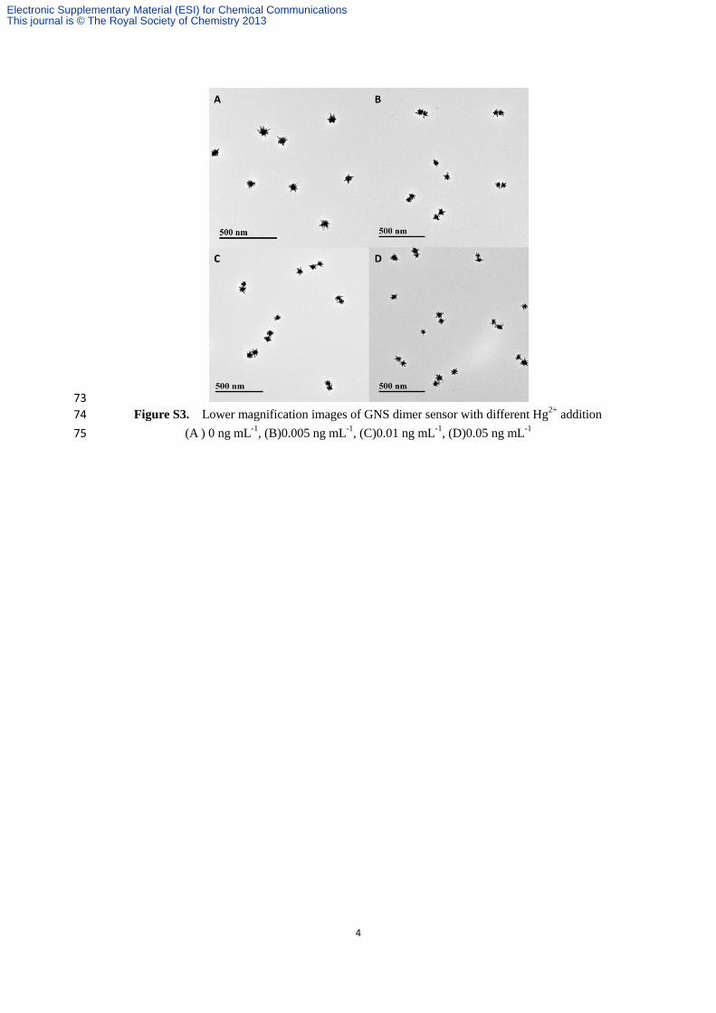
Figure S3. Lower magnification images of GNS dimer sensor with different Hg2+
addition 74
(A ) 0 ng mL-1
, (B)0.005 ng mL-1
, (C)0.01 ng mL-1
, (D)0.05 ng mL-1
75
Electronic Supplementary Material (ESI) for Chemical CommunicationsThis journal is © The Royal Society of Chemistry 2013


![LIAISON QuantiFERON-TB Gold Plus ( [REF] 311010) 1 ...](https://static.fdocument.org/doc/165x107/61b26a9e529835162559e41c/liaison-quantiferon-tb-gold-plus-ref-311010-1-.jpg)