Scaffolds of PDLLA/Bioglass 58S Produced via … 17(Suppl. 1) Scaffolds of PDLLA/Bioglass 58S...
6
2014; 17(Suppl. 1): 33-38 © 2014 Materials Research. DO D http//dx.doi.org/10.1590/S1516-14392014005000075 *e-mail: [email protected] Scaffolds of PDLLA/Bioglass 58S Produced via Selective Laser Sintering Rafaela do Vale Pereira a *, Gean Vitor Salmoria b , Marcela Oliveira Caldeira de Moura a , Águedo Aragones c , Márcio Celso Fredel a a CERMAT, Mechanical Engineering Department, Federal University of Santa Catarina – UFSC, Florianópolis, SC, Brazil b CIMJECT, Mechanical Engineering Department, Federal University of Santa Catarina – UFSC, Florianópolis, SC, Brazil c Odontology Department, Federal University of Santa Catarina – UFSC, Florianópolis, SC, Brazil Received: June 20, 2013; Revised: May 5, 2014 Scaffolds of PDLLA were produced to be implemented in maxilofacial surgeries inducing bone repair and regeneration. To prepare these scaffolds, bioglass (BG58S) was synthesized by sol-gel method, in order to be applied as osteoconductive dispersed particles in PDLLA matrix. Once presenting greater facility on parts fabrication, this polymeric matrix enables complex geometries production besides presenting compatible degradation rate for scaffold absorption and bone regeneration. Scaffolds production was performed by selective laser sintering in order to obtain tailored-made parts. FTIR and XRD analyses were carried out to observe the composition and evaluate the presence of crystalized phases in bioglass, obtaining Wollastonite. SEM was used to observe the BG particle distribution in PDLLA matrix and flexural test was performed to evaluate the composite mechanical properties. Results showed that was possible to obtain pieces using SLS method and with addition of 10%wt BG to polymeric matrix, flexural modulus and strength increased regarding to pure polymer. Keywords: scaffold, bioglass, PDLLA, selective laser sintering (SLS). 1. Introduction In Tissue engineering, materials science is combined with medicine and biology in order to make improvements in the human beings life by providing techniques to reconstruct damaged tissues and organs 1 . A common technique used for these purpose is scaffold implantation at the injured parts, using adequate materials that promote tissue regeneration. Biodegradable polymers are widely used, nevertheless, their degradation residues decrease the medium pH, turning it acid, promoting an undesirable inflammatory response. In order to minimize this inflammatory response, bioactive ceramic particles are incorporated to the matrix, stabilizing the medium pH. BG 58S (60%SiO 2 , 36%CaO, 4%P 2 O 5 (% mol)), a resorbable biomaterial, was used as dispersed particles in polymer matrix in order to promote osteoconduction and pH stabilization, aiding bone regeneration. These regeneration is due to calcium and phosphate ions release, which are present at BG composition and play an important role at bone metabolism (angiogenesis, grow and tissue mineralization) 2-6 . Scaffolds can be prepared by Selective Laser Sintering (SLS), a Rapid Prototyping (RP) manufacturing technology, which is able to produce tailored-made parts for each fracture. This method is based at material powder sinterization, layer-by-layer, by an infrared laser beam (CO 2 Laser), which produces a final part previously obtained by computer-aided design (CAD). Other advantages of this technique are high dimensional accuracy, allowing well defined details, and achievement of complex designs. Once scaffolds require controlled properties and structure (as interconnected porosity and 3D structure, to allow cell migration, nutrition and proliferation), SLS is a widely used fabrication method 7,8 . In order to optimize samples processing with SLS method, (optimizing microstructure, mechanical properties and surface characteristics of the scaffolds), some variables can be controlled: laser power, scan speed and spot diameter at focus. These variables estimate the laser energy density (Equation 1) that reaches the material surface 9-14 . ρ e =P/(v.d) (1) where ρ e (J/mm²) is the laser energy density, P (W) is the laser power, v (mm/s) is the scan speed and d (mm) is the spot diameter. This study presents scaffolds production to be used at non-load bearing areas, as, for example, facial skeleton 15 . To produce these scaffolds two biomaterial were combined, poly (D,L) lactide (PDLLA) and bioglass 58S (BG 58S). PDLLA was the composite matrix, hence is a biomaterial that can be applied in non-load bearing due to its mechanical characteristics and complete resorption after within 72 weeks of implantation 15 . Four compositions of sample (0%wt BG, 10%wt BG, 20%wt BG and 30%wt BG) were prepared and the aim
Transcript of Scaffolds of PDLLA/Bioglass 58S Produced via … 17(Suppl. 1) Scaffolds of PDLLA/Bioglass 58S...
2014; 17(Suppl. 1): 33-38 © 2014Materials Research. DOI:D httpI://dx.doi.org/10.1590/S1516-14392014005000075
*e-mail: [email protected]
Scaffolds of PDLLA/Bioglass 58S Produced via Selective Laser Sintering
Rafaela do Vale Pereiraa*, Gean Vitor Salmoriab,
Marcela Oliveira Caldeira de Mouraa, Águedo Aragonesc, Márcio Celso Fredela
aCERMAT, Mechanical Engineering Department, Federal University of Santa Catarina – UFSC, Florianópolis, SC, Brazil
bCIMJECT, Mechanical Engineering Department, Federal University of Santa Catarina – UFSC, Florianópolis, SC, Brazil
cOdontology Department, Federal University of Santa Catarina – UFSC, Florianópolis, SC, Brazil
Received: June 20, 2013; Revised: May 5, 2014
Scaffolds of PDLLA were produced to be implemented in maxilofacial surgeries inducing bone repair and regeneration. To prepare these scaffolds, bioglass (BG58S) was synthesized by sol-gel method, in order to be applied as osteoconductive dispersed particles in PDLLA matrix. Once presenting greater facility on parts fabrication, this polymeric matrix enables complex geometries production besides presenting compatible degradation rate for scaffold absorption and bone regeneration. Scaffolds production was performed by selective laser sintering in order to obtain tailored-made parts. FTIR and XRD analyses were carried out to observe the composition and evaluate the presence of crystalized phases in bioglass, obtaining Wollastonite. SEM was used to observe the BG particle distribution in PDLLA matrix and flexural test was performed to evaluate the composite mechanical properties. Results showed that was possible to obtain pieces using SLS method and with addition of 10%wt BG to polymeric matrix, flexural modulus and strength increased regarding to pure polymer.
Keywords: scaffold, bioglass, PDLLA, selective laser sintering (SLS).
1. IntroductionIn Tissue engineering, materials science is combined
with medicine and biology in order to make improvements in the human beings life by providing techniques to reconstruct damaged tissues and organs1. A common technique used for these purpose is scaffold implantation at the injured parts, using adequate materials that promote tissue regeneration.
Biodegradable polymers are widely used, nevertheless, their degradation residues decrease the medium pH, turning it acid, promoting an undesirable inflammatory response. In order to minimize this inflammatory response, bioactive ceramic particles are incorporated to the matrix, stabilizing the medium pH. BG 58S (60%SiO
2, 36%CaO,
4%P2O
5 (% mol)), a resorbable biomaterial, was used
as dispersed particles in polymer matrix in order to promote osteoconduction and pH stabilization, aiding bone regeneration. These regeneration is due to calcium and phosphate ions release, which are present at BG composition and play an important role at bone metabolism (angiogenesis, grow and tissue mineralization)2-6.
Scaffolds can be prepared by Selective Laser Sintering (SLS), a Rapid Prototyping (RP) manufacturing technology, which is able to produce tailored-made parts for each fracture. This method is based at material powder sinterization, layer-by-layer, by an infrared laser beam (CO
2
Laser), which produces a final part previously obtained by computer-aided design (CAD). Other advantages of
this technique are high dimensional accuracy, allowing well defined details, and achievement of complex designs. Once scaffolds require controlled properties and structure (as interconnected porosity and 3D structure, to allow cell migration, nutrition and proliferation), SLS is a widely used fabrication method7,8.
In order to optimize samples processing with SLS method, (optimizing microstructure, mechanical properties and surface characteristics of the scaffolds), some variables can be controlled: laser power, scan speed and spot diameter at focus. These variables estimate the laser energy density (Equation 1) that reaches the material surface9-14.
ρe=P/(v.d) (1)
where ρe (J/mm²) is the laser energy density, P (W) is the
laser power, v (mm/s) is the scan speed and d (mm) is the spot diameter.
This study presents scaffolds production to be used at non-load bearing areas, as, for example, facial skeleton15. To produce these scaffolds two biomaterial were combined, poly (D,L) lactide (PDLLA) and bioglass 58S (BG 58S). PDLLA was the composite matrix, hence is a biomaterial that can be applied in non-load bearing due to its mechanical characteristics and complete resorption after within 72 weeks of implantation15.
Four compositions of sample (0%wt BG, 10%wt BG, 20%wt BG and 30%wt BG) were prepared and the aim
34 Pereira et al. Materials Research
of this study was to produce samples by SLS method, confirm if the synthesized materials presented the correct composition and structure and investigate the influence of BG particles amount at the mechanical properties of these composites.
2. Material and MethodsPDLLA was synthesized (as described in16) and
provided by Biomaterials Laboratory from Medical and Biological Science Center of Pontifical Catholic University (PUC-Sorocaba, SP).
BG 58S was synthesized by the author via sol-gel process. For that, were used Tetraethyl orthosilicate (TEOS) (Sigma Aldrich, reagente grade 98%, 131903-1l, lot STBB8489V), to obtain silica. Triethyl phosphate (TEP) (Sigma Aldrich, 99.8%, 538728-1l, lot MKBC5090). Calcium nitrate tetrahydrate (Ca(NO
3)
2 * 4 H
2O) (Vetec, cod.
000663.08, lot 1006753). Nitric acid (HNO3) (Vetec, 68%,
cod. 000451.06, lot 0903829), to control solution pH and ethyl alcohol (Synth, P.A.-A.C.S., lot A1084.01.BJ), used as solution solvent.
2.1. Synthesis of sol-gel process
BG58S was synthesized by sol-gel process as follows. At first TEOS, TEP and (Ca(NO
3)
2*4H
2O) (with BG58S molar
ratio), ethanol and 2M HNO3 were added in an Erlenmeyer,
at 50 °C and constant agitation. Solution was placed in a stove (Drying and sterilizing stove, Nova Ética, 402/D) at 70 °C for 24h in order to dry the material and promote its gelation.
Subsequently the material was thermally treated at 600 °C and milled in an eccentric mill (BP Engenharia, CB2-T), producing a material with d
50= 12.15 µm
(Mastersizer 2000, Malvern Instruments), used as dispersed particles at the polymeric matrix of composite. Also, with this powder, FTIR (Spectrophotometer Bruker, Tensor 27) and XRD (Vertical diffractometer, Philips, PW1150) were performed.
2.2. Sample preparation at SLS
In order to prepare samples for mechanical analyses, PDLLA in pellets was milled in an industrial blender (Stainless steel Industrial Blender, TRON, 25000 rpm), with liquid nitrogen, achieving a range of particles between 150 µm and 300 µm (sieve analysis), which is indicated to obtain the necessary scaffolds porosity17,18.
PDLLA and BG powder were mixed at a Y-mixer for 45 min, in different proportions, 10%wt BG, 20%wt BG and 30%wt BG. The different compositions were processed at
SLS (Table 1) producing samples with rectangular shape (35.0 × 5.0 × 2.3 mm).
Laser power of 5.4W was used at the process, once lower power didn’t promote the particles sinterization and higher powder promoted polymer degradation.
These rectangular samples were tested at DMA (Q800, TA Instruments) with single cantilever at 30 °C with an increase stress rate of 2N/min, obtaining stress-strain curves.
2.3. Characterizations
In order to characterize the materials, some tests were realized. For BG 58S FTIR (Bruker Spectrophotometer, Tensor 27) was carried out with sample prepared as a pastille of BG and KBR. Also XRD (Vertical diffractometer, Philips, PW1150) in which BG powder was analyzed.
For PDLLA were carried out: Gel permeation chromatography (GPC) (Schmadzu, UFLC), polymer powder was solubilized at THF; FTIR (Bruker Spectrophotometer, Tensor 27), sample was prepared as a PDLLA pastille; Nuclear magnetic Resonance (NMR) (Varian, NMR AS 400) and Differential Scanning Calorimetry (DSC) (Shimadzu, DSC – 50) with initial temperature of 20 °C heating rate of 5 °C/min until 200 °C (N
2 purge).
3. Results and Discussion
3.1. BG Characterization
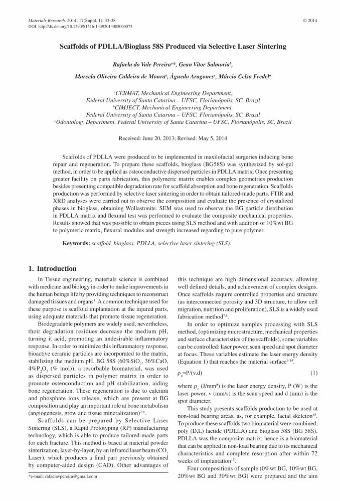
The observed infrared spectrum obtained for BG58S is presented at Figure 1. The peak at 465cm–1 refers to Si-O-Si bending mode, at 601 cm–1 refers to P-O bending vibration of PO
4–3 tetrahedral in crystalline HCA (carbonated
hydroxyapatite), probably resulting of a reaction from BG powder and atmospheric moisture. The slope about 930 cm–1 is associated to the Si-O-Ca vibration mode. Region around 1045 cm–1 refers to both Si-O-Si asymmetric stretching mode and P-O stretching vibration. 1395 and 1647 cm–1 present carbonates bands and 3410 cm–1 (not shown in graphic) refers to hydroxyl band2,19-21.
Table 1. Compositions and laser power for sample processing.
Composition Power (W) Sample name
Pure polymer 5.4 00BG5,4
10%wt of BG 5.4 10BG5,4
20%wt of BG 5.4 20BG5,4
30%wt of BG 5.4 30BG5,4 Figure 1. BG58S FTIR spectrum.
2014; 17(Suppl. 1) 35Scaffolds of PDLLA/Bioglass 58S Produced via Selective Laser Sintering
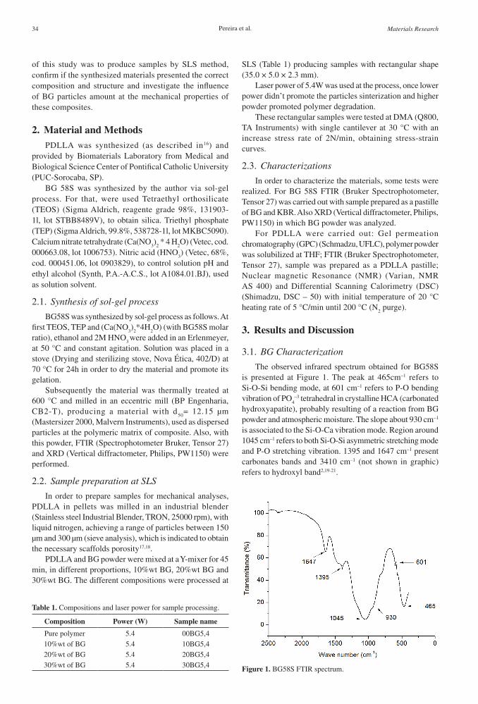
Figure 2 presents BG58S XRD spectrum, showing the amorphous region and a peak at 2θ equal to 34, indicating the presence of wollastonite (CaSiO
3) crystalline phase.
Wollastonite presents great influence at biomaterials activity, aiding at the formation of carbonated apatite layer, which provides the bonding with osseous tissue19,22.
3.2. PDLLA characterization
The composite matrix was produced with the synthesized PDLLA and its composition and properties were analyzed by GPC, FTIR, DSC and NMR.
Molecular mass was measured by gel permeation chromatography (GPC) and the values for Mn (78498 g/mol), Mw (153596 g/mol) and polydispersity index (1.95) were obtained.
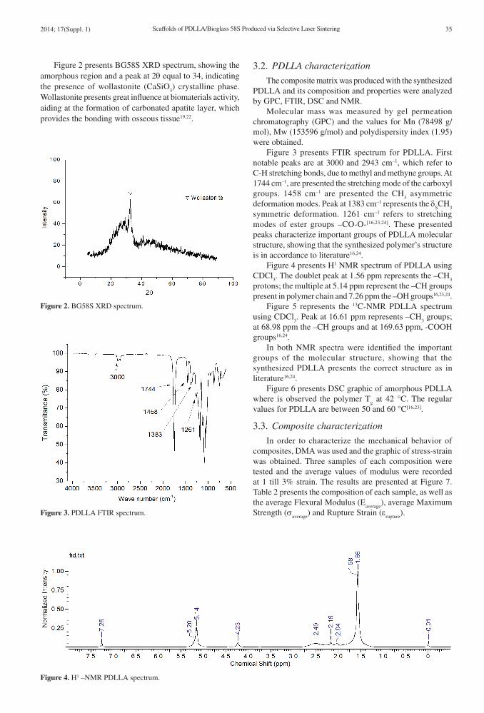
Figure 3 presents FTIR spectrum for PDLLA. First notable peaks are at 3000 and 2943 cm–1, which refer to C-H stretching bonds, due to methyl and methyne groups. At 1744 cm–1, are presented the stretching mode of the carboxyl groups. 1458 cm–1 are presented the CH
3 asymmetric
deformation modes. Peak at 1383 cm–1 represents the δSCH
3
symmetric deformation. 1261 cm–1 refers to stretching modes of ester groups –CO-O-[16,23,24]. These presented peaks characterize important groups of PDLLA molecular structure, showing that the synthesized polymer’s structure is in accordance to literature16,24.
Figure 4 presents H1 NMR spectrum of PDLLA using CDCl
3. The doublet peak at 1.56 ppm represents the –CH
3
protons; the multiple at 5.14 ppm represent the –CH groups present in polymer chain and 7.26 ppm the –OH groups16,23,24.
Figure 5 represents the 13C-NMR PDLLA spectrum using CDCl
3. Peak at 16.61 ppm represents –CH
3 groups;
at 68.98 ppm the –CH groups and at 169.63 ppm, -COOH groups16,24.
In both NMR spectra were identified the important groups of the molecular structure, showing that the synthesized PDLLA presents the correct structure as in literature16,24.
Figure 6 presents DSC graphic of amorphous PDLLA where is observed the polymer T
g at 42 °C. The regular
values for PDLLA are between 50 and 60 °C[16,23].
3.3. Composite characterization
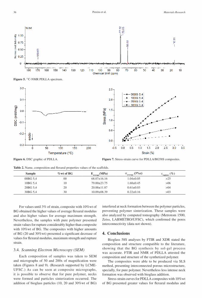
In order to characterize the mechanical behavior of composites, DMA was used and the graphic of stress-strain was obtained. Three samples of each composition were tested and the average values of modulus were recorded at 1 till 3% strain. The results are presented at Figure 7. Table 2 presents the composition of each sample, as well as the average Flexural Modulus (E
average), average Maximum
Strength (σaverage
) and Rupture Strain (εrupture
).
Figure 2. BG58S XRD spectrum.
Figure 3. PDLLA FTIR spectrum.
Figure 4. H1 –NMR PDLLA spectrum.
36 Pereira et al. Materials Research
For values until 3% of strain, composite with 10%wt of BG obtained the higher values of average flexural modulus and also higher values for average maximum strength. Nevertheless, the samples with pure polymer presented strain values for rupture considerably higher than composite with 10%wt of BG. The composites with higher amounts of BG (20 and 30%wt) presented a significant decrease of values for flexural modulus, maximum strength and rupture strain.
3.4. Scanning Electron Microscopy (SEM)
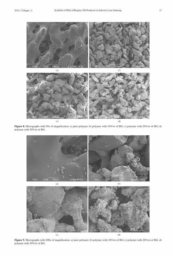
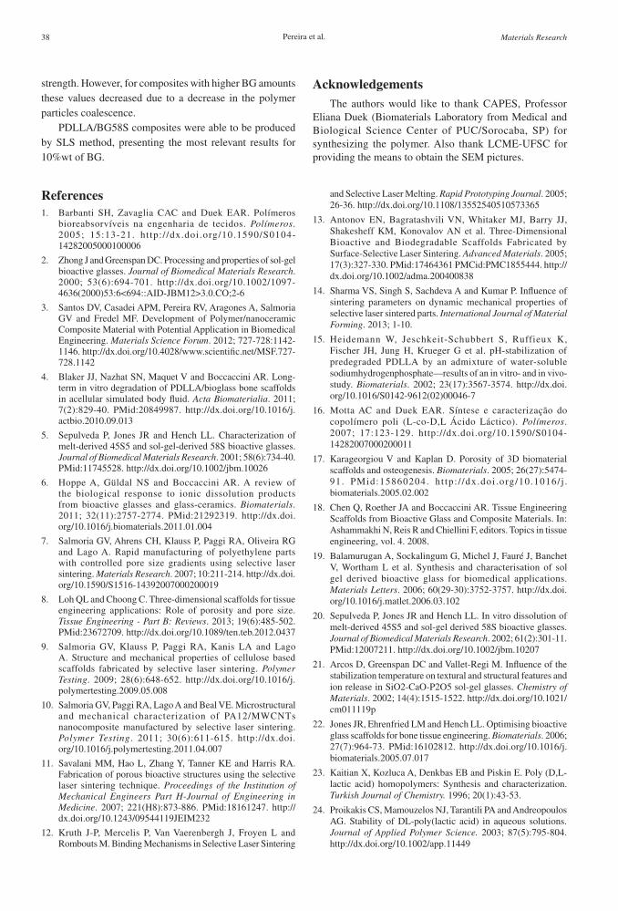
Each composition of samples was taken to SEM and micrographs of 50 and 200x of magnification were taken (Figures 8 and 9). (Research supported by LCME-UFSC.) As can be seen at composite micrographs, it is possible to observe that for pure polymer, necks were formed and particles sinterization occurred. The addition of bioglass particles (10, 20 and 30%wt of BG)
interfered at neck formation between the polymer particles, preventing polymer sinterization. These samples were also analyzed by computed tomography (Metrotom 1500, Zeiss, LABMETRO/UFSC), which confirmed the pores interconnectivity (data not shown).
4. ConclusionsBioglass 58S analyses by FTIR and XDR stated the
composition and structure compatible to the literature, showing that the BG synthesis by sol-gel process was accurate. FTIR and NMR of PDLLA attested the composition and structure of the synthetized polymer.
The composites were able to be produced via SLS method, presenting interconnected porous microstructure, specially, for pure polymer. Nevertheless less intense neck formation was observed with bioglass addition.
Stress-strain curves for PDLLA composites with 10%wt of BG presented greater values for flexural modulus and
Figure 5. 13C-NMR PDLLA spectrum.
Figure 6. DSC graphic of PDLLA. Figure 7. Stress-strain curve for PDLLA/BG58S composites.
Table 2. Name, composition and flexural properties values of the scaffolds.
Sample %wt of BG Eaverage(MPa) σaverage (3%ε) εrupture (%)
00BG 5,4 00 68.07±16.16 1.04±0.05 >25
10BG 5,4 10 79.00±23.75 1.68±0.45 >06
20BG 5,4 20 20.88±11.87 0.61±0.03 >04
30BG 5,4 30 10.09±08.39 0.22±0.16 >03
2014; 17(Suppl. 1) 37Scaffolds of PDLLA/Bioglass 58S Produced via Selective Laser Sintering
Figure 8. Micrographs with 50x of magnification. a) pure polymer; b) polymer with 10%wt of BG; c) polymer with 20%wt of BG; d) polymer with 30%wt of BG.
Figure 9. Micrographs with 200x of magnification. a) pure polymer; b) polymer with 10%wt of BG; c) polymer with 20%wt of BG; d) polymer with 30%wt of BG.
38 Pereira et al. Materials Research
strength. However, for composites with higher BG amounts these values decreased due to a decrease in the polymer particles coalescence.
PDLLA/BG58S composites were able to be produced by SLS method, presenting the most relevant results for 10%wt of BG.
AcknowledgementsThe authors would like to thank CAPES, Professor
Eliana Duek (Biomaterials Laboratory from Medical and Biological Science Center of PUC/Sorocaba, SP) for synthesizing the polymer. Also thank LCME-UFSC for providing the means to obtain the SEM pictures.
References1. Barbanti SH, Zavaglia CAC and Duek EAR. Polímeros
bioreabsorvíveis na engenharia de tecidos. Polímeros. 2005; 15:13-21. http:/ /dx.doi.org/10.1590/S0104-14282005000100006
2. Zhong J and Greenspan DC. Processing and properties of sol-gel bioactive glasses. Journal of Biomedical Materials Research. 2000; 53(6):694-701. http://dx.doi.org/10.1002/1097-4636(2000)53:6<694::AID-JBM12>3.0.CO;2-6
3. Santos DV, Casadei APM, Pereira RV, Aragones A, Salmoria GV and Fredel MF. Development of Polymer/nanoceramic Composite Material with Potential Application in Biomedical Engineering. Materials Science Forum. 2012; 727-728:1142-1146. http://dx.doi.org/10.4028/www.scientific.net/MSF.727-728.1142
4. Blaker JJ, Nazhat SN, Maquet V and Boccaccini AR. Long-term in vitro degradation of PDLLA/bioglass bone scaffolds in acellular simulated body fluid. Acta Biomaterialia. 2011; 7(2):829-40. PMid:20849987. http://dx.doi.org/10.1016/j.actbio.2010.09.013
5. Sepulveda P, Jones JR and Hench LL. Characterization of melt-derived 45S5 and sol-gel-derived 58S bioactive glasses. Journal of Biomedical Materials Research. 2001; 58(6):734-40. PMid:11745528. http://dx.doi.org/10.1002/jbm.10026
6. Hoppe A, Güldal NS and Boccaccini AR. A review of the biological response to ionic dissolution products from bioactive glasses and glass-ceramics. Biomaterials. 2011; 32(11):2757-2774. PMid:21292319. http://dx.doi.org/10.1016/j.biomaterials.2011.01.004
7. Salmoria GV, Ahrens CH, Klauss P, Paggi RA, Oliveira RG and Lago A. Rapid manufacturing of polyethylene parts with controlled pore size gradients using selective laser sintering. Materials Research. 2007; 10:211-214. http://dx.doi.org/10.1590/S1516-14392007000200019
8. Loh QL and Choong C. Three-dimensional scaffolds for tissue engineering applications: Role of porosity and pore size. Tissue Engineering - Part B: Reviews. 2013; 19(6):485-502. PMid:23672709. http://dx.doi.org/10.1089/ten.teb.2012.0437
9. Salmoria GV, Klauss P, Paggi RA, Kanis LA and Lago A. Structure and mechanical properties of cellulose based scaffolds fabricated by selective laser sintering. Polymer Testing. 2009; 28(6):648-652. http://dx.doi.org/10.1016/j.polymertesting.2009.05.008
10. Salmoria GV, Paggi RA, Lago A and Beal VE. Microstructural and mechanical characterization of PA12/MWCNTs nanocomposite manufactured by selective laser sintering. Polymer Testing. 2011; 30(6):611-615. http://dx.doi.org/10.1016/j.polymertesting.2011.04.007
11. Savalani MM, Hao L, Zhang Y, Tanner KE and Harris RA. Fabrication of porous bioactive structures using the selective laser sintering technique. Proceedings of the Institution of Mechanical Engineers Part H-Journal of Engineering in Medicine. 2007; 221(H8):873-886. PMid:18161247. http://dx.doi.org/10.1243/09544119JEIM232
12. Kruth J-P, Mercelis P, Van Vaerenbergh J, Froyen L and Rombouts M. Binding Mechanisms in Selective Laser Sintering
and Selective Laser Melting. Rapid Prototyping Journal. 2005; 26-36. http://dx.doi.org/10.1108/13552540510573365
13. Antonov EN, Bagratashvili VN, Whitaker MJ, Barry JJ, Shakesheff KM, Konovalov AN et al. Three-Dimensional Bioactive and Biodegradable Scaffolds Fabricated by Surface-Selective Laser Sintering. Advanced Materials. 2005; 17(3):327-330. PMid:17464361 PMCid:PMC1855444. http://dx.doi.org/10.1002/adma.200400838
14. Sharma VS, Singh S, Sachdeva A and Kumar P. Influence of sintering parameters on dynamic mechanical properties of selective laser sintered parts. International Journal of Material Forming. 2013; 1-10.
15. Heidemann W, Jeschkeit-Schubbert S, Ruffieux K, Fischer JH, Jung H, Krueger G et al. pH-stabilization of predegraded PDLLA by an admixture of water-soluble sodiumhydrogenphosphate—results of an in vitro- and in vivo-study. Biomaterials. 2002; 23(17):3567-3574. http://dx.doi.org/10.1016/S0142-9612(02)00046-7
16. Motta AC and Duek EAR. Síntese e caracterização do copolímero poli (L-co-D,L Ácido Láctico). Polímeros. 2007; 17:123-129. http://dx.doi.org/10.1590/S0104-14282007000200011
17. Karageorgiou V and Kaplan D. Porosity of 3D biomaterial scaffolds and osteogenesis. Biomaterials. 2005; 26(27):5474-91. PMid:15860204. h t tp : / /dx.doi .org/10.1016/ j .biomaterials.2005.02.002
18. Chen Q, Roether JA and Boccaccini AR. Tissue Engineering Scaffolds from Bioactive Glass and Composite Materials. In: Ashammakhi N, Reis R and Chiellini F, editors. Topics in tissue engineering, vol. 4. 2008.
19. Balamurugan A, Sockalingum G, Michel J, Fauré J, Banchet V, Wortham L et al. Synthesis and characterisation of sol gel derived bioactive glass for biomedical applications. Materials Letters. 2006; 60(29-30):3752-3757. http://dx.doi.org/10.1016/j.matlet.2006.03.102
20. Sepulveda P, Jones JR and Hench LL. In vitro dissolution of melt-derived 45S5 and sol-gel derived 58S bioactive glasses. Journal of Biomedical Materials Research. 2002; 61(2):301-11. PMid:12007211. http://dx.doi.org/10.1002/jbm.10207
21. Arcos D, Greenspan DC and Vallet-Regi M. Influence of the stabilization temperature on textural and structural features and ion release in SiO2-CaO-P2O5 sol-gel glasses. Chemistry of Materials. 2002; 14(4):1515-1522. http://dx.doi.org/10.1021/cm011119p
22. Jones JR, Ehrenfried LM and Hench LL. Optimising bioactive glass scaffolds for bone tissue engineering. Biomaterials. 2006; 27(7):964-73. PMid:16102812. http://dx.doi.org/10.1016/j.biomaterials.2005.07.017
23. Kaitian X, Kozluca A, Denkbas EB and Piskin E. Poly (D,L-lactic acid) homopolymers: Synthesis and characterization. Turkish Journal of Chemistry. 1996; 20(1):43-53.
24. Proikakis CS, Mamouzelos NJ, Tarantili PA and Andreopoulos AG. Stability of DL-poly(lactic acid) in aqueous solutions. Journal of Applied Polymer Science. 2003; 87(5):795-804. http://dx.doi.org/10.1002/app.11449


![Porous poly(α-hydroxyacid)/bioglass composite scaffolds ...application in tissue engineering [1-3]. Composite scaffolds may prove necessary for reconstruction of multi-tissue organs,](https://static.fdocument.org/doc/165x107/5e3f1725786dcc56c068fc14/porous-poly-hydroxyacidbioglass-composite-scaffolds-application-in-tissue.jpg)