Role of the WNT/ -...
143
NISHA BALSARA Role of the WNT/β-catenin signal pathway in idiopathic and experimental pulmonary fibrosis INAUGURAL-DISSERTATION zur Erlangung des Grades eines Dr. med. vet. beim Fachbereich Veterinärmedizin der Justus-Liebig-Universität Gießen VVB LAUFERSWEILER VERLAG édition scientifique
Transcript of Role of the WNT/ -...
NISH
A B
ALSA
RA
R
OLE O
F W
NT/β
-CA
TEN
IN
IN
IP
F
VVB
VVB LAUFERSWEILER VERLAGSTAUFENBERGRING 15D-35396 GIESSEN
Tel: 0641-5599888 Fax: [email protected]
VVB LAUFERSWEILER VERLAGédition scientifique
9 7 8 3 8 3 5 9 6 0 5 4 1
ISBN: 978-3-8359-6054-1
NISHA BALSARA
Role of the WNT/β-catenin signal pathway in
idiopathic and experimental pulmonary fibrosis
INAUGURAL-DISSERTATION zur Erlangung des Grades eines Dr. med. vet.
beim Fachbereich Veterinärmedizin der Justus-Liebig-Universität Gießen
VVB LAUFERSWEILER VERLAGédition scientifique

Das Werk ist in allen seinen Teilen urheberrechtlich geschützt.
Jede Verwertung ist ohne schriftliche Zustimmung des Autors oder des Verlages unzulässig. Das gilt insbesondere für Vervielfältigungen, Übersetzungen, Mikroverfilmungen
und die Einspeicherung in und Verarbeitung durch elektronische Systeme.
1. Auflage 2013
All rights reserved. No part of this publication may be reproduced, stored in a retrieval system, or transmitted,
in any form or by any means, electronic, mechanical, photocopying, recording, or otherwise, without the prior
written permission of the Author or the Publishers.
st1 Edition 2013
© 2013 by VVB LAUFERSWEILER VERLAG, Giessen
Printed in Germany
VVB LAUFERSWEILER VERLAG
STAUFENBERGRING 15, D-35396 GIESSENTel: 0641-5599888 Fax: 0641-5599890
email: [email protected]
www.doktorverlag.de
édition scientifique
aus dem Institut für Veterinär-Physiologie
Fachbereich Veterinärmedizin
Justus-Liebig-Universität Gießen
Betreuer: Prof. Dr. Rüdiger Gerstberger
und
aus dem Zentrum für Innere Medizin
Medizinische Klinik und Poliklinik II,
Fachbereich Humanmedizin
Universitätsklinikum Gießen und Marburg - Standort Gießen
Betreuer: Prof. Dr. Oliver Eickelberg
Role of the WNT/β-catenin signal pathway in
idiopathic and experimental pulmonary fibrosis
INAUGURAL-DISSERTATION
zur Erlangung des Grades eines
Dr. med. vet.
beim Fachbereich Veterinärmedizin
der Justus-Liebig-Universität Giessen
eingereicht von
Nisha Balsara Tierärztin aus Offenbach am Main
Giessen 2013

3
mit Genehmigung des Fachbereichs Veterinärmedizin
der Justus-Liebig-Universität Gießen
Dekan: Prof. Dr. Dr. h.c. M. Kramer
Gutachter: Prof. Dr. R. Gerstberger
Prof. Dr. O. Eickelberg
Tag der Disputation: 17.06.2013

4
FOR MY PARENTS

5
I TABLE OF CONTENTS
I TABLE OF CONTENTS............................................................................................................5
II LIST OF FIGURES ....................................................................................................................8
III LIST OF TABLES .................................................................................................................... 10
IV LIST OF ABBREVIATIONS................................................................................................... 11
V SUMMARY ............................................................................................................................... 19
VI ZUSAMMENFASSUNG........................................................................................................... 21
1 INTRODUCTION..................................................................................................................... 23
1.1 Diffuse parenchymal lung diseases........................................................................................ 23
1.2 Idiopathic pulmonary fibrosis................................................................................................ 24
1.2.1 Definition and epidemiology ..................................................................................... 24
1.2.2 Clinical features, radiological characteristics and therapy options ............................ 26
1.2.3 Histopathological characteristics ............................................................................... 27
1.2.4 Pathomechanism of idiopathic pulmonary fibrosis .................................................... 27
1.3 WNT signalling ..................................................................................................................... 31
1.3.1 The canonical WNT/β-catenin signalling pathway.................................................... 31
1.4 WNT signalling in the lung ................................................................................................... 34
1.4.1 Lung development and homeostasis .......................................................................... 34
1.4.2 Lung cancer................................................................................................................ 36
1.5 WNT signalling in fibrotic disorders ..................................................................................... 37
2 AIMS OF THE STUDY ............................................................................................................ 38
3 MATERIALS AND METHODS.............................................................................................. 39
3.1 Materials ................................................................................................................................ 39
3.1.1 Equipment.................................................................................................................. 39
3.1.2 Chemical reagents ...................................................................................................... 40
3.1.3 Antibodies .................................................................................................................. 42
3.1.4 Recombinant proteins ................................................................................................ 43
3.1.5 Human tissues ............................................................................................................ 43
3.1.6 Animal tissues............................................................................................................ 44
3.1.7 Primary cells .............................................................................................................. 44
3.2 Methods ................................................................................................................................. 44
3.2.1 Animal model of bleomycin-induced pulmonary fibrosis in mice............................. 44
3.2.2 WNT reporter mice (TOPGAL)................................................................................. 45
3.2.3 Isolation of primary mouse alveolar epithelial type II (ATII) cells ........................... 45
3.2.4 Supernatants............................................................................................................... 48
6
3.2.5 RNA isolation and measurement ............................................................................... 48
3.2.5.1 RNA isolation from lung homogenates................................................................. 48
3.2.5.2 RNA isolation from cultured cells......................................................................... 48
3.2.6 cDNA synthesis ......................................................................................................... 49
3.2.7 Quantitative real-time polymerase chain reaction (qRT-PCR) .................................. 50
3.2.8 DNA agarose gel electrophoresis............................................................................... 52
3.2.9 Immunohistochemistry .............................................................................................. 52
3.2.10 Immunocytochemistry ............................................................................................... 54
3.2.11 Western blot analysis ................................................................................................. 55
3.2.12 [3H]-thymidine proliferation assay............................................................................. 57
3.2.13 Whole genome microarray......................................................................................... 58
3.2.14 Enzyme linked-immuno-sorbent assay (ELISA) ....................................................... 58
3.2.15 Statistical analysis ...................................................................................................... 59
4 RESULTS................................................................................................................................... 60
4.1 Analysis of the canonical WNT/β-catenin signalling pathway in idiopathic pulmonary
fibrosis ................................................................................................................................... 60
4.1.1 Expression of the canonical WNT/β-catenin signalling pathway in idiopathic
pulmonary fibrosis...................................................................................................... 60
4.1.2 Localisation of the canonical WNT/β-catenin signalling pathway in idiopathic
pulmonary fibrosis...................................................................................................... 63
4.1.3 Activity of the canonical WNT/β-Catenin signalling pathway in idiopathic pulmonary
fibrosis........................................................................................................................ 72
4.2 Analysis of the canonical WNT/β-catenin signalling pathway in experimental pulmonary
fibrosis ................................................................................................................................... 74
4.2.1 Expression of the canonical WNT/β-catenin signalling pathway in experimental
pulmonary fibrosis...................................................................................................... 74
4.2.2 Localisation of the WNT/β-catenin signalling pathway in experimental pulmonary
fibrosis........................................................................................................................ 77
4.3 Functional analysis of the WNT/β-catenin signalling pathway ............................................. 87
4.3.1 Purity and phenotype of primary mouse alveolar epithelial type II (ATII) cells ....... 88
4.3.2 Expression of Wnt3a-induced target genes in primary mouse alveolar epithelial type
II (ATII) cells ............................................................................................................. 91
4.3.3 Proliferative effects of Wnt3a stimulation on primary mouse alveolar epithelial type
II (ATII) cells ............................................................................................................. 92
4.3.4 Whole genome microarray analysis on Wnt3a-induced primary mouse alveolar
epithelial type II (ATII) cells...................................................................................... 94
4.4 Analysis of the novel WNT target genes IL1β and IL6 in vitro and in vivo .......................... 98
4.4.1 ELISA analysis for quantification of IL1β and IL6 ................................................... 98
5 DISCUSSION .......................................................................................................................... 103
7
5.1 Conclusions and future perspectives.................................................................................... 116
6 APPENDIX .............................................................................................................................. 118
6.1 List of human (q)RT-PCR primers ...................................................................................... 118
6.2 List of mouse (q)RT-PCR primers ...................................................................................... 121
6.3 Characteristics of IPF patients ............................................................................................. 124
6.4 List of antibodies ................................................................................................................. 125
6.4.1 Primary antibodies ................................................................................................... 125
6.4.2 Secondary antibodies ............................................................................................... 126
6.5 Microarray gene list............................................................................................................. 127
7 REFERENCES........................................................................................................................ 129
8 DECLARATION..................................................................................................................... 139
9 ACKNOWLEDGEMENTS.................................................................................................... 140
8
II LIST OF FIGURES
figure 1.1: Classification scheme of diffuse parenchymal lung diseases
(DPLDs)
figure 1.2: Survival curves in different interstitial lung diseases (ILDs)
figure 1.3: Origins of myofibroblasts in idiopathic pulmonary fibrosis (IPF)
figure 1.4: Original and new hypotheses for the pathogenesis of idiopathic
pulmonary fibrosis (IPF)
figure 1.5: An overview of the canonical WNT signalling pathway
figure 4.1: Gene expression profile of canonical WNT signalling components
in idiopathic (IPF)
figure 4.2.1 +
figure 4.2.2: Localisation of WNT1 in idiopathic pulmonary fibrosis (IPF)
figure 4.3.1 +
figure 4.3.2: Localisation of WNT3A in idiopathic pulmonary fibrosis (IPF)
figure 4.4.1 +
figure 4.4.2: Localisation of GSK3β in idiopathic pulmonary fibrosis (IPF)
figure 4.5.1 +
figure 4.5.2: Localisation of CTNNB1 in idiopathic pulmonary fibrosis (IPF)
figure 4.6: Protein expression of canonical WNT signalling components in
idiopathic pulmonary fibrosis (IPF)
figure 4.7: Gene expression profile of canonical WNT signalling components
in experimental pulmonary fibrosis
figure 4.8.1 +
figure 4.8.2: Localisation of Wnt1 in experimental pulmonary fibrosis
figure 4.9.1 +
figure 4.9.2: Localisation of Wnt3a in experimental pulmonary fibrosis
figure 4.10.1 +
figure 4.10.2: Localisation of Gsk3β in experimental pulmonary fibrosis
figure 4.11.1 +
figure 4.11.2: Localisation of Ctnnb1 in experimental pulmonary fibrosis
9
figure 4.12: Gene expression profile of canonical WNT signalling components
in alveolar epithelial type II (ATII) cells from bleomycin-induced
mouse lungs
figure 4.13
(A+B): Purity and phenotype of primary mouse alveolar epithelial type II
(ATII) cells
figure 4.14: mRNA expression of Wnt3a-induced target genes of the canonical
WNT/β-catenin signalling pathway in primary mouse alveolar
epithelial type II (ATII) cells
figure 4.15: Proliferative effect induced by Wnt3a in primary mouse alveolar
epithelial type II (ATII) cells
figure 4.16: Whole genome expression analysis
figure 4.17: mRNA expression of potential Wnt3a-induced target genes in
primary mouse alveolar epithelial type II (ATII) cells
figure 4.18: Protein quantification in supernatants from Wnt3a-stimulated
primary mouse alveolar epithelial type II (ATII) cells
figure 4.19: Protein quantification in conditioned media from bleomycin-treated
primary mouse alveolar epithelial type II (ATII) cells
figure 4.20: Protein quantification in mouse bronchoalveolar lavage fluids
(BALFs)
figure 4.21: Protein quantification in human bronchoalveolar lavage fluids
(BALFs)
10
III LIST OF TABLES
table 3.1: Composition of culture media for isolation of primary mouse
alveolar epithelial type II (ATII) cells
table 3.2: Composition of mixture for complementary deoxyribonucleic acid
(cDNA) synthesis
table 3.3: Composition of mixture for quantitative reverse transcription real
time polymerase chain reaction ((q)RT-PCR)
table 6.1: Human primer sequences and amplicon sizes
table 6.2: Mouse primer sequences and amplicon sizes
table 6.3: Characteristics of idiopathic pulmonary fibrosis (IPF) patients with
usual interstitial pneumonia (UIP) pattern
table 6.4.1: List of primary antibodies
table 6.4.2: List of secondary antibodies
table 6.5: List of selected target genes found in the microarray
11
IV LIST OF ABBREVIATIONS
α Alpha (first letter of the Greek alphabet)
β Beta (second letter of the Greek alphabet)
γ Gamma (third letter of the Greek alphabet)
∆ Delta (forth letter of the Greek alphabet)
λ Lambda ( eleventh letter of the Greek alphabet)
µCi microcurie (1 µCi = 3.7x10⁴ dpm; measure/unit of
radioactivity)
µg microgram (10-6 g)
µl microlitre (10-6 l)
µm micrometre (10-6 m)
< less-than sign
- minus sign (mathematical symbol for negative/less)
+ plus sign (mathematical symbol for positive/more)
% percentage (per cent = “per hundred”)
A
A549 adenocarcinomic human alveolar basal epithelial cells
(a cell line)
ACTA2 alpha smooth muscle actin (α-SMA, a protein)
AIP acute interstitial pneumonia
ANGII angiotensin II (oligopeptide/hormone)
AP1 activating protein 1
APC adenomatous polyposis coli (protein)
APS ammonium persulfate
ATI alveolar epithelial type I cell (type I pneumocyte)
ATII alveolar epithelial type II cell (type II pneumocyte)
ATS American Thoracic Society
AXIN2 axis inhibition protein 2 or conductin (protein)
AZ “Aktenzeichen” (= file reference)
12
B
β-TrCP E3 ubiquitin ligase β-transducin repeat-containing
protein
BALF bronchoalveolar lavage fluid
bp base pair
BSA bovine serum albumin
BW body weight
C
C57BL/6 common inbred strain of laboratory mice
("C57 black 6")
°C degree Celsius
Ca2+ calcium
CamKII calmodulin kinase II
cAMP cyclic adenosine monophosphate
Cdh1 E-cadherin
CD cluster of differentiation (used for identification of
cell surface molecules)
cDNA complementary deoxyribonucleic acid
CFA cryptogenic fibrosing alveolitis
CK casein kinase
CK1γ casein kinase 1γ (serine/threonine protein kinase)
CO carbon monoxide
CO2 carbon dioxide
COP cryptogenic organising pneumonia
CRD cysteine-rich domain
CREB cAMP response element-binding (cellular
transcription factor)
Ct cycle of threshold in (q)RT-PCR
CTGF connective tissue growth factor
CTNNB1 β-catenin
Cy cyanine (synthetic fluorescent dyes)
13
D
DAPI 4´,6-diamidino-2-phenylindole
ddH2O double distilled water
DIP desquamative interstitial pneumonia
Dkk dickkopf
DLCO diffusing capacity of the lung for carbon monoxide
DLT double lung transplantation
DMEM Dulbecco's modified eagle medium
DNA deoxyribonucleic acid
DNase deoxyribonuclease (enzyme/nuclease)
dNTP deoxynucleoside triphosphate
DPBS Dulbecco's phosphate buffered saline
DPLD diffuse parenchymal lung disease
dpm disintegrations per minute (measure of radioactivity)
dsDNA doublestranded deoxyribonucleic acid
DVL dishevelled
E
ECAD E-cadherin
ECM extracellular matrix
EDTA ethylenediaminetetraacetic acid
e.g. for example
EGTA ethylene glycol tetraacetic acid
ELISA enzyme linked-immuno-sorbent assay
EMT epithelial-to-mesenchymal transition
ERS European Thoracic Society
et al. and others (et alii)
F
FAP familial adenomatous polyposis
FBS fetal bovine serum
f.c. final concentration
Fc fragment crystallizable region (region of an antibody)
FCS fetal calf serum
14
FZD frizzled
FITC fluorescein-5-isothiocyanate
G
g g-force (m/sec2, acceleration)/gramm
GBP GSK binding protein
GSK3β glycogen synthase kinase 3 beta
GTP guanosine-5'-triphosphate (purine nucleotide)
H
h hours 3H tritium/hydrogen-3 (a radioactive isotope)
HDAC histone deacetylases
HEPES 4-(2-hydroxyethyl)-1-piperazine ethane sulfonic acid
(buffering agent)
HPRT hypoxanthine phosphoribosyltransferase
HRCT high-resolution chest computed tomography
HRP horseradish peroxidase
I
IF immunofluorescence
IgG immunoglobulin G (antibody molecules)
IHC immunohistochemistry
IL interleukin
IL1β interleukin 1 beta
IL1R interleukin 1 receptor
IL1RA interleukin 1 receptor accessory protein
ILD interstitial lung disease
IIP idiopathic interstitial pneumonia
int-1 integration 1
i.p. intraperitoneal
IPF idiopathic pulmonary fibrosis
15
J
JNK c-Jun N-terminal kinase
K
kDA kilo Dalton
kg kilogram
KGF keratinocyte growth factor
L
l liter
LacZ structural gene encoding β-galactosidase
LAM lymphangioleiomyomatosis
LEF lymphoid enhancer-binding factor
LIP lymphocytic interstitial pneumonia
log logarithm
LRP lipoprotein receptor-related protein
M
mg milligram (10-3 g)
MgCl2 magnesium chloride
min. minutes
ml milliliter (10-3 l)
mM millimolar (a unit of concentration)
mm millimeter (10-3 m)
mmHg millimetre of mercury
MMP matrix metalloproteinase
MMTV mouse mammary tumor virus
mRNA messenger RNA
MuLVs murine leukemia viruses (retroviruses)
N
NaCl sodium chloride
NaOH sodium hydroxide
ng nanogram (10-9 g)
16
nm nanometer (10-9 m)
NOV nephroblastoma overexpressed
NSCLC non-small cell lung cancer
NSIP nonspecific interstitial pneumonia
O
O2 oxygen
OCLN occluding (tight junction protein)
OD optical density
P
p p-value (probability of obtaining a test statistic)
panCK pan-cytokeratin
PaCO2 partial pressure of carbon dioxide
PaO2 partial pressure of oxygen
Pbgd porphobilinogen deaminase
PBS phosphate-buffered saline
PBST phosphate-buffered saline + 0.1 % Tween 20
PCP planar cell polarity
PCR polymerase chain reaction
PFA paraform aldehyde
pg picogram (10-12 g)
pH measure of the acidity or basicity of an aqueous
solution
PKA protein kinase A
PKC protein kinase C
Q
qRT-PCR quantitative reverse transcription real time PCR
R
Rac protein of the family of GTPases
RB-ILD respiratory bronchiolitis-associated interstitial lung
disease
17
Rho protein of the family of GTPases
rmWnt3a recombinant mouse protein Wnt3a
RNA ribonucleic acid
RNase ribonuclease (enzyme/nuclease)
ROX an internal fluorescence dye
RT reverse transcriptase/transcription
RT-PCR reverse transcription PCR
S
SDS sodium dodecyl sulfate
SDS-PAGE sodium dodecyl sulfate polyacrylamide gel
electrophoresis
sem standard error of mean
SLT single lung transplantation
SMA smooth muscle actin
SP-C surfactant protein C
SPF specific pathogen-free
SPP1 secreted phosphoprotein 1/osteopontin
ssDNA single stranded
SYBR Green I N',N'-dimethyl-N-[1]-1-phenylquinolin-1-ium-2-yl]-
N-propylpropane-1,3-diamine (cyanine dye)
T
TAE tris-acetate-EDTA
TBST tris-buffered saline and Tween 20 (mixture)
TCF T-cellspecific transcription factor
TEMED N,N,N,N'-tetramethyl-ethane-1,2-diamine
TGFβ1 transforming growth factor β1
TIMPs tissue inhibitors of metalloproteinases
Tjp1 tight junction protein 1
TLC total lung capacity
TNF tumor necrosis factor
TRIS tris(hydroxymethyl)-aminomethan
t-test Student’s t-test (statistical hypothesis test)
18
Tween20 polysorbate 20 (polysorbate surfactant)
U
U units
UDG uracil-DNA glycosylase
UIP usual interstitial pneumonia
USA United States of America
UV ultraviolet (light)
V
VA alveolar volume
VC vital capacity
v/v volume fraction (volume/volume)
W
WB Western blot
Wg wingless
Wisp Wnt1-inducible signalling protein
Wnt signal protein (hybrid of Wg and Int)
w/v mass concentration (mass/volume)
X
Y
Z
Further, not listed abbreviations are taken from the valid IUPAC-Nomenclature.
19
V SUMMARY
Human idiopathic pulmonary fibrosis (IPF) is a progressive and fatal lung disease of
unknown origin, which is refractory to any currently available therapy. It is
characterised by initial alveolar epithelial cell injury and hyperplasia, enhanced
fibroblast/myofibroblast proliferation and activation and increased deposition of
extracellular matrix (ECM) in the lung interstitium. These key features, ultimately
lead to architectural distortion of the normal lung parenchyma and due to severe loss
of function, to respiratory failure. However, the molecular mechanisms underlying
alveolar epithelial type II (ATII) cell dysfunction are still poorly understood.
This study is based on the hypothesis that the WNT/β-catenin signalling pathway,
which is essential for organ development, is aberrantly activated in ATII cells in
idiopathic and experimental pulmonary fibrosis. The role of canonical WNT
signalling was elucidated by determining the expression, function and activity of the
pathway.
An increased expression of most WNT/β-catenin signalling components was
demonstrated in pulmonary fibrosis. The mRNA expression was analyzed in lung
homogenates of IPF and donor patients, as well as in lung homogenates and ATII
cells isolated from the lungs of bleomycin- and saline-treated mice.
Immunohistochemical analysis localized increased protein expression of WNT
signalling largely in alveolar and bronchial epithelium of IPF patients, as well as of
bleomycin-treated mice. This result was confirmed by quantitative real-time (q)RT-
PCR of primary human and mouse ATII cells, demonstrating an increase of cell-
specific functional WNT signalling. To elucidate active WNT/β-catenin signalling in
IPF, western blot analysis demonstrated that several WNT components were
expressed and functional in ATII cells. Furthermore, WNT3A treatment significantly
increased ATII cell proliferation. WNT3A treatment of ATII cells in vitro caused a
significantly increased expression of WNT target genes and, most interestingly, the
interleukins IL1β and IL6. The occurrence of IL1β and IL6 in conjunction with
increased WNT/β-catenin signalling was corroborated in primary ATII cells isolated
from the lungs of bleomycin- and saline-treated mice and in vivo in BALF by
orotracheal application of WNT3A. In addition, IL1β and IL6 were increased in
BALF samples from IPF patients.

20
In summary, this study shows that WNT signalling is expressed and localised in
idiopathic and experimental pulmonary fibrosis, in particular in alveolar ATII cells.
Reversal and/or inhibition predominantly of the WNT/β-catenin signalling pathway,
but equally the use of neutralising antibodies or inhibitors of IL1β/IL6 may represent
a valid therapeutic option in human lung fibrosis.
21
VI ZUSAMMENFASSUNG
Die häufigste und schwerwiegendste Form der idiopathischen interstitiellen
Pneumonien (IIP) beim Menschen ist die idiopathische pulmonale Fibrose (IPF). Es
handelt sich um eine therapierefraktäre, progressiv und tödlich verlaufende
Lungenerkrankung mit unbekannter Ätiologie. Die initiale Schädigung des
Alveolarepithels und eine daraus resultierende abnormale Wundheilung
charakterisieren diese Erkrankung. Eine verstärkte Proliferation und Aktivierung von
Fibroblasten/Myofibroblasten und die vermehrte Ansammlung von extrazellulärer
Matrix (ECM) im Lungeninterstitium sind zusätzliche Begleiterscheinungen. Dies
führt letztendlich zu einem kompletten Umbau bzw. einer Zerstörung des normalen
Lungengewebes und aufgrund des funktionellen Verlustes zum respiratorischen
Versagen. Die molekularen Mechanismen, die den veränderten Schädigungs- und
Reparaturprozessen des Alveolarepithels zugrunde liegen und demnach für die
Entwicklung von IPF verantwortlich sind, sind allerdings größtenteils noch unklar.
Der kanonische WNT Signaltransduktionsweg, benannt nach dem Liganden „WNT“,
hat in der Organentwicklung eine besondere Bedeutung, spielt aber auch eine
essentielle Rolle bei verschiedenen Erkrankungen. Das Signalprotein „WNT“ fungiert
als lokaler Mediator, und der Name setzt sich zusammen aus den Genen Wingless
(Wg) und Integration 1 (Int-1). Die vorliegende Arbeit basiert auf der Hypothese, dass
der WNT/β-catenin Signalweg in alveolaren Epithelzellen Typ II (ATII) in der
idiopathischen sowie experimentellen pulmonalen Fibrose differenziell reguliert und
aktiviert vorliegt.
In dieser Studie wurden neben humanem und murinem Lungenhomogenat, zusätzlich
ATII Zellen aus gesunden bzw. fibrotischen murinen Lungen isoliert und untersucht.
Mit Hilfe der quantitativen real-time reversen Transkriptase-Polymerase-
Kettenreaktion ((q)RT-PCR) konnte eine gesteigerte Expression des WNT
Signalweges nachgewiesen werden. Die verschiedenen WNT Komponenten, darunter
auch das Protoonkogen WNT1, waren auf Transkriptionsebene im humanen und
murinen fibrotischen Lungengewebe bzw. in daraus isolierten ATII Zellen zum Teil
signifikant reguliert. Dabei auffallend war allerdings eine deutlich verminderte
Expression des Proteins WNT3A, ein weiteres Mitglied der Sekretionsproteine aus
der WNT Familie. Darüber hinaus konnte mittels Immunohistochemie eine
22
gesteigerte Proteinexpression in Bronchial- und Alveolarepithelzellen von gesunden
und IPF Patienten sowie von gesunden und mit Bleomycin behandelten Mäusen,
lokalisiert werden. Neben WNT1 und WNT3A wurde auch β-catenin (CTNNB1), ein
intrazelluläres Schlüsselmolekül des WNT/β-catenin Signalweges, expremiert. Auch
der zusätzliche Nachweis in primären murinen ATII Zellen hebt die Zellspezifität des
Signalweges hervor. Ebenso konnte die Aktiverung des WNT/β-catenin Signalweges
in Lungengewebe von IPF Patienten durch Western Blot Analyse gezeigt werden.
Zusätzlich bewirkte der Einsatz von rekombinantem murinen Wnt3A Protein eine
zeitabhängige Regulation von bestimmten Wnt-spezifischen Zielgenen, wie cyclinD1
und WNT1-inducible-signaling pathway protein 1 (Wisp1), in primären murinen ATII
Zellen. Weitere Versuche ergaben, dass WNT3A eine signifikant erhöhte Expression
von bestimmten WNT-spezifischen Zielgenen bewirkte, welche unter anderem für die
Interleukine IL1β und IL6 kodieren. Der direkter Zusammenhang zwischen der
gesteigerten Expression von spezifischen Interleukinen und die Aktivierung des
WNT/β-catenin Signalweges konnte mit Hilfe eines Microarrays verdeutlicht werden.
Neben veränderten Genexpressionsprofilen, konnte bei primären murinen ATII
Zellen, die mit rekombinantem murinen Wnt3A Protein stimuliert wurden, auch eine
gesteigerte Zellproliferation beobachtet werden. Dabei wurden mittels [3H]-Thymidin
Inkorporation die Effekte auf Proliferationsebene untersucht. Diese Beobachtung
bestätigte sich in mit WNT3A stimulierten murinen ATII Zellen anhand von (q)RT-
PCR, sowie in bronchoalveolären Lavage (BALF) Proben von IPF Patienten und
Bleomycin behandelten Mäusen mittels ELISA.
Zusammenfassend konnte gezeigt werden, dass der WNT Signalweg bei der
idiopathischen und experimentellen Lungenfibrose, vornehmlich in ATII Zellen
lokalisiert sowie exprimiert wird. Demnach spielt dieser Signaltransduktionsweg eine
potentiell erhebliche Rolle im Rahmen der Pathogenese der Erkrankung. Eine
generelle Hemmung des WNT/β-catenin Signalweges, aber auch die spezifische
Verwendung von neutralisierenden IL1β/IL6 Antikörpern, könnte einen möglichen
therapeutischen Ansatz in der Behandlung der IPF darstellen.
23
1 INTRODUCTION
1.1 Diffuse parenchymal lung diseases
Diffuse parenchymal lung diseases (DPLDs), commonly also termed interstitial lung
diseases (ILDs), are a group of acute and chronic disorders that involve inflammation
and fibrosis of the alveoli, distal airways, and septal interstitium of the lungs,
subsequently leading to distortion of the normal lung architecture and respiratory
failure. In ILDs, the lung interstitium, primarily the space between the basement
membranes of epithelium and endothelium, is injured. ILDs consist of two different
subgroups: firstly, pulmonary disorders, which develop on the basis of known causes
and secondly, pulmonary disorders of unknown origin. Occupational or environmental
exposures, inherited conditions, drugs or infections are few of the possible known
causes associated with pulmonary disorders. Furthermore, ILDs occur in association
with systemical diseases, such as connective tissue diseases, collagen vascular
diseases, or sarcoidosis. The group of idiopathic pulmonary disorders comprises
eosinophilic pneumonia, pulmonary Langerhans’ cell histiocytosis/histiocytosis X and
lymphangioleiomyomatosis, as well as the more common idiopathic interstitial
pneumonias (IIPs) [1, 2].
The group of IIPs is divided into seven diverse entities: idiopathic pulmonary fibrosis
(IPF), non-specific interstitial pneumonia (NSIP), cryptogenic organizing pneumonia
(COP), acute interstitial pneumonia (AIP), respiratory bronchiololitis-associated
interstitial lung disease (RB-ILD), desquamative interstitial pneumonia (DIP) and
lymphoid interstitial pneumonia (LIP). The different forms are mainly differentiated
by clinical, radiological and histological features [1, 3].
24
1.2 Idiopathic pulmonary fibrosis
1.2.1 Definition and epidemiology
Idiopathic pulmonary fibrosis (IPF; also termed cryptogenic fibrosing alveolitis
(CFA)) is the most common and severe form among the IIPs. It is a chronically
progressive, devastating and life-threatening lung disease with unknown etiology, for
which no effective therapy exists at present [1, 4]. IPF occurs worldwide, more
commonly in male gender and the rates of prevalence and incidence are associated
predominantly with increasing age.
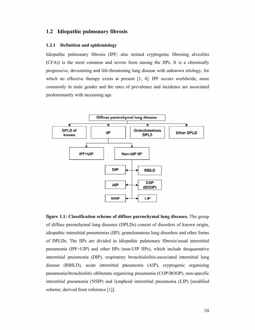
figure 1.1: Classification scheme of diffuse parenchymal lung diseases. The group
of diffuse parenchymal lung diseases (DPLDs) consist of disorders of known origin,
idiopathic interstitial pneumonias (IIP), granulomatous lung disorders and other forms
of DPLDs. The IIPs are divided in idiopathic pulmonary fibrosis/usual interstitial
pneumonia (IPF=UIP) and other IIPs (non-UIP IIPs), which include desquamative
interstitial pneumonia (DIP), respiratory bronchiololitis-associated interstitial lung
disease (RBILD), acute interstitial pneumonia (AIP), cryptogenic organizing
pneumonia/bronchiolitis obliterans organizing pneumonia (COP/BOOP), non-specific
interstitial pneumonia (NSIP) and lymphoid interstitial pneumonia (LIP) [modified
scheme; derived from reference [1]].
25
The rate of prevalence in the United States is estimated at 13 to 20 cases per 100,000
annually, suggesting that IPF is more common than previously assumed. The majority
of patients are between 50 to 70 years old (mean age over 65 years). The estimated
incidence rate per year is 7 to 10 cases per 100,000 in the United States, with
increasing tendency [5]. Many risk factors, like medication, chronic aspiration,
environmental exposures (e.g. metal/wood dust), infectious agents, bacterial or viral
infections (e.g. hepatitis C, adenovirus, Epstein - Barr virus), physical trauma
(mechanical ventilation) or genetic components have been suggested to correlate with
IPF. However, further investigations are required to reveal the coherence. There is no
evidence of race or ethnicity predispositions. At present, cigarette smoke, the most
potential risk factor, is strongly associated with IPF [4, 6-8]. IPF patients have a
significantly worse prognosis, due to unresponsiveness to currently available therapy
options. The median survival time is between 2.5 to 3.5 years from the time of
diagnosis [1, 9, 10].
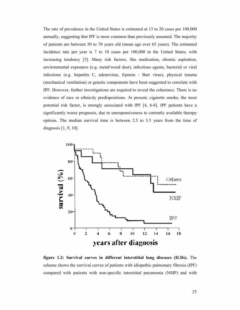
figure 1.2: Survival curves in different interstitial lung diseases (ILDs). The
scheme shows the survival curves of patients with idiopathic pulmonary fibrosis (IPF)
compared with patients with non-specific interstitial pneumonia (NSIP) and with
26
other diffuse parenchymal lung diseases. IPF patients had a significantly reduced
median survival time [modified scheme; derived from reference [3]].
1.2.2 Clinical features, radiological characteristics and therapy options
Clinically, the most prominent and typical symptoms an IPF patient presents are
dyspnoea and non-productive cough for a minimum period of 3 months. A chest
auscultation gives frequent evidence of Velcro-type crackles during the inspiration
phase in the basal parts of the lung. Often digital clubbing can be observed. An
analysis of bronchoalveolar lavage fluids (BALFs) shows a strong increase of
neutrophils [1, 4, 11, 12]. Recurrent respiratory infections and acute exacerbations are
frequent and often responsible for rapid deterioration. In the course of the disease, a
lung function test mostly depicts a restrictive pattern, as lung compliance is reduced
and impaired gas exchange is noticeable. These facts inevitably lead to decreased
physical capacity and loss of life quality. Respiratory failure is the most frequent
cause of death, and has been reported to account for over 80% of all fatalities;
bronchogenic carcinoma, ischemic heart disease, infection, and pulmonary embolism
are also common causes of mortality. In more advanced stages, IPF patients develop
severe pulmonary hypertension, peripheral edema and cor pulmonale, resulting in
right heart failure [1]. In chest radiographs and high-resolution chest computed
tomography (HRCT) peripheral, basal and subpleural parts of the lung are affected,
with involvement of other lung parts in advanced disease. A decreased lung volume,
bilateral reticular abnormalities, diffuse areas of “ground-glass” opacities associated
with traction bronchiectasis and peripheral “honeycombing” are characteristic
radiological findings in HRCT [1, 13-15].
The treatment strategies have been based on the concept that inflammation leads to
injury and fibrosis. Anti-inflammatory and immunosuppressive therapies are often
used in the treatment of IPF; however, such treatment has not been demonstrated to
improve survival or quality of life. Oral corticosteroids alone or in combination with
immunsuppressives, such as azathioprines and cyclophosphamides, usually used in
cases of other IIP forms, showed no or limited effects in IPF. Currently, as no other
alternatives are available, several studies preferably suggest a combination of low
dose prednisolone and azathioprine with concomitant antioxidant treatment [16].
Accordingly, a conventional therapy for IPF provides only marginal benefit [3, 17,
27
18]. It has to be pointed out that lung transplantation is currently the best and only
therapeutic intervention shown to prolong survival in IPF. Although the survival rate
is improved, many patients die, being waitlisted, due to rapidly progressive or severe
disease and additionally are often referred late in the course of their disease. Until
some years ago, single lung transplantation (SLT) has been the standard procedure for
patients with IPF and has produced good results. In the most recent years, a larger
application of double lung transplantation (DLT) has been observed worldwide,
probably related to higher survival rates. The mean life expectancy in patients with
IPF after lung transplantation is about 65-70% at 1 year and 40% at 5 years [19-22].
1.2.3 Histopathological characteristics
To diagnose IPF, it is important to perform a surgical lung biopsy [1]. IPF is
associated with a classic histopathological pattern - the usual interstitial pneumonia
(UIP). UIP is characterised by alternating areas of normal lung parenchyma parts,
interstitial inflammation, honeycomb cysts and fibrosis, resulting in dramatic
irreversible disruption of the normal lung architecture. The typical distribution of
pathological changes affects the subpleural, basal, and predominantly peripheral areas
of the lung [1, 23, 24]. Alveolar epithelial cell injury with hyperplastic type II cells
and abnormal regeneration/re-epithelialisation, as well as enhanced extracellular
matrix (ECM) deposition in the lung interstitium and increased
fibroblast/myofibroblast proliferation and activation, are observed as histological
abnormalities in IPF. Only mild inflammation is present in these areas. The hallmark
lesions of UIP/IPF are fibroblast foci, prominent aggregates of organised activated
myofibroblasts that localise next to hyperplastic type II cells. The number of
fibroblast foci has been reported, as an important factor for the individual prognosis of
a patient, to correlate with survival in IPF; increasing numbers are related to impaired
lung function, resulting in higher mortality [25, 26].
1.2.4 Pathomechanism of idiopathic pulmonary fibrosis
Even though the pathogenesis of IPF is still a topic of controversy, it is widely
accepted that the activated fibroblast/myofibroblast represents the key effector cell
type in IPF lungs [27, 28]. The role of fibroblasts implies the maintenance of matrix
28
homoeostasis, as well as synthesis and degradation of several extracellular molecules.
Myofibroblast accumulation, activation and impaired apoptosis are characteristic
features in the pathogenesis of IPF [29]. Finally, the structural remodelling leads to
the loss of alveolar function. Three hypotheses have been proposed, suggesting the
cellular origin of the activated myofibroblasts in IPF lungs. The relative contribution
of the possible pathways is still unclear [27, 30]. Initially, it was hypothesised that
profibrotic cytokines and growth factors influence resident lung fibroblasts to
proliferate and differentiate into myofibroblasts. Recently, a second possibility was
proposed, emphasising that epithelial cells undergo trans-differentiation into
fibroblasts by a process termed epithelial-mesenchymal transition (EMT). The third
theory suggests that circulating fibrocytes, derived from the bone marrow, may serve
as precursor cells for myofibroblasts. However, the evidence that fibrocytes are able
to differentiate into fully activated myofibroblasts is still insufficient [29].
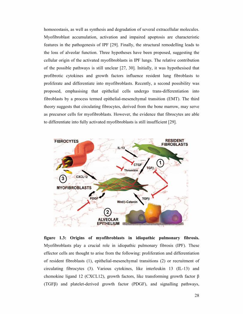
figure 1.3: Origins of myofibroblasts in idiopathic pulmonary fibrosis.
Myofibroblasts play a crucial role in idiopathic pulmonary fibrosis (IPF). These
effector cells are thought to arise from the following: proliferation and differentiation
of resident fibroblasts (1), epithelial-mesenchymal transitions (2) or recruitment of
circulating fibrocytes (3). Various cytokines, like interleukin 13 (IL-13) and
chemokine ligand 12 (CXCL12), growth factors, like transforming growth factor β
(TGFβ) and platelet-derived growth factor (PDGF), and signalling pathways,
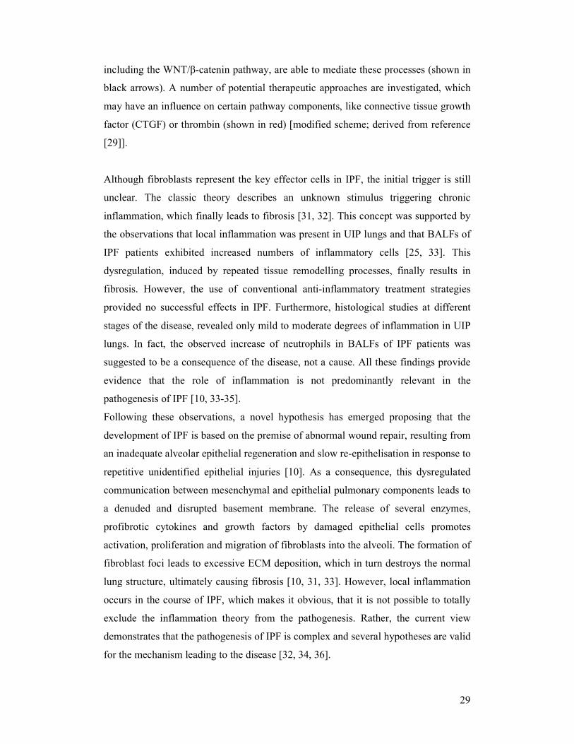
29
including the WNT/β-catenin pathway, are able to mediate these processes (shown in
black arrows). A number of potential therapeutic approaches are investigated, which
may have an influence on certain pathway components, like connective tissue growth
factor (CTGF) or thrombin (shown in red) [modified scheme; derived from reference
[29]].
Although fibroblasts represent the key effector cells in IPF, the initial trigger is still
unclear. The classic theory describes an unknown stimulus triggering chronic
inflammation, which finally leads to fibrosis [31, 32]. This concept was supported by
the observations that local inflammation was present in UIP lungs and that BALFs of
IPF patients exhibited increased numbers of inflammatory cells [25, 33]. This
dysregulation, induced by repeated tissue remodelling processes, finally results in
fibrosis. However, the use of conventional anti-inflammatory treatment strategies
provided no successful effects in IPF. Furthermore, histological studies at different
stages of the disease, revealed only mild to moderate degrees of inflammation in UIP
lungs. In fact, the observed increase of neutrophils in BALFs of IPF patients was
suggested to be a consequence of the disease, not a cause. All these findings provide
evidence that the role of inflammation is not predominantly relevant in the
pathogenesis of IPF [10, 33-35].
Following these observations, a novel hypothesis has emerged proposing that the
development of IPF is based on the premise of abnormal wound repair, resulting from
an inadequate alveolar epithelial regeneration and slow re-epithelisation in response to
repetitive unidentified epithelial injuries [10]. As a consequence, this dysregulated
communication between mesenchymal and epithelial pulmonary components leads to
a denuded and disrupted basement membrane. The release of several enzymes,
profibrotic cytokines and growth factors by damaged epithelial cells promotes
activation, proliferation and migration of fibroblasts into the alveoli. The formation of
fibroblast foci leads to excessive ECM deposition, which in turn destroys the normal
lung structure, ultimately causing fibrosis [10, 31, 33]. However, local inflammation
occurs in the course of IPF, which makes it obvious, that it is not possible to totally
exclude the inflammation theory from the pathogenesis. Rather, the current view
demonstrates that the pathogenesis of IPF is complex and several hypotheses are valid
for the mechanism leading to the disease [32, 34, 36].
30
As a result of the complex pathogenesis mentioned above, IPF can be described as a
disease of disturbed epithelial-mesenchymal crosstalk. Epithelial-mesenchymal
interactions are responsible for the maintenance of the alveolar unit, and are essential
for normal lung function and gas exchange. However, an impaired crosstalk between
alveolar epithelial type (AT) II cells and fibroblasts has recently been shown to
contribute to the pathomechanism of IPF [36]. Several soluble mediators, such as
transforming growth factor (TGF)β1, angiotensin II, or interleukin (IL)1β, were
released by ATII cells and have been assigned a clear pathogenic role in experimental
and idiopathic pulmonary fibrosis [37, 38]. Most recently, the WNT target gene
WISP1 has been identified as a novel mediator of epithelial-mesenchymal interaction
[39]. The WNT signalling pathway is essential to organ development, a process that
can be recapitulated in organ failure. An aberrant activation of the WNT/β-catenin
signalling pathway during adult homeostasis leads to pathological events resulting in
cancer, but may also be associated with the development of IPF.
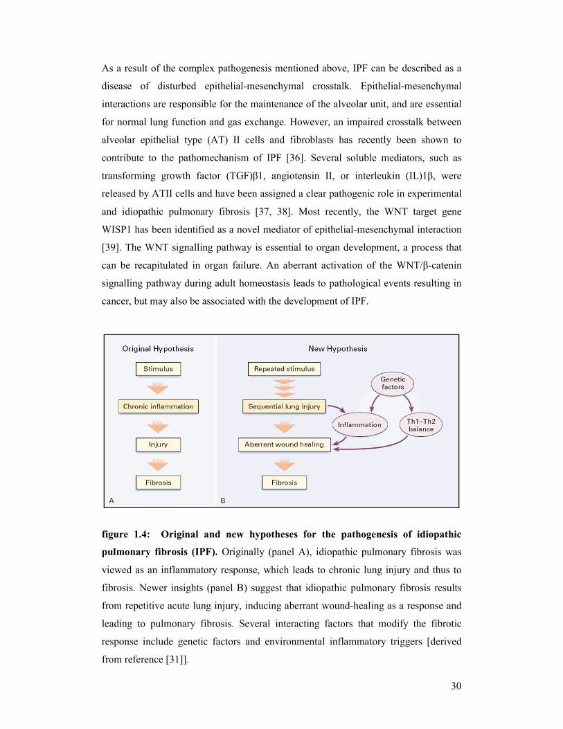
figure 1.4: Original and new hypotheses for the pathogenesis of idiopathic
pulmonary fibrosis (IPF). Originally (panel A), idiopathic pulmonary fibrosis was
viewed as an inflammatory response, which leads to chronic lung injury and thus to
fibrosis. Newer insights (panel B) suggest that idiopathic pulmonary fibrosis results
from repetitive acute lung injury, inducing aberrant wound-healing as a response and
leading to pulmonary fibrosis. Several interacting factors that modify the fibrotic
response include genetic factors and environmental inflammatory triggers [derived
from reference [31]].
31
1.3 WNT signalling
WNT signalling plays a pivotal role during development of organisms as well as in
maintenance and regeneration during adulthood. Signal transduction in the cytoplasm
of different target cells leads to altered expression of various target genes and
subsequently can influence several cell functions, such as proliferation, migration,
differentiation, polarity, growth and cell fate specification [40-43]. Developmental
defects are caused as a consequence of disregulated active WNT signalling during
embryogenesis. Aberrant WNT signal transduction in adult tissue promotes various
diseases, including degenerative and inflammatory disorders, cancer and fibrosis [39,
43-45]. Currently, four different WNT signalling pathways are known: first, the
canonical WNT/β-catenin signalling pathway, which triggers WNT ligand binding to
cell surface receptors, resulting in β-catenin stabilisation and translocation to the
nucleus for target gene expression. Second, the non-canonical WNT/calcium
(WNT/Ca2+) signalling pathway, which activates protein kinase C (PKC) and
calmodulin kinase (CamKII) II by intracellular Ca2+ release. Third, the planar cell
polarity (PCP) pathway, also termed c-Jun N-terminal kinase (JNK) pathway, which
is a β-catenin independent and non-canonical pathway, like the WNT/Ca2+ signalling
pathway. It acts through small GTPases, like Rho/Rac, resulting in activation of
activating protein1 (AP1) and is implicated in cytoskeletal organisation and epithelial
cell polarity. Recently, a fourth pathway was discovered, particularly regulating
muscle development. Thereby, the transcription factor CREB is phosphorylated by
protein kinase A (PKA) [42, 44].
1.3.1 The canonical WNT/β-catenin signalling pathway
The best characterised WNT signalling pathway is the β-catenin dependent or
canonical WNT signalling pathway, which is the first discovered and best understood
pathway. The transcriptional regulator β-catenin acts as an intracellular key molecule
[42, 44, 45].
The canonical WNT/β-catenin signalling pathway is modulated by multiple
extracellular, cytoplasmic and nuclear signalling molecules. The WNT signalling
molecules belong to a large family of secreted glycoproteins that play an important
role as ligands in receptor-mediated signalling pathways. WNT proteins, generally
32
350 - 400 amino acids long, contain a signal domain followed by a highly conserved
sequence of cysteines, and are usually quite insoluble and hydrophobic due to
extensive palmitoylation of the mature protein. This post-translational modification is
critical for proper WNT signalling [45, 46]. In mammals 19 WNT members are
identified at present: WNT1, 2, 2B, 3, 3A, 4, 5A, 5B, 6, 7A, 7B, 8A, 8B, 9A, 9B,
10A, 10B, 11 and 16. Many WNT proteins share a high amino-acid sequence
homology, in particular those that are grouped within similar subfamilies. Basically,
the WNT genes have been divided into two classes, those that induce β-catenin
signalling, and those that activate other pathways. Known WNT ligands that trigger
the canonical pathway are: WNT1, 2, 2B, 3, 3A, 7A, 7B, 8, 10 [41, 42]. The first
representative of the WNT gene family was int-1 in mice and wingless in fruit flies
(Drosophila melanogaster). Int1 was first identified in a retrovirus causing mammary
tumors in mice (mouse mammary tumor virus; MMTV). The preferred integration
place for MMTV was the int-1 gene, a proto-oncogene. Wingless was named after the
phenotype of the Drosophila mutant, where the gene is defect and results in wingless
flies. By cloning and sequencing both these genes, a homology could be shown and
therefore the terms int-1 and wingless (wg) were combined to the new term WNT [47-
49].
In the absence of WNT ligands, the cytoplasmic β-catenin level is low because the
molecule is destined to 26S proteasome-mediated degradation by a so-called
degradation/destruction complex. This “destruction complex” is comprised of the
scaffolding protein axin and the adenomatous polyposis coli protein (APC) as well as
the glycogen synthase kinase 3β (GSK3β) as central elements. The serine/threonine
kinases GSK3β and casein kinase 1γ (CK1γ) are responsible for constitutive
phosphorylation of β-catenin, which is recognised by the E3 ubiquitin ligase β-
transducin repeat-containing protein (β-TrCP) and targeted for ubiquitination and
final proteosomal degradation [42, 45]. As a consequence, in the nucleus prospective
target genes of the pathway are repressed by interaction with members of the T-cell-
specific transcription factor/lymphoid enhancer-binding factor (TCF/LEF) family.
The TCF/LEF transcription factors form a complex with the associated co-repressor
Groucho. These repressing effects are mediated by histone deacetylases (HDAC) and
accordingly avoid transcription [42, 43]. When WNT ligands are present and WNT/β-
catenin signalling is activated, the degradation pathway is inhibited. In this case,
WNT proteins are released from the surface of signalling cells and act on target cells
33
by binding to two distinct membrane receptors, the frizzled (FZD) and low density
lipoprotein receptor-related proteins 5 and 6 (LRP5/6) at the cell surface [42, 45].
Thereby, the seven transmembrane receptor FZD and single pass transmembrane co-
receptor LRP5/6 build a complex. The FZD proteins are the corresponding primary
cell surface receptors for the WNT ligands. These seven transmembrane spanning
receptors belong to the family of serpentine receptors and possess a long extracellular
N-terminal extension, termed as cysteine-rich domain (CRD). WNT proteins directly
bind to the CRD. There are 10 FZD members currently known to interact with WNT
ligands [40, 46, 50]. In addition to the interactions between WNT and FZD, WNT
signalling also requires the presence of the long single pass transmembrane co-
receptor LRP5/6, which builds a complex with FZD. They are composed of a small
intracellular domain and a large extracellular domain. A hallmark of LRP6 is a
cytoplasmic tail with a central proline-rich motif (PPPSP). After phosphorylation by
GSK3β and CK1γ, this part serves as binding site for axin [41, 43, 51].
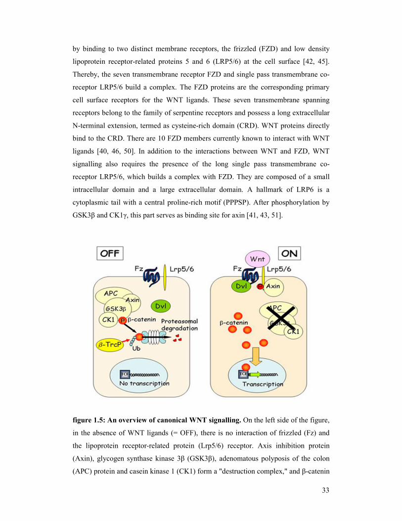
figure 1.5: An overview of canonical WNT signalling. On the left side of the figure,
in the absence of WNT ligands (= OFF), there is no interaction of frizzled (Fz) and
the lipoprotein receptor-related protein (Lrp5/6) receptor. Axis inhibition protein
(Axin), glycogen synthase kinase 3β (GSK3β), adenomatous polyposis of the colon
(APC) protein and casein kinase 1 (CK1) form a "destruction complex," and β-catenin
34
is phophorylated, which is recognised by the E3 ubiquitin ligase β-transducin repeat-
containing protein (β-TrcP) and targeted for ubiquitination (Ub) and final proteosomal
degradation. On the right side of the figure, in the presence of WNT ligands (= ON),
after binding to Fz, the signal is transduced to dishevelled (Dvl) and the Lrp5/6
receptor is activated. Axin is removed from the "destruction complex" and interacts
with Lrp5/6. Without Axin the degradation complex is inactive. β-catenin moves into
the nucleus, binds to the transcription factor TCF/LEF on DNA, and activates
transcription of a protein. "P" represents phosphate [scheme derived from
http://www.umcutrecht.nl/subsite/cmc-utrecht/People/Staff/Madelon-Maurice].
After the WNT ligands bind to the receptors, the signal is transduced to dishevelled
(DVL), another intracellular pathway protein with the ability to interact with Axin. In
detail, WNT activation promotes phosphorylation of LRP5/6 by GSK3β and CK1γ,
which leads to the recruitment of Axin to the plasma membrane, directly binding to
the cytoplasmic tail of LRP5/6. Without Axin the degradation complex is inactive. At
the same time GSK binding protein (GBP) binds to GSK3β and prevents the
interaction between GSK3β and Axin. β-catenin is not phosphorylated anymore and
therefore cannot be recognised by β-TrCP. Consequently, hypophosphorylated β-
catenin accumulates in the cytoplasm. The increased stability and elevated levels of
free β-catenin lead to its nuclear translocation and subsequently β-catenin induces
transcription of WNT target genes mediated by interactions with TCF/LEF
transcription factors. Once in the nucleus, β-catenin replaces Groucho and thereby
converts the “repressor complex” into a transcriptional “activator complex” [42, 43,
45, 52].
1.4 WNT signalling in the lung
1.4.1 Lung development and homeostasis
It has been demonstrated that WNT signalling in general plays an important role in
the development of various organ systems, such as brain, limb, mammary glands, skin
or cardiovascular system. Recent observations revealed that the WNT/β-catenin
signalling pathway additionally plays a fundamental role in lung development.
35
Several WNT components of the canonical pathway are expressed in a highly cell-
specific fashion in the developing lung. Recently, Gross and colleagues proved that
WNT/β-catenin signalling is of particular importance in lung regeneration.
Additionally, it was shown that inadequate expression of WNT2/2B in mouse
embryos leads to complete lung agenesis [53]. The knowledge of functional
importance of WNT signalling during the early development of lung epithelium is
largely derived from transgenic animal models. Transgenic mice with epithelial-
specific overexpression of WNT5A exhibited reduced epithelial branching
morphogenesis and distal air space enlargement [45, 54, 55]. In mice, loss of WNT7B
function leads to lung defects during different stages of development, which affects
epithelial and mesenchymal cells. These defects arise as a result of aberrant autocrine
and paracrine signalling mechanisms [56]. Additionally, it has been shown that
WNT7B expression has a positive influence on embryonic lung growth [57]. Several
studies revealed active canonical WNT signalling throughout lung development,
which subsequently disappeared over time [58, 59]. A lack of β-catenin has severe
consequences, as it is the intracellular key molecule of the canonical WNT pathway
and cell adhesion processes. It may not only result in disregulated WNT signalling,
but also distorted cell adhesion. An appropriate gas exchange is not ensured, as the
distal airways show misguided development [60, 61]. In general, various observations
reflect the relevance of WNT signalling in lung development and indicate its possibly
strong involvement in pathological processes in the adult organ.
Equally, in the adult lung, most WNT components, including canonical and
noncanonical WNT pathway molecules, are expressed. In addition, the expression of
several WNT proteins in lung epithelial cell lines was demonstrated [62]. Therefore,
WNT signalling may be of great importance not only during organ development, but
also in homeostasis. Aberrant WNT signalling inevitably results in abnormalities and
diseases. Large numbers of knock-out experiments in mice in the past revealed
diverse phenotypes, thereby implicating disregulated WNT/β-catenin signalling as a
possible consequence in various human diseases. An aberrant signalling pathway is
due to altered expression and function, mostly linked to constant activation, or
mutations in various components of the pathway. Thus, uncontrolled WNT/β-catenin
signalling may be a hallmark of several diseases [45].
36
1.4.2 Lung cancer
The role of WNT signalling in correlation with various types of cancer has been
widely investigated. The involvement of the canonical WNT signalling pathway is
obvious, as many WNT target genes are involved in proliferation, apoptosis and cell
cycle regulation, functions that are disregulated during cancerogenesis [43, 63, 64].
The most famous example is a mutation in the APC gene, which is of inherited nature
and leads to familial adenomatous polyposis (FAP), a disorder characterized by
precancerous polyps. A constitutive activation of the pathway can also be caused by
sporadic truncation of APC or mutations in β-catenin, a condition that inevitably leads
to colorectal cancer [44, 45, 65]. Axin2 is an example for mutations in intracellular
WNT pathway components that also lead to cancer. A predisposition to colon cancer
exists, due to activated β-catenin signalling [66]. Various mutations in WNT
signalling pathway components have been also shown in hepatocellular, pancreatic,
ovarian, prostate and breast cancer [44].
The role of canonical WNT/β-catenin signalling, especially in lung cancer has yet to
be established. Accordingly, disregulated WNT signalling in cancer has been largely
derived from initial colon cancer studies, and more recently focused especially on
non-small cell lung cancer (NSCLC). It has been shown that mutations of APC or β-
catenin are frequently associated with colon cancer, whereas such mutations seem to
be rare in lung cancer. In NSCLC, overexpression of WNT1 and 2, as well as DVL
was shown [67-71]. WNT5A gene expression was reported to be higher in squamous
cell carcinoma than adenocarcinoma, suggesting that WNT5A expression may be
responsible for more aggressive forms of NSCLC [72]. Additionally, some target
genes of the canonical pathway, including matrix metalloproteinases (MMPs), have
been reported to be upregulated in invasive cancer forms [73]. In general the
involvement of the WNT/β-catenin signalling pathway is suggested to play a role in
lung cancer, as inhibition of WNT signalling is able to affect and arrest cell growth in
lung cancer [74].
37
1.5 WNT signalling in fibrotic disorders
Furthermore WNT/β-catenin signalling contributes to the development of fibrotic
diseases that are characterised by pathologic tissue remodelling.
Recently it was shown, that WNT4 and canonical WNT signalling contributes to the
pathogenesis of renal fibrosis. Tumor progression was affected by inhibition of the
signalling cascade [75, 76]. The expression of WNT and FZD was recently
demonstrated in experimental liver fibrosis. The activation of hepatic stellate cells is a
key feature in the pathogenesis of liver fibrosis. However, WNT antagonists were able
to influence and inhibit this activation [77].
Chilosi and colleagues reported increased levels of β-catenin in ATII cells and
fibroblasts [78], indicating an aberrant activation of WNT signal transduction in IPF
[79]. Unbiased microarray screens have revealed an increased expression of several
WNT genes, WNT receptors and WNT regulators, in IPF lungs compared with
transplant donor lungs or other interstitial lung diseases [80-82]. In addition, several
WNT target genes, such as WNT1-inducible signalling protein (WISP) 1, matrix
metalloproteinase (MMP) 7, or osteopontin were identified in IPF lungs [39, 81, 83].
38
2 AIMS OF THE STUDY
Recent studies provided evidence for the reactivation of developmental programs in
IPF [82]. The contribution of WNT/β-catenin signalling to the development and
progression of IPF, however, remains to be elucidated. Further investigation is
required to evaluate the functional significance of active WNT/β-catenin signalling in
the diseased lung due to IPF and to identify the cell-specific mechanisms that may
drive fibrogenesis. This study aimed to answer the following key question:
“Is the WNT/β-Catenin pathway expressed and activated under conditions of IPF?”
To answer this question, the expression of canonical WNT pathway components was
assessed in lung samples obtained from human IPF patients and in a mouse model of
experimental pulmonary fibrosis. In addition, the functional effects of WNT
signalling on primary mouse alveolar epithelial type (AT) II cells were analysed.
The specific aims of this study were:
(1) to determine alterations of WNT/β-catenin signalling at both, gene and
protein expression levels in lung tissue of human patients suffering from
idiopathic pulmonary fibrosis and in experimental pulmonary fibrosis.
(2) to assess the localisation of WNT/β-catenin signalling in lung tissue of
human patients suffering from idiopathic pulmonary fibrosis and in
experimental pulmonary fibrosis.
(3) to analyse cellular functions of activated WNT/β-catenin signalling in
primary mouse AT II cells.
(4) to identify novel WNT target genes in primary mouse ATII cells performing
unbiased whole genome microarray analysis.
39
3 MATERIALS AND METHODS
3.1 Materials
The following equipment, chemicals reagents, antibodies and recombinant proteins
were purchased from the companies indicated.
3.1.1 Equipment
ABI PRISM 7500 detection system Applied Biosystems, USA
Bioanalyser 2100 Agilent Technologies, USA
Bacteriological petri dishes BD Biosciences, USA
Cover slides BD Falcon, USA
Culture slides BD Falcon, USA
Developing machine X Omat 2000 Kodak, USA
Electrophoresis chambers Bio-Rad, USA
Film cassette Sigma-Aldrich, Germany
Filter 10, 20, 100 µm Sefar, Germany
Fluorescence microscope LEICA AS MDW Leica, Germany
Fusion A153601 reader Packard Bioscience, Germany
Gel blotting paper 70 x 100 mm Bioscience, Germany
GenePix 4100A scanner Molecular Devices, USA
GS-800TM calibrated densitometer Bio-Rad, USA
Indwelling I.V. cannula 20 G11/4”, pink
(Vasocan®) B. Braun, Germany
Light microscope LEICA DMIL Leica, Germany
Light microscope Olympus BX51 Olympus, Germany
Liquid-β-scintillation counter
(1600 TR, Liquid scintillation analyser) Canberra Packard Central
Europe GmbH, Austria
Measuring vials Roth, Germany
MicroSprayer® aerosoliser - model IA-1C Penn-Century, USA
Mini spin centrifuge Eppendorf, Germany
40
Multifuge centrifuge, 3 s-R Heraeus, Germany
Nanodrop® ND-1000 Peqlab, Germany
Neubauer counting chamber Roth, Germany
PCR-thermocycler MJ Research, USA
Petri dishes Greiner Bio-One, Germany
Quantity One software Bio-Rad, USA
Radiographic film X-Omat LS Sigma-Aldrich, Germany
Stericup® filter unit 0.22 µm, 33 mm, 150 ml,
GP ExpressTM PLUS (PES) membrane Millipore, USA
Sterilizing filter unit 0.22 µm, Millex®-GP Millipore, USA
Tissue culture plates: 6, 12, 48 well Greiner Bio-One, Germany
Vortex machine Eppendorf, Germany
3.1.2 Chemical reagents
Agarose Invitrogen, UK
Agarose Roth, Germany
Bleomycin sulphate (Bleomycin HEXAL®) Hexal, Germany
Bovine serum albumin (BSA) Sigma-Aldrich, Germany
Citrate buffer 20x Invitrogen, UK
D-(+)-glucose Sigma-Aldrich, Germany
DAPI [4’,6-Diamidino-2-phenylindole
dihydrochloride] Sigma-Aldrich, Germany
Deoxyribonuclease I (DNase I) Sigma-Aldrich, Germany
Dispase BD Biosciences, USA
DNA ladder (100 bp, 1kb) Promega, USA
DNA loading buffer (blue/orange dye) 6x Promega, USA
Dulbecco’s Modified Eagle’s Medium (DMEM) Sigma-Aldrich, Germany
Dulbecco’s phosphate buffered saline 1×
(DPBS 1×) PAA Laboratories, Austria
Dulbecco’s phosphate buffered saline 10x
(DPBS 10×) PAA Laboratories, Austria
DuoSet® ELISA development kit; human IL-1β R & D Systems, USA
DuoSet® ELISA development kit; mouse IL-1β R & D Systems, USA
41
DuoSet® ELISA development kit; mouse IL-6 R & D Systems, USA
Ecotainer® NaCl 0.9 % B. Braun, Germany
EDTA/EGTA Promega, USA
Ethanol Roth, Germany
Ethidium bromide Bio-Rad, USA
Fluorescence mounting medium Dako, Denmark
Fetal bovine serum “GOLD” (FBS) PAA Laboratories, Austria
Hematoxylin (Mayer’s Hematoxylin) Sigma-Aldrich, Germany
Heparinsodium
(Heparin-Natrium-25000-ratiopharm®) Ratiopharm, Germany
HEPES [2-(-4-2-hydroxyethyl)-
piperazinyl-1-ethansulfonate] PAA Laboratories, Austria
Histostain® PLUS kit Zymed Laboratories, USA
Hydrogene peroxide (H2O2) Roth, Germany
Isofluran CP-Pharma, Germany
Ketamin hydrochloride (Ketavet®) Pfizer, USA
L-glutamine PAA Laboratories, Austria
Low input RNA T7 kit Agilent Technologies, USA
Methanol Roth, Germany
MgCl2 (25 mM) Invitrogen, Germany
MuLV reverse transcriptase Applied Biosystems, USA
Narcoren® (Pentobarbitalsodium) Merial, Germany
Nile Red Sigma-Aldrich, Germany
Paraformaldehyde (PFA) Roth, Germany
PCR nucleotide mix Promega, USA
PCR buffer II (without MgCl2)10x Applied Biosystems, USA
Penicillin/Streptomycin PAA Laboratories, Austria
Platinum® SYBR® green qPCR SuperMix-UDG Invitrogen, Germany
Proteome profilerTM; mouse cytokine array
Panel A array kit R & D Systems, USA
Quick start Bradford protein assay Bio-Rad, USA
Random hexamers Promega, USA
RNase-free DNase Set Qiagen, Germany
RNase inhibitor Applied Biosystems, USA
42
RNase ZAP Sigma-Aldrich, Germany
RNeasy mini kit Qiagen, Germany
Roti-quick-kit Roth, Germany
Rotiszint® Eco plus (scintillation liquid) Roth, Germany
Sodium chloride 0.9 % B. Braun, Germany
TAE buffer Roth, Germany
TEMED [Tetramethyl ethylene diamine] Bio-Rad, USA
Xylazin hydrochlorid (Rompun® 2 %) Bayer Vital, Germany
[3H] thymidine Amersham Bioscience, USA
3.1.3 Antibodies
Alexa Fluor 555 anti-mouse IgG Invitrogen, USA
Alexa Fluor 555 anti-rabbit IgG Invitrogen, USA
Alexa Fluor 555 anti-rat IgG Invitrogen, USA
Alpha (α)-smooth muscle actin [Acta2] Sigma-Aldrich, USA
β-catenin Cell Signaling Technology, USA
Biotinylated anti-mouse IgG Invitrogen, USA
Biotinylated anti-rabbit IgG Invitrogen, USA
CyclinD1 [CCND1] Millipore, USA
E-cadherin [Cdh1] BD Biosciences, USA
FITC-conjugated anti-mouse IgG Dako, USA
GSK3β Cell Signaling Technology, USA
HRP-conjugated anti-mouse IgG Thermo Scientific, USA
HRP-conjugated anti-rabbit IgG Thermo Scientific, USA
HRP-conjugated anti-rat IgG Thermo Scientific, USA
Lamin A/C Santa Cruz Biotechnology, USA
LRP6 Cell Signaling Technology, USA
Pan-cytokeratin (pan-CK) Dako, USA
Phospho-GSK3β Cell Signaling Technology, USA
Phospho-LRP6 Cell Signaling Technology, USA
Prosurfactant protein C [proSP-C] Chemicon International, USA
Purified rat anti-mouse CD16/CD32 BD Biosciences, USA
Purified rat anti-mouse CD45 BD Biosciences, USA
43
Tight junction protein 1 (ZO-1) Zymed Laboratories, USA
WNT1 Abcam, UK
WNT3A Invitrogen, USA
For further details concerning the use of the antibodies see appendix/chapter 6/table
6.4.1 and 6.4.2..
3.1.4 Recombinant proteins
KGF (a kind gift from Dr. Veronica
Grau (ULCG, Germany))
TGFβ1 R & D Systems, USA
WNT3A R & D Systems, USA
3.1.5 Human tissues
Lung tissue biopsies were obtained from nine control subjects (organ donors; four
females, five males; mean age 42 ± 10 years) and 12 IPF patients with histologically
identified usual interstitial pneumonia (UIP) pattern (two females, 10 males; mean age
= 58 ± 8 years; mean vital capacity (VC) = 48 % ± 7 %; mean total lung capacity
(TLC) = 50 % ± 5 %; mean diffusion capacity of the lung for carbon monoxide per
unit of alveolar volume (DLCO/VA) = 23 % ± 3 %; ventilated volume of oxygen per
minute (VO2) = 2 - 4 l/min; partial pressure of oxygen in the arterial blood (PaO2) = 49
- 71 mmHg, partial pressure of carbon dioxide in the arterial blood (PaCO2) = 33 - 65
mmHg). Individual patient characteristics are shown in detail (see appendix/table 6.3).
Samples were immediately snap-frozen in liquid nitrogen or placed in 4 % (w/v)
paraformaldehyde after explantation. The study protocol was approved by the Ethics
Committee of the Justus-Liebig-University of Giessen, School of Medicine (AZ
31/93). Informed consent was obtained in written form from each subject for the study
protocol.
44
3.1.6 Animal tissues
All animal studies in this project were performed in accordance with the guidelines of
the Ethics Committee of the Justus-Liebig-University of Giessen, School of Medicine
and approved by the local authorities (Regierungspräsidium Giessen, no. V54 – 19 c
20/15 (1) GI20/10 no. 49/2005). Six to eight week old specific pathogen-free (SPF)
adult female C57BL/6N mice, minimum 18-20 g in body weight, were used
throughout this study (supplied by Charles River, Sulzfeld, Germany). The mice were
held in groups at an average room temperature of 26 °C, a humidity of 50-60 % and
an alternating day and night light interval of 12 hours. The animals had free access to
water and rodent laboratory chow. After exposure to saline or bleomycin, lungs were
surgically excised and collected to process for embedding and sectioning, used for
isolation of primary alveolar epithelial type (AT) II cells or immediately snap-frozen
in liquid nitrogen for further analyses.
3.1.7 Primary cells
Primary mouse alveolar epithelial type (AT) II cells were isolated from saline- and
bleomycin-treated, as well as from healthy untreated animals, as described in 3.2.3.
3.2 Methods
3.2.1 Animal model of bleomycin-induced pulmonary fibrosis in mice
Bleomycin is an effective chemotherapy drug, used for the treatment of some types of
cancer. The possible side effects of repeated systemic administration may result in
lung inflammation that can progress to fibrosis. As bleomycin-induced pulmonary
fibrosis is easily reproduced in many different mammalian species (mice, rats etc.), an
experimental model using this drug was adopted with the aim to investigate the
cellular and molecular basis of interstitial lung fibrosis.
In order to perform the experiments, specific pathogen-free adult female C57BL/6N
mice were used. Anesthesia was initiated by intraperitoneal injection of a mixture of
ketamin (10 %), xylazin (2 %) and sodium chloride (NaCl) (1:1:2.5 - in a final volume
of 20 µl) and maintained by inhalation with isofluran (3.5 vol.%) for approximately
45
10-15 seconds. Then, mice were intubated under microscopic control with a self-made
tracheal tube, using a peripheral venous catheter cut to size. In further preparation
steps, bleomycin sulphate was dissolved in sterile 0.9 % saline solution (NaCl) (5
U/kg body weight (BW) → dissolved in 3 ml NaCl). According to exact body weight,
a single dose of 0.09 - 0.1 U was administered as a solution with NaCl in a total
volume of 200 µl per animal orotracheally via microsprayer on day 0. Control mice
received 200 µl of pure saline solution for comparison. Mice were euthanised at days
7, 14 and 21 after saline, or bleomycin exposure by an overdosed intraperitoneal (i.p.)
injection of a mixture, containing Narcoren® (16 %), Heparin® (5000 I.E.) and NaCl
(1:1:1 - in a final volume of 300 µl) and lung tissue was used for further analyses.
3.2.2 WNT reporter mice (TOPGAL)
Specific pathogen-free (SPF), four to eight week old mice were used in this study
(supplied by Jackson Laboratories, Bar Habor, USA). The mice had free access to
water and rodent laboratory chow. The TOPGAL mice were previously described in
detail [84]. The following primers were used for the identification of transgenic
animals: Lac(Z)-F5’-gttgcagtgcacggcagatacacttgctga-3´; Lac(Z)-R5´-
gccactggtgtgggccataattcattcgc-3´.
3.2.3 Isolation of primary mouse alveolar epithelial type II (ATII) cells
Primary mouse alveolar epithelial type II (ATII) cells were isolated from specific
pathogen-free adult female C57BL/6N mice.
Briefly, mice were euthanised by an overdosed intraperitoneal (i.p.) injection of a
mixture, containing Narcoren® (16 %), Heparin® (5000 I.E.) and NaCl (1:1:1 - in a
final volume of 300 µl). A midline incision was placed from the umbilicus to the chin.
The trachea was exposed by a midline neck incision; a shortened 21-gauge cannula
was inserted, firmly ligated, and lungs were lavaged twice with sterile saline (300 µl).
The abdomen was opened, the renal artery was separated and a pneumothorax was
induced by disruption of the diaphragm. The thoracic cavity was opened carefully to
avoid puncturing of the lung. To exclude a contamination with immune cells, the
thymus was carefully removed. An incision was placed at the left cardiac auricle and
the lung was manually perfused through the right heart ventricle with sterile saline
46
until it was visually free of blood. Next, a total volume of 1.5 ml sterile dispase (5000
caseinolytic units), a proteolytic enzyme, was instilled via the trachea to release ATII
cells. This was followed by a 400 µl instillation of 1% low-melting-point agarose in
DMEM, maintained in liquid form in a water bath at 55 °C. The agarose solution was
allowed to solidify for two minutes inside the lungs and the respiratory organ was
then extracted from the thorax, externally blood rinsed, placed in a falcon tube and
incubated in 1.0 ml dispase solution for 45 min. at room temperature. After this
incubation period, lungs were transferred into a petri dish with plus (+) medium (see
table 3.1) and were gently teased with forceps to aid cell separation. Therefore, the
lung tissue was carefully dissected from upper airways and large vessels and cells
were extracted by obliteration of the lungs. This was followed by subsequent washing
steps of the cell suspension with plus (+) medium and filtration through 100, 20 and
10 µm nylon mesh filters, and centrifugation for 10 minutes at 15 °C and 200 g to
collect cells and exclude debris. The resulting pellets were dispersed with minus (-)
medium (see table 3.1). Following dispase disaggregation, a purification method,
based on the differential adherence of immune cells and other contaminating cells to
IgG antibody-coated petri dishes was performed. Petri dishes were coated a day
before with a mixture of CD16/32 (15 µl/dish) and CD45 (15 µl/dish) specific
antibodies diluted in Dulbecco’s Modified Eagle’s Medium (DMEM), and incubated
overnight at 4 °C to allow the antibodies to adhere onto the petri dishes. For a
negative selection of lymphocytes and macrophages, the cell suspension was first
incubated on the coated dishes for 35 minutes at 37 °C for the attachment of
contaminating cells bearing Fc receptors. This was followed by a 45 minute
incubation of the cell suspension containing unattached cells at 37 °C in non-coated
cell culture dishes for negative selection and attachment of fibroblasts. The unattached
cells, mainly comprising ATII cells, were then collected and cell counting was
performed using Nile Red staining, which identifies lamellar bodies, a characteristic
organelle in ATII cells. The Nile Red staining was performed as follows: 20 µl of cell
suspension was dispensed in 500 µl DMEM containing a 1:20 dilution of a Nile Red
solution. The mix was vortexed and incubated for one minute at room temperature.
Stained cells were counted with a Neubauer chamber under a light microscope.
Freshly isolated ATII cells were directly analysed by immunofluorescence for
characteristics of ATII cell purity and morphology. Cell purity was routinely assessed
47
by positive staining for the ATII cell marker prosurfactant protein C (pro-SPC) and
negative staining for the fibroblast cell marker alpha-smooth muscle actin (Acta2).
The phenotypic cell characterization was investigated by the specific epithelial cell
marker proteins pan-CK, TJP1 and E-cadherin (ECAD). Finally, ATII cells were kept
in minus (-) medium, supplied with 10 % fetal bovine serum “GOLD” (FBS) for 24 to
30 hours to allow attachment and seeded on 6-, 12- and 48-well plates, or chamber
slides, depending on the experiment. All cultures were maintained in an atmosphere at
5 % CO2, 95 - 100 % humiditiy and 37 °C. After adherence time, cells were kept in
minus (-) medium with reduced FBS content of 0.1 % for at least 12 hours to
synchronize their metabolic activity before treatment.
Reagent Volume/weight Final
concentration
minus (-) medium DMEM 500 ml -
HEPES 26 ml 10 mM
D-(+)-glucose 1.8 g 3.6 mg/ml
Penicillin/streptomycin 5 ml 1 %
L-glutamin 10 ml 2 %
plus (+) medium DMEM 500 ml -
HEPES 26 ml 10 mM
D-(+)-glucose 1.8 g 3.6 mg/ml
Penicillin/streptomycin 5 ml 1 %
L-glutamin 10 ml 2 %
DNase I 20 mg 0.04 mg/ml
table 3.1: Composition of culture media for the isolation of primary mouse
alveolar epithelial type II (ATII) cells obtained from specific pathogen-free adult
female C57BL/6N mice.
48
3.2.4 Supernatants
Primary mouse alveolar epithelial type II (ATII) cells were isolated and cultured in
minus (-) medium supplemented with 10 % FBS for 24 hours. After adherence time,
culture media and non-adherent cells were removed; attached cells were washed twice
with DPBS, and replenished with minus (-) medium at reduced FBS content of 0.1 %
for at least 12 hours to synchronize their metabolic activity before treatment with
recombinant WNT3A. After indicated time-points of treatment, supernatant was
collected and centrifuged for 5 min. at 4 °C and 1.500 g to remove cellular debris.
Finally, supernatants were transferred to sterile eppendorf tubes and used for ELISA
analysis.
3.2.5 RNA isolation and measurement
In order to isolate total RNA from human or animal tissue samples and animal cells,
two different methods were applied.
3.2.5.1 RNA isolation from lung homogenates
Human and mouse lung tissue were ground into a powder under liquid nitrogen with a
mortar and pestle, and total RNA was extracted using Roti-quick-kit from Roth®.
Further steps were performed according to the manufacturer’s protocol.
3.2.5.2 RNA isolation from cultured cells
Extraction of total RNA from primary mouse ATII cells was performed according to
the manufacturer’s protocol using the RNeasy mini kit from QIAGEN®. During the
isolation procedure, a RNase-free DNase set was used, as DNase digestion is
recommended to avoid contamination with DNA in RNA solutions. DNA removal is
of importance for certain applications (e.g. (q)RT-PCR) that are sensitive to very
small amounts of DNA.
The concentration and quality of isolated RNA was determined by measurement of
the optical density of the obtained sample, using a NanoDrop® ND-1000
spectrophotometer. The maximum absorbance of nucleic acids is at a wavelength of
260 nm (A260), whereas that of proteins at a wavelength of 280 nm (A280). An
absorbance of one unit at 260 nm corresponds to 44 µg of RNA per ml (A260 = 1 →
44 µg/ml). The ratio of absorbance at 260 and 280nm (A260/280) is used to assess the
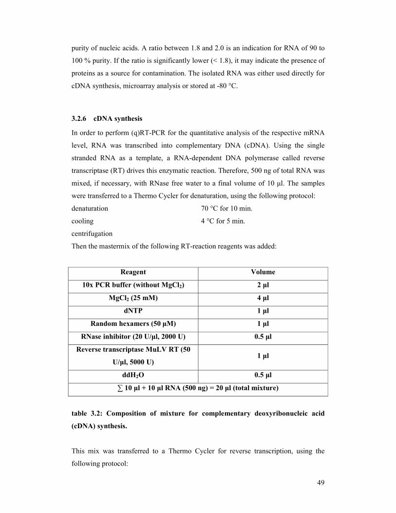
49
purity of nucleic acids. A ratio between 1.8 and 2.0 is an indication for RNA of 90 to
100 % purity. If the ratio is significantly lower (< 1.8), it may indicate the presence of
proteins as a source for contamination. The isolated RNA was either used directly for
cDNA synthesis, microarray analysis or stored at -80 °C.
3.2.6 cDNA synthesis
In order to perform (q)RT-PCR for the quantitative analysis of the respective mRNA
level, RNA was transcribed into complementary DNA (cDNA). Using the single
stranded RNA as a template, a RNA-dependent DNA polymerase called reverse
transcriptase (RT) drives this enzymatic reaction. Therefore, 500 ng of total RNA was
mixed, if necessary, with RNase free water to a final volume of 10 µl. The samples
were transferred to a Thermo Cycler for denaturation, using the following protocol:
denaturation 70 °C for 10 min.
cooling 4 °C for 5 min.
centrifugation
Then the mastermix of the following RT-reaction reagents was added:
Reagent Volume
10x PCR buffer (without MgCl2) 2 µl
MgCl2 (25 mM) 4 µl
dNTP 1 µl
Random hexamers (50 µM) 1 µl
RNase inhibitor (20 U/µl, 2000 U) 0.5 µl
Reverse transcriptase MuLV RT (50
U/µl, 5000 U) 1 µl
ddH2O 0.5 µl
∑ 10 µl + 10 µl RNA (500 ng) = 20 µl (total mixture)
table 3.2: Composition of mixture for complementary deoxyribonucleic acid
(cDNA) synthesis.
This mix was transferred to a Thermo Cycler for reverse transcription, using the
following protocol:
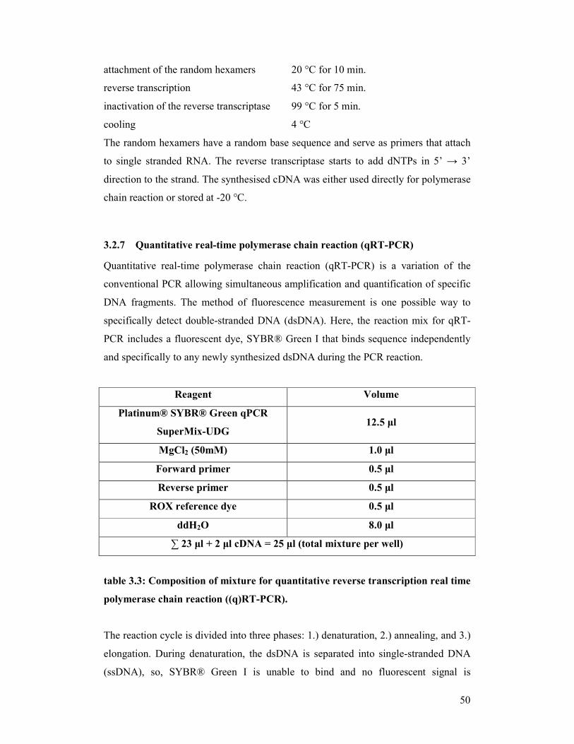
50
attachment of the random hexamers 20 °C for 10 min.
reverse transcription 43 °C for 75 min.
inactivation of the reverse transcriptase 99 °C for 5 min.
cooling 4 °C
The random hexamers have a random base sequence and serve as primers that attach
to single stranded RNA. The reverse transcriptase starts to add dNTPs in 5’ → 3’
direction to the strand. The synthesised cDNA was either used directly for polymerase
chain reaction or stored at -20 °C.
3.2.7 Quantitative real-time polymerase chain reaction (qRT-PCR)
Quantitative real-time polymerase chain reaction (qRT-PCR) is a variation of the
conventional PCR allowing simultaneous amplification and quantification of specific
DNA fragments. The method of fluorescence measurement is one possible way to
specifically detect double-stranded DNA (dsDNA). Here, the reaction mix for qRT-
PCR includes a fluorescent dye, SYBR® Green I that binds sequence independently
and specifically to any newly synthesized dsDNA during the PCR reaction.
Reagent Volume
Platinum® SYBR® Green qPCR
SuperMix-UDG 12.5 µl
MgCl2 (50mM) 1.0 µl
Forward primer 0.5 µl
Reverse primer 0.5 µl
ROX reference dye 0.5 µl
ddH2O 8.0 µl
∑ 23 µl + 2 µl cDNA = 25 µl (total mixture per well)
table 3.3: Composition of mixture for quantitative reverse transcription real time
polymerase chain reaction ((q)RT-PCR).
The reaction cycle is divided into three phases: 1.) denaturation, 2.) annealing, and 3.)
elongation. During denaturation, the dsDNA is separated into single-stranded DNA
(ssDNA), so, SYBR® Green I is unable to bind and no fluorescent signal is
51
detectable. During the annealing phase, primers bind to the complementary ssDNA
and SYBR® Green I binds directly to the short dsDNA segments, resulting in steadily
increasing fluorescence intensity. In the elongation phase, the complete product is
synthesized by a thermostable DNA-dependent DNA polymerase.
The maximum of fluorescent signal and accumulation of target product is detected
after each cycle at the end of elongation, as the level of fluorescence produced by the
dye is proportional to the amount of amplified DNA.
In order to perform the experiments, all components were mixed as follows and the
volume was adjusted to 23 µl with distilled, autoclaved water.
The (q)RT-PCR reaction was performed for 45 cycles according to the following
protocol, using the appropriate primers indicated (table 6.1 and 6.2).
activation of polymerase enzyme 50 °C for 2 min.
first denaturation 95 °C for 5 min.
second denaturation 95 °C for 5 sec.
annealing 59 °C for 5 sec.
extension 72 °C for 30 sec.
dissociation step 1 95 °C for 15 sec.
dissociation step 2 60 °C for 1 min.
dissociation step 3 95 °C for 15 sec.
dissociation step 4 60 °C for 15 sec.
The gene expression was analysed using the AB 7500 fast real-time PCR system. To
obtain a relative measure for the quantification of the mRNA level of the gene of
interest, all results were normalised to the relative expression of an ubiquitously,
constantly and equally expressed gene that is free of pseudogenes (= reference gene).
Hypoxanthine phosphoribosyltransferase (HPRT) 1 in human tissue/cells and
porphobilinogen deaminase (Pbgd) in mouse tissue/cells, respectively, were used as
reference genes in all (q)RT-PCR determinations. The relative mRNA level of a target
gene was presented in ∆Ct values (∆Ct = Ct reference gene - Ct target gene). A
commonly-employed method of DNA quantification by real-time PCR relies on
plotting fluorescence against the number of cycles on a logarithmic scale. A threshold
for detection of DNA-based fluorescence is set slightly above background. The
number of cycles at which the fluorescence exceeds the threshold is called the the
threshold cycle (Ct). During the exponential amplification phase, the sequence of the
52
DNA target doubles every cycle. Relative changes in transcript levels compared to
healthy/untreated controls were shown as ∆∆Ct values (∆∆Ct = ∆Ct disease/treated -
∆Ct healthy/untreated control). All ∆∆Ct values correspond approximately to the
binary logarithm of the fold change. The specific amplification of the PCR products
was confirmed by melting curve analysis and gel electrophoresis.
3.2.8 DNA agarose gel electrophoresis
Agarose gel electrophoresis is a method to separate and visualize DNA fragments
according to their size. Negatively charged nucleic acids are moved through the
electric field in the gel. The shorter a molecule is, the faster it moves through the gel,
what means, that after a certain time period short molecules have covered a longer
distance than longer ones.
This method was used to assess the products of (q)RT-PCR. Agarose gels (2 %) were
prepared, by mixing 3 g of agarose with 150 µl TAE buffer 1× and heated up for 3
minutes. Then 0.5 µg/µl ethidium bromide was added to make the products visible, as
it is a fluorescent dye that intercalates with the DNA. Then the gel was cooled down
to solidify. The DNA samples and empty control samples were mixed with 6x DNA
loading buffer, before loading them onto the gel. A 100 bp and 1000 bp ladder were
used for size control. Then electrophoresis was run constantly for 60 - 90 min. at 100
V/cm in 1× TAE buffer. Separated nucleic acids were visualized at short wavelength
with UV-light (λ = 257 nm).
3.2.9 Immunohistochemistry
Immunohistochemistry (IHC) represents a histological method to localise cell- and
tissue-specific antigens at the level of thin tissue sections. In order to detect and
visualise e.g. cellular expression of particular proteins, antibodies/antisera specifically
directed against these antigenes are used in situ. Serial sections of respective tissue
samples are incubated with an antigen-specific primary antibody. A biotinylated
secondary antibody is used against the primary antibody. The subsequent incubation
with streptavidin creates an enzyme-conjugated complex, which can be visualised by
addition of a chromogen substrate after its oxidative conversion. The experiments
were performed according to the following protocol. At first, 3 µm thin sections by
53
using a microtom were sliced from 4 % paraformaldehyde-fixed, paraffin embedded
human and mouse lung tissue samples and mounted on slides. In order to remove
paraffin before immuno-staining, slides were kept for incubation in an oven at 50 °C
overnight and subsequently were transferred to xylene three times for 10 minutes
each. After rehydration in decreasing ethanol concentrations (100 %, 95 % and 70 %),
each twice for 5 minutes, and a washing step with Dulbecco’s phosphate buffered
saline (DPBS) 1x twice for 5 minutes each, tissue sections were treated for
paraformaldehyde removal and antigen retrieval was performed. Therefore, slides
were placed in slide boxes with citrate buffer 1x (pH 6.0, 6.5 mM) and then placed on
a cooker in a pot with water boiled for 25 minutes at 300 °C. They were then kept for
another 10 minutes at 50 °C, followed by a cooling step for five minutes. After
cooling down, slides were washed twice for five minutes in DPBS 1x again.
Endogenous peroxidase activity was quenched by incubation with 3 % (v/v) H2O2
twice for 10 minutes each. After another washing step with DPBS 1x (as described
earlier), serum blocking solution (Histostain PLUS kit), derived from a species
different than the source of the intended primary antibody, was put onto the slides for
10 minutes and kept at room temperature, Finally, the primary antibodies were
applied to the slides, diluted appropriately in blocking solution and DPBS 1x, and the
slides were stored at 4 °C overnight.
dilution for primary antibody:
DPBS 1x
serum blocking solution
primary antibody solution (depending on the respective dilutions for each antibody
used in the experiment (see appendix/table 6.4.1)).
The following day, the slides were first kept for 30 minutes at room temperature and
then washed twice for 5 minutes in DPBS 1x. Immune complexes were visualized by
incubation, using the species-appropriate secondary biotinylated antibodies for 10
minutes at room temperature (Histostain PLUS kit; see appendix/table 6.4.2). Slides
were then incubated with HRP-enzyme conjugated streptavidin for 10 minutes at
room temperature. The tissue was stained for a maximum of 10 minutes at room
temperature, by using AEC Single Solution chromogen, until red colour staining was
visible under a microscope. After each incubation step, a washing step in DPBS 1x
was performed as described earlier. Slides were counterstained with hematoxylin for
54
eight minutes at room temperature, followed by a washing step in running tap water
for 10 minutes. Finally, tissue sections were covered with glycerol gelatine (mounting
medium) and a cover slide. Slides were stored at room temperature and light protected
until use. The stained tissue sections were viewed with an Olympus BX51 light
microscope and pictures were taken with an attached DP25 digital camera at 2
different magnifications (10x and 40x) and analysed.
3.2.10 Immunocytochemistry
Immunocytochemistry (ICC) is applied to visualise the localisation of particular
antigens at the level of single cells, grown as primary cultures or derived from cell
lines, by using enzymes or fluorophores (fluorescent dyes), again coupled to the
respective secondary antibodies. This method is based on antigen-specific binding of
primary antibodies to the respective antigen and the use of specific fluorochrome-
labelled secondary antibodies against the primary antibodies. For the experiments,
freshly isolated primary mouse ATII (ATII) cells were plated at a density of 15 x
104/well in 8-well chamber slides in 10 % FCS culture medium (500 µl/well).
Attachment, synchronisation and stimulation of ATII cells were performed according
to the protocol (see chapter 3.2.3). Cells were then washed three times with DPBS 1x
and fixed with ice-cold 100 % methanol (200 µl/well) for five minutes. After each
incubation step, a washing step was performed. To block unspecific antibody binding
sites, cells were incubated with 3 % (m/vol) BSA in DPBS 1x (200 µl/well) for a
minimum of two hours. Cells were then incubated with the respective primary
antibody, diluted in 0.1 % (m/vol) BSA in DPBS 1x (200 µl/well), at 4 °C overnight.
dilution for primary antibody:
DPBS 1x
BSA
primary antibody solution (depending on the respective dilutions for each antibody
used in the experiment (see table 6.4.1)).
Indirect immunofluorescence was performed the following day by incubation with
FITC- or Alexa Fluor 555-conjugated secondary antibodies, diluted in DPBS 1x
containing 0.1 % BSA (200 µl/well), which were used at room temperature and light
protected for two hours.
55
dilution for secondary antibody:
DPBS 1x
BSA
secondary antibody solution (depending on the respective dilutions for each antibody
used in the experiment (see table 6.4.2)).
Nuclei were visualized by 4’,6-diamidino-2-phenylindole dihydrochloride (DAPI)
staining (1:1000 dilution). Cells were incubated with DAPI, diluted in DPBS 1x
containing 0.1 % BSA (100 µl/well), at room temperature, light protected for five
minutes. After incubation with DAPI, the cells were then fixed with 4 % (m/vol)
paraformaldehyde (PFA) (100 µl/well) at room temperature, light protected for seven
minutes. Finally, the chamber of the culture slide was removed and cells were covered
with glycerol gelatine (mounting medium) and a cover slide. Slides were stored at 4
°C and light protected until use. The stained cells were viewed with a fluorescence
microscope equipped with appropriate filter sets, and pictures were taken and image
analysis was performed with the Leica Q Win program.
3.2.11 Western blot analysis
The Western blot represents a semi-quantitative analytical technique, which is used
for protein detection in crude samples of tissue homogenates and allows identification
of specific proteins by exposing all proteins present on a SDS-gel after electrophoretic
separation to a specific antibody.
In this study, Western blot analysis was performed on whole protein extracts of lung
homogenates from donor transplants and IPF patients. For protein extraction, the
human lung tissue was homogenized in liquid nitrogen by a pestle and 1 ml tissue
lysis buffer per 0.1 g of tissue was added. After five minutes of incubation, the tissue
lysate was passed several times through an RNase-free syringe with a 0.9 mm needle
for further homogenization. The samples were incubated on ice for 60 minutes and
meanwhile vortexed every five minutes. Protein extracts were clarified by
centrifugation for 15 minutes at 4 °C and 16000 g; the resulting supernatant
containing the proteins was collected and either used directly or stored at -20 °C.
lysis buffer:
20 mM Tris pH 7.5
56
150 mM NaCl
1 mM EDTA
1 mM EGTA
1% Triton X-100
2 mM Na3VO4
1:25 Complete™, protease inhibitor mix
The protein quantification of each sample was determined by using the Quick start
Bradford protein assay (a colorimetrical protein assay). For measurement, each
protein sample was diluted 1:100 in ddH2O, of which 10 µl was mixed with 200 µl of
the Quick start Bradford dye reagent and transferred respectively to a 96-well plate.
Additionally, different dilutions of bovine serum albumin (BSA) (0.05, 0.1, 0.2, 0.3,
0.4, 0.5 and 0.6 µg/µl) were used as protein standards. Equally, negative controls
containing the Quick start Bradford dye reagent and 1:100 diluted protein lysis buffer
were applied at the same 96-well plate. After an incubation period of 15 minutes, a
spectrophotometric measurement was performed with a Fusion A153601 Reader at a
wavelength of 570 nm, to measure the absorption. Protein samples, as well as the
BSA standards and controls were measured as double values and averaged. The
corresponding protein concentrations of each sample were calculated by comparing
protein sample values to the BSA standard curve via interpolation. Proteins were
separated according to their molecular weight by SDS polyacrylamide gel
electrophoresis (SDS-PAGE). For separation, a 10 % resolving gel mixture was
poured between two glass plates separated by teflon spacers, to allow polymerisation.
The stacking gel was poured on top of the resolving gel, and a teflon comb was
inserted into the gel to form wells. Ammonium persulfate (APS) and N,N,N,N,N`
tetramethylenediamine (TEMED) were added at last to initiate the polymerisation of
the gels. Prior to loading onto the gel, 20 µg of each protein sample was mixed with
10x SDS gel-loading buffer and denaturated at 95 °C for five minutes. Samples were
loaded onto the polymerized gel, which consisted of a resolving gel with a 15-well
stacking gel on top. 5 µl of protein standards run at the same gel were used as
molecular weight size markers.In between the working steps, proteins were kept on
ice at all times. After loading the proteins onto the gel, electrophoresis was performed
in an electrophoresis chamber filled with 1x SDS-running buffer at 120 V for 90
minutes. After separation of the proteins by SDS-Page, immunoblotting was
57
performed to detect and visualize specific proteins. The separated proteins were
transferred from the polyacrylamide gel onto a nitrocellulose membrane. The protein
transfer was performed in transfer buffer at 100 V for 60 minutes The membrane was
incubated in blocking solution for 60 minutes at room temperature followed by
incubation with the appropriate primary antibodies (table 6.4.1) diluted in blocking
buffer at 4 °C overnight. As blocking buffer, either PBST with 5 % non-fat dry milk
or TBST with 5 % BSA were used, depending on the recommendations for the
corresponding antibody. The membrane was then washed three times for 10 minutes
each with washing buffer and incubated with appropriate horseradish proxidase-
labelled (HRP) secondary antibodies for 60 minutes at room temperature (table 6.4.2).
The membrane was washed five times for 10 minutes. After the washing step, the
protein detection was performed using chemiluminescence by enhanced
chemiluminescent immunoblotting system and exposure to radiographic film. For
subsequent application of different antibodies, the membrane was incubated in
stripping buffer for seven minutes at 52 °C, repeatedly washed and blocked. Protein
visualization was performed as previously described.
3.2.12 [3H]-thymidine proliferation assay
The most commonly cellular proliferation assay used is based on DNA cleavage-
dependent incorporation of thymidine, labeled with β-emitting tritium [3H]. An
indirect quantification can be done by measuring the incorporated radioactive
thymidine. Therefore, freshly isolated primary mouse ATII cells were plated at a
density of 15 x 104/well on 48-well plates in 500 µl of 10 % FBS culture medium
(minus (-) medium with FBS). After 24-30 hours the ATII cells became adherent,
were subsequently synchronized in 0.1 % FBS medium for further 12 hours and then
stimulated with recombinant mouse Wnt3a protein in 200 µl/well of 0.1 % FCS
medium for 18-24 hours. As [3H]-thymidine (0.5 µCi/ml) is being incorporated into
the DNA during cell proliferation, it was added to the medium for the last six hours of
the experiment. After washing twice with 1x PBS, cells were lysed with 300 µl/well
sodium hydroxide (NaOH) and placed on a shaker for a minimum of 30 minutes.
Samples were transferred to separate measuring vials, adding 8 ml of scintillation
liquid to each. The relative radioactivity was determined by a liquid-β-scintilllation
counter, to assess the proliferation rate.
58
3.2.13 Whole genome microarray
In order to perform whole genome microarray analysis, primary mouse ATII cells
were isolated from healthy mouse lungs, treated for 8 and 24 hours with recombinant
mouse Wnt3a protein according to the stimulation protocol and directly used. Each
isolation obtained primary ATII cells pooled from 6 different mice. Total RNA was
extracted as described from three independent groups and the quality assessed by
capillary electrophoresis using the Bioanalyzer 2100. All samples contained 0.3 - 1.0
µg RNA, which was preamplified and labelled using the Low input RNA T7 kit
according to the manufacturer’s instructions. Three samples each (untreated and
Wnt3a-treated ATII cells) were labeled with Cy3 and Cy5. The labeled RNA was
hybridized overnight to 44K 60mer oligonucleotide spotted microarray slides. The
slides were washed with different stringencies, dried by gentle centrifugation, and
scanned using the GenePix 4100A scanner. Data analysis was performed with
GenePix Pro 5.0 software, and calculated fore- and background intensities for all
spots were saved as GenePix results files.
3.2.14 Enzyme linked-immuno-sorbent assay (ELISA)
Enzyme linked-immuno-sorbent assays (ELISA) are used for the quantitative
determination of selected soluble proteins. The experiments were performed
according to the manufacturer’s protocol using the DuoSet® ELISA development kits
from R & D Systems. The following sample types were used to measure natural and
recombinant human and mouse interleukin 1β (IL1β), and mouse interleukin 6 (IL6):
cell culture supernatants from primary mouse ATII cells, untreated and treated with
recombinant Wnt3a protein; conditioned media from primary mouse ATII cells,
obtained from saline- and bleomycin-treated mice; bronchoalveolar lavage fluid
(BALF) from saline- and bleomycin-treated mice, as well as from donor transplants
and IPF patients.
At first a 96-well microplate was coated with the appropriate capture antibody, sealed
and incubated overnight at room temperature. The following day, the 96-well
microplate was washed three times, followed by a drying step to completely remove
liquid and unbound material. Next, blocking buffer was added and the 96-well
microplate was incubated for one hour at room temperature. The wash/drying step
was repeated after each incubation step as before. The samples and standards were
59
diluted in reagent diluent and added in duplicates to the 96-well microplate, which
was sealed and incubated for two hours at room temperature. After removing unbound
material, the appropriate detection antibody was added to the 96-well microplate,
which was sealed and incubated for two hours at room temperature. Streptavidin-HRP
was added and the 96-well microplate was incubated for 20 minutes, light protected at
room temperature. After incubation with streptavidin-HRP, the 96-well microplate
was incubated with substrate solution for 20 minutes, light protected at room
temperature. Finally, a stop solution was added and the optical density of each well
was determined using a spectrophotometer. The absorption was measured at a
wavelength of 450 nm. Additionally, in order to correct optical imperfections in the
96-well microplate, the absorption was measured at 570 nm and substracted from
readings at 450 nm. The corresponding protein concentrations were calculated by
interpolation using the standard curve.
3.2.15 Statistical analysis
All results were presented as means ± standard error of mean (s.e.m.), if not otherwise
stated, and were considered statistically significant when the probability by rejecting
the null hypothesis is less than 0.05 (* p < 0.05). The mean of indicated groups were
compared using the Student´s t-test. All ∆Ct values obtained from quantitative RT-
PCR were analyzed for normal distribution using the Shapiro-Wilk test. All ∆∆Ct
values were analyzed using the two-tailed, one-sample t-test. Intergroup differences of
∆Ct values from IPF patients and bleomycin-treated mice were derived using an one-
tailed, two-sample t-test. Proliferation assay data were analyzed using the Wilcoxon
rank-sum test and singed-rank test. All p values obtained from multiple tests were
adjusted using the procedure from Benjamini & Hochberg.
60
4 RESULTS
4.1 Analysis of the canonical WNT/β-catenin signalling pathway in
idiopathic pulmonary fibrosis
4.1.1 Expression of the canonical WNT/β-catenin signalling pathway in
idiopathic pulmonary fibrosis
In order to investigate the role of the canonical WNT/β-catenin signalling pathway in
IPF, quantitative real time polymerase chain reaction ((q)RT-PCR) was performed to
compare the mRNA expression levels of canonical WNT/β-catenin signalling
components in lung tissue samples of healthy transplant donors (= controls) and IPF
patients. The total lung homogenates were obtained from twelve (n = 12) healthy
transplant donor and IPF lungs, respectively.
First, the canonical WNT ligands WNT1, 2, 3A, 7B and 10B were analysed in this
study. As depicted in figure 4.1 (A), WNT1, 2, 3A and 7B were expressed at similar
low levels in the human lung, while WNT10B expression in particular was minimal in
lung tissue obtained from healthy transplant donors. In IPF lung samples, there was a
significant upregulation of WNT1, 7B and 10B mRNA levels compared with healthy
transplant donor lung samples (log fold change: 0.90 ± 0.30, 1.00 ± 0.36 and 1.58 ±
0.50, respectively). However, WNT3A proved to be significantly downregulated in
IPF lung samples as compared to healthy transplant donor lung samples (log fold
change: -1.93 ± 0.58). Subsequently, the expression profile of the common WNT
receptors FZD1, 2, 3 and 4; as well as the WNT co-receptors LRP5 and 6 was
determined. As seen in figure 4.1 (B), the most abundant receptors in the human lung
were FZD1 and 4, as well as the co-receptors LRP5 and 6. However, their expression
levels were similar in healthy transplant donor and IPF lungs. FZD2 and 3 specific
mRNAs were detected at low levels in healthy transplant donor and IPF lungs, but
significantly increased under pathological conditions of IPF as compared to healthy
transplant donor lung samples (log fold change: 1.04 ± 0.28 and 1.41 ± 0.27,
respectively). Figure 4.1 (C) shows the expression of the WNT cytoplasmic factors
AXIN1 and 2, as well as DVL1, 2 and 3, and GSK3β.
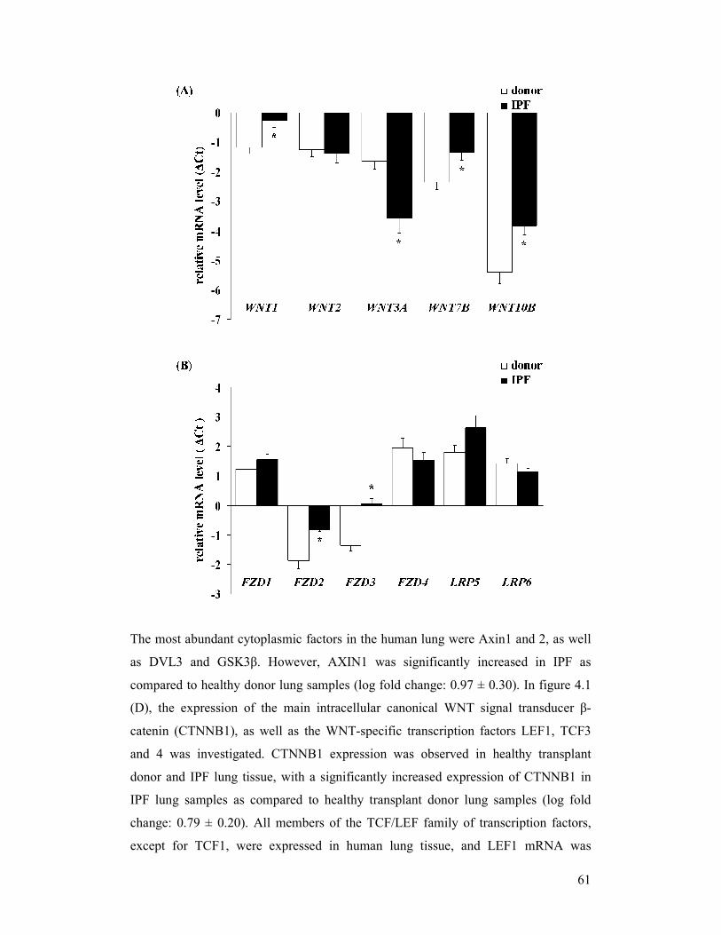
61
The most abundant cytoplasmic factors in the human lung were Axin1 and 2, as well
as DVL3 and GSK3β. However, AXIN1 was significantly increased in IPF as
compared to healthy donor lung samples (log fold change: 0.97 ± 0.30). In figure 4.1
(D), the expression of the main intracellular canonical WNT signal transducer β-
catenin (CTNNB1), as well as the WNT-specific transcription factors LEF1, TCF3
and 4 was investigated. CTNNB1 expression was observed in healthy transplant
donor and IPF lung tissue, with a significantly increased expression of CTNNB1 in
IPF lung samples as compared to healthy transplant donor lung samples (log fold
change: 0.79 ± 0.20). All members of the TCF/LEF family of transcription factors,
except for TCF1, were expressed in human lung tissue, and LEF1 mRNA was
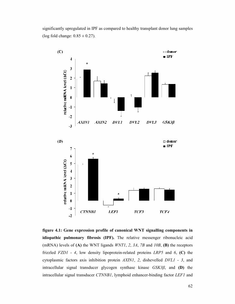
62
significantly upregulated in IPF as compared to healthy transplant donor lung samples
(log fold change: 0.85 ± 0.27).
figure 4.1: Gene expression profile of canonical WNT signalling components in
idiopathic pulmonary fibrosis (IPF). The relative messenger ribonucleic acid
(mRNA) levels of (A) the WNT ligands WNT1, 2, 3A, 7B and 10B, (B) the receptors
frizzled FZD1 - 4, low density lipoprotein-related proteins LRP5 and 6, (C) the
cytoplasmic factors axis inhibition protein AXIN1, 2, dishevelled DVL1 - 3, and
intracellular signal transducer glycogen synthase kinase GSK3β, and (D) the
intracellular signal transducer CTNNB1, lymphoid enhancer-binding factor LEF1 and
63
T-cell-specific transcription factors TCF3 and 4 were assessed in healthy transplant
donor and IPF total lung homogenates by quantitative real time polymerase chain
reaction ((q)RT-PCR). Data were presented in a bar graph; plotting the results as ∆Ct
(∆Ct = Ct reference gene - Ct target gene); statistical significance (p/p-value) was
presented as * (* = p < 0.05), samples of 12 individual isolations were used (n = 12).
The resulting data for LRP5 and 6 were kindly provided by Eva Pfaff.
4.1.2 Localisation of the canonical WNT/β-catenin signalling pathway in
idiopathic pulmonary fibrosis
In order to detect the various cell types, which are capable of expressing specific
canonical WNT/β-catenin signalling pathway components, immunohistochemistry
(IHC) was performed. The WNT ligands WNT1 and 3A and the main canonical
intracellular WNT signal transducers CTNNB1 and GSK3β were assessed in
paraformaldehyde-fixed and paraffin embedded lung tissue sections prepared from
biopsies of healthy transplant donors (= controls) and IPF patients. The lung tissue
sections were obtained from three (n = 3) healthy transplant donor and IPF lungs,
respectively.
As shown in figure 4.2.1 (A) and 4.2.2 (A), in healthy transplant donor lung sections
WNT1 was mainly expressed in bronchial and alveolar epithelium, with strong
staining of alveolar epithelial type II (ATII) cells. Additionally, WNT1 was detected
in vascular smooth muscle cells. In IPF lung sections (see figure 4.2.1 (B) and 4.2.2
(B)), WNT1 staining was observed in bronchial epithelial cells and hyperplastic ATII
cells, identifiable by their cubic shape. In bronchial epithelial cells of IPF lungs
WNT1 showed an apical staining pattern as compared to healthy transplant donor
lungs, which indicates increased secretion. WNT1 was also expressed in endothelial
cells in IPF tissues. Figure 4.3.1 and 4.3.2 (A and B) shows, that WNT3A protein
expression was predominantly detected in ATII cells and selected ciliated bronchial
epithelial cells in both, healthy transplant donor and IPF lung tissue sections. In figure
4.4.1 (A) and 4.4.2 (A), GSK3β exhibits a predominant staining in bronchial epithelial
and ATII cells, as well as endothelial cells in healthy transplant donor lung tissue. In
figure 4.4.1 (B) and 4.4.2 (B), GSK3β expression was observed in basal bronchial
epithelial and hyperplastic ATII cells in IPF lung tissues.
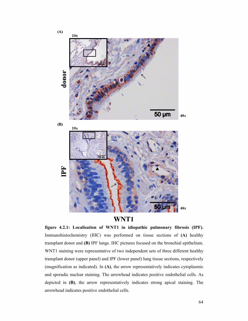
64
figure 4.2.1: Localisation of WNT1 in idiopathic pulmonary fibrosis (IPF).
Immunohistochemistry (IHC) was performed on tissue sections of (A) healthy
transplant donor and (B) IPF lungs. IHC pictures focused on the bronchial epithelium.
WNT1 staining were representative of two independent sets of three different healthy
transplant donor (upper panel) and IPF (lower panel) lung tissue sections, respectively
(magnification as indicated). In (A), the arrow representatively indicates cytoplasmic
and sporadic nuclear staining. The arrowhead indicates positive endothelial cells. As
depicted in (B), the arrow representatively indicates strong apical staining. The
arrowhead indicates positive endothelial cells.
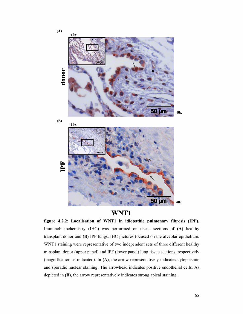
65
figure 4.2.2: Localisation of WNT1 in idiopathic pulmonary fibrosis (IPF).
Immunohistochemistry (IHC) was performed on tissue sections of (A) healthy
transplant donor and (B) IPF lungs. IHC pictures focused on the alveolar epithelium.
WNT1 staining were representative of two independent sets of three different healthy
transplant donor (upper panel) and IPF (lower panel) lung tissue sections, respectively
(magnification as indicated). In (A), the arrow representatively indicates cytoplasmic
and sporadic nuclear staining. The arrowhead indicates positive endothelial cells. As
depicted in (B), the arrow representatively indicates strong apical staining.
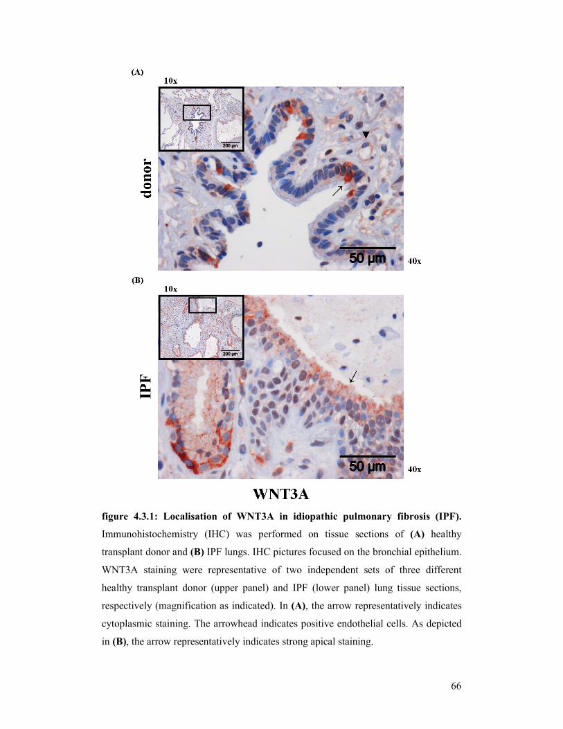
66
figure 4.3.1: Localisation of WNT3A in idiopathic pulmonary fibrosis (IPF).
Immunohistochemistry (IHC) was performed on tissue sections of (A) healthy
transplant donor and (B) IPF lungs. IHC pictures focused on the bronchial epithelium.
WNT3A staining were representative of two independent sets of three different
healthy transplant donor (upper panel) and IPF (lower panel) lung tissue sections,
respectively (magnification as indicated). In (A), the arrow representatively indicates
cytoplasmic staining. The arrowhead indicates positive endothelial cells. As depicted
in (B), the arrow representatively indicates strong apical staining.
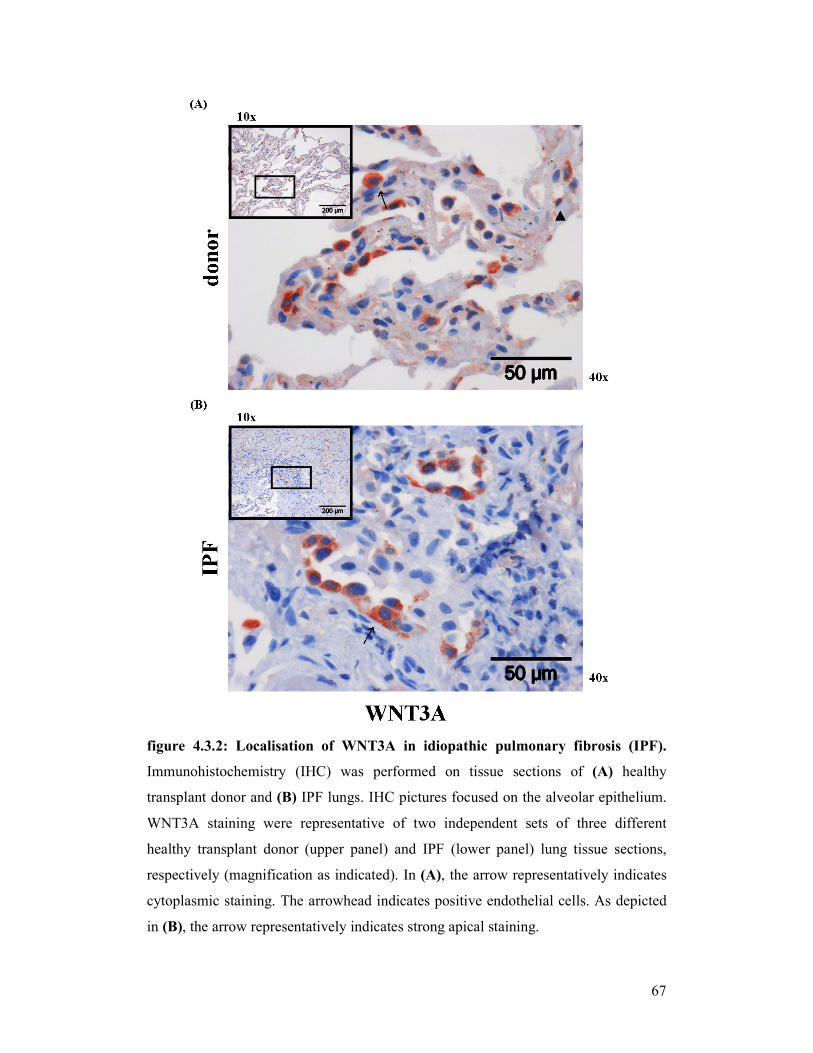
67
figure 4.3.2: Localisation of WNT3A in idiopathic pulmonary fibrosis (IPF).
Immunohistochemistry (IHC) was performed on tissue sections of (A) healthy
transplant donor and (B) IPF lungs. IHC pictures focused on the alveolar epithelium.
WNT3A staining were representative of two independent sets of three different
healthy transplant donor (upper panel) and IPF (lower panel) lung tissue sections,
respectively (magnification as indicated). In (A), the arrow representatively indicates
cytoplasmic staining. The arrowhead indicates positive endothelial cells. As depicted
in (B), the arrow representatively indicates strong apical staining.
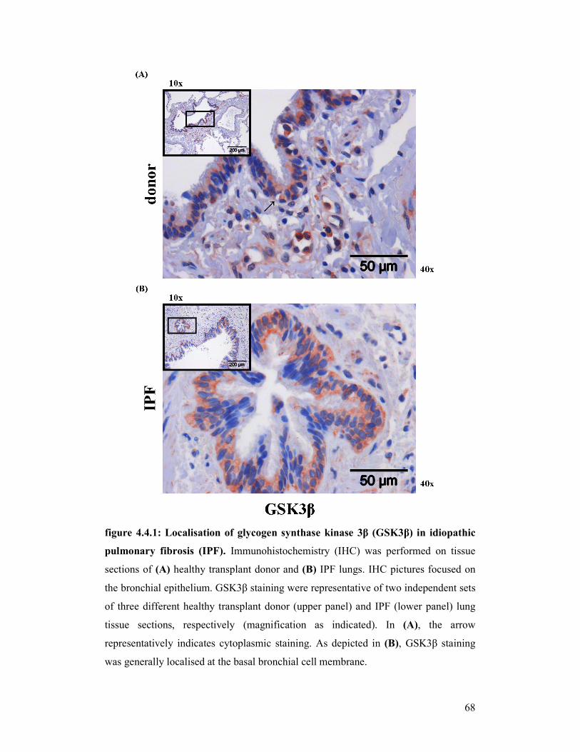
68
figure 4.4.1: Localisation of glycogen synthase kinase 3β (GSK3β) in idiopathic
pulmonary fibrosis (IPF). Immunohistochemistry (IHC) was performed on tissue
sections of (A) healthy transplant donor and (B) IPF lungs. IHC pictures focused on
the bronchial epithelium. GSK3β staining were representative of two independent sets
of three different healthy transplant donor (upper panel) and IPF (lower panel) lung
tissue sections, respectively (magnification as indicated). In (A), the arrow
representatively indicates cytoplasmic staining. As depicted in (B), GSK3β staining
was generally localised at the basal bronchial cell membrane.
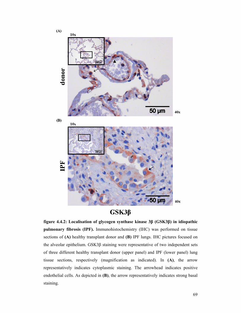
69
figure 4.4.2: Localisation of glycogen synthase kinase 3β (GSK3β) in idiopathic
pulmonary fibrosis (IPF). Immunohistochemistry (IHC) was performed on tissue
sections of (A) healthy transplant donor and (B) IPF lungs. IHC pictures focused on
the alveolar epithelium. GSK3β staining were representative of two independent sets
of three different healthy transplant donor (upper panel) and IPF (lower panel) lung
tissue sections, respectively (magnification as indicated). In (A), the arrow
representatively indicates cytoplasmic staining. The arrowhead indicates positive
endothelial cells. As depicted in (B), the arrow representatively indicates strong basal
staining.
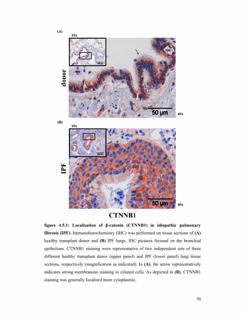
70
figure 4.5.1: Localisation of β-catenin (CTNNB1) in idiopathic pulmonary
fibrosis (IPF). Immunohistochemistry (IHC) was performed on tissue sections of (A)
healthy transplant donor and (B) IPF lungs. IHC pictures focused on the bronchial
epithelium. CTNNB1 staining were representative of two independent sets of three
different healthy transplant donor (upper panel) and IPF (lower panel) lung tissue
sections, respectively (magnification as indicated). In (A), the arrow representatively
indicates strong membranous staining in ciliated cells. As depicted in (B), CTNNB1
staining was generally localised more cytoplasmic.
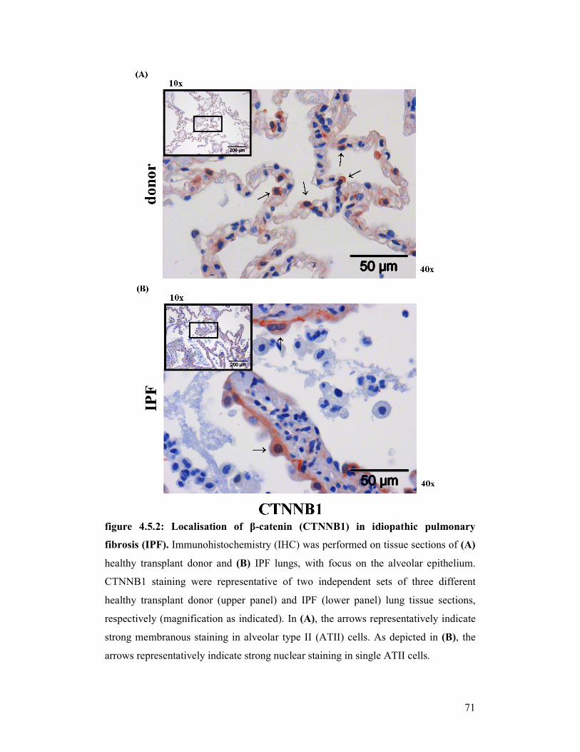
71
figure 4.5.2: Localisation of β-catenin (CTNNB1) in idiopathic pulmonary
fibrosis (IPF). Immunohistochemistry (IHC) was performed on tissue sections of (A)
healthy transplant donor and (B) IPF lungs, with focus on the alveolar epithelium.
CTNNB1 staining were representative of two independent sets of three different
healthy transplant donor (upper panel) and IPF (lower panel) lung tissue sections,
respectively (magnification as indicated). In (A), the arrows representatively indicate
strong membranous staining in alveolar type II (ATII) cells. As depicted in (B), the
arrows representatively indicate strong nuclear staining in single ATII cells.
72
As depicted in figure 4.5.1 (A) and 4.5.2 (A), strong membranous and cytoplasmic
CTNNB1 expression was observed in bronchial epithelial and ATII cells, as well as in
endothelial cells of healthy transplant donor lungs. Importantly, in IPF lungs (see
figure 4.5.1 (B) and 4.5.2 (B)), CTNNB1 showed less membranous staining. An
enhanced expression was detected in the cytoplasm. Additionally, clear nuclear
staining was observed in single cells. Especially in bronchial areas and in alveolar
type II (ATII) cells a strong CTNNB1 staining was noticed.
In summary, the canonical WNT/β-catenin signalling pathway components in general
were largely expressed in the bronchial and alveolar epithelium of healthy transplant
donor and IPF lung tissue.
4.1.3 Activity of the canonical WNT/β-Catenin signalling pathway in idiopathic
pulmonary fibrosis
To determine, whether the canonical WNT/β-catenin signalling pathway was
activated at the protein level in IPF, Western blot (WB) analysis was performed.
Therefore, total lung homogenates were obtained from five donor and IPF patients,
respectively.
As presented in figure 4.6, increased phosphorylation of GSK3β and LRP6 in IPF
patients was observed compared with transplant donors (=control). This is consistent
with elevated protein levels of total CTNNB1, indicating activated canonical WNT/β-
catenin signalling. The analysis revealed upregulated protein levels of cyclinD1, a
known target gene of the canonical WNT/β-catenin signalling pathway. An increased
level of alpha-smooth muscle actin (ACTA2 = α-SMA) was detected in IPF lung
homogenates compared with donor lung homogenates. Total GSK3β, total LRP6 and
lamin A/C served as loading controls.
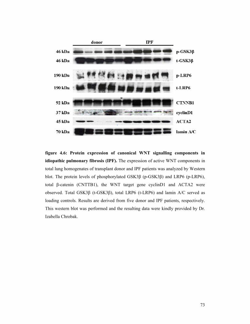
73
figure 4.6: Protein expression of canonical WNT signalling components in
idiopathic pulmonary fibrosis (IPF). The expression of active WNT components in
total lung homogenates of transplant donor and IPF patients was analyzed by Western
blot. The protein levels of phosphorylated GSK3β (p-GSK3β) and LRP6 (p-LRP6),
total β-catenin (CNTTB1), the WNT target gene cyclinD1 and ACTA2 were
observed. Total GSK3β (t-GSK3β), total LRP6 (t-LRP6) and lamin A/C served as
loading controls. Results are derived from five donor and IPF patients, respectively.
This western blot was performed and the resulting data were kindly provided by Dr.
Izabella Chrobak.
74
4.2 Analysis of the canonical WNT/β-catenin signalling pathway in
experimental pulmonary fibrosis
4.2.1 Expression of the canonical WNT/β-catenin signalling pathway in
experimental pulmonary fibrosis
The mouse model of bleomycin-induced pulmonary fibrosis was used to determine
whether the expression of canonical WNT/β-catenin signalling components was
regulated and activated during this process. To induce an experimental model of
pulmonary fibrosis, mice were subjected to saline (= control) and to bleomycin
treatment for 14 days, respectively. Initially, qRT-PCR was performed to quantify the
mRNA expression of canonical WNT/β-catenin signalling components in lung tissue
samples of saline- and bleomycin-treated mice. The total lung homogenates were
obtained from six saline- and bleomycin-treated lungs, respectively.
As depicted in figure 4.7 (A), the WNT ligands Wnt2, 3a, 7b and 10b were detected at
similar levels in the mouse lung, whereas Wnt1 was much lower expressed in lung
tissue from saline-treated mice. In bleomycin-treated lung tissue however, a
significant increase of Wnt1 expression was observed (log fold change: 2.90 ± 0.75).
Whereas, Wnt2, 7b and 10b showed a marked decrease of mRNA levels in
bleomycin-treated total lung homogenates as compared with control lung
homogenates (log fold change: -0.71 ± 0.22, -1.07 ± 0.22 and -1.28 ± 0.18,
respectively). Next, the expression profile of the common WNT receptors, Fzd1, 2, 3
and 4; and the WNT co-receptors, Lrp5 and 6 was determined. As seen in figure 4.7
(B), the co-receptors Lrp5 and 6 were expressed at similar high mRNA levels in
saline- and bleomycin-treated lungs. Interestingly, 14 days after bleomycin treatment,
there was a significant downregulation of Fzd2 and 4 (log fold change: -0.69 ± 0.20
and -0.94 ± 0.25, respectively). Figure 4.7 (C) shows the expression of the WNT
cytoplasmic factors, Axin1 and 2, as well as Dvl1, 2 and 3, and Gsk3β. High
expression levels of all these components were detected in saline- and bleomycin-
treated lungs. Thereby, Axin2 was significantly decreased at 14 days after bleomycin
treatment as compared to saline treatment (log fold change: -1.04 ± 0.21).
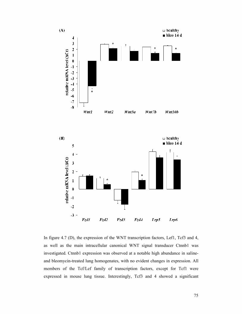
75
In figure 4.7 (D), the expression of the WNT transcription factors, Lef1, Tcf3 and 4,
as well as the main intracellular canonical WNT signal transducer Ctnnb1 was
investigated. Ctnnb1 expression was observed at a notable high abundance in saline-
and bleomycin-treated lung homogenates, with no evident changes in expression. All
members of the Tcf/Lef family of transcription factors, except for Tcf1 were
expressed in mouse lung tissue. Interestingly, Tcf3 and 4 showed a significant
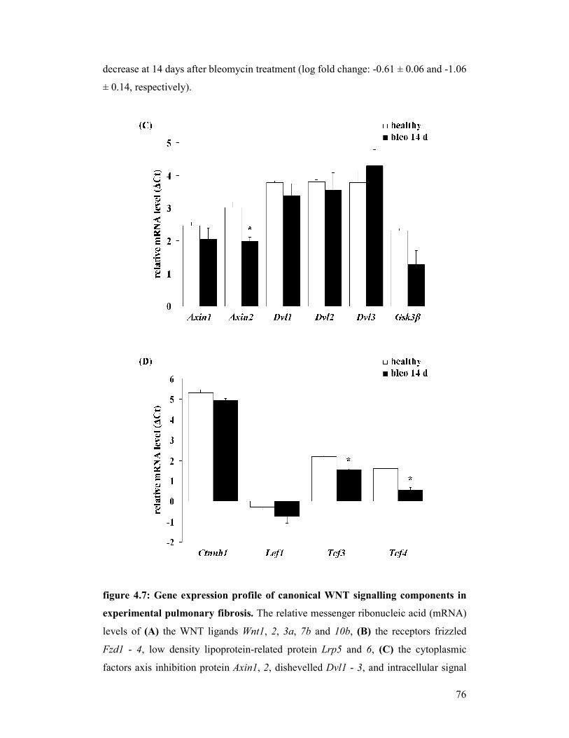
76
decrease at 14 days after bleomycin treatment (log fold change: -0.61 ± 0.06 and -1.06
± 0.14, respectively).
figure 4.7: Gene expression profile of canonical WNT signalling components in
experimental pulmonary fibrosis. The relative messenger ribonucleic acid (mRNA)
levels of (A) the WNT ligands Wnt1, 2, 3a, 7b and 10b, (B) the receptors frizzled
Fzd1 - 4, low density lipoprotein-related protein Lrp5 and 6, (C) the cytoplasmic
factors axis inhibition protein Axin1, 2, dishevelled Dvl1 - 3, and intracellular signal
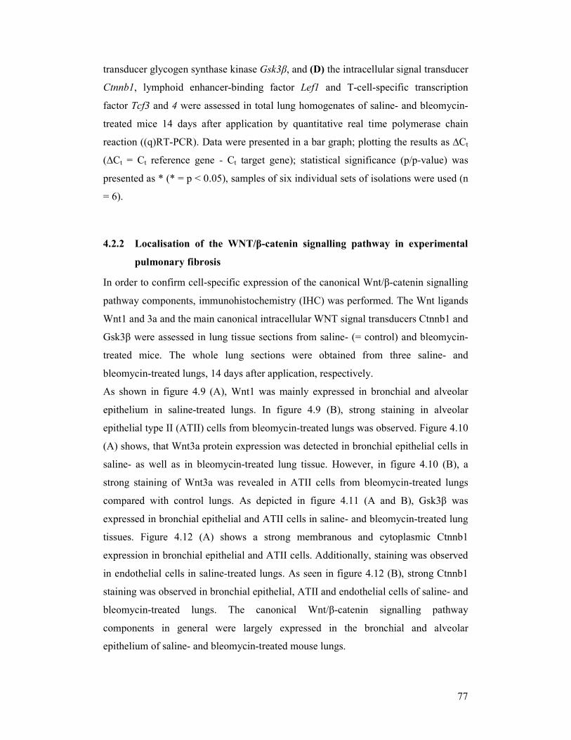
77
transducer glycogen synthase kinase Gsk3β, and (D) the intracellular signal transducer
Ctnnb1, lymphoid enhancer-binding factor Lef1 and T-cell-specific transcription
factor Tcf3 and 4 were assessed in total lung homogenates of saline- and bleomycin-
treated mice 14 days after application by quantitative real time polymerase chain
reaction ((q)RT-PCR). Data were presented in a bar graph; plotting the results as ∆Ct
(∆Ct = Ct reference gene - Ct target gene); statistical significance (p/p-value) was
presented as * (* = p < 0.05), samples of six individual sets of isolations were used (n
= 6).
4.2.2 Localisation of the WNT/β-catenin signalling pathway in experimental
pulmonary fibrosis
In order to confirm cell-specific expression of the canonical Wnt/β-catenin signalling
pathway components, immunohistochemistry (IHC) was performed. The Wnt ligands
Wnt1 and 3a and the main canonical intracellular WNT signal transducers Ctnnb1 and
Gsk3β were assessed in lung tissue sections from saline- (= control) and bleomycin-
treated mice. The whole lung sections were obtained from three saline- and
bleomycin-treated lungs, 14 days after application, respectively.
As shown in figure 4.9 (A), Wnt1 was mainly expressed in bronchial and alveolar
epithelium in saline-treated lungs. In figure 4.9 (B), strong staining in alveolar
epithelial type II (ATII) cells from bleomycin-treated lungs was observed. Figure 4.10
(A) shows, that Wnt3a protein expression was detected in bronchial epithelial cells in
saline- as well as in bleomycin-treated lung tissue. However, in figure 4.10 (B), a
strong staining of Wnt3a was revealed in ATII cells from bleomycin-treated lungs
compared with control lungs. As depicted in figure 4.11 (A and B), Gsk3β was
expressed in bronchial epithelial and ATII cells in saline- and bleomycin-treated lung
tissues. Figure 4.12 (A) shows a strong membranous and cytoplasmic Ctnnb1
expression in bronchial epithelial and ATII cells. Additionally, staining was observed
in endothelial cells in saline-treated lungs. As seen in figure 4.12 (B), strong Ctnnb1
staining was observed in bronchial epithelial, ATII and endothelial cells of saline- and
bleomycin-treated lungs. The canonical Wnt/β-catenin signalling pathway
components in general were largely expressed in the bronchial and alveolar
epithelium of saline- and bleomycin-treated mouse lungs.
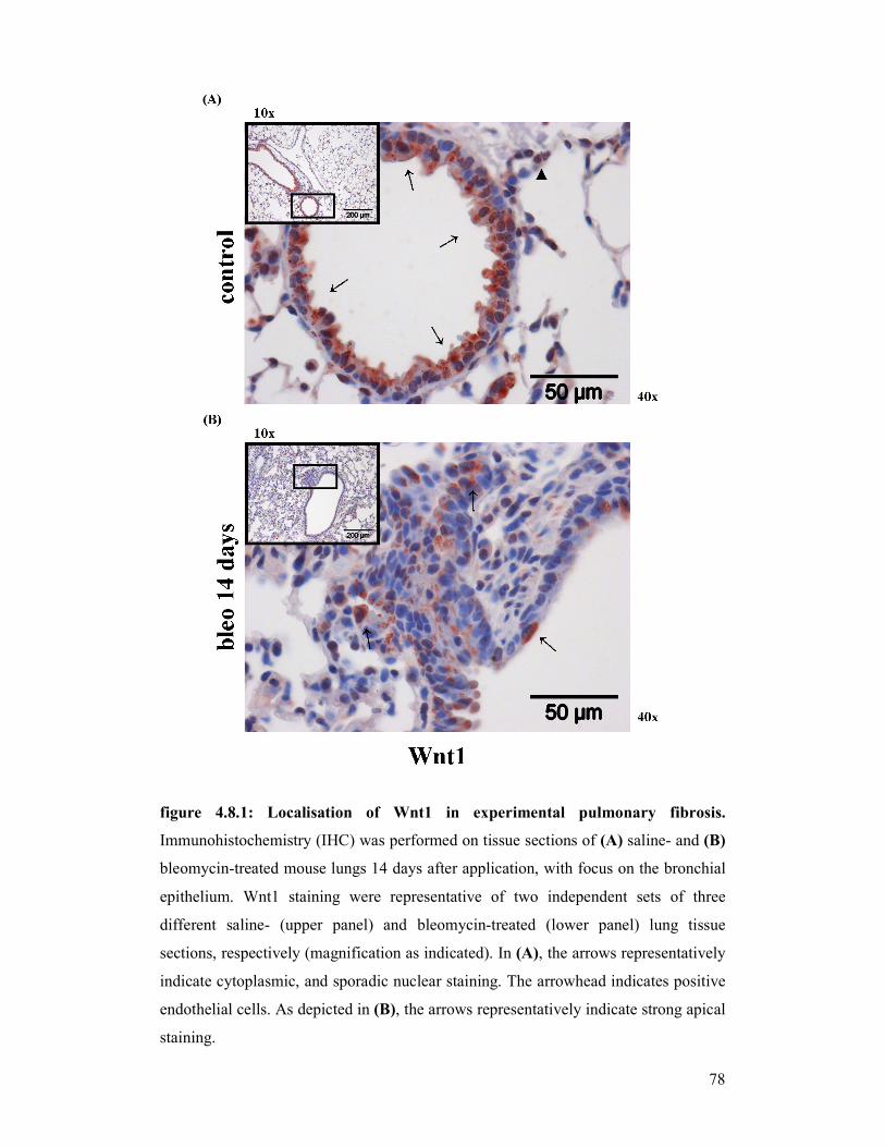
78
figure 4.8.1: Localisation of Wnt1 in experimental pulmonary fibrosis.
Immunohistochemistry (IHC) was performed on tissue sections of (A) saline- and (B)
bleomycin-treated mouse lungs 14 days after application, with focus on the bronchial
epithelium. Wnt1 staining were representative of two independent sets of three
different saline- (upper panel) and bleomycin-treated (lower panel) lung tissue
sections, respectively (magnification as indicated). In (A), the arrows representatively
indicate cytoplasmic, and sporadic nuclear staining. The arrowhead indicates positive
endothelial cells. As depicted in (B), the arrows representatively indicate strong apical
staining.
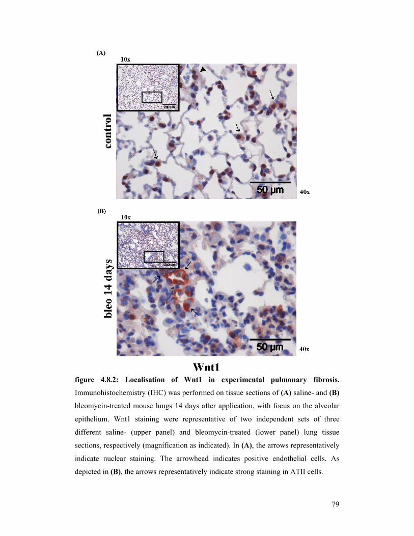
79
figure 4.8.2: Localisation of Wnt1 in experimental pulmonary fibrosis.
Immunohistochemistry (IHC) was performed on tissue sections of (A) saline- and (B)
bleomycin-treated mouse lungs 14 days after application, with focus on the alveolar
epithelium. Wnt1 staining were representative of two independent sets of three
different saline- (upper panel) and bleomycin-treated (lower panel) lung tissue
sections, respectively (magnification as indicated). In (A), the arrows representatively
indicate nuclear staining. The arrowhead indicates positive endothelial cells. As
depicted in (B), the arrows representatively indicate strong staining in ATII cells.
80
figure 4.9.1: Localisation of Wnt3a in experimental pulmonary fibrosis.
Immunohistochemistry (IHC) was performed on tissue sections of (A) saline- and (B)
bleomycin-treated mouse lungs 14 days after application, with focus on the bronchial
epithelium. Wnt3a staining were representative of two independent sets of three
different saline- (upper panel) and bleomycin-treated (lower panel) lung tissue
sections, respectively (magnification as indicated). In (A), the arrows representatively
indicate cytoplasmic, and sporadic nuclear staining. As depicted in (B), the arrows
representatively indicate strong apical staining.
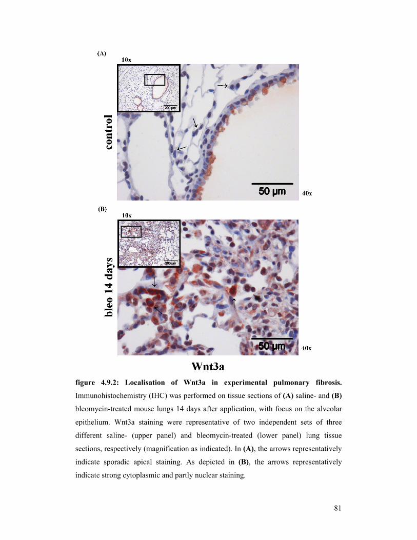
81
figure 4.9.2: Localisation of Wnt3a in experimental pulmonary fibrosis.
Immunohistochemistry (IHC) was performed on tissue sections of (A) saline- and (B)
bleomycin-treated mouse lungs 14 days after application, with focus on the alveolar
epithelium. Wnt3a staining were representative of two independent sets of three
different saline- (upper panel) and bleomycin-treated (lower panel) lung tissue
sections, respectively (magnification as indicated). In (A), the arrows representatively
indicate sporadic apical staining. As depicted in (B), the arrows representatively
indicate strong cytoplasmic and partly nuclear staining.
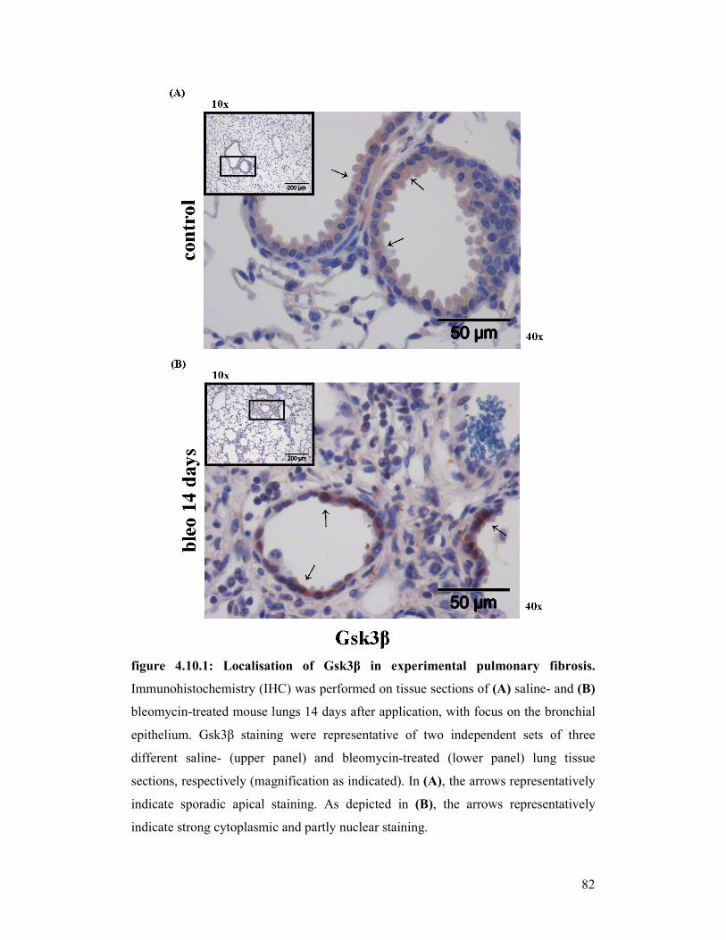
82
figure 4.10.1: Localisation of Gsk3β in experimental pulmonary fibrosis.
Immunohistochemistry (IHC) was performed on tissue sections of (A) saline- and (B)
bleomycin-treated mouse lungs 14 days after application, with focus on the bronchial
epithelium. Gsk3β staining were representative of two independent sets of three
different saline- (upper panel) and bleomycin-treated (lower panel) lung tissue
sections, respectively (magnification as indicated). In (A), the arrows representatively
indicate sporadic apical staining. As depicted in (B), the arrows representatively
indicate strong cytoplasmic and partly nuclear staining.
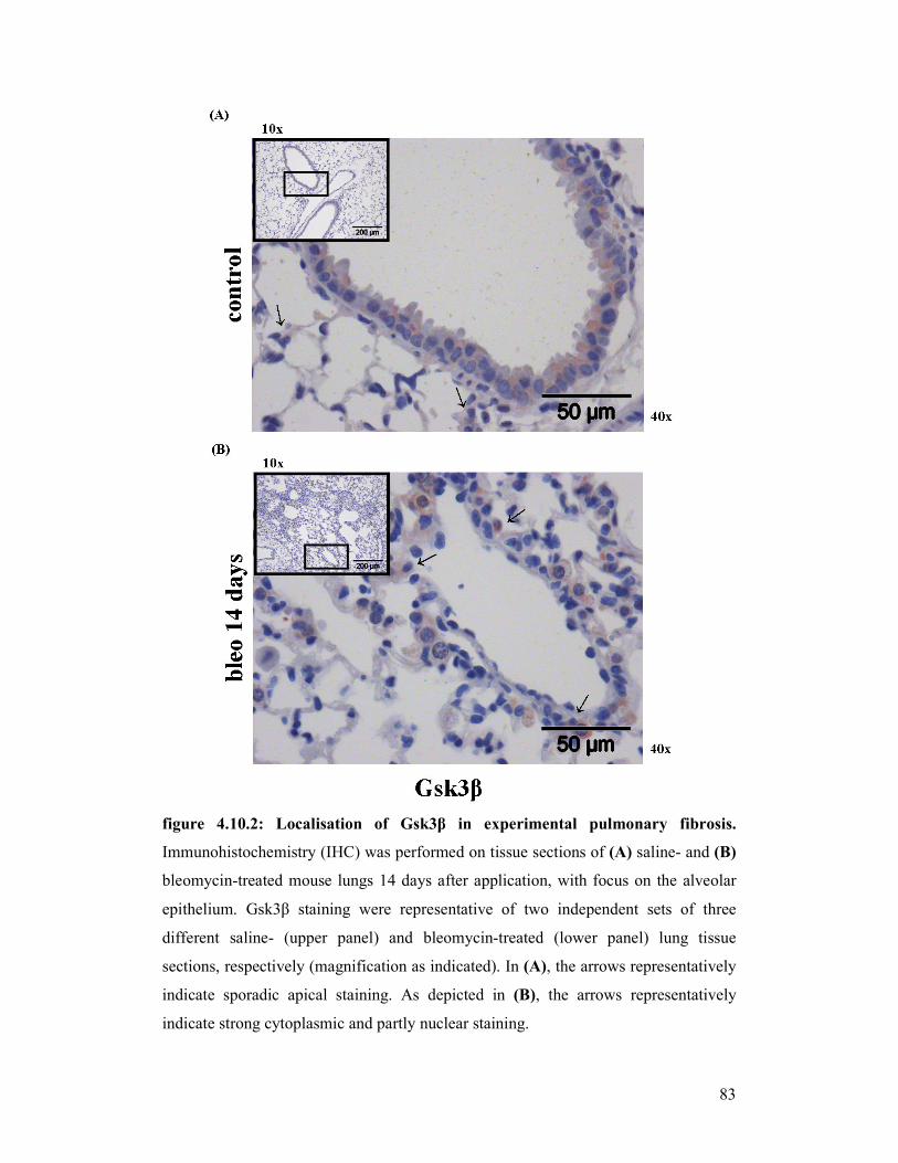
83
figure 4.10.2: Localisation of Gsk3β in experimental pulmonary fibrosis.
Immunohistochemistry (IHC) was performed on tissue sections of (A) saline- and (B)
bleomycin-treated mouse lungs 14 days after application, with focus on the alveolar
epithelium. Gsk3β staining were representative of two independent sets of three
different saline- (upper panel) and bleomycin-treated (lower panel) lung tissue
sections, respectively (magnification as indicated). In (A), the arrows representatively
indicate sporadic apical staining. As depicted in (B), the arrows representatively
indicate strong cytoplasmic and partly nuclear staining.
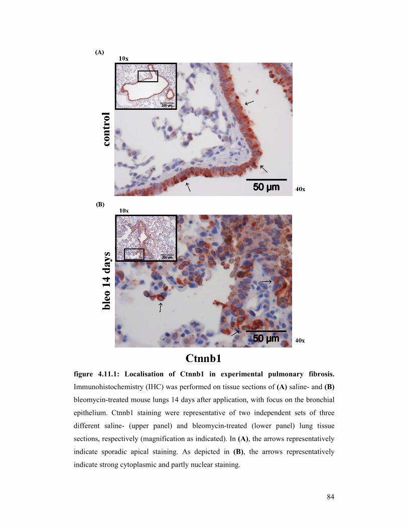
84
figure 4.11.1: Localisation of Ctnnb1 in experimental pulmonary fibrosis.
Immunohistochemistry (IHC) was performed on tissue sections of (A) saline- and (B)
bleomycin-treated mouse lungs 14 days after application, with focus on the bronchial
epithelium. Ctnnb1 staining were representative of two independent sets of three
different saline- (upper panel) and bleomycin-treated (lower panel) lung tissue
sections, respectively (magnification as indicated). In (A), the arrows representatively
indicate sporadic apical staining. As depicted in (B), the arrows representatively
indicate strong cytoplasmic and partly nuclear staining.
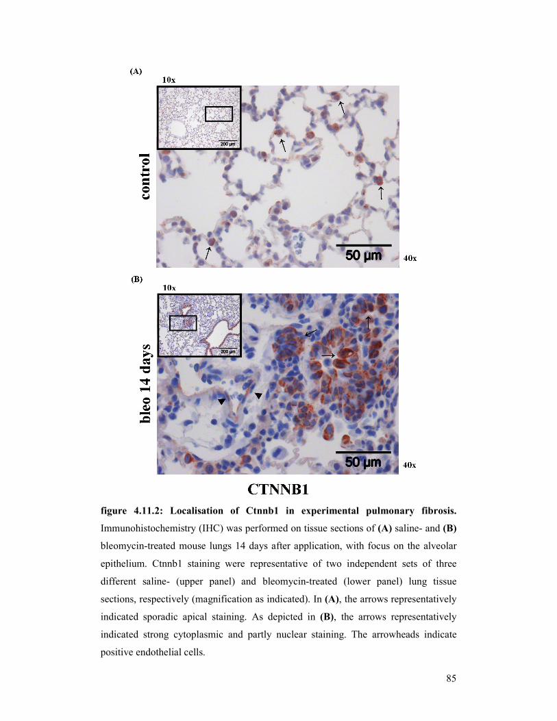
85
figure 4.11.2: Localisation of Ctnnb1 in experimental pulmonary fibrosis.
Immunohistochemistry (IHC) was performed on tissue sections of (A) saline- and (B)
bleomycin-treated mouse lungs 14 days after application, with focus on the alveolar
epithelium. Ctnnb1 staining were representative of two independent sets of three
different saline- (upper panel) and bleomycin-treated (lower panel) lung tissue
sections, respectively (magnification as indicated). In (A), the arrows representatively
indicated sporadic apical staining. As depicted in (B), the arrows representatively
indicated strong cytoplasmic and partly nuclear staining. The arrowheads indicate
positive endothelial cells.
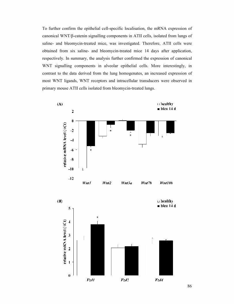
86
To further confirm the epithelial cell-specific localisation, the mRNA expression of
canonical WNT/β-catenin signalling components in ATII cells, isolated from lungs of
saline- and bleomycin-treated mice, was investigated. Therefore, ATII cells were
obtained from six saline- and bleomycin-treated mice 14 days after application,
respectively. In summary, the analysis further confirmed the expression of canonical
WNT signalling components in alveolar epithelial cells. More interestingly, in
contrast to the data derived from the lung homogenates, an increased expression of
most WNT ligands, WNT receptors and intracellular transducers were observed in
primary mouse ATII cells isolated from bleomycin-treated lungs.
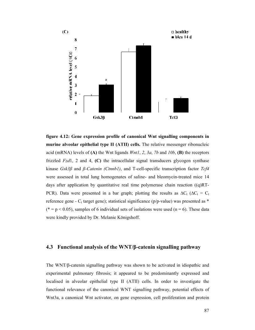
87
figure 4.12: Gene expression profile of canonical Wnt signalling components in
murine alveolar epithelial type II (ATII) cells. The relative messenger ribonucleic
acid (mRNA) levels of (A) the Wnt ligands Wnt1, 2, 3a, 7b and 10b, (B) the receptors
frizzled Fzd1, 2 and 4, (C) the intracellular signal transducers glycogen synthase
kinase Gsk3β and β-Catenin (Ctnnb1), and T-cell-specific transcription factor Tcf4
were assessed in total lung homogenates of saline- and bleomycin-treated mice 14
days after application by quantitative real time polymerase chain reaction ((q)RT-
PCR). Data were presented in a bar graph; plotting the results as ∆Ct (∆Ct = Ct
reference gene - Ct target gene); statistical significance (p/p-value) was presented as *
(* = p < 0.05), samples of 6 individual sets of isolations were used (n = 6). These data
were kindly provided by Dr. Melanie Königshoff.
4.3 Functional analysis of the WNT/β-catenin signalling pathway
The WNT/β-catenin signalling pathway was shown to be activated in idiopathic and
experimental pulmonary fibrosis; it appeared to be predominantly expressed and
localised in alveolar epithelial type II (ATII) cells. In order to investigate the
functional relevance of the canonical WNT signalling pathway, potential effects of
Wnt3a, a canonical Wnt activator, on gene expression, cell proliferation and protein
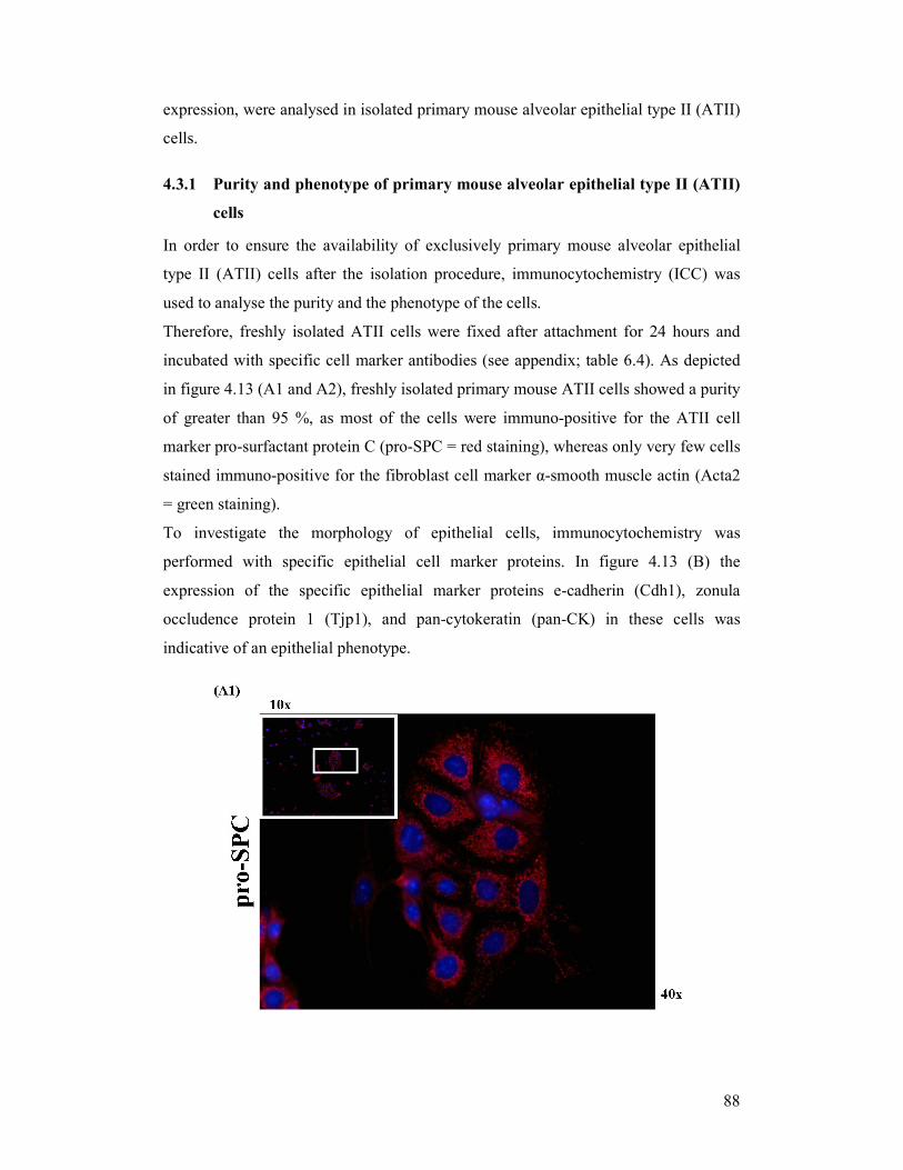
88
expression, were analysed in isolated primary mouse alveolar epithelial type II (ATII)
cells.
4.3.1 Purity and phenotype of primary mouse alveolar epithelial type II (ATII)
cells
In order to ensure the availability of exclusively primary mouse alveolar epithelial
type II (ATII) cells after the isolation procedure, immunocytochemistry (ICC) was
used to analyse the purity and the phenotype of the cells.
Therefore, freshly isolated ATII cells were fixed after attachment for 24 hours and
incubated with specific cell marker antibodies (see appendix; table 6.4). As depicted
in figure 4.13 (A1 and A2), freshly isolated primary mouse ATII cells showed a purity
of greater than 95 %, as most of the cells were immuno-positive for the ATII cell
marker pro-surfactant protein C (pro-SPC = red staining), whereas only very few cells
stained immuno-positive for the fibroblast cell marker α-smooth muscle actin (Acta2
= green staining).
To investigate the morphology of epithelial cells, immunocytochemistry was
performed with specific epithelial cell marker proteins. In figure 4.13 (B) the
expression of the specific epithelial marker proteins e-cadherin (Cdh1), zonula
occludence protein 1 (Tjp1), and pan-cytokeratin (pan-CK) in these cells was
indicative of an epithelial phenotype.
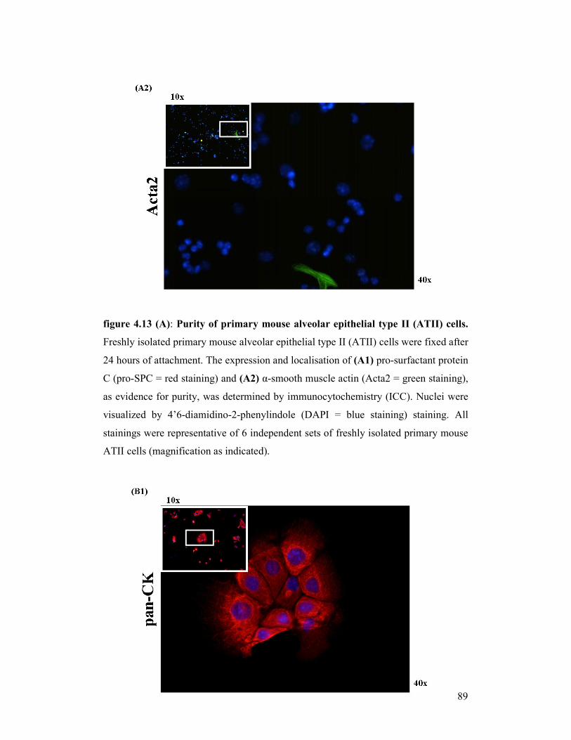
89
figure 4.13 (A): Purity of primary mouse alveolar epithelial type II (ATII) cells.
Freshly isolated primary mouse alveolar epithelial type II (ATII) cells were fixed after
24 hours of attachment. The expression and localisation of (A1) pro-surfactant protein
C (pro-SPC = red staining) and (A2) α-smooth muscle actin (Acta2 = green staining),
as evidence for purity, was determined by immunocytochemistry (ICC). Nuclei were
visualized by 4’6-diamidino-2-phenylindole (DAPI = blue staining) staining. All
stainings were representative of 6 independent sets of freshly isolated primary mouse
ATII cells (magnification as indicated).

90
figure 4.13(B): Phenotype of primary mouse alveolar epithelial type II (ATII)
cells. Freshly isolated primary mouse alveolar epithelial type II (ATII) cells were
fixed after 24 hours of attachment. The expression and localisation of (B1) pan-
cytokeratin (panCK = red staining), (B2) zonula occludence protein 1 (Tjp1 = red
staining), and (B3) e-cadherin (Cdh1 = green staining), as evidence for an epithelial
cellular phenotype, was determined by immunocytochemistry. Nuclei were visualized
by 4’6-diamidino-2-phenylindole (DAPI = blue staining) staining. All stainings were
representative of 6 independent sets of freshly isolated primary mouse ATII cells
(magnification as indicated).
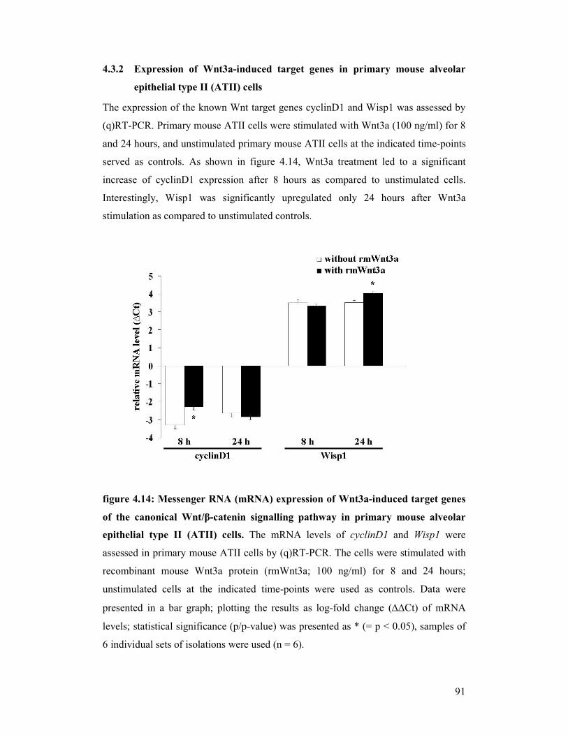
91
4.3.2 Expression of Wnt3a-induced target genes in primary mouse alveolar
epithelial type II (ATII) cells
The expression of the known Wnt target genes cyclinD1 and Wisp1 was assessed by
(q)RT-PCR. Primary mouse ATII cells were stimulated with Wnt3a (100 ng/ml) for 8
and 24 hours, and unstimulated primary mouse ATII cells at the indicated time-points
served as controls. As shown in figure 4.14, Wnt3a treatment led to a significant
increase of cyclinD1 expression after 8 hours as compared to unstimulated cells.
Interestingly, Wisp1 was significantly upregulated only 24 hours after Wnt3a
stimulation as compared to unstimulated controls.
figure 4.14: Messenger RNA (mRNA) expression of Wnt3a-induced target genes
of the canonical Wnt/β-catenin signalling pathway in primary mouse alveolar
epithelial type II (ATII) cells. The mRNA levels of cyclinD1 and Wisp1 were
assessed in primary mouse ATII cells by (q)RT-PCR. The cells were stimulated with
recombinant mouse Wnt3a protein (rmWnt3a; 100 ng/ml) for 8 and 24 hours;
unstimulated cells at the indicated time-points were used as controls. Data were
presented in a bar graph; plotting the results as log-fold change (∆∆Ct) of mRNA
levels; statistical significance (p/p-value) was presented as * (= p < 0.05), samples of
6 individual sets of isolations were used (n = 6).
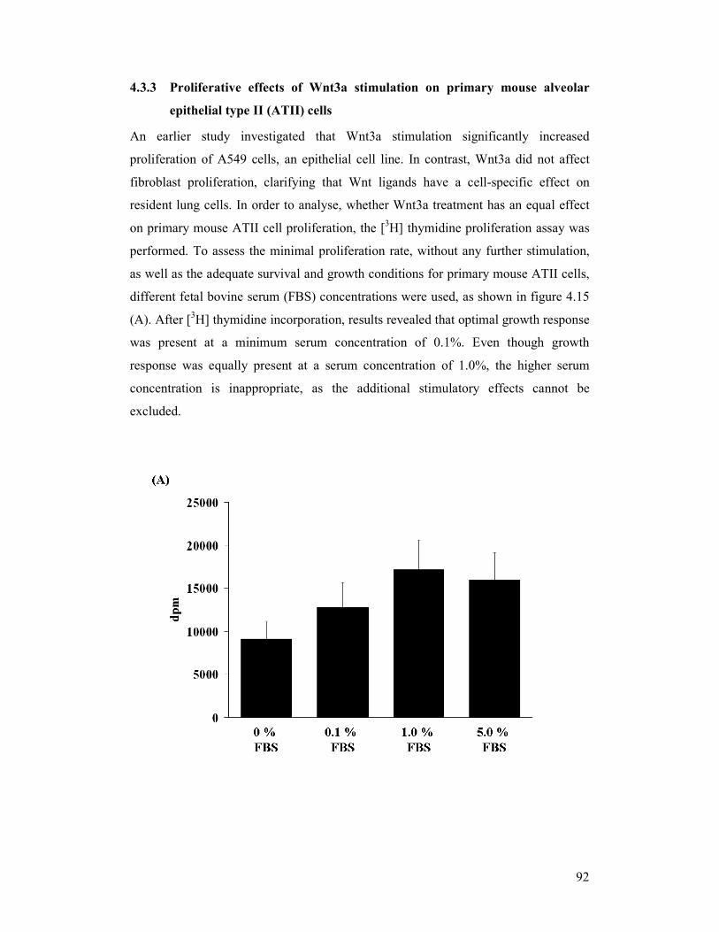
92
4.3.3 Proliferative effects of Wnt3a stimulation on primary mouse alveolar
epithelial type II (ATII) cells
An earlier study investigated that Wnt3a stimulation significantly increased
proliferation of A549 cells, an epithelial cell line. In contrast, Wnt3a did not affect
fibroblast proliferation, clarifying that Wnt ligands have a cell-specific effect on
resident lung cells. In order to analyse, whether Wnt3a treatment has an equal effect
on primary mouse ATII cell proliferation, the [3H] thymidine proliferation assay was
performed. To assess the minimal proliferation rate, without any further stimulation,
as well as the adequate survival and growth conditions for primary mouse ATII cells,
different fetal bovine serum (FBS) concentrations were used, as shown in figure 4.15
(A). After [3H] thymidine incorporation, results revealed that optimal growth response
was present at a minimum serum concentration of 0.1%. Even though growth
response was equally present at a serum concentration of 1.0%, the higher serum
concentration is inappropriate, as the additional stimulatory effects cannot be
excluded.
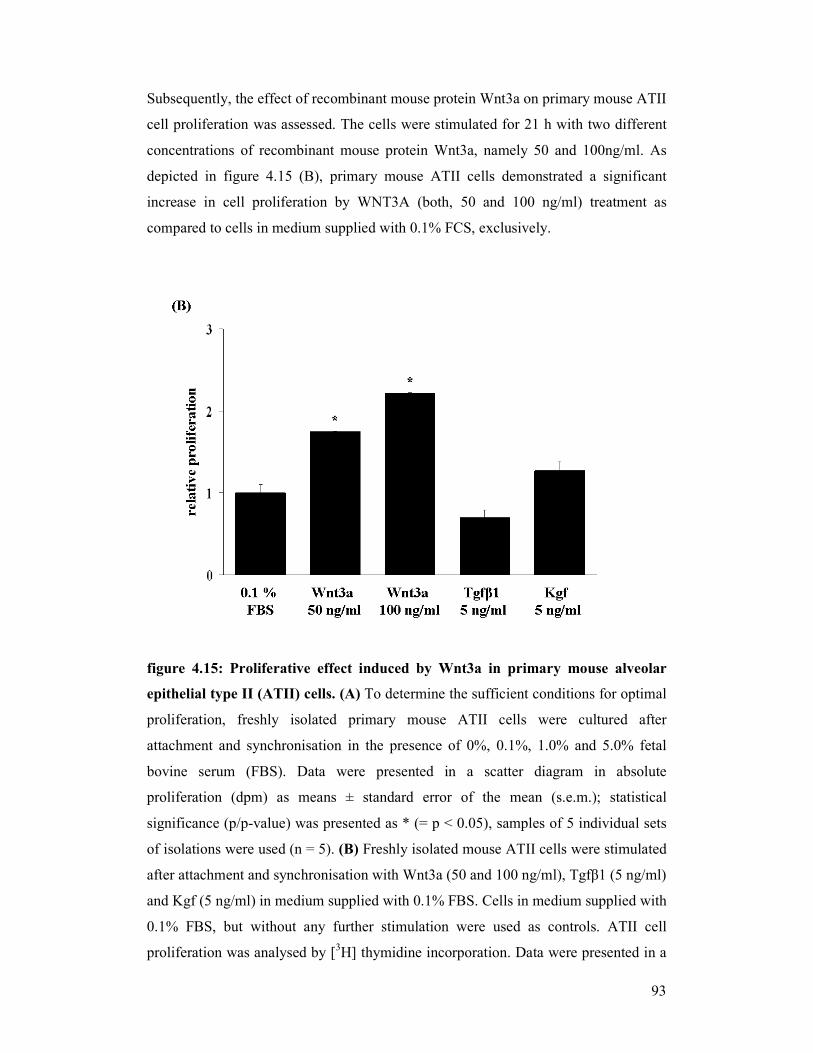
93
Subsequently, the effect of recombinant mouse protein Wnt3a on primary mouse ATII
cell proliferation was assessed. The cells were stimulated for 21 h with two different
concentrations of recombinant mouse protein Wnt3a, namely 50 and 100ng/ml. As
depicted in figure 4.15 (B), primary mouse ATII cells demonstrated a significant
increase in cell proliferation by WNT3A (both, 50 and 100 ng/ml) treatment as
compared to cells in medium supplied with 0.1% FCS, exclusively.
figure 4.15: Proliferative effect induced by Wnt3a in primary mouse alveolar
epithelial type II (ATII) cells. (A) To determine the sufficient conditions for optimal
proliferation, freshly isolated primary mouse ATII cells were cultured after
attachment and synchronisation in the presence of 0%, 0.1%, 1.0% and 5.0% fetal
bovine serum (FBS). Data were presented in a scatter diagram in absolute
proliferation (dpm) as means ± standard error of the mean (s.e.m.); statistical
significance (p/p-value) was presented as * (= p < 0.05), samples of 5 individual sets
of isolations were used (n = 5). (B) Freshly isolated mouse ATII cells were stimulated
after attachment and synchronisation with Wnt3a (50 and 100 ng/ml), Tgfβ1 (5 ng/ml)
and Kgf (5 ng/ml) in medium supplied with 0.1% FBS. Cells in medium supplied with
0.1% FBS, but without any further stimulation were used as controls. ATII cell
proliferation was analysed by [3H] thymidine incorporation. Data were presented in a
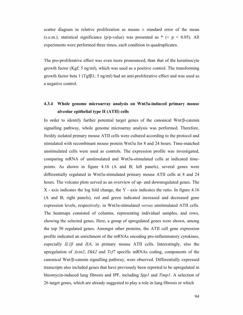
94
scatter diagram in relative proliferation as means ± standard error of the mean
(s.e.m.); statistical significance (p/p-value) was presented as * (= p < 0.05). All
experiments were performed three times, each condition in quadruplicates.
The pro-proliferative effect was even more pronounced, than that of the keratinocyte
growth factor (Kgf; 5 ng/ml), which was used as a positive control. The transforming
growth factor beta 1 (Tgfβ1; 5 ng/ml) had an anti-proliferative effect and was used as
a negative control.
4.3.4 Whole genome microarray analysis on Wnt3a-induced primary mouse
alveolar epithelial type II (ATII) cells
In order to identify further potential target genes of the canonical Wnt/β-catenin
signalling pathway, whole genome microarray analysis was performed. Therefore,
freshly isolated primary mouse ATII cells were cultured according to the protocol and
stimulated with recombinant mouse protein Wnt3a for 8 and 24 hours. Time-matched
unstimulated cells were used as controls. The expression profile was investigated,
comparing mRNA of unstimulated and Wnt3a-stimulated cells at indicated time-
points. As shown in figure 4.16 (A and B; left panels), several genes were
differentially regulated in Wnt3a-stimulated primary mouse ATII cells at 8 and 24
hours. The volcano plots served as an overview of up- and downregulated genes. The
X - axis indicates the log fold change, the Y - axis indicates the ratio. In figure 4.16
(A and B; right panels), red and green indicated increased and decreased gene
expression levels, respectively, in Wnt3a-stimulated versus unstimulated ATII cells.
The heatmaps consisted of columns, representing individual samples, and rows,
showing the selected genes. Here, a group of upregulated genes were shown, among
the top 50 regulated genes. Amongst other proteins, the ATII cell gene expression
profile indicated an enrichment of the mRNAs encoding pro-inflammatory cytokines,
especially IL1β and IL6, in primary mouse ATII cells. Interestingly, also the
upregulation of Axin2, Dkk2 and Tcf7 specific mRNAs coding, components of the
canonical Wnt/β-catenin signalling pathway, were observed. Differentially expressed
transcripts also included genes that have previously been reported to be upregulated in
bleomycin-induced lung fibrosis and IPF, including Spp1 and Timp1. A selection of
26 target genes, which are already suggested to play a role in lung fibrosis or which
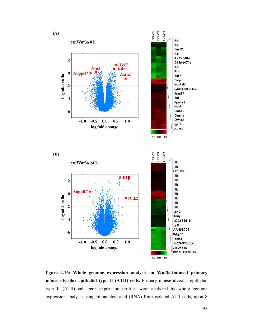
95
figure 4.16: Whole genome expression analysis on Wnt3a-induced primary
mouse alveolar epithelial type II (ATII) cells. Primary mouse alveolar epithelial
type II (ATII) cell gene expression profiles were analyzed by whole genome
expression analysis using ribonucleic acid (RNA) from isolated ATII cells, upon 8
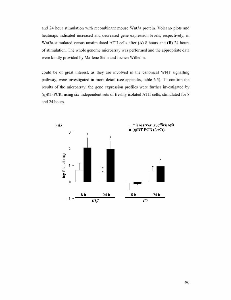
96
and 24 hour stimulation with recombinant mouse Wnt3a protein. Volcano plots and
heatmaps indicated increased and decreased gene expression levels, respectively, in
Wnt3a-stimulated versus unstimulated ATII cells after (A) 8 hours and (B) 24 hours
of stimulation. The whole genome microarray was performed and the appropriate data
were kindly provided by Marlene Stein and Jochen Wilhelm.
could be of great interest, as they are involved in the canonical WNT signalling
pathway, were investigated in more detail (see appendix, table 6.5). To confirm the
results of the microarray, the gene expression profiles were further investigated by
(q)RT-PCR, using six independent sets of freshly isolated ATII cells, stimulated for 8
and 24 hours.
97
figure 4.17: Messenger RNA (mRNA) expression of potential Wnt3a-induced
target genes in primary mouse alveolar epithelial type II (ATII) cells. The
confirmation of the microarray results for special selected genes was performed by
quantitative real time polymerase chain reaction (q)RT-PCR, using isolated primary
mouse alveolar epithelial type II (ATII) cells, stimulated with recombinant mouse
protein Wnt3a protein for 8 and 24 hours. The mRNA levels of (A) the cytokines,
interleukin 1β (IL1β) and interleukin 6 (IL6), as well as of (B) axis inhibition protein 2
(Axin2) and dickkopf-related protein 2 (Dkk2), inhibitors of the canonical Wnt/β-
catenin signalling pathway were assessed. Data were presented in a bar graph; plotting
the results as log-fold change (∆∆Ct) of mRNA levels, comparing the microarray
results with the (q)RT-PCR results; statistical significance (p/p-value) was presented
as * (= p < 0.05), samples of 6 individual isolations were used (n = 6).
Out of 26 selected genes, 20 genes were finally confirmed. In turn, out of these genes,
significantly upregulated expression of the cytokines IL1β and IL6 in Wnt3a-
stimulated primary mouse ATII cells were of special interest, as they are known to be
relevant in lung fibrosis. As shown in figure 4.17 (A), IL1β mRNA expression level
was significantly increased after stimulation with recombinant mouse protein Wnt3a
protein for 8 and 24 hours, while IL6 mRNA expression level was detectable and
increased only after 24 hours of stimulation. Equally interesting, as depicted in figure
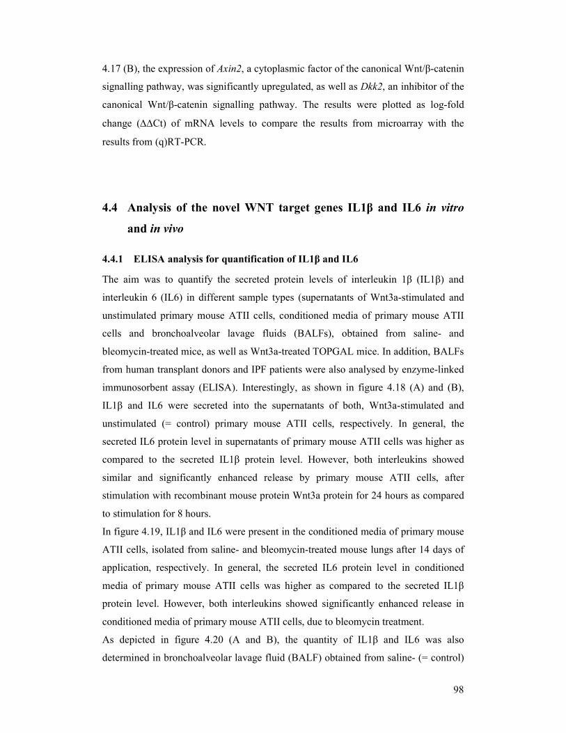
98
4.17 (B), the expression of Axin2, a cytoplasmic factor of the canonical Wnt/β-catenin
signalling pathway, was significantly upregulated, as well as Dkk2, an inhibitor of the
canonical Wnt/β-catenin signalling pathway. The results were plotted as log-fold
change (∆∆Ct) of mRNA levels to compare the results from microarray with the
results from (q)RT-PCR.
4.4 Analysis of the novel WNT target genes IL1β and IL6 in vitro
and in vivo
4.4.1 ELISA analysis for quantification of IL1β and IL6
The aim was to quantify the secreted protein levels of interleukin 1β (IL1β) and
interleukin 6 (IL6) in different sample types (supernatants of Wnt3a-stimulated and
unstimulated primary mouse ATII cells, conditioned media of primary mouse ATII
cells and bronchoalveolar lavage fluids (BALFs), obtained from saline- and
bleomycin-treated mice, as well as Wnt3a-treated TOPGAL mice. In addition, BALFs
from human transplant donors and IPF patients were also analysed by enzyme-linked
immunosorbent assay (ELISA). Interestingly, as shown in figure 4.18 (A) and (B),
IL1β and IL6 were secreted into the supernatants of both, Wnt3a-stimulated and
unstimulated (= control) primary mouse ATII cells, respectively. In general, the
secreted IL6 protein level in supernatants of primary mouse ATII cells was higher as
compared to the secreted IL1β protein level. However, both interleukins showed
similar and significantly enhanced release by primary mouse ATII cells, after
stimulation with recombinant mouse protein Wnt3a protein for 24 hours as compared
to stimulation for 8 hours.
In figure 4.19, IL1β and IL6 were present in the conditioned media of primary mouse
ATII cells, isolated from saline- and bleomycin-treated mouse lungs after 14 days of
application, respectively. In general, the secreted IL6 protein level in conditioned
media of primary mouse ATII cells was higher as compared to the secreted IL1β
protein level. However, both interleukins showed significantly enhanced release in
conditioned media of primary mouse ATII cells, due to bleomycin treatment.
As depicted in figure 4.20 (A and B), the quantity of IL1β and IL6 was also
determined in bronchoalveolar lavage fluid (BALF) obtained from saline- (= control)
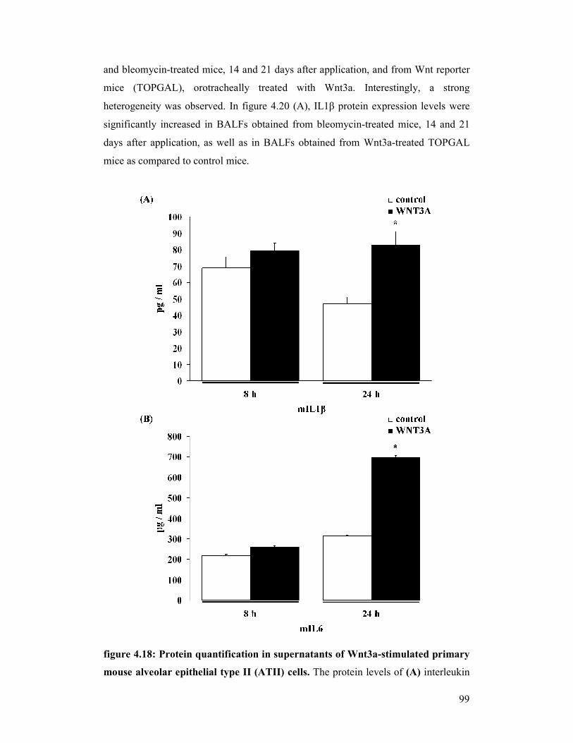
99
and bleomycin-treated mice, 14 and 21 days after application, and from Wnt reporter
mice (TOPGAL), orotracheally treated with Wnt3a. Interestingly, a strong
heterogeneity was observed. In figure 4.20 (A), IL1β protein expression levels were
significantly increased in BALFs obtained from bleomycin-treated mice, 14 and 21
days after application, as well as in BALFs obtained from Wnt3a-treated TOPGAL
mice as compared to control mice.
figure 4.18: Protein quantification in supernatants of Wnt3a-stimulated primary
mouse alveolar epithelial type II (ATII) cells. The protein levels of (A) interleukin
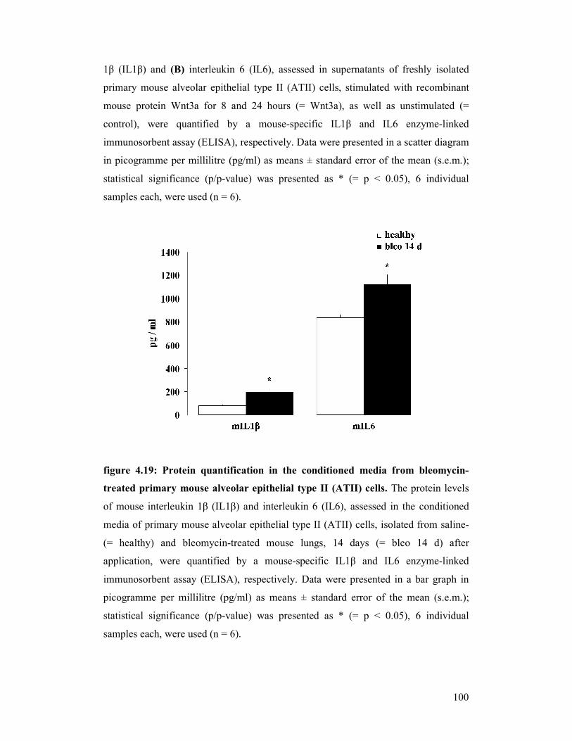
100
1β (IL1β) and (B) interleukin 6 (IL6), assessed in supernatants of freshly isolated
primary mouse alveolar epithelial type II (ATII) cells, stimulated with recombinant
mouse protein Wnt3a for 8 and 24 hours (= Wnt3a), as well as unstimulated (=
control), were quantified by a mouse-specific IL1β and IL6 enzyme-linked
immunosorbent assay (ELISA), respectively. Data were presented in a scatter diagram
in picogramme per millilitre (pg/ml) as means ± standard error of the mean (s.e.m.);
statistical significance (p/p-value) was presented as * (= p < 0.05), 6 individual
samples each, were used (n = 6).
figure 4.19: Protein quantification in the conditioned media from bleomycin-
treated primary mouse alveolar epithelial type II (ATII) cells. The protein levels
of mouse interleukin 1β (IL1β) and interleukin 6 (IL6), assessed in the conditioned
media of primary mouse alveolar epithelial type II (ATII) cells, isolated from saline-
(= healthy) and bleomycin-treated mouse lungs, 14 days (= bleo 14 d) after
application, were quantified by a mouse-specific IL1β and IL6 enzyme-linked
immunosorbent assay (ELISA), respectively. Data were presented in a bar graph in
picogramme per millilitre (pg/ml) as means ± standard error of the mean (s.e.m.);
statistical significance (p/p-value) was presented as * (= p < 0.05), 6 individual
samples each, were used (n = 6).
101
Generally, IL6 was detected at lower protein quantity levels than IL1β in mouse
BALFs. In contrast, as depicted in figure 4.20 (B), IL6 showed a significant
upregulation in BALFs obtained from Wnt3a-treated TOPGAL mice compared with
BALFs obtained from control mice.
figure 4.20: Protein quantification in mouse bronchoalveolar lavage fluids
(BALFs). The protein levels of (A) mouse interleukin 1β (IL1β) and (B) mouse
102
interleukin 6 (IL6), assessed in bronchoalveolar lavage fluids (BALFs) obtained from
saline- (= control) and bleomycin-treated mice, 14 and 21 days (= bleo 14 d and 21 d)
after application, and transgenic Wnt reporter/Wnt3a-treated TOPGAL (Tg Fos LacZ)
mice were quantified by a mouse-specific IL1β and IL6 enzyme-linked
immunosorbent assay (ELISA), respectively. Data were presented in a scatter diagram
in picogramme per millilitre (pg/ml) as means ± standard error of the mean (s.e.m.);
statistical significance (p/p-value) was presented as * (= p < 0.05), 12 individual
samples each, were used (n = 12).
In figure 4.21, the protein expression level of IL1β was measured in human samples.
Similar protein expression levels of IL1β were detected in BALFs from transplant
donors and IPF patients.
figure 4.21: Protein quantification in human bronchoalveolar lavage fluids
(BALFs). The protein levels of interleukin 1β (IL1β), assessed in bronchoalveolar
lavage fluids (BALFs) obtained from transplant donor (= control) and idiopathic
pulmonary fibrosis (IPF) patients were quantified by a human interleukin 1β (huIL1β)
enzyme-linked immunosorbent assay (ELISA). Data were presented in a scatter
diagram in picogramme per millilitre (pg/ml) as means ± standard error of the mean
(s.e.m.); 12 individual samples were used (n = 12).
103
5 DISCUSSION
The current study revealed that the canonical WNT/β-catenin signalling pathway
might at least partially be involved in the pathogenesis of lung fibrosis. It was
demonstrated that functional components of WNT/β-catenin signalling were present
in both, experimental murine and human idiopathic pulmonary fibrosis (IPF). The
expression and activity of the WNT signalling pathway was mainly detected in
bronchial and alveolar epithelium of the lung. In particular, this study identified
alveolar epithelial type II (ATII) cells as a major source and target of WNT signalling
and demonstrated putative autocrine effects of WNT ligands on cell proliferation and
WNT target gene expression in ATII cells. Additionally, it was identified that active
WNT/β-catenin signalling led to an increased expression of proinflammatory
cytokines, such as interleukin 1β (IL1β) and interleukin 6 (IL6), in ATII cells. The
interaction of WNT signalling and IL1β/IL6 is suggested to possibly play a crucial
role in the progression of IPF.
IPF, the most common form of the idiopathic interstitial pneumonias (IIP) in humans,
is a progressive and fatal lung disease, which is characterized by distorted lung
architecture and severe loss of respiratory function. IPF exhibits a poor prognosis due
to unresponsiveness to currently available therapies [24, 36]. Known to be active
during developmental processes, the canonical WNT/β-catenin signalling pathway has
been suggested to be reactivated in chronic diseases, which are characterised by
pathologic tissue remodelling [82]. In 2006, Douglas and colleagues showed that
active WNT/β-catenin signalling occurs in the fibro-proliferative phase after acute
lung inflammation, using a mouse model of oxidant-induced injury. Thus, the WNT
signalling pathway is suggested to be involved in lung regeneration after injury [85].
In this respect, the WNT signalling pathway is of special interest.
In recent studies, unbiased microarray screens revealed increased expression levels of
several WNT signalling genes, including WNT2 and 5A, FZD6, 7 and 10, and LEF1,
in IPF lungs as compared to transplant donor lungs [82, 86]. Most recently, it was
demonstrated that canonical WNT signalling induces skin fibrosis. Thus, transgenic
mice overexpressing Wnt10b proved to be an activator for the development of dermal
104
fibrosis, increased collagen deposition, fibroblast activation and myofibroblast
accumulation. These new transgenic mice represent a novel animal model, useful for
further studies of WNT signalling in fibrosis [87]. In 2003, Chilosi and colleagues
reported increased nuclear localisation of β-catenin (CTNNB1) in ATII cells and in
interstitial fibroblasts in IPF lungs [78], indicative of active WNT signalling in IPF
[79].
In the current study, a comprehensive analysis of the mRNA expression of canonical
WNT/β-catenin signalling components was performed in total lung homogenates from
IPF patients and transplant donors, demonstrating different expression of WNT
signalling components in IPF lung epithelium. An immunohistochemical analysis
revealed particularly bronchial and alveolar epithelium as specific cell types capable
of WNT signalling. Several WNT signal pathway components were largely expressed
in donor and IPF lung tissue, with increased immunohistochemical staining in
bronchial epithelial and hyperplastic ATII cells at sites of bronchiolisation under
conditions of IPF. Interestingly, also DKK1, a WNT regulator, was predominantly
localised in basal bronchial epithelial cells and in hyperplastic alveolar epithelial cells
in IPF, which may be a suitable therapeutic target for IPF [88].
Normally, β-catenin levels are tightly regulated in the developing and healthy adult
lung, with low cytoplasmic/nuclear β-catenin [89]. Several studies investigated WNT
signalling in lung morphogenesis demonstrating, that a deletion of β-catenin in
epithelial cells impairs lung morphogenesis, arrests the differentiation of alveolar
epithelial cells and leaves the lung containing mainly conducting airways.
Consistently, aberrant activation of β-catenin in epithelial cells of the developing lung
causes enlarged air space, atypical expression of ATII cells and goblet cell
hyperplasia [61, 90]. In this study only rare nuclear staining of β-catenin was
observed; however, Chilosi and colleagues identified nuclear localisation of β-catenin
by immunhistochemistry [78]. Recent investigations have provided evidence
supporting the sparse nuclear localisation of β-catenin, as it plays a key role in
canonical WNT signalling and only a minority of the intracellular β-catenin content in
epithelial cells is responsible for WNT signalling, while the majority of β-catenin
molecules is present at the localisation of cell-cell contacts [91, 92]. Although nuclear
β-catenin expression is difficult to determine in general, different tissue preparation
techniques or antibodies used for IHC could also be responsible for the observed
105
differences. It was shown that in vivo constitutive β-catenin signalling is not exhibited
in adult and healthy ATII cells, but rather, this pathway is activated during lung
injury, known to promote alveolar epithelial cell survival, migration, and
differentiation towards an ATI-like cell phenotype. Accordingly, these results
suggested that ATII cells may function as facultative progenitor cells [93].
In general, it was evident that β-catenin-mediated WNT signalling is central to
mechanisms of lung healing in stem cell maintenance, as well as in several diseases
like fibrosis and cancer. In detail, β-catenin signalling is known to stimulate cell fate,
migration, differentiation, wound closure and tissue remodelling, as well as tissue
destruction through several downstream mediators like MMPs, IL1β, IL6 or TGF-β.
Furthermore, β-catenin, an essential mediator of the canonical WNT signalling
pathway, promotes the expression of cyclinD1, which additionally indicates cell
proliferation [94]. Most recently, Kim and colleagues [95] observed that the use of β-
catenin siRNA was able to block the WNT/β-catenin pathway. While Henderson and
his working group [96] inhibited the WNT pathway at the nuclear transcription level,
in this study the effector molecule was inhibited at the cytoplasm level. Thereby,
intratracheal instillation of β-catenin siRNA was utilised, which selectively and
effectively inhibited β-catenin and reduced TGF-β expression. Bleomycin-induced
pulmonary fibrosis was attenuated as an outcome of this. These findings reconfirmed
that using specific siRNA to target WNT/β-catenin signalling, may be a safe and
effective therapeutic approach in the treatment of IPF. However, a noninvasive
procedure for intrapulmonary delivery of aerosolized siRNA poses a major challenge
[95]. It was shown in former studies that the use of siRNA to block specific key
molecules by gene silencing has significant effects. There have been several
preclinical and clinical trials for siRNA treatment in various lung diseases [97].
In this study, functional WNT signalling in IPF was further assessed at the protein
level. Increased phosphorylation of LRP6 and GSK3β, combined with increased
expression of total β-catenin in total lung homogenates of IPF patients as compared to
transplant donors, indicates activation of the WNT/β-catenin signalling pathway.
Recently it was demonstrated that increased phosphorylation of LRP6 and GSK3β are
the most sensitive indicators of WNT activity in tissue sections [98, 99]. Additionally,
an earlier study showed that gene expression analysis of WNT signalling components
in primary human ATII cells from IPF patients further corroborated the results [100].
106
WNT1, however was not regulated in the freshly isolated primary ATII cells, but in
the lung homogenates from IPF patients. Therefore, other cell types, such as
endothelial cells or bronchial smooth muscle cells may serve as possible sources of
WNT1 expression in IPF.
Subsequently, the WNT expression and activity were assessed in an experimental
model of lung fibrosis. Different models of pulmonary fibrosis have been developed
over the years. However, it is not possible to display all features of IPF, especially the
progressive and irreversible nature of this disease [101]. The bleomycin-induced
mouse model of experimental lung fibrosis, a well-studied model of IPF, is generally
used to study the pathogenetic mechanisms of this disease. As a single intratracheal
administration of bleomycin, an anti-neoplastic agent, induces peribronchial fibrosis
in the mouse, this condition comparatively reproduces the histological features of IPF
in humans [102]. The instillation of bleomycin is easy to handle, reproducible and
easily available. Overall, the model of bleomycin-induced pulmonary fibrosis is
widely accepted to investigate the molecular mechanisms in vivo and to identify new
targets for medication. However, this model has its limits, as bleomycin-induced
fibrosis is reversible, and as such, has to be carefully compared with IPF [101].
To further elucidate whether WNT/β-catenin activation is also present in experimental
lung fibrosis, a comprehensive analysis was performed by qRT-PCR. The current
study shows the occurrence of essential WNT/β-catenin signalling components in the
mouse lung, and regulation thereof in ATII cells. These findings are in agreement
with the results observed in IPF and donor transplant tissue, suggesting ATII cells as
target cells for expression of canonical WNT signalling components. A recent study
has demonstrated that mice treated with freshly isolated primary ATII cells, by
intratracheal instillation, show reduced manifestations of bleomycin-induced
pulmonary fibrosis [103]. Immunohistochemical analysis confirmed these results,
showing enhanced staining of alveolar epithelial cells in bleomycin-treated mouse
lungs compared with saline-treated mouse lungs. Additionally, in 2009, Liu and
colleagues reported increased nuclear localisation of β-catenin in epithelial cells of
remodelled and fibrotic lung areas, indicating active WNT signalling in bleomycin-
induced pulmonary fibrosis [104]. Importantly, a recent study used TOPGAL reporter
mice to localise the activation of the WNT/β-catenin pathway in vivo in experimental
107
lung fibrosis. Accordingly, mice were treated orotracheally with either recombinant
Wnt3a to demonstrate the capability of the lung to activate WNT/β-catenin signalling
or with bleomycin to induce lung fibrosis. It has been shown that bronchial and
alveolar epithelial cells stained for β-galactosidase (β-GAL), in response to Wnt3a or
bleomycin treatment [39]. WNT ligands are mainly secreted by lung epithelial cells,
indicating that WNT signalling in the adult lung initiates from the epithelium and may
act mainly in an autocrine fashion on epithelial cells in the early phase of lung fibrosis
development. Liu and colleagues observed that WNT/β-catenin signalling is activated
in murine bleomycin-induced lung fibrosis. Accordingly, target genes like cyclinD1,
Mmp7, and Tgfβ were expressed and localised in bleomycin-treated transgenic Wnt
reporter mice [104]. Recently, another study reconfirmed that bleomycin exposure
induces aberrant activation of Wnt/β-catenin signalling. Additionally, it was
demonstrated that administration of ICG-001, a small molecule, which inhibits T-cell
factor/β-catenin transcription, selectively blocks the β-catenin/CBP interaction and
with it reduces the expression of WNT target genes. The administration of ICG-001
concurrent with bleomycin not only prevented, but attenuated and reversed
established pulmonary fibrosis, enhanced tissue repair and significantly improved
survival. As canonical WNT signalling is known to be of particular importance in the
pathogenesis of IPF, the selective blockade of the pathway displays a further potential
therapeutic approach [96].
In sum, increased WNT/β-catenin signalling occurs at sites of alveolar epithelial cell
injury and hyperplasia in response to bleomycin, underlining the impact of this
pathway on impaired alveolar repair in the progress of the disease. Thus, activation of
WNT signalling may be important in the course of IPF.
In the present study, the autocrine effects of WNT signalling, particulary the role of
WNT signalling on ATII cell proliferation, was analysed. The importance of ATII cell
proliferation in lung fibrosis has been demonstrated in experimental as well as human
idiopathic fibrosis. ATII cells from bleomycin-treated mice reveal a significant
increase in cell proliferation compared with ATII cells from saline-treated control
mice. These findings point out that initial lung injury may lead to ATII cell
proliferation, which represents an important pathomechanism in experimental lung
fibrosis [39]. Interestingly, a recent study showed that higher concentrations of DKK1
led to a reduction of WNT3A-induced effects, while no significant effect on epithelial
108
cell proliferation was observed after stimulation of human bronchial and epithelial cell
lines with DKK1 alone [88]. The dose-dependent exertion of DKK1 can attenuate
WNT signalling effects, and therefore may act as a potential therapeutic agent.
Epithelial restitution involves acute inflammatory response, survival, apoptosis,
proliferation and migration of cells to restore cell numbers and differentiation to
recover function. But regional differences within the lung when coupled with
persistent injury may contribute to the pathology of several diseases. Abnormalities in
these epithelial cell functions contribute to the pathogenesis of IPF [36, 94]. Active
WNT signalling is known to cause epithelial cell hyperplasia in non-pulmonary
epithelia. Therefore, WNT signalling may be a mediator for ATII cell hyperplasia and
increased bronchial epithelial cell proliferation in IPF [44].
WNT3A was used in this study to assess the functional effects of canonical WNT
signalling in epithelial cells. Although WNT3A was the only ligand, which was not
upregulated in IPF, several in vitro studies reported WNT3A as a potent inducer of
WNT/β-catenin signalling [105], which is also recognized as the prototypic WNT
ligand for in vitro stimulations [42]. It was demonstrated, using a TCF/LEF-driven
reporter gene assay that WNT3A, but not WNT7A, initiates a potent canonical
WNT/β-catenin response [100]. A recent study revealed that WNT3A increased levels
of N-cadherin, α-catenin and β-catenin in primary cultured chondrocytes. In addition,
WNT3A caused a significant translocation of β-catenin to the nucleus and increased
the transcriptional activity of the β-catenin-TCF/LEF complex [106]. Importantly,
WNT3A belongs to one of the few WNT ligands, which is available in active and
recombinant form. Other WNT ligands used in in vitro studies, such as WNT1, are
commonly either overexpressed by viral transduction, or supplied in the form of
conditioned media harvested from WNT1 overexpressing cell lines [107, 108]. The
current study demonstrates that stimulation with the canonical murine WNT ligand
Wnt3a leads to a significant increase in ATII cell proliferation. ATII cells are
generally considered to be the facultative progenitor cells for the recovery of the
pneumocyte pools, with the capability to differentiate into ATI cells following lung
injury. However, it should be pointed out that the evidence in support of the role of
ATII cells as progenitors is based in part on earlier studies showing increased ATII
cell proliferation following injury and the incorporation of [3H]-thymidine in ATII
cells [94]. In the lung, the two most abundant epithelial cell types are responsible for
109
forming the lung air barrier. The ATI cell is distinguished by its flattened shape and
large surface area, which presumably facilitates the diffusion of oxygen and carbon
dioxide. The ATII cell produces surfactants, which reduce surface tension, among
other functions; given the number of alveoli and their extensive surface area [109-
111]. These data are in accordance with studies, reporting induction of proliferation in
neural progenitor cells in response to WNT3A [112]. In addition, these results have
been confirmed in A549 lung epithelial cells, showing a strong increase of cell count,
upon WNT3A stimulation [100]. This study focused on canonical WNT signalling.
Interestingly, a recent study showed increased expression of WNT5A on mRNA and
protein levels in fibroblasts isolated from UIP lung tissue compared with normal
fibroblasts, pointing to a difference in canonical and non-canonical WNT signalling
pathway. WNT5A was identified as a regulator of fibroblast proliferation [86]. These
observations suggest that the secretion and function of WNT ligands occur in a cell-
specific manner.
Additionally, potential target genes of the WNT/β-catenin signalling pathway in ATII
cells were identified in this study using whole genome microarray analysis. Several
other study groups have observed active Wnt/β-catenin signalling and increased
expression of pathway components by placing primary ATII cells in culture [113,
114]. Initially, it was investigated that primary mouse ATII cells were responsive to
Wnt3a stimulation as they expressed known WNT target genes, such as cyclinD1 and
Wnt1-inducible signalling protein 1 (Wisp1). WISP1, which belongs to the CCN
family of growth factors, was identified as a direct target gene of the WNT/β-catenin
signalling pathway, having anti-apoptotic and proliferative effects on epithelial and
mesenchymal cells [115, 116]. Interestingly, WISP1 has been recently identified to
play a crucial role in experimental and human pulmonary fibrosis. Increased mRNA
and protein expression levels of WISP1 were observed in ATII cells from IPF lungs
compared with ATII cells from transplant donor lungs. Furthermore, autocrine effects
of WISP1 were reported, leading to ATII cell hyperplasia and expression of
profibrotic cytokines [39]. In addition, MMP7, a known key mediator of pulmonary
fibrosis and found on the surface of lung epithelial cells, is one of several MMPs that
activate TGF-β. To come full circle, TGF-β has been identified as a “master switch”
in the induction of EMT in several tissues including the lung, and, specifically,
together with β-catenin, in ATII cells, which reconfirms the crucial role of WNT/β-
110
catenin signalling in IPF. This activation directly or indirectly stimulates many of the
proinflammatory cytokines, like IL1β, that participate in inflammation-mediated
tissue destruction and elaboration of ECM [94]. In spite of these facts, the MMP
expression levels in this study were unchanged.
Normally, the WNT signalling system is tightly controlled in a spatiotemporal manner
by different secreted WNT regulators. The unbiased microarray revealed increased
expression levels of the WNT regulators Axin2 and dickkopf2 (Dkk2) have been
observed in Wnt3a-stimulated ATII cells. Axin2, which is the human homologue of
mouse conductin and rat axil, is known to be a component of WNT signalling, which
directly interacts with β-catenin, GSK3β and APC to regulate phosphorylation and
stability of β-catenin and is implicated in downregulation of WNT signalling.
Overexpression of Axin destabilises β-catenin, induced by active WNT signalling and
therefore acts in a negative feedback loop [117, 118]. Recent studies indicated that
isolated and cultured ATII cells may recapitulate key aspects of an in vivo response to
alveolar lung injury. Interestingly, Axin2 expression was detected in ATII cells of
saline- treated control mice, raising suspicion that a low level of WNT/β-catenin
signalling is present in a distinct cell population [93]. Among the dickkopf (Dkk)
family, four different Dkk proteins (Dkk1-4) have been discovered, which share
conserved cysteinerich domains. Dkk2, an extracellular protein, belongs to the group
of WNT regulators, which modulates WNT signalling by binding to the WNT binding
domain of LRP6. Dkk2 can function either as an activator or inhibitor of WNT
signalling, depending on its cellular context and the availability of WNT and co-
receptors. Additionally, Dkk proteins are expressed in response to active WNT/β-
catenin signalling and bind with high affinity to a second class of receptors, termed
Kremen (Krm). The complex formation of DKK, KRM, and LRP6 is thought to lead
to endocytosis of the whole receptor complex, which ultimately potentiates the ability
of DKK to regulate and block WNT signalling [119-121]. Most recently, a study
demonstrated altered expression of WNT regulators in IPF. In whole lung
homogenates from IPF patients increased expression of DKK1, DKK4 and KRM1
was detected. In addition, DKK1 was expressed with a significantly increased amount
in BALF of IPF patients.
These observations suggested that DKK proteins may be crucial for lung epithelial
cell injury and repair mechanisms in IPF. Further studies are needed to elucidate the
effects of DKK proteins on different cell types to reveal the potential therapeutic
111
capability in IPF [88]. Recent studies demonstrated that the blockade of the WNT
signalling pathway, using recombinant DKK proteins, inhibited renal and liver
fibrosis [77, 122].
In this context, recent studies showed that aberrant WNT/β-catenin signalling is also
linked to the pathogenesis of renal fibrosis and that blockage of the signal
transduction leads to intervention of disease progression. Certain qualities of some
currently available drugs have modulating effects on the signalling pathway [123].
One aim would be to develop orally active WNT inhibitors, providing a potentially
important pharmacological resource for further investigation of the WNT/β-catenin
signalling, as well as offering a possible novel therapeutic target in pulmonary
fibrosis.
Soluble factors, like growth factors (TGF-β, KGF), cytokines (IL1β, IL6, IL8, IL-13),
chemokines (MCP-1), prostaglandins (PGE2) and matrix components participate in
epithelial repair [94]. In addition, the microarray screen in this study identified
interleukin1β (IL1β) and interleukin (IL6) 6 as novel WNT target genes. Of all genes
differentially expressed in Wnt3a-stimulated murine ATII cells, this study focused on
the cytokines IL1β and IL6, for the following reasons: First, a study from Kolb and
colleagues showed that IL1β plays a crucial role in the development of experimental
pulmonary fibrosis [37]. A recombinant replication-deficient adenovirus vector was
used to transfer and overexpress the gene for human IL1β in epithelial cells of rodent
lungs. The extended expression of IL1β leads to acute alveolar injury, subsequently
inducing TGF-β expression and severe progressive lung fibrosis. Matrix deposition
and fibroblast foci appeared similar to those seen in human pulmonary fibrosis.
Additionally, a significant accumulation of extracellular collagen and fibronectin was
observed. IL1β is known to directly induce acute and chronic inflammation,
enlargement of distal airspaces, and airway fibrosis in the adult mouse [124] and is
suggested to be a therapeutic target in diseases associated with fibrosis and tissue
remodelling, such as IPF. Second, it has been reported that IL1β is produced in
injured and apoptotic lung epithelial cells upon bleomycin administration. For optimal
IL1β production, autocrine or paracrine mechanisms are discussed that amplify the
response. IL1β is known to stimulate the induction of other pro-inflammatory
cytokines. Accordingly, significantly increased concentrations of IL6 and TNFα were
observed in bronchoalveolar lavage fluid (BALF) from treated rodents [37, 125].
112
Third, IL6 is known to be involved in the pathogenesis of various inflammatory
diseases. Lung pathology after bleomycin exposure showed significant fibrotic
changes with increased collagen content in IL6-deficient mice compared with wild-
type mice. These results indicate that IL6 may play an important role in the
pathogenesis of bleomycin-induced lung injury and subsequent fibrotic changes [126].
Forth, several studies observed that IL1β in general is able to regulate WNT
signalling. One group tested the expression of various WNT molecules, using IL1β in
primary culture chondrocytes obtained from rabbits. IL1β led to upregulated β-catenin
levels, finally resulting in a significant increase of Wnt5a and 7a expression levels in
primary culture chondrocytes [127]. Newest studies demonstrated that fibrotic tissue
remodelling is induced by distinct cytokine-dependent mechanisms. Thereby, the
effector cytokines IL13 and IL17A played central roles. Moreover, these findings
suggested that TGF-β and proinflammatory mediators like IL1β promote fibrosis by
regulating and increasing the production of IL17A [128]. Hence, the question arises,
whether IL1β and / or IL6 accordingly mediate the fibrotic effects of WNT signalling.
ATII cells, one of the effector cells in IPF have been shown to be a major source of
cytokines and growth factors, including interleukins (ILs) [129-131]. ILs also termed
as cytokines, are non-structural, highly active proteins. IL1 family members alter the
host response to an inflammatory, infectious or immunological challenge and are
involved in various disorders of the lung. IL1α and IL1β are inflammatory cytokines,
which bind with similar affinity to the IL1 receptor (IL1R) and the IL1R accessory
protein (IL1RA). The IL1RA acts as a natural antagonist of IL1α/IL1β by trapping
IL1R1 molecules. IL1β is synthesized as an inactive cytoplasmic precursor (pro-
IL1β), which essentially lacks biological effects. The active, mature IL1β is produced
upon cleavage of pro-IL1β by a specific IL1β converting enzyme (ICE or caspase-1)
or by proteases [132]. IL1β is a pro-inflammatory cytokine primarily produced by
activated macrophages and monocytes; as well as by fibroblasts, neutrophils,
bronchial and alveolar epithelial cells. As a central cytokine it is involved in the
initiation and persistence of inflammation [133]. IL1β induces the production of IL6
or TNFα, thereby leading to neutrophil recruitment and fibroblast proliferation, which
may elicits potent fibrotic responses. These findings suggest that IL1β may be a
potent inducer and mediator of several other members of the cytokine family.
Analyses of bleomycin-treated whole mouse lung homogenates revealed significantly
113
increased levels of several IL17A-promoting cytokines, most strikingly the mediators
IL1β and IL6. IL1β mRNA expression and cytokine production correlated with the
degree of fibrosis after intratracheal administration of bleomycin [128]. This
observation has been confirmed in several recent studies, suggesting an important role
for IL1β in the development of bleomycin-induced pulmonary fibrosis [125, 134-
136]. In primary rat ATII cells, as well as in A549 cells, as a model of human ATII
cells, IL1β stimulation induced a significant increase of IL6 [137]. Recent studies
reported that ATII cells secreted several cytokines, such as monocyte chemotactic
protein (MCP)-1, macrophage inflammatory proteins (MIP)-1α and -2, interleukin
(IL) 8, and chemokine (C-C motif) ligand (CCL) 5 in response to IL1β [129, 131]. A
number of human and animal studies have revealed the presence of IL1β in tissues
undergoing fibrogenesis with accumulation of myofibroblasts and matrix deposition,
such as chronic fibrotic lung diseases [132, 138, 139].
The family of IL6-type cytokines, including IL6, activates target genes, which are
involved in differentiation, survival, apoptosis and proliferation. IL6 binds to plasma
membrane receptor complexes containing the common signal transducing receptor
chain glycoprotein 130 (gp 130) [140]. IL6 is released by a variety of cell types
including monocytes, macrophages, fibroblasts, endothelial and epithelial cells,
induced by inflammatory and profibrotic mediators, such as IL1β, TNFα, PDGF and
TGFβ. Accordingly, it is suggested that IL6 contributes to the onset and maintenance
of several ILDs [141, 142]. Moreover it has been shown that IL1β induced IL6, IL8
and MCP1 expression in bronchial epithelial cells or fibroblasts [128].
Both, IL1β and IL6 were upregulated upon WNT3A stimulation in vitro as well as in
vivo. Importantly, IL1β and IL6 were increased in the supernantants of ATII cells
isolated from fibrotic mouse lungs, as well as in the BALF from these mice. These
results conform to the investigation that IL1β was also increased in the BALF of IPF
patients. Additionally, IL17A expression levels were increased, indicting that these
cytokines may be involved in the development of IPF [128]. In accordance, a recent
study showed activated WNT/β-catenin signalling, with increased WNT7B expression
levels, in rheumatoid arthritis and osteoarthritis. Interestingly, synovium from
rheumatoid arthritis patients and WNT7B-transfected normal synovial cells showed
increased levels of IL1β, IL6 and TNFα compared with normal samples [143].
114
Most recently, studies investigated the correlation of several cytokine levels with
cellular characteristics in BALFs and lung function parameters in different ILDs.
Interestingly, increased IL1 receptor antagonist (IL1RA) expression was observed in
IPF. Additionally, increased interleukin (IL)8 expression is suggested to correlate
with prominent fibrosis and strong lung function decline [144, 145]. In addition,
significantly increased levels of IL6 have been detected in BALFs of different
pathologic conditions, including IPF [146]. Interestingly, in this study, no IL6
expression levels were detectable in BALFs from IPF patients. Possible reasons may
be discrepancies in the BALF extraction methods or preparation techniques. Several
studies investigated different animal models of acute lung injury and pulmonary
fibrosis, by application of bleomycin, silica or irradiation, leading to increased
cytokine expression [138, 139]. It has been shown that the expression of IL1β and IL6
in bleomycin-induced pulmonary fibrosis is influencing fibroblast activation,
proliferation and collagen deposition [147]. In addition, it was shown that exogenous
rmIL1β recapitulates acute inflammation induced by bleomycin with alveolar tissue
destruction and remodelling, resulting in progressive interstitial fibrosis, confirming
the proinflammatory and profibrotic effects of IL1β [37, 125]. These observations
were confirmed and amplified by showing that IL1β-mediated inflammation and
fibrosis is dependent on IL17A, a critical downstream mediator. IL17A deficiency
significantly reduced bleomycin-induced fibrosis in wildtype mice, as shown by
analysis of collagen expression in the BALF and collagen deposition in the lung. In
bleomycin-treated IL17A-/- mice compared with wildtype animals reduced
inflammation and fibrosis, associated with decreased IL1β expression was observed.
In vitro experiments with cultured fibroblasts demonstrated that IL17A promotes IL1β
production and vice versa [128, 148, 149].
In sum, it has been suggested that IL17A and IL1β cross-regulate each other, which
explains their additive roles in bleomycin-induced fibrosis.
Furthermore, in IL6-deficient mice attenuated fibrotic changes were observed
compared with wild-type mice, upon bleomycin treatment. These results indicate that
IL6 may play an important role in the pathogenesis of bleomycin-induced lung
fibrosis [126]. These cytokines have been demonstrated to be produced at the sites of
active fibrosis where they appear to be expressed by activated inflammatory cells,
such as macrophages. More interestingly, other non-inflammatory lung cells,
115
including myofibroblasts and epithelial cells, have been found to be significant
sources as well. The regulation of diverse functions of lung inflammatory and
epithelial cells can further influence the fibrotic process by autocrine and paracrine
mechanisms [150]. Latest findings assign IL13 an impact with fibrotic activity;
suggesting a role in bleomycin-induced pulmonary fibrosis. It has been indicated that
IL13 triggers fibrosis by inducing and activating TGF-β signalling. Of special interest
is a recently described IL1β-driven model of experimental fibrosis, which mimics
bleomycin-induced pulmonary fibrosis. Therefore, IL1β is suggested to be a critical
mediator of pulmonary fibrosis, highly dependent on IL17A by stimulating the
production [128].
In summary, the WNT/β-catenin signaling pathway is one of the core signal
transduction pathways. This study demonstrates that WNT/β-catenin signalling is
expressed in human and mouse lung epithelium. These results provided evidence that
WNT signalling pathway components are mainly increased and active in the bronchial
and alveolar epithelium in IPF and experimental lung fibrosis, and may influence
alveolar epithelial cell injury and hyperplasia due to significant dysregulation of the
tightly controlled WNT/β-catenin pathway. Therefore it is strongly suggested that
especially WNT3A, which is aberrantly secreted by ATII cells in IPF, takes a crucial
role in the development of the disease and modification of WNT signalling may
represent a therapeutic option in IPF.
IL17A was identified as a critical mediator of pulmonary fibrosis after bleomycin
administration, a blockade of IL17A could act as a potential treatment of pulmonary
fibrosis [128]. It will be important to translate those approaches, identified from
studies of cultured cells, into studies of human cells. There is a significant need for
understanding how these varied and sometimes disparate signals are integrated into a
coordinated response. This will require increased understanding of the source of the
mediators, the pathways that become activated, the timing of their activation, their
spatial targeting, and the factors that regulate their responsiveness.
Anti-inflammatory therapies including corticosteroids have limited efficacy in this
ultimately fatal disorder; an important unmet need is to identify new agents that
interact with key molecular pathways involved in the pathogenesis of pulmonary
fibrosis to prevent progression or reverse fibrosis in these patients. Because no
effective treatment for IPF exists, selective regulation of WNT/β-catenin-dependent
transcription suggests a potential previously unexplored therapeutic approach for IPF.
116
Alveolar cell death is one of the chief causes, which impairs lung diseases, like
fibrosis. Therefore, several studies suggested that the use of pharmacological
activators of the canonical WNT pathway possibly stimulates tissue reconstruction,
and especially alveolar epithelial cell repair [151, 152]. Flozak and colleagues used
adenoviral vectors to express proteins in ATII cultures that would constitutively
activate or inhibit β-catenin signalling. Activation of this pathway can be achieved by
expressing a point mutant version of β-catenin, resistant to degradation. Conversely,
inhibition of this pathway can be achieved through expression of ICAT, a polypeptide
that specifically antagonizes β-catenin transcription through binding β-catenin and
competing for its interactions with both TCF/p300 [93]. Interestingly, a recent study
observed reduced expression of the WNT/β-catenin signalling pathway in two
different mouse models of experimental chronic obstructive pulmonary disease
(COPD) and emphysema, using cigarette smoke exposure and elastase instillation in
wildtype and TOPGAL reporter mice, respectively. In general, COPD is characterised
by parenchymal tissue destruction and impaired repair capacity, resulting from an
imbalance between lung injury and repair processes. Furthermore, LiCl, a well-known
activator of WNT/β-catenin signalling, was used and demonstrated enhanced
activation of the signal pathway in lung epithelial cells, which led to a significant
reduction of experimental emphysema with restored alveolar epithelial structure and
function. These results not only suggest that reduced activity of WNT/β-catenin
signalling is a common and relevant feature of COPD and emphysema development,
but also that activation may be a preventive and effective therapeutic approach [153].
5.1 Conclusions and future perspectives
Among all idiopathic interstitial pneumonias (IIP), idiopathic pulmonary fibrosis
(IPF) is the most common and most severe form. As of now, the pathomechanisms are
not completely understood, which leads to the fact that appropriate therapies do not
yet exist. In this study, evidence for the reactivation of WNT/β-catenin signalling,
including several WNT target molecules, which contribute to the development and
progression of fibrosis, is provided. The key finding of the present study is that
canonical WNT signalling/WNT pathway components are expressed and operative in
adult lung epithelium of experimental and idiopathic pulmonary fibrosis. In addition,
117
the interleukins, IL1β and IL6 were identified as novel WNT target genes expressed
by the alveolar epithelium in lung fibrosis, likewise presumed to be involved in the
pathogenesis of the disease. Future studies analyzing the autocrine and paracrine
effects of IL1β/IL6 and crosstalk with WNT/β-Catenin pathway, in particular
WNT3A in alveolar epithelial cells, are required to reveal the functional effects.
Detailed in vivo and in vitro will shed more light into the relevance of this proposed
signalling pathway.
In light of recent research regarding IPF and WNT signalling this detection strongly
suggests WNT/β-catenin signalling and its effectors to be activated and expressed in
ATII cells during the pathogenesis of IPF. Inhibiting WNT/β-catenin signal
transduction more specifically or antagonizing its downstream effectors, using novel
small molecules, could possibly prevent the progression of the disease. In summary,
the aim of further investigation is to elucidate the downstream mechanisms and the
effects of WNT in more detail. Thus, modification of WNT signalling and better
understanding of the cytokines and the cytokine networks involved in IPF leads to the
possibility of new therapeutic approaches.
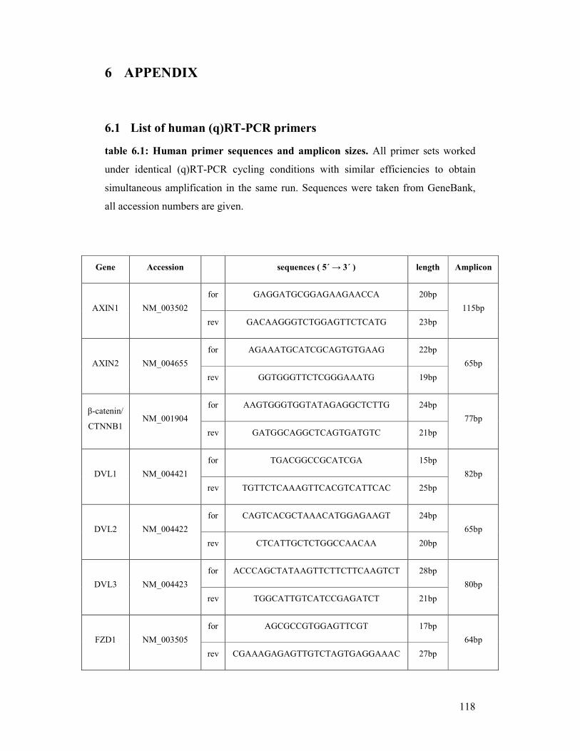
118
6 APPENDIX
6.1 List of human (q)RT-PCR primers
table 6.1: Human primer sequences and amplicon sizes. All primer sets worked
under identical (q)RT-PCR cycling conditions with similar efficiencies to obtain
simultaneous amplification in the same run. Sequences were taken from GeneBank,
all accession numbers are given.
Gene Accession sequences ( 5´ → 3´ ) length Amplicon
for GAGGATGCGGAGAAGAACCA 20bp
AXIN1 NM_003502
rev GACAAGGGTCTGGAGTTCTCATG 23bp
115bp
for AGAAATGCATCGCAGTGTGAAG 22bp
AXIN2 NM_004655
rev GGTGGGTTCTCGGGAAATG 19bp
65bp
for AAGTGGGTGGTATAGAGGCTCTTG 24bp β-catenin/
CTNNB1 NM_001904
rev GATGGCAGGCTCAGTGATGTC 21bp
77bp
for TGACGGCCGCATCGA 15bp
DVL1 NM_004421
rev TGTTCTCAAAGTTCACGTCATTCAC 25bp
82bp
for CAGTCACGCTAAACATGGAGAAGT 24bp
DVL2 NM_004422
rev CTCATTGCTCTGGCCAACAA 20bp
65bp
for ACCCAGCTATAAGTTCTTCTTCAAGTCT 28bp
DVL3 NM_004423
rev TGGCATTGTCATCCGAGATCT 21bp
80bp
for AGCGCCGTGGAGTTCGT 17bp
FZD1 NM_003505
rev CGAAAGAGAGTTGTCTAGTGAGGAAAC 27bp
64bp
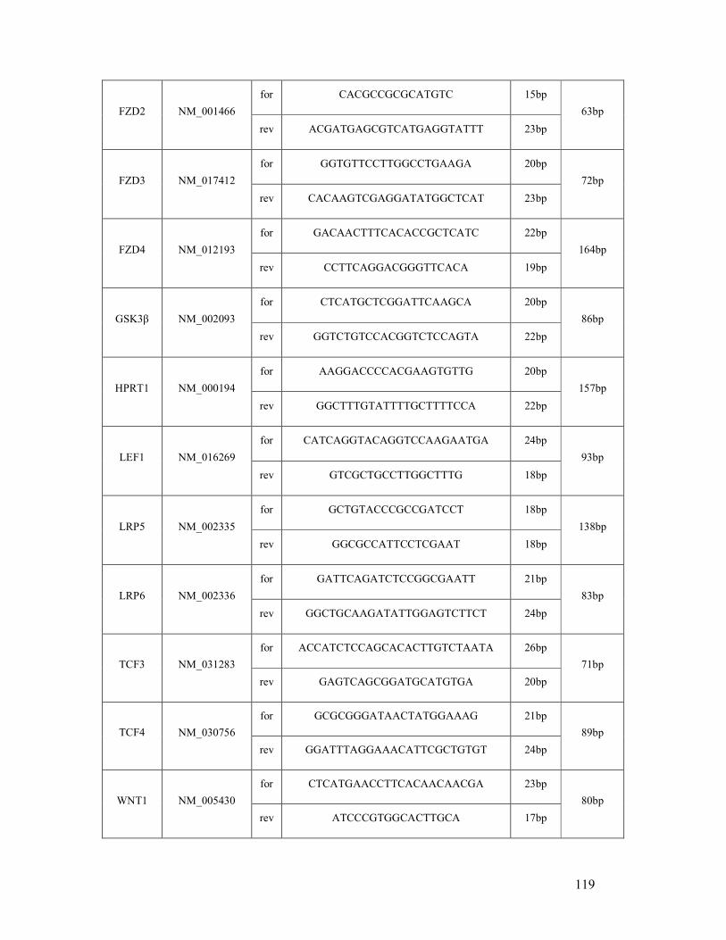
119
for CACGCCGCGCATGTC 15bp
FZD2 NM_001466
rev ACGATGAGCGTCATGAGGTATTT 23bp
63bp
for GGTGTTCCTTGGCCTGAAGA 20bp
FZD3 NM_017412
rev CACAAGTCGAGGATATGGCTCAT 23bp
72bp
for GACAACTTTCACACCGCTCATC 22bp
FZD4
NM_012193
rev CCTTCAGGACGGGTTCACA 19bp
164bp
for CTCATGCTCGGATTCAAGCA 20bp
GSK3β NM_002093
rev GGTCTGTCCACGGTCTCCAGTA 22bp
86bp
for AAGGACCCCACGAAGTGTTG 20bp
HPRT1 NM_000194
rev GGCTTTGTATTTTGCTTTTCCA 22bp
157bp
for CATCAGGTACAGGTCCAAGAATGA 24bp
LEF1 NM_016269
rev GTCGCTGCCTTGGCTTTG 18bp
93bp
for GCTGTACCCGCCGATCCT 18bp
LRP5
NM_002335
rev GGCGCCATTCCTCGAAT 18bp
138bp
for GATTCAGATCTCCGGCGAATT 21bp
LRP6
NM_002336
rev GGCTGCAAGATATTGGAGTCTTCT 24bp
83bp
for ACCATCTCCAGCACACTTGTCTAATA 26bp
TCF3
NM_031283
rev GAGTCAGCGGATGCATGTGA 20bp
71bp
for GCGCGGGATAACTATGGAAAG 21bp
TCF4
NM_030756
rev GGATTTAGGAAACATTCGCTGTGT 24bp
89bp
for CTCATGAACCTTCACAACAACGA 23bp
WNT1
NM_005430
rev ATCCCGTGGCACTTGCA 17bp
80bp
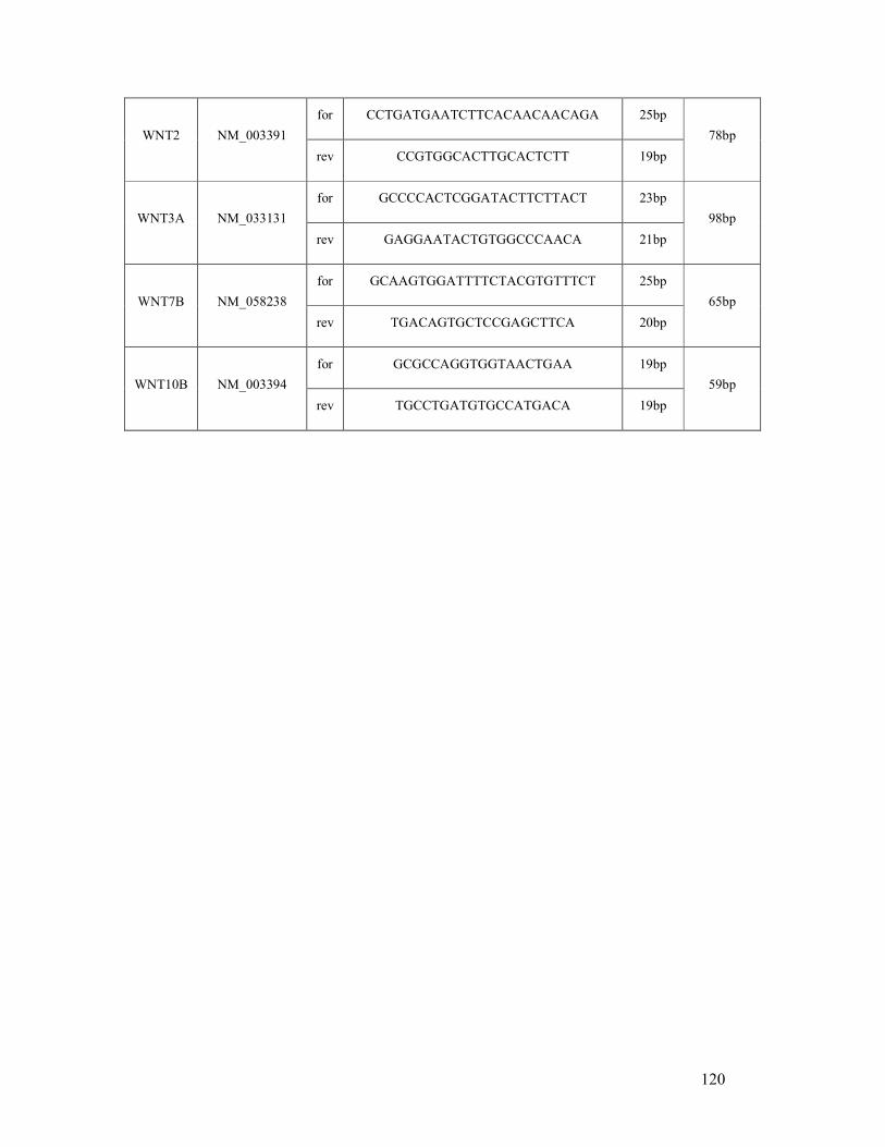
120
for CCTGATGAATCTTCACAACAACAGA 25bp
WNT2
NM_003391
rev CCGTGGCACTTGCACTCTT 19bp
78bp
for GCCCCACTCGGATACTTCTTACT 23bp
WNT3A NM_033131
rev GAGGAATACTGTGGCCCAACA 21bp
98bp
for GCAAGTGGATTTTCTACGTGTTTCT 25bp
WNT7B NM_058238
rev TGACAGTGCTCCGAGCTTCA 20bp
65bp
for GCGCCAGGTGGTAACTGAA 19bp
WNT10B NM_003394
rev TGCCTGATGTGCCATGACA 19bp
59bp
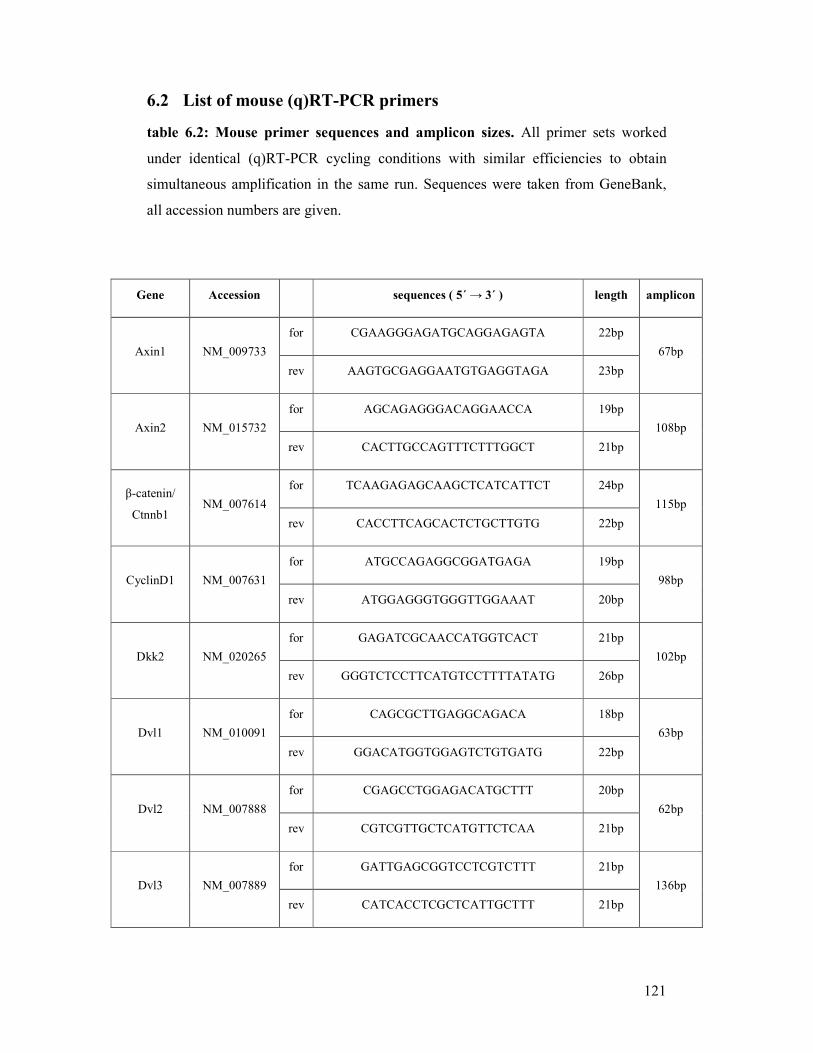
121
6.2 List of mouse (q)RT-PCR primers
table 6.2: Mouse primer sequences and amplicon sizes. All primer sets worked
under identical (q)RT-PCR cycling conditions with similar efficiencies to obtain
simultaneous amplification in the same run. Sequences were taken from GeneBank,
all accession numbers are given.
Gene Accession sequences ( 5´ → 3´ ) length amplicon
for CGAAGGGAGATGCAGGAGAGTA 22bp
Axin1 NM_009733
rev AAGTGCGAGGAATGTGAGGTAGA 23bp
67bp
for AGCAGAGGGACAGGAACCA 19bp
Axin2 NM_015732
rev CACTTGCCAGTTTCTTTGGCT 21bp
108bp
for TCAAGAGAGCAAGCTCATCATTCT 24bp β-catenin/
Ctnnb1 NM_007614
rev CACCTTCAGCACTCTGCTTGTG 22bp
115bp
for ATGCCAGAGGCGGATGAGA 19bp
CyclinD1
NM_007631
rev ATGGAGGGTGGGTTGGAAAT 20bp
98bp
for GAGATCGCAACCATGGTCACT 21bp
Dkk2 NM_020265
rev GGGTCTCCTTCATGTCCTTTTATATG 26bp
102bp
for CAGCGCTTGAGGCAGACA 18bp
Dvl1 NM_010091
rev GGACATGGTGGAGTCTGTGATG 22bp
63bp
for CGAGCCTGGAGACATGCTTT 20bp
Dvl2 NM_007888
rev CGTCGTTGCTCATGTTCTCAA 21bp
62bp
for GATTGAGCGGTCCTCGTCTTT 21bp
Dvl3 NM_007889
rev CATCACCTCGCTCATTGCTTT 21bp
136bp
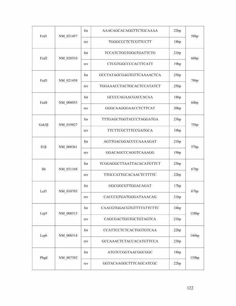
122
for AAACAGCACAGGTTCTGCAAAA 22bp
Fzd1 NM_021457
rev TGGGCCCTCTCGTTCCTT 18bp
58bp
for TCCATCTGGTGGGTGATTCTG 21bp
Fzd2 NM_020510
rev CTCGTGGCCCCACTTCATT 19bp
66bp
for GCCTATAGCGAGTGTTCAAAACTCA 25bp
Fzd3 NM_021458
rev TGGAAACCTACTGCACTCCATATCT 25bp
78bp
for GCCCCAGAACGACCACAA 18bp
Fzd4 NM_008055
rev GGGCAAGGGAACCTCTTCAT 20bp
64bp
for TTTGAGCTGGTACCCTAGGATGA 23bp
Gsk3β NM_019827
rev TTCTTCGCTTTCCGATGCA 19bp
75bp
for AGTTGACGGACCCCAAAAGAT 21bp
Il1β NM_008361
rev GGACAGCCCAGGTCAAAGG 19bp
57bp
for TCGGAGGCTTAATTACACATGTTCT 25bp
Il6 NM_031168
rev TTGCCATTGCACAACTCTTTTC 22bp
67bp
for GGCGGCGTTGGACAGAT 17bp
Lef1 NM_010703
rev CACCCGTGATGGGATAAACAG 21bp
67bp
for CAACGTGGACGTGTTTTATTCTTC 24bp
Lrp5
NM_008513
rev CAGCGACTGGTGCTGTAGTCA 21bp
138bp
for CCATTCCTCTCACTGGTGTCAA 22bp
Lrp6
NM_008514
rev GCCAAACTCTACCACATGTTCCA 23bp
146bp
for ATGTCCGGTAACGGCGGC 18bp
Pbgd
NM_007392
rev GGTACAAGGCTTTCAGCATCGC 22bp
139bp
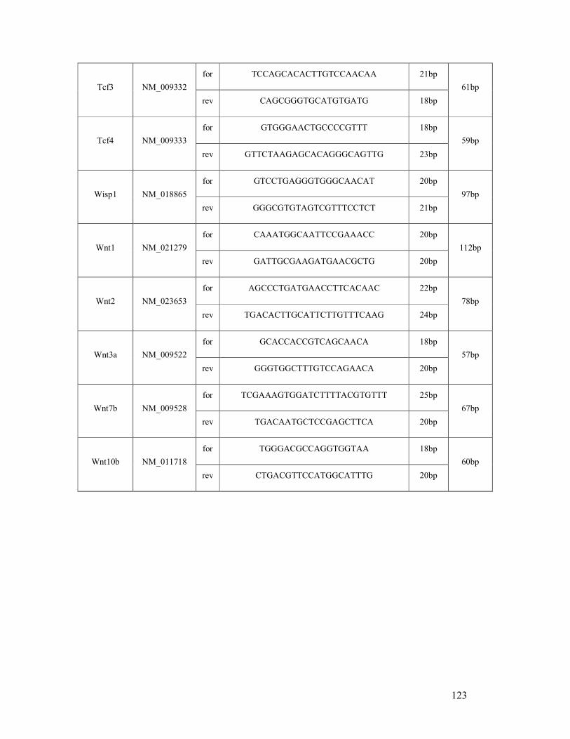
123
for TCCAGCACACTTGTCCAACAA 21bp
Tcf3
NM_009332
rev CAGCGGGTGCATGTGATG 18bp
61bp
for GTGGGAACTGCCCCGTTT 18bp
Tcf4
NM_009333
rev GTTCTAAGAGCACAGGGCAGTTG 23bp
59bp
for GTCCTGAGGGTGGGCAACAT 20bp
Wisp1 NM_018865
rev GGGCGTGTAGTCGTTTCCTCT 21bp
97bp
for CAAATGGCAATTCCGAAACC 20bp
Wnt1
NM_021279
rev GATTGCGAAGATGAACGCTG 20bp
112bp
for AGCCCTGATGAACCTTCACAAC 22bp
Wnt2 NM_023653
rev TGACACTTGCATTCTTGTTTCAAG 24bp
78bp
for GCACCACCGTCAGCAACA 18bp
Wnt3a NM_009522
rev GGGTGGCTTTGTCCAGAACA 20bp
57bp
for TCGAAAGTGGATCTTTTACGTGTTT 25bp
Wnt7b NM_009528
rev TGACAATGCTCCGAGCTTCA 20bp
67bp
for TGGGACGCCAGGTGGTAA 18bp
Wnt10b NM_011718
rev CTGACGTTCCATGGCATTTG 20bp
60bp
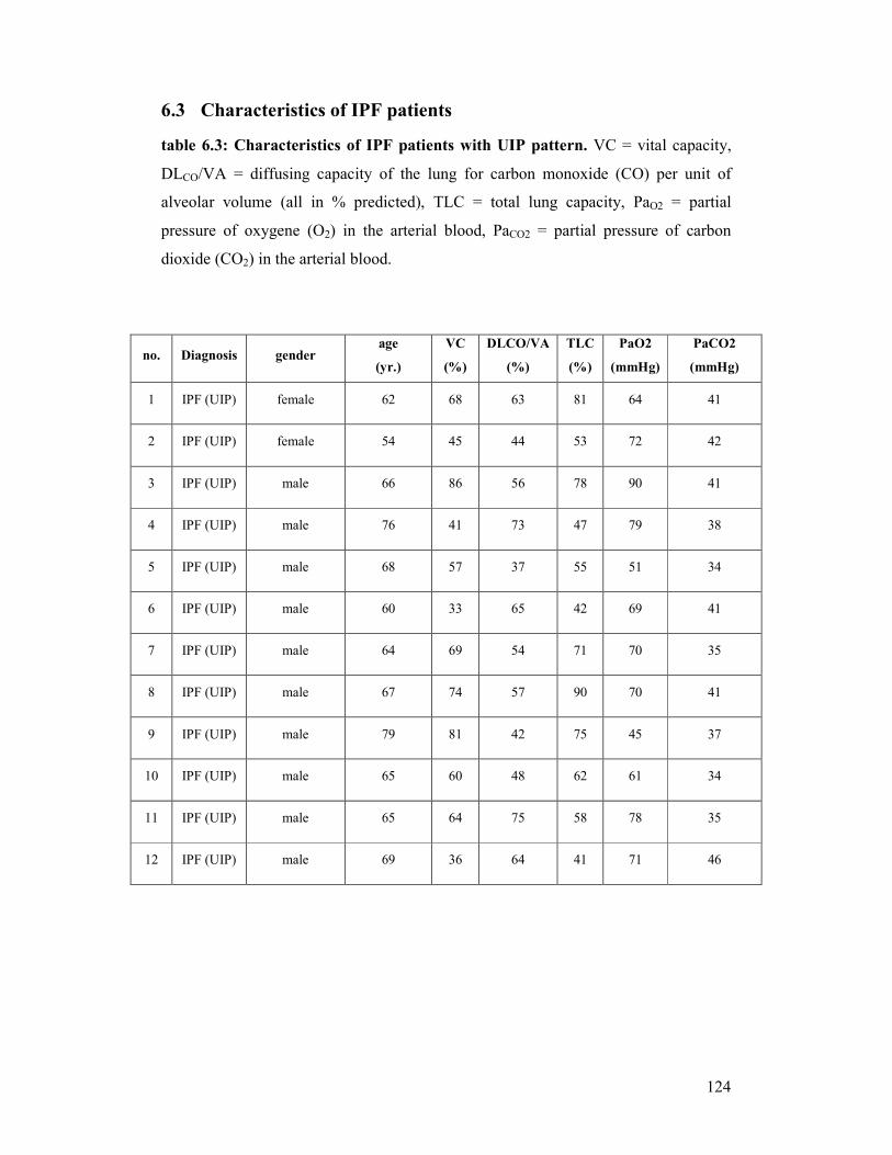
124
6.3 Characteristics of IPF patients
table 6.3: Characteristics of IPF patients with UIP pattern. VC = vital capacity,
DLCO/VA = diffusing capacity of the lung for carbon monoxide (CO) per unit of
alveolar volume (all in % predicted), TLC = total lung capacity, PaO2 = partial
pressure of oxygene (O2) in the arterial blood, PaCO2 = partial pressure of carbon
dioxide (CO2) in the arterial blood.
no. Diagnosis gender age
(yr.)
VC
(%)
DLCO/VA
(%)
TLC
(%)
PaO2
(mmHg)
PaCO2
(mmHg)
1 IPF (UIP) female 62 68 63 81 64 41
2 IPF (UIP) female 54 45 44 53 72 42
3 IPF (UIP) male 66 86 56 78 90 41
4 IPF (UIP) male 76 41 73 47 79 38
5 IPF (UIP) male 68 57 37 55 51 34
6 IPF (UIP) male 60 33 65 42 69 41
7 IPF (UIP) male 64 69 54 71 70 35
8 IPF (UIP) male 67 74 57 90 70 41
9 IPF (UIP) male 79 81 42 75 45 37
10 IPF (UIP) male 65 60 48 62 61 34
11 IPF (UIP) male 65 64 75 58 78 35
12 IPF (UIP) male 69 36 64 41 71 46
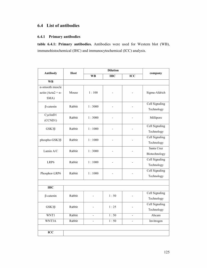
125
6.4 List of antibodies
6.4.1 Primary antibodies
table 6.4.1: Primary antibodies. Antibodies were used for Western blot (WB),
immunohistochemical (IHC) and immunocytochemical (ICC) analysis.
Dilution Antibody Host
WB IHC ICC company
WB
α-smooth muscle
actin (Acta2 = α-
SMA)
Mouse 1 : 100 - - Sigma-Aldrich
β-catenin Rabbit 1 : 3000 - - Cell Signaling
Technology
CyclinD1
(CCND1) Rabbit 1 : 3000 - - Millipore
GSK3β Rabbit 1 : 1000 - - Cell Signaling
Technology
phospho-GSK3β Rabbit 1 : 1000 - - Cell Signaling
Technology
Lamin A/C Rabbit 1 : 3000 - - Santa Cruz
Biotechnology
LRP6 Rabbit 1 : 1000 - - Cell Signaling
Technology
Phosphor-LRP6 Rabbit 1 : 1000 - - Cell Signaling
Technology
IHC
β-catenin Rabbit - 1 : 50 - Cell Signaling
Technology
GSK3β Rabbit - 1 : 25 - Cell Signaling
Technology
WNT1 Rabbit - 1 : 50 - Abcam
WNT3A Rabbit - 1 : 50 - Invitrogen
ICC
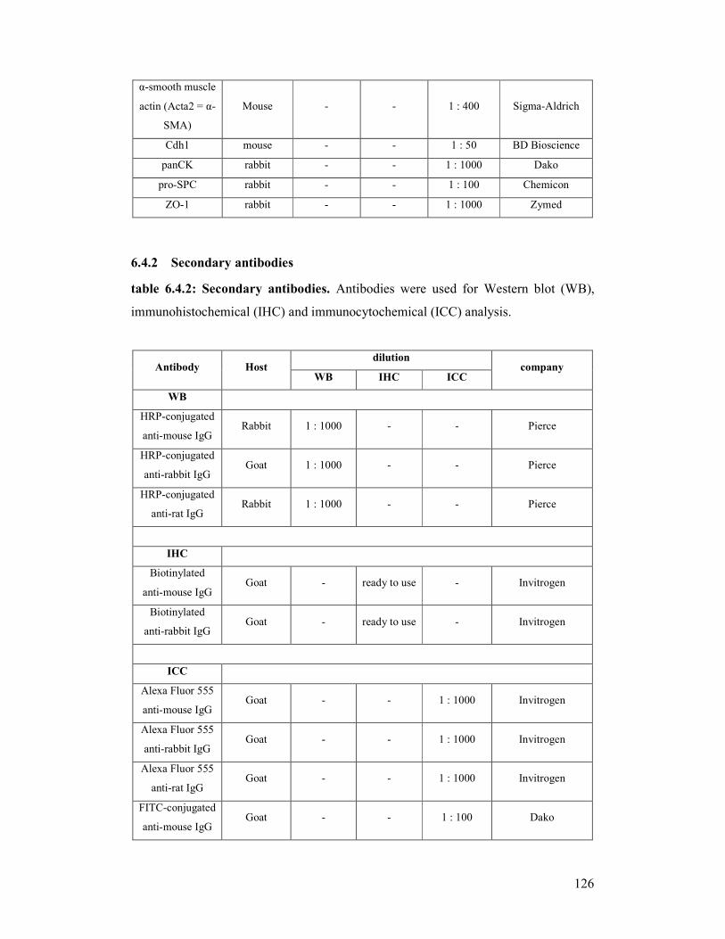
126
α-smooth muscle
actin (Acta2 = α-
SMA)
Mouse - - 1 : 400 Sigma-Aldrich
Cdh1 mouse - - 1 : 50 BD Bioscience
panCK rabbit - - 1 : 1000 Dako
pro-SPC rabbit - - 1 : 100 Chemicon
ZO-1 rabbit - - 1 : 1000 Zymed
6.4.2 Secondary antibodies
table 6.4.2: Secondary antibodies. Antibodies were used for Western blot (WB),
immunohistochemical (IHC) and immunocytochemical (ICC) analysis.
dilution Antibody Host
WB IHC ICC company
WB
HRP-conjugated
anti-mouse IgG Rabbit 1 : 1000 - - Pierce
HRP-conjugated
anti-rabbit IgG Goat 1 : 1000 - - Pierce
HRP-conjugated
anti-rat IgG Rabbit 1 : 1000 - - Pierce
IHC
Biotinylated
anti-mouse IgG Goat - ready to use - Invitrogen
Biotinylated
anti-rabbit IgG Goat - ready to use - Invitrogen
ICC
Alexa Fluor 555
anti-mouse IgG Goat - - 1 : 1000 Invitrogen
Alexa Fluor 555
anti-rabbit IgG Goat - - 1 : 1000 Invitrogen
Alexa Fluor 555
anti-rat IgG Goat - - 1 : 1000 Invitrogen
FITC-conjugated
anti-mouse IgG Goat - - 1 : 100 Dako

127
6.5 Microarray gene list
table 6.5: List of selected target genes found in the microarray. Genes were partly
confirmed by (q)RT-PCR
Symbol accession no. Wnt3a
( 8 h )
Wnt3a
( 24 h )
coefficient 0,58 0,49 Angptl4 NM_020581
ddCT 0,18 0,64
coefficient -0,65 -0,67 Angptl7 NM_001039554
ddCT -1,21 -1,25
coefficient -0,33 -0,48 Aqp1 NM_007472
ddCT -0,04 -0,34
coefficient -0,40 0,13 Arg1 NM_007482
ddCT -0,80 -0,42
coefficient 0,92 0,49 Axin2 NM_015732
ddCT 1,31 0,83
coefficient -0,50 -0,13 Col5a2 NM_007737
ddCT -0,19 -0,27
coefficient -0,43 0,22 Col7a1 NM_007738
ddCT -0,32 -0,95
coefficient -0,16 -0,46 Dcn NM_007833
ddCT -0,17 -0,10
coefficient 0,21 0,94 Dkk2 NM_020265
ddCT 0,91 1,87
coefficient -0,56 -0,21 Fn1 NM_010233
ddCT -0,42 0,29
coefficient -0,20 0,49 Grem2 NM_011825
ddCT -0,09 0,30
coefficient 0,42 0,52 Has2 NM_008216
ddCT 0,37 0,48
coefficient 0,70 0,57 Il1β NM_008361
ddCT 1,91 2,19
coefficient -0,08 0,64 Il6 NM_031168
ddCT 0,21 0,63
coefficient 0,50 -0,15 Kdr NM_010612
ddCT 0,54 -0,07
128
coefficient 0,07 0,65 Lcn2 NM_008491
ddCT 2,41 1,45
coefficient -0,34 -0,52 Magoh NM_010760
ddCT 0,56 0,21
coefficient 0,77 0,28 Nkd1 NM_027280
ddCT 1,15 0,42
coefficient 0,41 0,34 Prickle1 NM_001033217
ddCT 0,56 0,41
coefficient 0,36 -0,47 Rock1 NM_009071
ddCT 0,40 -0,31
coefficient -0,48 0,00 Spp1 NM_009263
ddCT -0,20 -0,26
coefficient 0,48 0,08 Tcf7 NM_009331
ddCT 0,94 0,71
coefficient 0,01 0,46 Timp1 NM_011593
ddCT -0,45 0,25
coefficient -0,19 0,41 Tagln NM_011526
ddCT 0,10 0,18
coefficient -0,23 0,49 Zyx NM_0117772
ddCT 0,05 0,22
129
7 REFERENCES
1. American Thoracic Society/European Respiratory Society International Multidisciplinary Consensus Classification of the Idiopathic Interstitial Pneumonias. This joint statement of the American Thoracic Society (ATS), and the European Respiratory Society (ERS) was adopted by the ATS board of directors, June 2001 and by the ERS Executive Committee, June 2001. Am J
Respir Crit Care Med 2002: 165(2): 277-304. 2. Bourke SJ. Interstitial lung disease: progress and problems. Postgrad Med J
2006: 82(970): 494-499. 3. King TE, Jr. Clinical advances in the diagnosis and therapy of the interstitial lung
diseases. Am J Respir Crit Care Med 2005: 172(3): 268-279. 4. Meltzer EB, Noble PW. Idiopathic pulmonary fibrosis. Orphanet J Rare Dis
2008: 3: 8. 5. Raghu G, Weycker D, Edelsberg J, Bradford WZ, Oster G. Incidence and
prevalence of idiopathic pulmonary fibrosis. Am J Respir Crit Care Med 2006: 174(7): 810-816.
6. American Thoracic Society. Idiopathic pulmonary fibrosis: diagnosis and treatment. International consensus statement. American Thoracic Society (ATS), and the European Respiratory Society (ERS). Am J Respir Crit Care Med 2000: 161(2 Pt 1): 646-664.
7. Baumgartner KB, Samet JM, Stidley CA, Colby TV, Waldron JA. Cigarette smoking: a risk factor for idiopathic pulmonary fibrosis. Am J Respir Crit Care
Med 1997: 155(1): 242-248. 8. Dempsey OJ. Clinical review: idiopathic pulmonary fibrosis--past, present and
future. Respir Med 2006: 100(11): 1871-1885. 9. Collard HR, King TE, Jr., Bartelson BB, Vourlekis JS, Schwarz MI, Brown KK.
Changes in clinical and physiologic variables predict survival in idiopathic pulmonary fibrosis. Am J Respir Crit Care Med 2003: 168(5): 538-542.
10. Selman M, King TE, Pardo A. Idiopathic pulmonary fibrosis: prevailing and evolving hypotheses about its pathogenesis and implications for therapy. Ann
Intern Med 2001: 134(2): 136-151. 11. Kim DS, Collard HR, King TE, Jr. Classification and natural history of the
idiopathic interstitial pneumonias. Proc Am Thorac Soc 2006: 3(4): 285-292. 12. Martinez FJ, Safrin S, Weycker D, Starko KM, Bradford WZ, King TE, Jr.,
Flaherty KR, Schwartz DA, Noble PW, Raghu G, Brown KK. The clinical course of patients with idiopathic pulmonary fibrosis. Ann Intern Med 2005: 142(12 Pt 1): 963-967.
13. Hunninghake GW, Lynch DA, Galvin JR, Gross BH, Muller N, Schwartz DA, King TE, Jr., Lynch JP, 3rd, Hegele R, Waldron J, Colby TV, Hogg JC. Radiologic findings are strongly associated with a pathologic diagnosis of usual interstitial pneumonia. Chest 2003: 124(4): 1215-1223.
14. Lynch DA, Godwin JD, Safrin S, Starko KM, Hormel P, Brown KK, Raghu G, King TE, Jr., Bradford WZ, Schwartz DA, Richard Webb W. High-resolution computed tomography in idiopathic pulmonary fibrosis: diagnosis and prognosis. Am J Respir Crit Care Med 2005: 172(4): 488-493.
15. Raghu G, Brown KK, Costabel U, Cottin V, du Bois RM, Lasky JA, Thomeer M, Utz JP, Khandker RK, McDermott L, Fatenejad S. Treatment of idiopathic
130
pulmonary fibrosis with etanercept: an exploratory, placebo-controlled trial. Am J
Respir Crit Care Med 2008: 178(9): 948-955. 16. Maher TM, Wells AU, Laurent GJ. Idiopathic pulmonary fibrosis: multiple
causes and multiple mechanisms? Eur Respir J 2007: 30(5): 835-839. 17. Daniels CE, Ryu JH. Treatment of idiopathic pulmonary fibrosis. Semin Respir
Crit Care Med 2006: 27(6): 668-676. 18. Walter N, Collard HR, King TE, Jr. Current perspectives on the treatment of
idiopathic pulmonary fibrosis. Proc Am Thorac Soc 2006: 3(4): 330-338. 19. Nathan SD. Lung transplantation: disease-specific considerations for referral.
Chest 2005: 127(3): 1006-1016. 20. Rinaldi M, Sansone F, Boffini M, El Qarra S, Solidoro P, Cavallo N, Ruffini E,
Baldi S. Single versus double lung transplantation in pulmonary fibrosis: a debated topic. Transplant Proc 2008: 40(6): 2010-2012.
21. Thabut G, Mal H, Castier Y, Groussard O, Brugiere O, Marrash-Chahla R, Leseche G, Fournier M. Survival benefit of lung transplantation for patients with idiopathic pulmonary fibrosis. J Thorac Cardiovasc Surg 2003: 126(2): 469-475.
22. Williams TJ, Wilson JW. Challenges in pulmonary fibrosis: 7--Novel therapies and lung transplantation. Thorax 2008: 63(3): 277-284.
23. Katzenstein AL, Myers JL. Idiopathic pulmonary fibrosis: clinical relevance of pathologic classification. Am J Respir Crit Care Med 1998: 157(4 Pt 1): 1301-1315.
24. Visscher DW, Myers JL. Histologic spectrum of idiopathic interstitial pneumonias. Proc Am Thorac Soc 2006: 3(4): 322-329.
25. King TE, Jr., Schwarz MI, Brown K, Tooze JA, Colby TV, Waldron JA, Jr., Flint A, Thurlbeck W, Cherniack RM. Idiopathic pulmonary fibrosis: relationship between histopathologic features and mortality. Am J Respir Crit Care Med 2001: 164(6): 1025-1032.
26. Nicholson AG, Fulford LG, Colby TV, du Bois RM, Hansell DM, Wells AU. The relationship between individual histologic features and disease progression in idiopathic pulmonary fibrosis. Am J Respir Crit Care Med 2002: 166(2): 173-177.
27. Hinz B, Phan SH, Thannickal VJ, Galli A, Bochaton-Piallat ML, Gabbiani G. The myofibroblast: one function, multiple origins. Am J Pathol 2007: 170(6): 1807-1816.
28. Phan SH. The myofibroblast in pulmonary fibrosis. Chest 2002: 122(6 Suppl): 286S-289S.
29. Scotton CJ, Chambers RC. Molecular targets in pulmonary fibrosis: the myofibroblast in focus. Chest 2007: 132(4): 1311-1321.
30. Willis BC, duBois RM, Borok Z. Epithelial origin of myofibroblasts during fibrosis in the lung. Proc Am Thorac Soc 2006: 3(4): 377-382.
31. Gross TJ, Hunninghake GW. Idiopathic pulmonary fibrosis. N Engl J Med 2001: 345(7): 517-525.
32. Strieter RM. Pathogenesis and natural history of usual interstitial pneumonia: the whole story or the last chapter of a long novel. Chest 2005: 128(5 Suppl 1): 526S-532S.
33. Noble PW, Homer RJ. Back to the future: historical perspective on the pathogenesis of idiopathic pulmonary fibrosis. Am J Respir Cell Mol Biol 2005: 33(2): 113-120.
34. Gauldie J, Kolb M, Sime PJ. A new direction in the pathogenesis of idiopathic pulmonary fibrosis? Respir Res 2002: 3: 1.
131
35. Selman M, Pardo A. Idiopathic pulmonary fibrosis: an epithelial/fibroblastic cross-talk disorder. Respir Res 2002: 3: 3.
36. Horowitz JC, Thannickal VJ. Epithelial-mesenchymal interactions in pulmonary fibrosis. Semin Respir Crit Care Med 2006: 27(6): 600-612.
37. Kolb M, Margetts PJ, Anthony DC, Pitossi F, Gauldie J. Transient expression of IL-1beta induces acute lung injury and chronic repair leading to pulmonary fibrosis. J Clin Invest 2001: 107(12): 1529-1536.
38. Konigshoff M, Wilhelm A, Jahn A, Sedding D, Amarie OV, Eul B, Seeger W, Fink L, Gunther A, Eickelberg O, Rose F. The angiotensin II receptor 2 is expressed and mediates angiotensin II signaling in lung fibrosis. Am J Respir Cell
Mol Biol 2007: 37(6): 640-650. 39. Konigshoff M, Kramer M, Balsara N, Wilhelm J, Amarie OV, Jahn A, Rose F,
Fink L, Seeger W, Schaefer L, Gunther A, Eickelberg O. WNT1-inducible signaling protein-1 mediates pulmonary fibrosis in mice and is upregulated in humans with idiopathic pulmonary fibrosis. J Clin Invest 2009: 119(4): 772-787.
40. Gordon MD, Nusse R. Wnt signaling: multiple pathways, multiple receptors, and multiple transcription factors. J Biol Chem 2006: 281(32): 22429-22433.
41. Mikels AJ, Nusse R. Wnts as ligands: processing, secretion and reception. Oncogene 2006: 25(57): 7461-7468.
42. Moon RT, Kohn AD, De Ferrari GV, Kaykas A. WNT and beta-catenin signalling: diseases and therapies. Nat Rev Genet 2004: 5(9): 691-701.
43. Nusse R. Wnt signaling in disease and in development. Cell Res 2005: 15(1): 28-32.
44. Johnson ML, Rajamannan N. Diseases of Wnt signaling. Rev Endocr Metab
Disord 2006: 7(1-2): 41-49. 45. Logan CY, Nusse R. The Wnt signaling pathway in development and disease.
Annu Rev Cell Dev Biol 2004: 20: 781-810. 46. Cadigan KM, Liu YI. Wnt signaling: complexity at the surface. J Cell Sci 2006:
119(Pt 3): 395-402. 47. Nusse R, van Ooyen A, Cox D, Fung YK, Varmus H. Mode of proviral activation
of a putative mammary oncogene (int-1) on mouse chromosome 15. Nature 1984: 307(5947): 131-136.
48. Nusse R, Varmus HE. Many tumors induced by the mouse mammary tumor virus contain a provirus integrated in the same region of the host genome. Cell 1982: 31(1): 99-109.
49. Nusslein-Volhard C, Wieschaus E. Mutations affecting segment number and polarity in Drosophila. Nature 1980: 287(5785): 795-801.
50. Kikuchi A, Yamamoto H, Kishida S. Multiplicity of the interactions of Wnt proteins and their receptors. Cell Signal 2007: 19(4): 659-671.
51. MacDonald BT, Yokota C, Tamai K, Zeng X, He X. Wnt signal amplification via activity, cooperativity, and regulation of multiple intracellular PPPSP motifs in the Wnt co-receptor LRP6. J Biol Chem 2008: 283(23): 16115-16123.
52. Clevers H. Wnt/beta-catenin signaling in development and disease. Cell 2006: 127(3): 469-480.
53. Goss AM, Tian Y, Tsukiyama T, Cohen ED, Zhou D, Lu MM, Yamaguchi TP, Morrisey EE. Wnt2/2b and beta-catenin signaling are necessary and sufficient to specify lung progenitors in the foregut. Dev Cell 2009: 17(2): 290-298.
54. Levay-Young BK, Navre M. Growth and developmental regulation of wnt-2 (irp) gene in mesenchymal cells of fetal lung. Am J Physiol 1992: 262(6 Pt 1): L672-683.
132
55. Li L, Mao J, Sun L, Liu W, Wu D. Second cysteine-rich domain of Dickkopf-2 activates canonical Wnt signaling pathway via LRP-6 independently of dishevelled. J Biol Chem 2002: 277(8): 5977-5981.
56. Shu W, Jiang YQ, Lu MM, Morrisey EE. Wnt7b regulates mesenchymal proliferation and vascular development in the lung. Development 2002: 129(20): 4831-4842.
57. Rajagopal J, Carroll TJ, Guseh JS, Bores SA, Blank LJ, Anderson WJ, Yu J, Zhou Q, McMahon AP, Melton DA. Wnt7b stimulates embryonic lung growth by coordinately increasing the replication of epithelium and mesenchyme. Development 2008: 135(9): 1625-1634.
58. Maretto S, Cordenonsi M, Dupont S, Braghetta P, Broccoli V, Hassan AB, Volpin D, Bressan GM, Piccolo S. Mapping Wnt/beta-catenin signaling during mouse development and in colorectal tumors. Proc Natl Acad Sci U S A 2003: 100(6): 3299-3304.
59. Okubo T, Hogan BL. Hyperactive Wnt signaling changes the developmental potential of embryonic lung endoderm. J Biol 2004: 3(3): 11.
60. Dean CH, Miller LA, Smith AN, Dufort D, Lang RA, Niswander LA. Canonical Wnt signaling negatively regulates branching morphogenesis of the lung and lacrimal gland. Dev Biol 2005: 286(1): 270-286.
61. Mucenski ML, Wert SE, Nation JM, Loudy DE, Huelsken J, Birchmeier W, Morrisey EE, Whitsett JA. beta-Catenin is required for specification of proximal/distal cell fate during lung morphogenesis. J Biol Chem 2003: 278(41): 40231-40238.
62. Winn RA, Marek L, Han SY, Rodriguez K, Rodriguez N, Hammond M, Van Scoyk M, Acosta H, Mirus J, Barry N, Bren-Mattison Y, Van Raay TJ, Nemenoff RA, Heasley LE. Restoration of Wnt-7a expression reverses non-small cell lung cancer cellular transformation through frizzled-9-mediated growth inhibition and promotion of cell differentiation. J Biol Chem 2005: 280(20): 19625-19634.
63. Kikuchi A. Tumor formation by genetic mutations in the components of the Wnt signaling pathway. Cancer Sci 2003: 94(3): 225-229.
64. Paul S, Dey A. Wnt signaling and cancer development: therapeutic implication. Neoplasma 2008: 55(3): 165-176.
65. Polakis P. Wnt signaling and cancer. Genes Dev 2000: 14(15): 1837-1851. 66. Lammi L, Arte S, Somer M, Jarvinen H, Lahermo P, Thesleff I, Pirinen S,
Nieminen P. Mutations in AXIN2 cause familial tooth agenesis and predispose to colorectal cancer. Am J Hum Genet 2004: 74(5): 1043-1050.
67. Chen S, Guttridge DC, You Z, Zhang Z, Fribley A, Mayo MW, Kitajewski J, Wang CY. Wnt-1 signaling inhibits apoptosis by activating beta-catenin/T cell factor-mediated transcription. J Cell Biol 2001: 152(1): 87-96.
68. He B, Jablons DM. Wnt signaling in stem cells and lung cancer. Ernst Schering
Found Symp Proc 2006(5): 27-58. 69. Mazieres J, He B, You L, Xu Z, Jablons DM. Wnt signaling in lung cancer.
Cancer Lett 2005: 222(1): 1-10. 70. Uematsu K, He B, You L, Xu Z, McCormick F, Jablons DM. Activation of the
Wnt pathway in non small cell lung cancer: evidence of dishevelled overexpression. Oncogene 2003: 22(46): 7218-7221.
71. You L, He B, Xu Z, Uematsu K, Mazieres J, Mikami I, Reguart N, Moody TW, Kitajewski J, McCormick F, Jablons DM. Inhibition of Wnt-2-mediated signaling induces programmed cell death in non-small-cell lung cancer cells. Oncogene
2004: 23(36): 6170-6174.
133
72. Huang CL, Liu D, Nakano J, Ishikawa S, Kontani K, Yokomise H, Ueno M. Wnt5a expression is associated with the tumor proliferation and the stromal vascular endothelial growth factor--an expression in non-small-cell lung cancer. J
Clin Oncol 2005: 23(34): 8765-8773. 73. Pongracz JE, Stockley RA. Wnt signalling in lung development and diseases.
Respir Res 2006: 7: 15. 74. Kim D, Rath O, Kolch W, Cho KH. A hidden oncogenic positive feedback loop
caused by crosstalk between Wnt and ERK pathways. Oncogene 2007: 26(31): 4571-4579.
75. Surendran K, McCaul SP, Simon TC. A role for Wnt-4 in renal fibrosis. Am J
Physiol Renal Physiol 2002: 282(3): F431-441. 76. Surendran K, Schiavi S, Hruska KA. Wnt-dependent beta-catenin signaling is
activated after unilateral ureteral obstruction, and recombinant secreted frizzled-related protein 4 alters the progression of renal fibrosis. J Am Soc Nephrol 2005: 16(8): 2373-2384.
77. Cheng JH, She H, Han YP, Wang J, Xiong S, Asahina K, Tsukamoto H. Wnt antagonism inhibits hepatic stellate cell activation and liver fibrosis. Am J Physiol
Gastrointest Liver Physiol 2008: 294(1): G39-49. 78. Chilosi M, Poletti V, Zamo A, Lestani M, Montagna L, Piccoli P, Pedron S,
Bertaso M, Scarpa A, Murer B, Cancellieri A, Maestro R, Semenzato G, Doglioni C. Aberrant Wnt/beta-catenin pathway activation in idiopathic pulmonary fibrosis. Am J Pathol 2003: 162(5): 1495-1502.
79. Morrisey EE. Wnt signaling and pulmonary fibrosis. Am J Pathol 2003: 162(5): 1393-1397.
80. Kaminski N, Rosas IO. Gene expression profiling as a window into idiopathic pulmonary fibrosis pathogenesis: can we identify the right target genes? Proc Am
Thorac Soc 2006: 3(4): 339-344. 81. Selman M, Pardo A, Barrera L, Estrada A, Watson SR, Wilson K, Aziz N,
Kaminski N, Zlotnik A. Gene expression profiles distinguish idiopathic pulmonary fibrosis from hypersensitivity pneumonitis. Am J Respir Crit Care
Med 2006: 173(2): 188-198. 82. Selman M, Pardo A, Kaminski N. Idiopathic pulmonary fibrosis: aberrant
recapitulation of developmental programs? PLoS Med 2008: 5(3): e62. 83. Lewis CC, Yang JY, Huang X, Banerjee SK, Blackburn MR, Baluk P, McDonald
DM, Blackwell TS, Nagabhushanam V, Peters W, Voehringer D, Erle DJ. Disease-specific gene expression profiling in multiple models of lung disease. Am
J Respir Crit Care Med 2008: 177(4): 376-387. 84. DasGupta R, Fuchs E. Multiple roles for activated LEF/TCF transcription
complexes during hair follicle development and differentiation. Development
1999: 126(20): 4557-4568. 85. Douglas IS, Diaz del Valle F, Winn RA, Voelkel NF. Beta-catenin in the
fibroproliferative response to acute lung injury. Am J Respir Cell Mol Biol 2006: 34(3): 274-285.
86. Vuga LJ, Ben-Yehudah A, Kovkarova-Naumovski E, Oriss T, Gibson KF, Feghali-Bostwick C, Kaminski N. WNT5A is a regulator of fibroblast proliferation and resistance to apoptosis. Am J Respir Cell Mol Biol 2009: 41(5): 583-589.
87. Wei J, Melichian D, Komura K, Hinchcliff M, Lam AP, Lafyatis R, Gottardi CJ, Macdougald OA, Varga J. Canonical Wnt signaling induces skin fibrosis and
134
subcutaneous lipoatrophy: A novel mouse model for scleroderma? Arthritis
Rheum: 63(6): 1707-1717. 88. Pfaff EM, Becker S, Gunther A, Konigshoff M. Dickkopf proteins influence lung
epithelial cell proliferation in idiopathic pulmonary fibrosis. Eur Respir J: 37(1): 79-87.
89. Zemke AC, Teisanu RM, Giangreco A, Drake JA, Brockway BL, Reynolds SD, Stripp BR. beta-Catenin is not necessary for maintenance or repair of the bronchiolar epithelium. Am J Respir Cell Mol Biol 2009: 41(5): 535-543.
90. Weng T, Liu L. The role of pleiotrophin and beta-catenin in fetal lung development. Respir Res: 11: 80.
91. Brembeck FH, Rosario M, Birchmeier W. Balancing cell adhesion and Wnt signaling, the key role of beta-catenin. Curr Opin Genet Dev 2006: 16(1): 51-59.
92. Perez-Moreno M, Fuchs E. Catenins: keeping cells from getting their signals crossed. Dev Cell 2006: 11(5): 601-612.
93. Flozak AS, Lam AP, Russell S, Jain M, Peled ON, Sheppard KA, Beri R, Mutlu GM, Budinger GR, Gottardi CJ. Beta-catenin/T-cell factor signaling is activated during lung injury and promotes the survival and migration of alveolar epithelial cells. J Biol Chem: 285(5): 3157-3167.
94. Crosby LM, Waters CM. Epithelial repair mechanisms in the lung. Am J Physiol
Lung Cell Mol Physiol: 298(6): L715-731. 95. Kim TH, Kim SH, Seo JY, Chung H, Kwak HJ, Lee SK, Yoon HJ, Shin DH,
Park SS, Sohn JW. Blockade of the Wnt/beta-catenin pathway attenuates bleomycin-induced pulmonary fibrosis. Tohoku J Exp Med: 223(1): 45-54.
96. Henderson WR, Jr., Chi EY, Ye X, Nguyen C, Tien YT, Zhou B, Borok Z, Knight DA, Kahn M. Inhibition of Wnt/beta-catenin/CREB binding protein (CBP) signaling reverses pulmonary fibrosis. Proc Natl Acad Sci U S A: 107(32): 14309-14314.
97. Shrivastava N, Srivastava A. RNA interference: an emerging generation of biologicals. Biotechnol J 2008: 3(3): 339-353.
98. Bilic J, Huang YL, Davidson G, Zimmermann T, Cruciat CM, Bienz M, Niehrs C. Wnt induces LRP6 signalosomes and promotes dishevelled-dependent LRP6 phosphorylation. Science 2007: 316(5831): 1619-1622.
99. Forde JE, Dale TC. Glycogen synthase kinase 3: a key regulator of cellular fate. Cell Mol Life Sci 2007: 64(15): 1930-1944.
100. Konigshoff M, Balsara N, Pfaff EM, Kramer M, Chrobak I, Seeger W, Eickelberg O. Functional Wnt signaling is increased in idiopathic pulmonary fibrosis. PLoS One 2008: 3(5): e2142.
101. Moeller A, Ask K, Warburton D, Gauldie J, Kolb M. The bleomycin animal model: a useful tool to investigate treatment options for idiopathic pulmonary fibrosis? Int J Biochem Cell Biol 2008: 40(3): 362-382.
102. Adamson IY, Bowden DH. The pathogenesis of bloemycin-induced pulmonary fibrosis in mice. Am J Pathol 1974: 77(2): 185-197.
103. Serrano-Mollar A, Nacher M, Gay-Jordi G, Closa D, Xaubet A, Bulbena O. Intratracheal transplantation of alveolar type II cells reverses bleomycin-induced lung fibrosis. Am J Respir Crit Care Med 2007: 176(12): 1261-1268.
104. Liu L, Carron B, Yee HT, Yie TA, Hajjou M, Rom W. Wnt pathway in pulmonary fibrosis in the bleomycin mouse model. J Environ Pathol Toxicol
Oncol 2009: 28(2): 99-108.
135
105. Lu D, Zhao Y, Tawatao R, Cottam HB, Sen M, Leoni LM, Kipps TJ, Corr M, Carson DA. Activation of the Wnt signaling pathway in chronic lymphocytic leukemia. Proc Natl Acad Sci U S A 2004: 101(9): 3118-3123.
106. Hwang SG, Yu SS, Lee SW, Chun JS. Wnt-3a regulates chondrocyte differentiation via c-Jun/AP-1 pathway. FEBS Lett 2005: 579(21): 4837-4842.
107. Szeto W, Jiang W, Tice DA, Rubinfeld B, Hollingshead PG, Fong SE, Dugger DL, Pham T, Yansura DG, Wong TA, Grimaldi JC, Corpuz RT, Singh JS, Frantz GD, Devaux B, Crowley CW, Schwall RH, Eberhard DA, Rastelli L, Polakis P, Pennica D. Overexpression of the retinoic acid-responsive gene Stra6 in human cancers and its synergistic induction by Wnt-1 and retinoic acid. Cancer Res
2001: 61(10): 4197-4205. 108. Taneyhill L, Pennica D. Identification of Wnt responsive genes using a murine
mammary epithelial cell line model system. BMC Dev Biol 2004: 4: 6. 109. Daniels CB, Orgeig S. Pulmonary surfactant: the key to the evolution of air
breathing. News Physiol Sci 2003: 18: 151-157. 110. George CL, Goss KL, Meyerholz DK, Lamb FS, Snyder JM. Surfactant-
associated protein A provides critical immunoprotection in neonatal mice. Infect
Immun 2008: 76(1): 380-390. 111. Wofford JA, Wright JR. Surfactant protein A regulates IgG-mediated
phagocytosis in inflammatory neutrophils. Am J Physiol Lung Cell Mol Physiol
2007: 293(6): L1437-1443. 112. Lee SM, Tole S, Grove E, McMahon AP. A local Wnt-3a signal is required for
development of the mammalian hippocampus. Development 2000: 127(3): 457-467.
113. Cheng CW, Yeh JC, Fan TP, Smith SK, Charnock-Jones DS. Wnt5a-mediated non-canonical Wnt signalling regulates human endothelial cell proliferation and migration. Biochem Biophys Res Commun 2008: 365(2): 285-290.
114. Goodwin AM, Sullivan KM, D'Amore PA. Cultured endothelial cells display endogenous activation of the canonical Wnt signaling pathway and express multiple ligands, receptors, and secreted modulators of Wnt signaling. Dev Dyn
2006: 235(11): 3110-3120. 115. Brigstock DR. The CCN family: a new stimulus package. J Endocrinol 2003:
178(2): 169-175. 116. Pennica D, Swanson TA, Welsh JW, Roy MA, Lawrence DA, Lee J, Brush J,
Taneyhill LA, Deuel B, Lew M, Watanabe C, Cohen RL, Melhem MF, Finley GG, Quirke P, Goddard AD, Hillan KJ, Gurney AL, Botstein D, Levine AJ. WISP genes are members of the connective tissue growth factor family that are up-regulated in wnt-1-transformed cells and aberrantly expressed in human colon tumors. Proc Natl Acad Sci U S A 1998: 95(25): 14717-14722.
117. Behrens J, Jerchow BA, Wurtele M, Grimm J, Asbrand C, Wirtz R, Kuhl M, Wedlich D, Birchmeier W. Functional interaction of an axin homolog, conductin, with beta-catenin, APC, and GSK3beta. Science 1998: 280(5363): 596-599.
118. Kikuchi A. Regulation of beta-catenin signaling in the Wnt pathway. Biochem
Biophys Res Commun 2000: 268(2): 243-248. 119. Mao B, Niehrs C. Kremen2 modulates Dickkopf2 activity during Wnt/LRP6
signaling. Gene 2003: 302(1-2): 179-183. 120. Niehrs C. Function and biological roles of the Dickkopf family of Wnt
modulators. Oncogene 2006: 25(57): 7469-7481.
136
121. Wu W, Glinka A, Delius H, Niehrs C. Mutual antagonism between dickkopf1 and dickkopf2 regulates Wnt/beta-catenin signalling. Curr Biol 2000: 10(24): 1611-1614.
122. He W, Dai C, Li Y, Zeng G, Monga SP, Liu Y. Wnt/beta-catenin signaling promotes renal interstitial fibrosis. J Am Soc Nephrol 2009: 20(4): 765-776.
123. Hwang I, Seo EY, Ha H. Wnt/beta-catenin signaling: a novel target for therapeutic intervention of fibrotic kidney disease. Arch Pharm Res 2009: 32(12): 1653-1662.
124. Lappalainen U, Whitsett JA, Wert SE, Tichelaar JW, Bry K. Interleukin-1beta causes pulmonary inflammation, emphysema, and airway remodeling in the adult murine lung. Am J Respir Cell Mol Biol 2005: 32(4): 311-318.
125. Gasse P, Mary C, Guenon I, Noulin N, Charron S, Schnyder-Candrian S, Schnyder B, Akira S, Quesniaux VF, Lagente V, Ryffel B, Couillin I. IL-1R1/MyD88 signaling and the inflammasome are essential in pulmonary inflammation and fibrosis in mice. J Clin Invest 2007: 117(12): 3786-3799.
126. Saito F, Tasaka S, Inoue K, Miyamoto K, Nakano Y, Ogawa Y, Yamada W, Shiraishi Y, Hasegawa N, Fujishima S, Takano H, Ishizaka A. Role of interleukin-6 in bleomycin-induced lung inflammatory changes in mice. Am J
Respir Cell Mol Biol 2008: 38(5): 566-571. 127. Hwang SG, Ryu JH, Kim IC, Jho EH, Jung HC, Kim K, Kim SJ, Chun JS. Wnt-
7a causes loss of differentiated phenotype and inhibits apoptosis of articular chondrocytes via different mechanisms. J Biol Chem 2004: 279(25): 26597-26604.
128. Wilson MS, Madala SK, Ramalingam TR, Gochuico BR, Rosas IO, Cheever AW, Wynn TA. Bleomycin and IL-1beta-mediated pulmonary fibrosis is IL-17A dependent. J Exp Med: 207(3): 535-552.
129. Manzer R, Wang J, Nishina K, McConville G, Mason RJ. Alveolar epithelial cells secrete chemokines in response to IL-1beta and lipopolysaccharide but not to ozone. Am J Respir Cell Mol Biol 2006: 34(2): 158-166.
130. Thorley AJ, Ford PA, Giembycz MA, Goldstraw P, Young A, Tetley TD. Differential regulation of cytokine release and leukocyte migration by lipopolysaccharide-stimulated primary human lung alveolar type II epithelial cells and macrophages. J Immunol 2007: 178(1): 463-473.
131. Witherden IR, Vanden Bon EJ, Goldstraw P, Ratcliffe C, Pastorino U, Tetley TD. Primary human alveolar type II epithelial cell chemokine release: effects of cigarette smoke and neutrophil elastase. Am J Respir Cell Mol Biol 2004: 30(4): 500-509.
132. Dinarello CA. Biologic basis for interleukin-1 in disease. Blood 1996: 87(6): 2095-2147.
133. Dinarello CA. The interleukin-1 family: 10 years of discovery. Faseb J 1994: 8(15): 1314-1325.
134. Hoshino T, Okamoto M, Sakazaki Y, Kato S, Young HA, Aizawa H. Role of proinflammatory cytokines IL-18 and IL-1beta in bleomycin-induced lung injury in humans and mice. Am J Respir Cell Mol Biol 2009: 41(6): 661-670.
135. Ortiz LA, Dutreil M, Fattman C, Pandey AC, Torres G, Go K, Phinney DG. Interleukin 1 receptor antagonist mediates the antiinflammatory and antifibrotic effect of mesenchymal stem cells during lung injury. Proc Natl Acad Sci U S A
2007: 104(26): 11002-11007.
137
136. Wang Z, Shu W, Lu MM, Morrisey EE. Wnt7b activates canonical signaling in epithelial and vascular smooth muscle cells through interactions with Fzd1, Fzd10, and LRP5. Mol Cell Biol 2005: 25(12): 5022-5030.
137. Crestani B, Cornillet P, Dehoux M, Rolland C, Guenounou M, Aubier M. Alveolar type II epithelial cells produce interleukin-6 in vitro and in vivo. Regulation by alveolar macrophage secretory products. J Clin Invest 1994: 94(2): 731-740.
138. Johnston CJ, Piedboeuf B, Rubin P, Williams JP, Baggs R, Finkelstein JN. Early and persistent alterations in the expression of interleukin-1 alpha, interleukin-1 beta and tumor necrosis factor alpha mRNA levels in fibrosis-resistant and sensitive mice after thoracic irradiation. Radiat Res 1996: 145(6): 762-767.
139. Phan SH, Kunkel SL. Lung cytokine production in bleomycin-induced pulmonary fibrosis. Exp Lung Res 1992: 18(1): 29-43.
140. Heinrich PC, Behrmann I, Haan S, Hermanns HM, Muller-Newen G, Schaper F. Principles of interleukin (IL)-6-type cytokine signalling and its regulation. Biochem J 2003: 374(Pt 1): 1-20.
141. Shahar I, Fireman E, Topilsky M, Grief J, Kivity S, Spirer Z, Ben Efraim S. Effect of IL-6 on alveolar fibroblast proliferation in interstitial lung diseases. Clin
Immunol Immunopathol 1996: 79(3): 244-251. 142. Vaillant P, Menard O, Vignaud JM, Martinet N, Martinet Y. The role of
cytokines in human lung fibrosis. Monaldi Arch Chest Dis 1996: 51(2): 145-152. 143. Nakamura Y, Nawata M, Wakitani S. Expression profiles and functional analyses
of Wnt-related genes in human joint disorders. Am J Pathol 2005: 167(1): 97-105.
144. Vasakova M, Sterclova M, Kolesar L, Slavcev A, Pohunek P, Sulc J, Skibova J, Striz I. Bronchoalveolar lavage fluid cellular characteristics, functional parameters and cytokine and chemokine levels in interstitial lung diseases. Scand
J Immunol 2009: 69(3): 268-274. 145. Vasakova M, Sterclova M, Kolesar L, Slavcev A, Pohunek P, Sulc J, Striz I.
Cytokine gene polymorphisms and BALF cytokine levels in interstitial lung diseases. Respir Med 2009: 103(5): 773-779.
146. Takizawa H, Satoh M, Okazaki H, Matsuzaki G, Suzuki N, Ishii A, Suko M, Okudaira H, Morita Y, Ito K. Increased IL-6 and IL-8 in bronchoalveolar lavage fluids (BALF) from patients with sarcoidosis: correlation with the clinical parameters. Clin Exp Immunol 1997: 107(1): 175-181.
147. Coker RK, Laurent GJ. Pulmonary fibrosis: cytokines in the balance. Eur Respir
J 1998: 11(6): 1218-1221. 148. Beklen A, Ainola M, Hukkanen M, Gurgan C, Sorsa T, Konttinen YT. MMPs,
IL-1, and TNF are regulated by IL-17 in periodontitis. J Dent Res 2007: 86(4): 347-351.
149. Sutton CE, Lalor SJ, Sweeney CM, Brereton CF, Lavelle EC, Mills KH. Interleukin-1 and IL-23 induce innate IL-17 production from gammadelta T cells, amplifying Th17 responses and autoimmunity. Immunity 2009: 31(2): 331-341.
150. Zhang K, Phan SH. Cytokines and pulmonary fibrosis. Biol Signals 1996: 5(4): 232-239.
151. Budinger GR, Mutlu GM, Eisenbart J, Fuller AC, Bellmeyer AA, Baker CM, Wilson M, Ridge K, Barrett TA, Lee VY, Chandel NS. Proapoptotic Bid is required for pulmonary fibrosis. Proc Natl Acad Sci U S A 2006: 103(12): 4604-4609.

138
152. Sheppard D. Pulmonary fibrosis: a cellular overreaction or a failure of communication? J Clin Invest 2001: 107(12): 1501-1502.
153. Kneidinger N, Yildirim AO, Callegari J, Takenaka S, Stein MM, Dumitrascu R, Bohla A, Bracke KR, Morty RE, Brusselle GG, Schermuly RT, Eickelberg O, Konigshoff M. Activation of the WNT/beta-catenin pathway attenuates experimental emphysema. Am J Respir Crit Care Med: 183(6): 723-733.
139
8 DECLARATION
I declare that I have completed this dissertation single-handedly without the
unauthorized help of a second party and only with the assistance acknowledged
therein. I have appropriately acknowledged and referenced all text passages that are
derived literally from or are based on the content of published or unpublished work of
others, and all information that relates to verbal communications. I have abided by the
principles of good scientific conduct laid down in the charter of the Justus-Liebig-
University of Giessen in carrying out the investigations described in the dissertation.

140
9 ACKNOWLEDGEMENTS
Last, but not least I would like to dedicate this closing chapter to all those people, who
have been involved into this period of life with my doctoral thesis in every possible
way.
I am thankful to Prof. Dr. Werner Seeger for giving young graduates, like me, from
different natural scientific and medical branches the opportunity to enter the exciting
world of science, providing an insight into research and teaching us to be
cosmopolitan.
I would like to express my sincere gratitude to my supervisor Prof. Dr. Oliver
Eickelberg, for giving me the opportunity to work in his laboratory and allowing me
to be part of an international research group. I thank him especially for his knowledge
and research insights, as well as for his encouragement and guidance throughout my
doctoral thesis. During this time, Prof. Dr. Eickelberg has given me the exceptional
opportunity to participate in various national and international conferences,
supporting my work and self-confidence.
I am deeply grateful to my veterinary supervisor Prof. Dr. Rüdiger Gerstberger for his
constant support, encouragement and especially for being receptive to all my
questions/problems. His exceptional guidance and patience throughout my doctoral
thesis incited me to have an agenda.
Special thanks to Dr. Melanie Königshoff for her constant support, knowledge,
enthusiasm and making it clear to me that to be optimistic about achieving a target is
the best way to “survive” in the laboratory. And I would like to thank her for the
efforts in proof-reading this thesis.
Many, many thanks to my friends Simone Becker and Andreas Jahn for all their help,
outstanding technical assistance and all the wonderful and cheerful moments that we
had together.
I would also like to thank Marlene Stein and Jochen Wilhelm for their special help
and technical assistance.
Special thanks to my friends and “WNT team” members Nele Rensing, Eva Pfaff,
Monika Kramer, Izabella Chrobak and Nikolaus Kneidinger for their professional, as

141
well as emotional support and all the fun, which also made these years of research
work in lab and in private life so unique.
I thank my soulmate and “bahen (sister)” Aparna Jayachandran for all her help,
support and friendship for life.
I want to thank all the other colleagues of my and collaborating laboratories for
fruitful discussions and the opportunity to work with them. There are so many people,
very good friends from school and university days who accompanied me during this
period of life and I would like to thank all of them by name, but unfortunately this
would go beyond the scope - THANKS A LOT TO EVERYONE!!!
I thank my family here and in India for all their love, support and prayers.
Finally, I would like to express my gratitude especially to my parents - the most
lovable mom and dad on earth - for their unlimited love, encouragement and support.
Without them, especially my mom, I wouldn’t be there, where I am today, reached my
aims and let my dreams come true. To both of them I dedicate this thesis, and
especially to my beloved dad, who always watched over me from “above”, giving me
strength and faith in myself. THANK YOU!!!

NISH
A B
ALSA
RA
R
OLE O
F W
NT/β
-CA
TEN
IN
IN
IP
F
VVB
VVB LAUFERSWEILER VERLAGSTAUFENBERGRING 15D-35396 GIESSEN
Tel: 0641-5599888 Fax: [email protected]
VVB LAUFERSWEILER VERLAGédition scientifique
9 7 8 3 8 3 5 9 6 0 5 4 1
ISBN: 978-3-8359-6054-1
NISHA BALSARA
Role of the WNT/β-catenin signal pathway in
idiopathic and experimental pulmonary fibrosis
INAUGURAL-DISSERTATION zur Erlangung des Grades eines Dr. med. vet.
beim Fachbereich Veterinärmedizin der Justus-Liebig-Universität Gießen
VVB LAUFERSWEILER VERLAGédition scientifique