Wnt/²-Catenin Signaling and Disease
14
Leading Edge Review Wnt/ b -Catenin Signaling and Disease Hans Clevers 1, * and Roel Nusse 2 1 Hubrecht Institute, KNAW and University Medical Center Utrecht, Uppsalalaan 8, 3584 CT Utrecht, The Netherlands 2 Howard Hughes Medical Institute and Department of Developmental Biology, Stanford University School of Medicine, Stanford, CA 94305, USA *Correspondence: [email protected] DOI 10.1016/j.cell.2012.05.012 The WNT signal transduction cascade controls myriad biological phenomena throughout develop- ment and adult life of all animals. In parallel, aberrant Wnt signaling underlies a wide range of pathol- ogies in humans. In this Review, we provide an update of the core Wnt/b-catenin signaling pathway, discuss how its various components contribute to disease, and pose outstanding questions to be addressed in the future. A Brief History of the Field The Wnt1 gene, originally named Int-1, was identified in 1982 as a gene activated by integration of mouse mammary tumor virus proviral DNA in virally induced breast tumors (Nusse and Varmus, 1982). The Wnt1 proto-oncogene encodes a secreted, cysteine-rich protein. The fly Wingless (wg) gene, which controls segment polarity during larval development (Nu ¨ sslein-Volhard and Wieschaus, 1980), was later shown to be a homolog of Wnt1 (Rijsewijk et al., 1987). By 1994, epis- tasis experiments examining the relationships among segment polarity mutations delineated the core of this developmental signal transduction cascade in Drosophila (e.g., porcupine, dishevelled, armadillo (b-catenin), and zeste-white 3/GSK3 gene (Noordermeer et al., 1994; Peifer et al., 1994; Siegfried et al., 1992). Injection of mouse Wnt1 mRNA into early frog embryos caused a duplication of the body axis in Xenopus, providing an assay to study the Wnt pathway in vertebrates (McMahon and Moon, 1989). The combined observations from Drosophila and Xenopus unveiled a highly conserved signaling pathway, commonly referred to as the canonical Wnt cascade. A few years later, major gaps in Wnt signal trans- duction were closed with the identification of TCF/LEF tran- scription factors as Wnt nuclear effectors (Behrens et al., 1996; Molenaar et al., 1996) and Frizzleds as Wnt receptors (Bhanot et al., 1996), which work together with LRPs/Arrow as coreceptors (Wehrli et al., 2000). The first direct connection between the Wnt pathway and human disease came in the early 1990s. The adenomatous pol- yposis coli (APC) gene was discovered independently in a hereditary cancer syndrome termed familial adenomatous polyposis (FAP; Kinzler et al., 1991; Nishisho et al., 1991). Soon thereafter, the large cytoplasmic APC protein was found to interact with b-catenin (Rubinfeld et al., 1993; Su et al., 1993). Many additional pathway components and disease connections were uncovered over the last two decades. Below, we discuss these, taking the reader from Wnt secretion through Wnt reception and signal transduction to the nuclear response of the recipient cell. Wnt Proteins Are Lipid Modified: Wnt Secretion Is Complex and Involves a Dedicated Machinery Most mammalian genomes, including the human genome, harbor 19 Wnt genes, falling into 12 conserved Wnt subfamilies. At least 11 of these subfamilies occur in the genome of a Cnidaria (the sea anemone Nematostella vectensis), emphasizing the crucial role that Wnt proteins play in organismal patterning throughout the animal kingdom (Kusserow et al., 2005). Even sponges contain a few Wnt genes, whereas single-cell organ- isms do not, suggesting that Wnt signaling may have been instru- mental in the evolutionary origin of multicellular animals (Petersen and Reddien, 2009), and mutations of six Wnt genes have been identified in a variety of hereditary conditions (Table 1). Wnt proteins are 40 kDa in size and contain many conserved cysteines (Tanaka et al., 2002). Despite the initial discovery of Wnt nearly 30 years ago, efficient production and biochemical characterization of Wnt proteins remain challenging. The first successful purification of active mouse Wnt3A revealed that Wnts are lipid modified (Willert et al., 2003). One of these is a mono-unsaturated fatty acid (palmitoleic acid) attached to a conserved serine (Takada et al., 2006). The lipids on Wnts are required for efficient signaling and may be important for Wnt secretion (Franch-Marro et al., 2008a; Kur- ayoshi et al., 2007; Willert et al., 2003). Most recently, the struc- ture of the Xenopus Wnt8 protein as bound to Frizzled was solved, revealing two domains on Wnt that interact with the receptor (Janda et al., 2012). Interestingly, one of these domains contains the palmitoleic acid lipid, which projects into a pocket in the Frizzled CRD, a configuration that reinforces the importance of the lipid for signaling. The role of the lipid is also reflected by the requirement for Porcupine (Porc; Figure 1), a dedicated and highly conserved component of the Wnt pathway active only in Wnt-producing cells. Porc is a multipass transmembrane O-acyltransferase in the ER that is essential for Wnt palmitoyla- tion and maturation (Hofmann, 2000; Kadowaki et al., 1996). Loss of Porcupine leads to retention of Wnt3A in the ER (Takada et al., 2006) and a defect in Wg secretion in the Drosophila 1192 Cell 149, June 8, 2012 ª2012 Elsevier Inc.
Transcript of Wnt/²-Catenin Signaling and Disease
Leading Edge
Review
Wnt/b-Catenin Signaling and DiseaseHans Clevers1,* and Roel Nusse21Hubrecht Institute, KNAW and University Medical Center Utrecht, Uppsalalaan 8, 3584 CT Utrecht, The Netherlands2Howard Hughes Medical Institute and Department of Developmental Biology, Stanford University School of Medicine, Stanford,CA 94305, USA*Correspondence: [email protected] 10.1016/j.cell.2012.05.012
The WNT signal transduction cascade controls myriad biological phenomena throughout develop-ment and adult life of all animals. In parallel, aberrantWnt signaling underlies awide range of pathol-ogies in humans. In this Review, we provide an update of the coreWnt/b-catenin signaling pathway,discuss how its various components contribute to disease, and pose outstanding questions to beaddressed in the future.
A Brief History of the FieldThe Wnt1 gene, originally named Int-1, was identified in 1982as a gene activated by integration of mouse mammary tumorvirus proviral DNA in virally induced breast tumors (Nusseand Varmus, 1982). The Wnt1 proto-oncogene encodesa secreted, cysteine-rich protein. The fly Wingless (wg) gene,which controls segment polarity during larval development(Nusslein-Volhard and Wieschaus, 1980), was later shown tobe a homolog of Wnt1 (Rijsewijk et al., 1987). By 1994, epis-tasis experiments examining the relationships among segmentpolarity mutations delineated the core of this developmentalsignal transduction cascade in Drosophila (e.g., porcupine,dishevelled, armadillo (b-catenin), and zeste-white 3/GSK3gene (Noordermeer et al., 1994; Peifer et al., 1994; Siegfriedet al., 1992). Injection of mouse Wnt1 mRNA into early frogembryos caused a duplication of the body axis in Xenopus,providing an assay to study the Wnt pathway in vertebrates(McMahon and Moon, 1989). The combined observationsfrom Drosophila and Xenopus unveiled a highly conservedsignaling pathway, commonly referred to as the canonicalWnt cascade. A few years later, major gaps in Wnt signal trans-duction were closed with the identification of TCF/LEF tran-scription factors as Wnt nuclear effectors (Behrens et al.,1996; Molenaar et al., 1996) and Frizzleds as Wnt receptors(Bhanot et al., 1996), which work together with LRPs/Arrowas coreceptors (Wehrli et al., 2000).
The first direct connection between the Wnt pathway andhuman disease came in the early 1990s. The adenomatous pol-yposis coli (APC) gene was discovered independently ina hereditary cancer syndrome termed familial adenomatouspolyposis (FAP; Kinzler et al., 1991; Nishisho et al., 1991).Soon thereafter, the large cytoplasmic APC protein was foundto interact with b-catenin (Rubinfeld et al., 1993; Su et al.,1993). Many additional pathway components and diseaseconnections were uncovered over the last two decades. Below,we discuss these, taking the reader from Wnt secretion throughWnt reception and signal transduction to the nuclear responseof the recipient cell.
Wnt Proteins Are Lipid Modified: Wnt Secretion IsComplex and Involves a Dedicated MachineryMost mammalian genomes, including the human genome,harbor 19 Wnt genes, falling into 12 conserved Wnt subfamilies.At least 11 of these subfamilies occur in the genome of a Cnidaria(the sea anemone Nematostella vectensis), emphasizing thecrucial role that Wnt proteins play in organismal patterningthroughout the animal kingdom (Kusserow et al., 2005). Evensponges contain a few Wnt genes, whereas single-cell organ-isms do not, suggesting thatWnt signalingmay have been instru-mental in the evolutionary origin of multicellular animals(Petersen and Reddien, 2009), and mutations of six Wntgenes have been identified in a variety of hereditary conditions(Table 1).Wnt proteins are!40 kDa in size and contain many conserved
cysteines (Tanaka et al., 2002). Despite the initial discovery ofWnt nearly 30 years ago, efficient production and biochemicalcharacterization of Wnt proteins remain challenging. The firstsuccessful purification of active mouse Wnt3A revealed thatWnts are lipid modified (Willert et al., 2003). One of these isa mono-unsaturated fatty acid (palmitoleic acid) attached toa conserved serine (Takada et al., 2006).The lipids on Wnts are required for efficient signaling and may
be important for Wnt secretion (Franch-Marro et al., 2008a; Kur-ayoshi et al., 2007; Willert et al., 2003). Most recently, the struc-ture of the Xenopus Wnt8 protein as bound to Frizzled wassolved, revealing two domains on Wnt that interact with thereceptor (Janda et al., 2012). Interestingly, one of these domainscontains the palmitoleic acid lipid, which projects into a pocket inthe Frizzled CRD, a configuration that reinforces the importanceof the lipid for signaling. The role of the lipid is also reflected bythe requirement for Porcupine (Porc; Figure 1), a dedicatedand highly conserved component of the Wnt pathway activeonly in Wnt-producing cells. Porc is a multipass transmembraneO-acyltransferase in the ER that is essential for Wnt palmitoyla-tion and maturation (Hofmann, 2000; Kadowaki et al., 1996).Loss of Porcupine leads to retention of Wnt3A in the ER (Takadaet al., 2006) and a defect in Wg secretion in the Drosophila
1192 Cell 149, June 8, 2012 ª2012 Elsevier Inc.
embryo (Kadowaki et al., 1996). The human gene (PORCN) islocated on the X chromosome, and mutations lead to the raregenetic disorder focal dermal hypoplasia (Table 1). This diseaseis characterized by skin abnormalities and other developmentaldefects (Grzeschik et al., 2007; Wang et al., 2007). Mutations inthe X-linked PORCN gene are lethal in males, consistent withthe early embryonic lethality due to gastrulation defectsobserved in mouse knockouts (Barrott et al., 2011; Biecheleet al., 2011). Females survive with focal defects due to randomX inactivation.The seven-transmembrane Wntless (Wls) protein provides an
essential though less understood function inWnt secretion (Ban-ziger et al., 2006; Bartscherer et al., 2006; Goodman et al., 2006).Wls localizes to the Golgi network, endosomes, and the plasmamembrane and bindsWnt proteins (Figure 1). InWlsmutant cells,Wg accumulates in the Golgi (Port et al., 2008). Wls is thoughtto act as a sorting receptor, taking Wnt from the Golgi to theplasma membrane. Intriguingly, there is evidence for Wls- andWg-containing secreted vesicles in the Drosophila neuromus-cular junction, where the Wg protein is tethered to the outsideof the vesicles (Korkut et al., 2009). In this configuration, theWg protein interacts with its receptor on the receiving muscle.
Whether this mode of Wg transport and signaling operates inother contexts is presently unknown.Several studies in C. elegans have revealed that the retromer,
an intracellular trafficking complex, is also required for Wntsignaling (Coudreuse et al., 2006; Prasad and Clark, 2006).One of the key functions of the retromer complex involves theretrograde transport of specific endocytosed transmembraneproteins back to the trans-Golgi network. Current evidence indi-cates that the retromer retrieves endosomal Wls, which is other-wise destined to be degraded in lysosomes, trafficking it to thetrans-Golgi network by retrograde transport (Belenkaya et al.,2008; Franch-Marro et al., 2008b; Port et al., 2008; Yang et al.,2008; Figure 1).
In Most Contexts, Wnts Signal over a Short DistanceIn the current literature, it is often taken for granted that Wntsignals are morphogens, molecules that exert their action acrossa distance in tissues. A classical morphogen forms a gradientthat determines cell fate in a concentration-dependent manner.The best-known example of morphogen signaling by a Wnt isin the wing imaginal disk of Drosophila, where the Winglessprotein (Wg) is produced by a thin line of cells. The morphogen
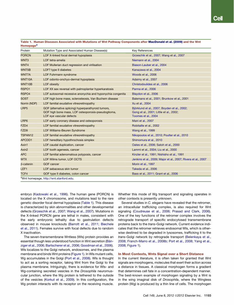
Table 1. Human Diseases Associated with Mutations of Wnt Pathway Components after MacDonald et al. (2009) and the WntHomepagea
Protein Mutation Type and Associated Human Disease(s) Key References
PORCN LOF X-linked focal dermal hypoplasia Grzeschik et al., 2007; Wang et al., 2007
WNT3 LOF tetra-amelia Niemann et al., 2004
WNT4 LOF Mullerian duct regression and virilisation Biason-Lauber et al., 2004
WNT5B LOF? type II diabetes Kanazawa et al., 2004
WNT7A LOF Fuhrmann syndrome Woods et al., 2006
WNT10A LOF odonto-onchyo-dermal hypoplasia Adaimy et al., 2007
WNT10B LOF obesity Christodoulides et al., 2006
RSPO1 LOF XX sex reversal with palmoplantar hyperkaratosis Parma et al., 2006
RSPO4 LOF autosomal-recessive anonychia and hyponychia congenita Blaydon et al., 2006
SOST LOF high bone mass, sclerosteosis, Van Buchem disease Balemans et al., 2001; Brunkow et al., 2001
Norrin (NDP) LOF familial exudative vitreoretinopathy Xu et al., 2004
LRP5 GOF (alternative splicing) hyperparathyroid tumors,
GOF high bone mass, LOF osteoporosis-pseudoglioma,
LOF eye vascular defects
Bjorklund et al., 2007; Boyden et al., 2002;
Gong et al., 2001; Little et al., 2002;
Toomes et al., 2004
LRP6 LOF early coronary disease and osteoporosis Mani et al., 2007
FZD4 LOF familial exudative vitreoretinopathy Robitaille et al., 2002
FZD9 LOF Williams-Beuren Syndrome Wang et al., 1999
TSPAN12 LOF familial exudative vitreoretinopathy Nikopoulos et al., 2010; Poulter et al., 2010
APCDD1 LOF hereditary hypothrochosis simplex Shimomura et al., 2010
Axin1 LOF caudal duplication, cancer Oates et al., 2006; Satoh et al., 2000
Axin2 LOF tooth agenesis, cancer Lammi et al., 2004; Liu et al., 2000
APC LOF familial adenomatous polyposis, cancer Kinzler et al., 1991; Nishisho et al., 1991
WTX LOF Wilms tumor, LOF OCTS Jenkins et al., 2009; Major et al., 2007; Rivera et al., 2007
b-catenin GOF cancer Morin et al., 1997
LEF1 LOF sebaceous skin tumor Takeda et al., 2006
TCF4 GOF type II diabetes, colon cancer Bass et al., 2011; Grant et al., 2006aWnt homepage, http://wnt.stanford.edu.
Cell 149, June 8, 2012 ª2012 Elsevier Inc. 1193

spreads out over the tissue, controlling gene expression ata distance. In this context, the Wg protein may act in associationwith lipoprotein particles (Panakova et al., 2005; Zecca et al.,1996). Alternatively, long-range signaling by lipid-modified Wgin thewing is facilitated by interactions with specific binding part-ners, such as the Swim protein (Mulligan et al., 2012).
It should be emphasized, however, that even in Drosophila,Wg rarely acts as a long-range signal in organs other than thewing. In the vast majority of the tissues, Wg mediates contact-dependent signaling. In the embryo, Wg interacts withEngrailed-positive cells that are adjacent to Wg-secreting cells(van den Heuvel et al., 1989). Likewise, Wg acts as a short-rangesignal in the neuromuscular junction (Korkut et al., 2009). In otheranimals, Wnt signaling appears to occur predominantly betweencells that are close to each other, for example, in adult stem cellniches (Strand and Micchelli, 2011; Sato et al., 2010). Wepropose, therefore, that Wnts are not classical morphogensbut signals that mediate close-range signaling.
Wnt Receptors Consist of a Heterodimeric ComplexWhen interacting with target cells, Wnt proteins bind a heterodi-meric receptor complex, consisting of a Frizzled (Fz) and anLRP5/6 protein (Figure 2). The ten mammalian Fz proteins areseven-transmembrane (7TM) receptors and have large extracel-lular N-terminal cysteine-rich domains (CRD; Bhanot et al., 1996)that provide a primary platform for Wnt binding (Dann et al.,2001; Janda et al., 2012). The structure of the CRD as boundto Wnt shows multiple binding surfaces, one of them containing
a hydrophobic groove that interacts with a lipid on theWnt mole-cule (Janda et al., 2012). The Wnt-Fz interaction is promiscuous:a single Wnt can bind multiple Fz proteins (e.g., Bhanot et al.,1996) and vice versa, which is also borne out by the structureof the Wnt-CRD complex (Janda et al., 2012). Fzs cooperatewith a single-pass transmembrane molecule of the LRP familyknown as Arrow in Drosophila (Wehrli et al., 2000) and LRP5and -6 in vertebrates (Pinson et al., 2000; Tamai et al., 2000).A recent study describes two monoclonal antibodies againstLRP6 with the unexpected ability to inhibit signaling by someWnt proteins and enhance signaling by others. As these anti-bodies bind nonoverlapping regions of LRP6 protein, these find-ings suggest that Lrp6 contains separate binding sites fordifferent classes of Wnt proteins (Gong et al., 2010).Like many signal transduction pathways, signaling by dimeric
Wnt receptors includes a ligand-induced conformational changeof the receptors followed by phosphorylation of key targetproteins. A crucial step in signaling is binding of Axin to the cyto-plasmic tail of LRP6 (Mao et al., 2001; Figure 2). Axin-LRP6binding is regulated by phosphorylation of the LRP6 tail (Heet al., 2004; Tamai et al., 2004) by at least two separate kinases,GSK3 and CK1g. GSK3 phosphorylates the serine in the PPPSPmotif found in a number of Wnt signaling components, includingb-catenin, Axin, APC, and potentially LRPs (Zeng et al., 2005).CK1g is anchored in the membrane by its palmitoylated Cterminus, and it phosphorylates these same proteins on residuesadjacent to the PPPSPmotif (Davidson et al., 2005). It is not clearwhether or how Wnt activates these protein kinases, but addingWnt protein to cells leads to CK1g-induced phosphorylationwithin minutes, suggesting a direct response to the signal.Relatively little is known on the role of Fz in Wnt reception. The
cytoplasmic part of Fz interacts with Dishevelled (Dsh; Chenet al., 2003; Figure 2), facilitating interaction between the LRPtail and Axin. The DIX domain on Dsh is similar to a region inAxin, and these two DIX domains can bind each other directly(Fiedler et al., 2011; Schwarz-Romond et al., 2007). Multimersof receptor-bound Dsh and Axin molecules might encouragethe formation of the LRP-Fz dimer. Higher-order complexes con-taining Wnts, receptors, and Dsh, as well as small particles ofmultimerized Dsh molecules, have been detected in cells(Schwarz-Romond et al., 2005).Importantly, the Wnt pathway transduces signals differently
than most other pathways. In many signaling cascades, proteinphosphorylation amplifies the signal, as individual kinase mole-cules catalyze the modification of multiple substrate molecules.In contrast, Wnt-induced LRP6 phosphorylation titrates awaya negative regulator, Axin, providing a stoichiometric ratherthan a catalytic mechanism of signal transduction. Likewise, ithas been proposed that the regulation of GSK3 by Wnt is stoi-chiometric, caused by sequestration of the enzyme inside multi-vesicular endosomes (Taelman et al., 2010).
Natural Wnt Inhibitors Act in Various Ways on WntReceptorsWnt/b-catenin signaling is regulated at many levels, including bysecreted proteins that antagonize the ligand. Among these aresecreted Frizzled-related proteins (sFRPs) and Wnt inhibitoryprotein (WIF), both of which can bind Wnts, thereby inhibiting
Figure 1. The Wnt Secretion MachineryWnt proteins become lipid modified in the endoplasmic reticulum by thePorcupine (Porc) enzyme. Further transport and secretion is dependent on theEvi/Wntless multiple pass transmembrane protein. The Retromer complex isnecessary for recycling of the Evi/Wntless endosomal vesicles.
1194 Cell 149, June 8, 2012 ª2012 Elsevier Inc.
interactions between Wnt and Wnt receptors (Bovolenta et al.,2008). Other Wnt inhibitors include proteins of the Dickkopf(DKK) (Glinka et al., 1998) and the WISE/SOST families, whichantagonize signaling by binding LRP5/6. Recent biochemicaland genetic studies have argued that DKK1 disrupts Wnt-induced Fz-LRP6 complex formation (Ellwanger et al., 2008;Semenov et al., 2008). Like DKK1, SOST can disrupt Wnt-induced Fz-LRP6 complexes in vitro (Semenov et al., 2005). Asa final example, APCDD1 is a membrane-bound glycoproteinthat inhibits Wnt signaling by binding both Wnt and LRP. It ismutated in hereditary hypotrichosis simplex, a condition charac-terized by hair follicle miniaturization (Shimomura et al., 2010;Table 1). Of interest, no natural secreted inhibitors have beenidentified in flies.
Mutations in Wnt Receptors and Their AntagonistsImplicate Wnt Signaling in Bone DiseaseA fast-growing field connects Wnt signaling with bone biologyand disease (Table 1; reviewed in Monroe et al., 2011). Thislink was first established by the discovery of LRP5 mutationsassociated with osteoporosis pseudoglioma syndrome(OPPG), a hereditary disorder characterized by low bone massand abnormal eye vasculature (Gong et al., 2001). Subsequently,patients with distinct types of hereditary high bone massdiseases were found to carry mutations in the LRP5 extracellulardomain (Boyden et al., 2002; Little et al., 2002), which renderLRP5 resistant to binding of the antagonist SOST (Ellies et al.,
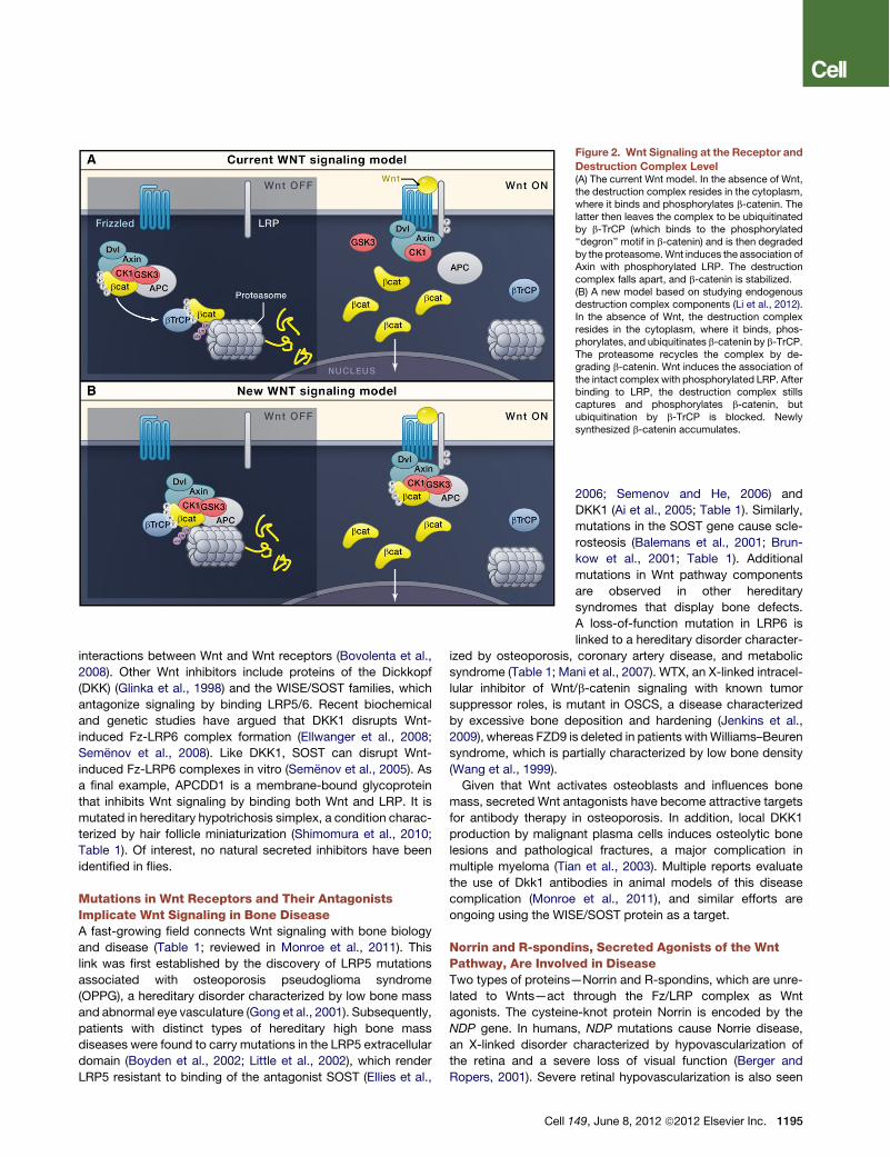
Figure 2. Wnt Signaling at the Receptor andDestruction Complex Level(A) The current Wnt model. In the absence of Wnt,the destruction complex resides in the cytoplasm,where it binds and phosphorylates b-catenin. Thelatter then leaves the complex to be ubiquitinatedby b-TrCP (which binds to the phosphorylated‘‘degron’’ motif in b-catenin) and is then degradedby the proteasome.Wnt induces the association ofAxin with phosphorylated LRP. The destructioncomplex falls apart, and b-catenin is stabilized.(B) A new model based on studying endogenousdestruction complex components (Li et al., 2012).In the absence of Wnt, the destruction complexresides in the cytoplasm, where it binds, phos-phorylates, and ubiquitinates b-catenin by b-TrCP.The proteasome recycles the complex by de-grading b-catenin. Wnt induces the association ofthe intact complex with phosphorylated LRP. Afterbinding to LRP, the destruction complex stillscaptures and phosphorylates b-catenin, butubiquitination by b-TrCP is blocked. Newlysynthesized b-catenin accumulates.
2006; Semenov and He, 2006) andDKK1 (Ai et al., 2005; Table 1). Similarly,mutations in the SOST gene cause scle-rosteosis (Balemans et al., 2001; Brun-kow et al., 2001; Table 1). Additionalmutations in Wnt pathway componentsare observed in other hereditarysyndromes that display bone defects.A loss-of-function mutation in LRP6 islinked to a hereditary disorder character-
ized by osteoporosis, coronary artery disease, and metabolicsyndrome (Table 1; Mani et al., 2007). WTX, an X-linked intracel-lular inhibitor of Wnt/b-catenin signaling with known tumorsuppressor roles, is mutant in OSCS, a disease characterizedby excessive bone deposition and hardening (Jenkins et al.,2009), whereas FZD9 is deleted in patients withWilliams–Beurensyndrome, which is partially characterized by low bone density(Wang et al., 1999).Given that Wnt activates osteoblasts and influences bone
mass, secreted Wnt antagonists have become attractive targetsfor antibody therapy in osteoporosis. In addition, local DKK1production by malignant plasma cells induces osteolytic bonelesions and pathological fractures, a major complication inmultiple myeloma (Tian et al., 2003). Multiple reports evaluatethe use of Dkk1 antibodies in animal models of this diseasecomplication (Monroe et al., 2011), and similar efforts areongoing using the WISE/SOST protein as a target.
Norrin and R-spondins, Secreted Agonists of the WntPathway, Are Involved in DiseaseTwo types of proteins—Norrin and R-spondins, which are unre-lated to Wnts—act through the Fz/LRP complex as Wntagonists. The cysteine-knot protein Norrin is encoded by theNDP gene. In humans, NDP mutations cause Norrie disease,an X-linked disorder characterized by hypovascularization ofthe retina and a severe loss of visual function (Berger andRopers, 2001). Severe retinal hypovascularization is also seen
Cell 149, June 8, 2012 ª2012 Elsevier Inc. 1195
in humans carrying a homozygous loss-of-function mutation inLrp5 (Gong et al., 2001). A milder retinal hypovascularization(familial exudative vitreoretinopathy or FEVR) occurs in patientsheterozygous for mutations in either Lrp5 (Robitaille et al.,2002) or Fz4 (Toomes et al., 2004; Table 1).
Norrin is a direct ligand for the Frizzled-4/Lrp5 complex. Thevascular phenotypes in the retina of mouse knockout modelsfor Norrin (Richter et al., 1998), Frizzled-4 (Wang et al., 2001),and Lrp5(Kato et al., 2002; Xia et al., 2008) resemble each other.In addition, Norrin binds with high affinity and specificity toFrizzled-4, whereas coexpression of Norrin, Frizzled-4, andLrp5 potently activates Wnt/b-catenin signaling (Xu et al.,2004). Biochemical evidence and analyses of mice carryingmutations in the tetraspanin family member Tspan12 provideevidence that Tspan12 is a Norrin-specific coreceptor (Jungeet al., 2009). Indeed, several FEVR families were subsequentlyfound to carry mutations in the TSPAN12 gene (Nikopouloset al., 2010; Poulter et al., 2010).
Vertebrate genomes encode four R-spondin (Rspo) proteins,small secreted proteins defined by two N-terminal furin domainsand a thrombospondin domain. The first evidence that Rspoproteins potently enhance Wnt/b-catenin signals came fromXenopus (Kazanskaya et al., 2004). Rspo1 was subsequentlyfound to feed into the canonical Wnt pathway, stronglypromoting intestinal crypt proliferation in vivo (Kim et al., 2005)and in vitro (Sato et al., 2009). Together, these results supporta crucial role for R-spondins in Wnt/b-catenin signaling. Rspomutations have been found in two hereditary syndromes inhumans (Table 1). RSPO1 is the gene disrupted in a recessivesyndrome characterized by XX sex reversal, a skin abnormalitycalled palmoplantar hyperkeratosis, and predisposition to squa-mous cell carcinomas (Parma et al., 2006). Mutations in theRSPO4 gene are linked to congenital anonychia, severe hypo-plasia of finger- and toenails, (Blaydon et al., 2006). Mutationsin RSPO2 have not been observed in humans, but rspo2 mousemutants exhibit a variety of developmental defects involvinglimbs (Nam et al., 2007), lung (Bell et al., 2008), and craniofacialanatomy (Yamada et al., 2009).
Lgr Molecules are R-Spondin Receptors that EnhanceWnt SignalingRecent studies have uncovered a small family of 7-TM receptors,the Lgr5 family, whichmediate Rspo input into the canonical Wntpathway (Carmon et al., 2011; de Lau et al., 2011; Glinka et al.,2011). All three studies demonstrate that the Lgr receptorsbind R-spondins with high affinity and are essential for signalenhancement of low-dose Wnt. Lgr4 and Lgr5 proteins physi-cally reside within Frizzled/LRP receptor complexes (de Lauet al., 2011).
What was known previously about the Lgr proteins? Uponthe cloning of these receptors, it was noted that Lgr4, Lgr5,and Lgr6 were related to the G-protein-coupled receptors(GPCRs) for thyroid-stimulating hormone (TSH), follicle-stimu-lating hormone (FSH), and luteinizing hormone (LH; Hsu et al.,2000). These receptors contain a large N-terminal extracellularleucine-rich repeat domain that binds the glycoproteinhormones. Similarly, the Lgr proteins bind R-spondins throughtheir N-terminal ectodomain, but current evidence indicates
that they do not utilize G proteins (Carmon et al., 2011; de Lauet al., 2011).Gene knockout studies revealed that Lgr4 as well as Lgr5
mutant mice are neonatal lethal. Pleiotropic phenotypes wereobserved for Lgr4 in male reproductive organs, eye, gall bladder,kidney, hair follicles, and a variety of other organs, whereas Lgr5mutants displayed a single abnormality of the lower jaw andtongue (Barker and Clevers, 2010). It was subsequently foundthat Lgr5 is a Wnt target gene in colon cancer and that it marksadult stem cells in a number of actively self-renewing organs,including the intestinal tract and the hair follicle (Barker et al.,2009, 2010; Jaks et al., 2008). Of note, a strong genetic interac-tion exists between Lgr4 and Lgr5, as seen in the gut of double-mutant mice (de Lau et al., 2011; Mustata et al., 2011). Lgr6similarly marks a rare, primitive stem cell that generates alllineages of the skin (Snippert et al., 2010). The finding that theLgr proteins act as receptors for R-spondins reinforces theintimate connection between Wnt signaling and activation ofadult stem cells (see below).
The Cytoplasmic APC/Axin Destruction ComplexRegulatesWnt Pathway Output by Controlling b-CateninStabilityThe destruction complex regulates the stability of cytoplasmicb-catenin, playing a key role in the signaling output of thecanonical Wnt cascade. The tumor suppressor protein Axinacts as the scaffold of the destruction complex, interactingwith b-catenin, the tumor suppressor proteins APC and WTX,and two constitutively active serine-threonine kinases (CK1a/dand GSK3a/b; Figure 2). APC is a large protein that interactswith both b-catenin and Axin. It contains three Axin-bindingmotifs that are interspersed between a series of 15 and 20amino acid repeats that bind b-catenin. Although it is clearfrom studies on colorectal cancer that APC is essential fordestruction complex function, its specific molecular activityremains unresolved. Another tumor suppressor protein involvedin destruction complex function is WTX. It is mutated in somecases of Wilms tumor, a pediatric kidney cancer (Rivera et al.,2007). WTX occurs in the destruction complex in which itpromotes b-catenin degradation, which would make its tumor-suppressive properties equivalent to those of Apc and Axin(Major et al., 2007). Its exact molecular functions in the Wntpathway, however, remain in debate (Regimbald-Dumas andHe, 2011).When Fz/LRP receptors are not engaged, CK1 and GSK3
sequentially phosphorylate Axin-bound b-catenin at a series ofregularly spaced N-terminal Ser/Thr residues. The phosphory-lated ‘‘degron’’ motif is then recognized by the F box/WD repeatprotein b-TrCP, part of an E3 ubiquitin ligase complex. Asa consequence, b-catenin is ubiquitinated and targeted for rapiddestruction by the proteasome (Aberle et al., 1997), preventingactivation of b-catenin target genes in the nucleus. Uponreceptor activation byWNT ligands, Axin is recruited to the phos-phorylated tail of LRP. Recent data show that, through thisrelocalization, the Wnt signal leads to inhibition of b-cateninubiquitination that normally occurs within the complex. Subse-quently, the complex becomes saturated by the phosphorylatedform of b-catenin, leading newly synthesized b-catenin to
1196 Cell 149, June 8, 2012 ª2012 Elsevier Inc.
accumulate and translocate to the nucleus to activate targetgenes (Figure 2 and Li et al., 2012).In addition to its role in Wnt pathway, b-catenin performs
a second, unrelated role in simple epithelia. It is an essentialbinding partner for the cytoplasmic tail of various cadherins,such as E-cadherin in adhesion junctions (Peifer et al., 1992).Though the half-life of the signaling pool of b-catenin is in theorder of minutes, the adherens junction pool is highly stable.The adhesive and signaling properties of b-catenin are mostlikely independent. Indeed, in C. elegans, the two functions ofb-catenin are performed by two different b-catenin homologs(Korswagen et al., 2000).
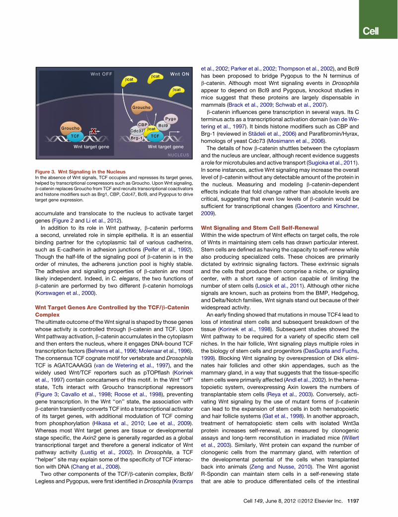
Wnt Target Genes Are Controlled by the TCF/b-CateninComplexThe ultimate outcome of theWnt signal is shaped by those geneswhose activity is controlled through b-catenin and TCF. UponWnt pathway activation, b-catenin accumulates in the cytoplasmand then enters the nucleus, where it engages DNA-bound TCFtranscription factors (Behrens et al., 1996; Molenaar et al., 1996).The consensus TCF cognate motif for vertebrate and DrosophilaTCF is AGATCAAAGG (van de Wetering et al., 1997), and thewidely used Wnt/TCF reporters such as pTOPflash (Korineket al., 1997) contain concatamers of this motif. In the Wnt ‘‘off’’state, Tcfs interact with Groucho transcriptional repressors(Figure 3; Cavallo et al., 1998; Roose et al., 1998), preventinggene transcription. In the Wnt ‘‘on’’ state, the association withb-catenin transiently converts TCF into a transcriptional activatorof its target genes, with additional modulation of TCF comingfrom phosphorylation (Hikasa et al., 2010; Lee et al., 2009).Whereas most Wnt target genes are tissue or developmentalstage specific, the Axin2 gene is generally regarded as a globaltranscriptional target and therefore a general indicator of Wntpathway activity (Lustig et al., 2002). In Drosophila, a TCF‘‘helper’’ site may explain some of the specificity of TCF interac-tion with DNA (Chang et al., 2008).Two other components of the TCF/b-catenin complex, Bcl9/
Legless and Pygopus, were first identified inDrosophila (Kramps
et al., 2002; Parker et al., 2002; Thompson et al., 2002), and Bcl9has been proposed to bridge Pygopus to the N terminus ofb-catenin. Although most Wnt signaling events in Drosophilaappear to depend on Bcl9 and Pygopus, knockout studies inmice suggest that these proteins are largely dispensable inmammals (Brack et al., 2009; Schwab et al., 2007).b-catenin influences gene transcription in several ways. Its C
terminus acts as a transcriptional activation domain (van de We-tering et al., 1997). It binds histone modifiers such as CBP andBrg-1 (reviewed in Stadeli et al., 2006) and Parafibromin/Hyrax,homologs of yeast Cdc73 (Mosimann et al., 2006).The details of how b-catenin shuttles between the cytoplasm
and the nucleus are unclear, although recent evidence suggestsa role for microtubules and active transport (Sugioka et al., 2011).In some instances, active Wnt signaling may increase the overalllevel of b-catenin without any detectable amount of the protein inthe nucleus. Measuring and modeling b-catenin-dependenteffects indicate that fold change rather than absolute levels arecritical, suggesting that even low levels of b-catenin would besufficient for transcriptional changes (Goentoro and Kirschner,2009).
Wnt Signaling and Stem Cell Self-RenewalWithin the wide spectrum of Wnt effects on target cells, the roleof Wnts in maintaining stem cells has drawn particular interest.Stem cells are defined as having the capacity to self-renew whilealso producing specialized cells. These choices are primarilydictated by extrinsic signaling factors. These extrinsic signalsand the cells that produce them comprise a niche, or signalingcenter, with a short range of action capable of limiting thenumber of stem cells (Losick et al., 2011). Although other nichesignals are known, such as proteins from the BMP, Hedgehog,and Delta/Notch families, Wnt signals stand out because of theirwidespread activity.An early finding showed that mutations in mouse TCF4 lead to
loss of intestinal stem cells and subsequent breakdown of thetissue (Korinek et al., 1998). Subsequent studies showed theWnt pathway to be required for a variety of specific stem cellniches. In the hair follicle, Wnt signaling plays multiple roles inthe biology of stem cells and progenitors (DasGupta and Fuchs,1999). Blocking Wnt signaling by overexpression of Dkk elimi-nates hair follicles and other skin appendages, such as themammary gland, in a way that suggests that the tissue-specificstem cells were primarily affected (Andl et al., 2002). In the hema-topoietic system, overexpressing Axin lowers the numbers oftransplantable stem cells (Reya et al., 2003). Conversely, acti-vating Wnt signaling by the use of mutant forms of b-catenincan lead to the expansion of stem cells in both hematopoieticand hair follicle systems (Gat et al., 1998). In another approach,treatment of hematopoietic stem cells with isolated Wnt3aprotein increases self-renewal, as measured by clonogenicassays and long-term reconstitution in irradiated mice (Willertet al., 2003). Similarly, Wnt protein can expand the number ofclonogenic cells from the mammary gland, with retention ofthe developmental potential of the cells when transplantedback into animals (Zeng and Nusse, 2010). The Wnt agonistR-Spondin can maintain stem cells in a self-renewing statethat are able to produce differentiated cells of the intestinal
Figure 3. Wnt Signaling in the NucleusIn the absence of Wnt signals, TCF occupies and represses its target genes,helped by transcriptional corepressors such as Groucho. Upon Wnt signaling,b-catenin replaces Groucho from TCF and recruits transcriptional coactivatorsand histone modifiers such as Brg1, CBP, Cdc47, Bcl9, and Pygopus to drivetarget gene expression.
Cell 149, June 8, 2012 ª2012 Elsevier Inc. 1197
epithelium (Sato et al., 2010). AndWnt canmediate maintenanceof pluripotency in mouse embryonic stem cells (Ten Berge et al.,2011). Despite all of these examples, it should bementioned thatthere are clear cases in which there is no role forWnts in stem cellmaintenance, particularly in the male and female germlines ofDrosophila (Losick et al., 2011).
In parallel, powerful genetic tools and lineage tracing haveprovided further evidence for Wnts as critical stem cell signals.These experiments have been based on identifying Wnt targetgenes such as LGR5 and Axin2 in tissues, some of them beingexpressed in putative stem cells (Snippert and Clevers, 2011).Labeled stem cells have been detected in organs ranging fromthe intestine to the stomach, the skin, the hair follicle, and themammary gland (Barker et al., 2007; Barker et al., 2010; vanAmerongen et al., 2012). Combined with the cell culture andgenetic data described above, the lineage tracing experimentsmake a compelling case for Wnts as self-renewal signals formany different varieties of adult stem cells.
What is the mechanism by which Wnt signals maintain stem-ness? Is there a common underlying mode of action? Althoughwe cannot yet definitively answer this question, we can invokethe fundamental concept that stem cells are intrinsically destinedto differentiate. It seems, therefore, that Wnt signals could blockthe default step, the differentiation of cells, possibly by sup-pressing differentiation-specific genes. There are indeed a fewexamples of genes that are downregulated rather than activatedby Wnt signals, and gene suppression could be part of a globalmechanism of regulation controlled by Wnts. It is likely thatepigenetic control lies at the root of the mechanism for influ-encing gene expression, enabling the stable propagation ofthe state of the cells. At the same time, a conditional signal-dependent state is required to make cells continuously depen-dent on the signal to maintain stemness. Combined with thelimited range of the lipid-modified Wnt proteins (Willert et al.,2003), this fits with an essential hallmark of the niche, a con-strained environment sustaining a small number of stem cells.
There are clear implications of the connection between Wntand stem cells, including the use ofWnt proteins or Wnt pathwayagonists to maintain stem cells in culture. Though stem cells aredifficult to expand in a self-renewing state, adding Wnts orR-Spondins has overcome some of these problems (Satoet al., 2010; Zeng and Nusse, 2010; Ten Berge et al., 2011).Likewise, Wnt reagents may have clinical applications for thetreatment of degenerative diseases.
Wnt Signaling in CancerGiven the importance of Wnt signaling for adult stem cellbiology, it is not surprising that Wnt pathway mutations arefrequently observed in cancer, most notably of tissues that nor-mally depend on Wnt for self-renewal or repair. Germline muta-tions in the APC gene cause a hereditary cancer syndrometermed familiar adenomatous polyposis (FAP; Kinzler et al.,1991; Nishisho et al., 1991). FAP patients carry heterozygousAPC mutations. The second allele is frequently lost in individualcells, which grow into colon adenomas, polyps, in early adult-hood. Additional mutations in genes like kras, tp53, and smad4induce some of these polyps to progress toward malignancy.Most cases of sporadic colorectal cancer result from loss of
both APC alleles (Kinzler and Vogelstein, 1996). Loss of APCfunction leads to the inappropriate stabilization of b-cateninand the formation of constitutive complexes between b-cateninand the intestinal TCF family member Tcf4 (Korinek et al., 1997).In rare cases of colorectal cancers wild-type for APC, Axin2is mutated (Liu et al., 2000), or activating point mutations inb-catenin remove the regulatory N-terminal Ser/Thr residues(Morin et al., 1997). Patients with hereditary Axin2 mutationsdisplay a predisposition to colon cancer in addition to toothagenesis (Lammi et al., 2004). Recent global exome-sequencingefforts have confirmed that the overwhelming majority of colo-rectal cancers carry inactivating APC mutations (Wood et al.,2007). In addition, this approach has revealed rare but recurrentfusions between VTl1A and Tcf7l2, the gene encoding Tcf4,adding yet another Wnt pathway member to the list of coloncancer genes (Bass et al., 2011).
Activating Wnt Pathway Mutations Are Not Restricted toCancer of the IntestineLoss-of-function mutations in Axin have also been found inhepatocellular carcinomas, whereas oncogenic b-catenin muta-tions, first described in colon cancer and melanoma (Rubinfeldet al., 1997), occur in a wide variety of solid tumors (reviewedin Reya and Clevers, 2005). Inactivating mutations in the TCFfamily member Lef1 occur in sebaceous skin tumors (Takedaet al., 2006). The link between stem cell biology and colon canceris further reinforced by several recent reports demonstratinga link between Wnt signal strength, stem cell signature, andcancer stem cell behavior (Merlos-Suarez et al., 2011; Vermeu-len et al., 2010).
Wnt Signaling Components and Metabolic DiseasesA rapidly emerging field involves the link between Wnt signalingand metabolism. Increased risk for type II diabetes has beenlinked to specific SNPs in WNT5B (Kanazawa et al., 2004),WNT10B (Christodoulides et al., 2006), and TCF7L2 (Grantet al., 2006). The Tcf7l association occurs in multiple ethnicgroups, and changes in Tcf7l2 define the strongest genetic riskdeterminant for this disease (Tong et al., 2009). Although themolecular and physiological mechanism underlying this linkremains a matter of debate, a recent transgenic study in micesuggests that metabolic risk is directly proportional to Tcf7l2gene expression (Savic et al., 2011).
Small-Molecule Wnt ModulatorsThe broad involvement of Wnt signaling in disease, as outlined inthis Review, has driven extensive research efforts at targetingthe Wnt pathway with small molecules. Table 2 lists many ofthe published molecules. From the perspective of blockingWnt signaling in cancer, the most effective target would be thecomplex between TCF and b-catenin, as it is a critical junctionin signaling and operates downstream in the pathway. Disap-pointingly, this complex has proven to be an elusive target. Itsstructure reveals a large binding surface that is not readily dis-lodged by chemical entities (Huber et al., 1997). Indeed, thoughseveral compounds, such as PKF115-854 and CGP049090,have been suggested to target Wnt signaling at this level (Lep-ourcelet et al., 2004), their specificity and efficacy remain to be
1198 Cell 149, June 8, 2012 ª2012 Elsevier Inc.
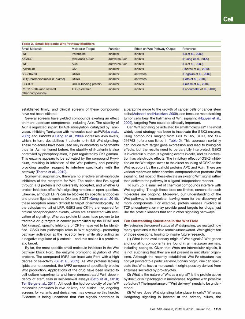
established firmly, and clinical screens of these compoundshave not been initiated.Several screens have yielded compounds exerting an effect
on more upstream components, including Axin. The stability ofAxin is regulated, in part, by ADP ribosylation, catalyzed by Tank-yrase. Inhibiting Tankyrase withmolecules such as IWR (Lu et al.,2009) and XAV939 (Huang et al., 2009) increases Axin levels,which, in turn, destabilizes b-catenin to inhibit Wnt signaling.These molecules have been used only in laboratory experimentsthus far. As mentioned before, the stability of b-catenin is alsocontrolled by phosphorylation, in part regulated by CK1 gamma.This enzyme appears to be activated by the compound Pyrvi-nium, resulting in inhibition of the Wnt pathway and possiblyproviding another reagent to interfere specifically with thepathway (Thorne et al., 2010).Somewhat surprisingly, there are no effective small-molecule
inhibitors of the receptors for Wnt. The notion that Fzs signalthrough a G protein is not universally accepted, and whether Gprotein inhibitors affect Wnt signaling remains an open question.Likewise, although LRPs can be blocked by specific antibodiesand protein ligands such as Dkk and SOST (Gong et al., 2010),these receptors remain difficult to target pharmacologically. Atthe cytoplasmic tail of LRP, GSK3 and CK1-g are required forcritical phosphorylation events, which are associated with acti-vation of signaling. Whereas protein kinases have proven to betractable drug targets in cancer (exemplified by the BRAF andAbl kinases), specific inhibitors of CK1-g have yet to be identi-fied. GSK3 has pleiotropic roles in Wnt signaling—promotingpathway activation at the receptor level while also acting asa negative regulator of b-catenin—and this makes it a problem-atic target.By far, the most specific small-molecule inhibitors in the Wnt
pathway block Porc, the enzyme promoting acylation of Wntproteins. The compound IWP2 can inactivate Porc with a highdegree of selectivity (Lu et al., 2009). As Wnt proteins lackinglipids are not secreted, the IWP2 compound specifically blocksWnt production. Applications of the drug have been limited tocell culture experiments and have demonstrated Wnt depen-dency of stem cells in self-renewal assays (Sato et al., 2010;Ten Berge et al., 2011). Although the hydrophobicity of the IWPmolecules precludes in vivo delivery and clinical use, ongoingscreens for variants and derivatives may overcome this hurdle.Evidence is being unearthed that Wnt signals contribute in
a paracrine mode to the growth of cancer cells or cancer stemcells (Malanchi and Huelsken, 2009), and becausemetastasizingtumor cells bear the hallmarks of Wnt signaling (Nguyen et al.,2009), targeting Porc could be clinically important.Can Wnt signaling be activated by small molecules? The most
widely used strategy has been to inactivate the GSK3 enzyme,using compounds ranging from LiCl to Bio, CHIR, and SB-216763 (references listed in Table 2). This approach certainlycan induce Wnt target gene expression and lead to biologicaleffects, but the results need to be carefully interpreted. GSK3is involved in numerous signaling events in cells, and its inactiva-tion has pleiotropic effects. The inhibitory effect of GSK3 inhibi-tion on theWnt signal owes to the direct coupling of GSK3 to theWnt receptors by the scaffold proteins APC and Axin. There arevarious reports on other chemical compounds that promote Wntsignaling, but most of these elevate an existing Wnt signal ratherthan activate the pathway in a ligand-independent manner.To sum up, a small set of chemical compounds interfere with
Wnt signaling. Though these tools are limited, screens for suchmolecules are ongoing. Moreover, our understanding of theWnt pathway is incomplete, leaving room for the discovery ofmore components. For example, protein kinases involved inTCF phosphorylation may provide good targets for drugs, justlike the protein kinases that act in other signaling pathways.
Ten Outstanding Questions in the Wnt FieldBy reviewing the current status of Wnt signaling, we realized howmany questions in this field remain unanswered. We highlight tenof those questions, hoping to inspire future research.(1) What is the evolutionary origin of Wnt signals? Wnt genes
and signaling components are found in all metazoan animals,including sponges. Given that Wnts are intercellular signals, itis not surprising that they are not present in unicellular organ-isms. Although the recently established Wnt-Fz structure hasnot yet pointed to a particular evolutionary origin, one can spec-ulate that Wnts have amore ancient origin, possibly derived fromenzymes secreted by prokaryotes.(2) What is the nature of Wnt as a signal? Is the protein active
by itself, or is it packaged in membranes, together with possiblecofactors? The importance of ‘‘Wnt delivery’’ needs to be under-stood.(3) Where does Wnt signaling take place in cells? Whereas
Hedgehog signaling is located at the primary cilium, the
Table 2. Small-Molecule Wnt Pathway Modifiers
Small Molecule Molecular Target Function Effect on Wnt Pathway Output Reference
IWP Porcupine inhibitor inhibits (Lu et al., 2009)
XAV939 tankyrase 1/Axin activates Axin inhibits (Huang et al., 2009)
IWR Axin activates Axin inhibits (Lu et al., 2009)
Pyrvinium CK1 inhibitor inhibits (Thorne et al., 2010)
SB-216763 GSK3 inhibitor activates (Coghlan et al., 2000)
BIO(6-bromoindirubin-30-oxime) GSK3 inhibitor activates (Sato et al., 2004)
ICG-001 CREB-binding protein inhibitor inhibits (Emami et al., 2004)
PKF115-584 (and several
other compounds)
TCF/b-catenin inhibitor inhibits (Lepourcelet et al., 2004)
Cell 149, June 8, 2012 ª2012 Elsevier Inc. 1199
subcellular location of Wnt signaling events remains unknown,though the endosomal compartment has been implicated asa signaling center.
(4) Besides the well-studied Frizzled and LRP receptors, thereare other mechanisms for Wnt reception that involve the tyrosinekinases ROR and RYK. There is very little insight into the mech-anisms of action of these receptors, and they deserve moreintense study.
(5) How is the stabilized form of b-catenin ferried into thenucleus? Is there a role for active microtubule-based transport?
(6) How does Wnt signaling coordinate cell fate changes withchanges in cell shape and polarity? This is a key question inmany developmental contexts of Wnt signaling, including stemcells.
(7) Many different kinds of stem cells are controlled byWnts, insuch a way that self-renewal and the developmental potential ofthe cells are preserved by the Wnt signal. Is there a universal‘‘stemness’’ property conferred to cells by Wnts?
(8) How much of the genome is Wnt controlled across variouscell types? Given the broad effects of Wnt signaling and manyWnt target cells, the total number of Wnt-controlled genes couldbe significant.
(9) Are cancer stem cell behaviors controlled byWnt signaling?The definition of cancer stem cells as being able to self-renewthe tumorigenic state but also able to differentiate suggeststhat their behavior is dependent on external signals, suchas Wnts.
(10) Can we identify bona fide and effective Wnt inhibitors?Though several molecules have been described to inhibit Wntsignaling in cells, there is a great need for additional reagentsinterfering with the Wnt pathway.
ACKNOWLEDGMENTS
The authors thank Vivian Li for providing figures, Janny van Eldik for editorial
assistance, and Rik Korswagen for critically reading the manuscript.
REFERENCES
Aberle, H., Bauer, A., Stappert, J., Kispert, A., and Kemler, R. (1997). beta-
catenin is a target for the ubiquitin-proteasome pathway. EMBO J. 16,
3797–3804.
Adaimy, L., Chouery, E., Megarbane, H., Mroueh, S., Delague, V., Nicolas, E.,
Belguith, H., de Mazancourt, P., and Megarbane, A. (2007). Mutation in
WNT10A is associated with an autosomal recessive ectodermal dysplasia:
the odonto-onycho-dermal dysplasia. Am. J. Hum. Genet. 81, 821–828.
Ai, M., Holmen, S.L., Van Hul, W., Williams, B.O., and Warman, M.L. (2005).
Reduced affinity to and inhibition by DKK1 form a common mechanism
by which high bone mass-associated missense mutations in LRP5 affect
canonical Wnt signaling. Mol. Cell. Biol. 25, 4946–4955.
Andl, T., Reddy, S.T., Gaddapara, T., and Millar, S.E. (2002). WNT signals are
required for the initiation of hair follicle development. Dev. Cell 2, 643–653.
Balemans, W., Ebeling, M., Patel, N., Van Hul, E., Olson, P., Dioszegi, M.,
Lacza, C., Wuyts, W., Van Den Ende, J., Willems, P., et al. (2001). Increased
bone density in sclerosteosis is due to the deficiency of a novel secreted
protein (SOST). Hum. Mol. Genet. 10, 537–543.
Banziger, C., Soldini, D., Schutt, C., Zipperlen, P., Hausmann, G., and Basler,
K. (2006). Wntless, a conserved membrane protein dedicated to the secretion
of Wnt proteins from signaling cells. Cell 125, 509–522.
Barker, N., and Clevers, H. (2010). Leucine-rich repeat-containing G-protein-
coupled receptors as markers of adult stem cells. Gastroenterology 138,
1681–1696.
Barker, N., van Es, J.H., Kuipers, J., Kujala, P., van den Born, M., Cozijnsen,
M., Haegebarth, A., Korving, J., Begthel, H., Peters, P.J., and Clevers, H.
(2007). Identification of stem cells in small intestine and colon by marker
gene Lgr5. Nature 449, 1003–1007.
Barker, N., Ridgway, R.A., van Es, J.H., van de Wetering, M., Begthel, H., van
den Born, M., Danenberg, E., Clarke, A.R., Sansom, O.J., and Clevers, H.
(2009). Crypt stem cells as the cells-of-origin of intestinal cancer. Nature
457, 608–611.
Barker, N., Huch, M., Kujala, P., van de Wetering, M., Snippert, H.J., van Es,
J.H., Sato, T., Stange, D.E., Begthel, H., van den Born, M., et al. (2010).
Lgr5(+ve) stem cells drive self-renewal in the stomach and build long-lived
gastric units in vitro. Cell Stem Cell 6, 25–36.
Barrott, J.J., Cash, G.M., Smith, A.P., Barrow, J.R., andMurtaugh, L.C. (2011).
Deletion of mouse Porcn blocks Wnt ligand secretion and reveals an ecto-
dermal etiology of human focal dermal hypoplasia/Goltz syndrome. Proc.
Natl. Acad. Sci. USA 108, 12752–12757.
Bartscherer, K., Pelte, N., Ingelfinger, D., and Boutros, M. (2006). Secretion
of Wnt ligands requires Evi, a conserved transmembrane protein. Cell 125,
523–533.
Bass, A.J., Lawrence, M.S., Brace, L.E., Ramos, A.H., Drier, Y., Cibulskis, K.,
Sougnez, C., Voet, D., Saksena, G., Sivachenko, A., et al. (2011). Genomic
sequencing of colorectal adenocarcinomas identifies a recurrent VTI1A-
TCF7L2 fusion. Nat. Genet. 43, 964–968.
Behrens, J., von Kries, J.P., Kuhl, M., Bruhn, L., Wedlich, D., Grosschedl, R.,
and Birchmeier, W. (1996). Functional interaction of beta-catenin with the
transcription factor LEF-1. Nature 382, 638–642.
Belenkaya, T.Y., Wu, Y., Tang, X., Zhou, B., Cheng, L., Sharma, Y.V., Yan, D.,
Selva, E.M., and Lin, X. (2008). The retromer complex influencesWnt secretion
by recycling wntless from endosomes to the trans-Golgi network. Dev. Cell 14,
120–131.
Bell, S.M., Schreiner, C.M., Wert, S.E., Mucenski, M.L., Scott, W.J., and Whit-
sett, J.A. (2008). R-spondin 2 is required for normal laryngeal-tracheal, lung
and limb morphogenesis. Development 135, 1049–1058.
Berger, W., and Ropers, H.H. (2001). Norrie disease. In Metabolic and Molec-
ular Bases of Inherited Disease, C.R. Scriver, A.L. Beaudet, W.S. Sly, and D.
Valle, eds. (New York: McGraw Hill), pp. 5977–5986.
Bhanot, P., Brink, M., Samos, C.H., Hsieh, J.C., Wang, Y., Macke, J.P.,
Andrew, D., Nathans, J., and Nusse, R. (1996). A new member of the frizzled
family fromDrosophila functions as aWingless receptor. Nature 382, 225–230.
Biason-Lauber, A., Konrad, D., Navratil, F., and Schoenle, E.J. (2004). AWNT4
mutation associated with Mullerian-duct regression and virilization in a 46,XX
woman. N. Engl. J. Med. 351, 792–798.
Biechele, S., Cox, B.J., and Rossant, J. (2011). Porcupine homolog is required
for canonical Wnt signaling and gastrulation in mouse embryos. Dev. Biol. 355,
275–285.
Bjorklund, P., Akerstrom, G., and Westin, G. (2007). An LRP5 receptor with
internal deletion in hyperparathyroid tumors with implications for deregulated
WNT/beta-catenin signaling. PLoS Med. 4, e328.
Blaydon, D.C., Ishii, Y., O’Toole, E.A., Unsworth, H.C., Teh, M.T., Ruschen-
dorf, F., Sinclair, C., Hopsu-Havu, V.K., Tidman, N., Moss, C., et al. (2006).
The gene encoding R-spondin 4 (RSPO4), a secreted protein implicated in
Wnt signaling, is mutated in inherited anonychia. Nat. Genet. 38, 1245–1247.
Bovolenta, P., Esteve, P., Ruiz, J.M., Cisneros, E., and Lopez-Rios, J. (2008).
Beyond Wnt inhibition: new functions of secreted Frizzled-related proteins in
development and disease. J. Cell Sci. 121, 737–746.
Boyden, L.M., Mao, J., Belsky, J., Mitzner, L., Farhi, A., Mitnick, M.A., Wu, D.,
Insogna, K., and Lifton, R.P. (2002). High bone density due to a mutation in
LDL-receptor-related protein 5. N. Engl. J. Med. 346, 1513–1521.
Brack, A.S., Murphy-Seiler, F., Hanifi, J., Deka, J., Eyckerman, S., Keller, C.,
Aguet, M., and Rando, T.A. (2009). BCL9 is an essential component of
1200 Cell 149, June 8, 2012 ª2012 Elsevier Inc.
canonical Wnt signaling that mediates the differentiation of myogenic progen-
itors during muscle regeneration. Dev. Biol. 335, 93–105.
Brunkow, M.E., Gardner, J.C., Van Ness, J., Paeper, B.W., Kovacevich, B.R.,
Proll, S., Skonier, J.E., Zhao, L., Sabo, P.J., Fu, Y., et al. (2001). Bone dysplasia
sclerosteosis results from loss of the SOST gene product, a novel cystine
knot-containing protein. Am. J. Hum. Genet. 68, 577–589.
Carmon, K.S., Gong, X., Lin, Q., Thomas, A., and Liu, Q. (2011). R-spondins
function as ligands of the orphan receptors LGR4 and LGR5 to regulate
Wnt/beta-catenin signaling. Proc. Natl. Acad. Sci. USA 108, 11452–11457.
Cavallo, R.A., Cox, R.T., Moline, M.M., Roose, J., Polevoy, G.A., Clevers, H.,
Peifer, M., and Bejsovec, A. (1998). Drosophila Tcf and Groucho interact to
repress Wingless signalling activity. Nature 395, 604–608.
Chang, M.V., Chang, J.L., Gangopadhyay, A., Shearer, A., and Cadigan, K.M.
(2008). Activation of wingless targets requires bipartite recognition of DNA by
TCF. Curr. Biol. 18, 1877–1881.
Chen,W., ten Berge, D., Brown, J., Ahn, S., Hu, L.A., Miller, W.E., Caron, M.G.,
Barak, L.S., Nusse, R., and Lefkowitz, R.J. (2003). Dishevelled 2 recruits beta-
arrestin 2 to mediate Wnt5A-stimulated endocytosis of Frizzled 4. Science
301, 1391–1394.
Christodoulides, C., Scarda, A., Granzotto, M., Milan, G., Dalla Nora, E.,
Keogh, J., De Pergola, G., Stirling, H., Pannacciulli, N., Sethi, J.K., et al.
(2006). WNT10B mutations in human obesity. Diabetologia 49, 678–684.
Coghlan, M.P., Culbert, A.A., Cross, D.A., Corcoran, S.L., Yates, J.W., Pearce,
N.J., Rausch, O.L., Murphy, G.J., Carter, P.S., Roxbee Cox, L., et al. (2000).
Selective small molecule inhibitors of glycogen synthase kinase-3 modulate
glycogen metabolism and gene transcription. Chem. Biol. 7, 793–803.
Coudreuse, D.Y., Roel, G., Betist, M.C., Destree, O., and Korswagen, H.C.
(2006). Wnt gradient formation requires retromer function in Wnt-producing
cells. Science 312, 921–924.
Dann, C.E., Hsieh, J.C., Rattner, A., Sharma, D., Nathans, J., and Leahy, D.J.
(2001). Insights into Wnt binding and signalling from the structures of two
Frizzled cysteine-rich domains. Nature 412, 86–90.
DasGupta, R., and Fuchs, E. (1999). Multiple roles for activated LEF/TCF
transcription complexes during hair follicle development and differentiation.
Development 126, 4557–4568.
Davidson, G., Wu, W., Shen, J., Bilic, J., Fenger, U., Stannek, P., Glinka, A.,
and Niehrs, C. (2005). Casein kinase 1 gamma couplesWnt receptor activation
to cytoplasmic signal transduction. Nature 438, 867–872.
de Lau, W., Barker, N., Low, T.Y., Koo, B.K., Li, V.S., Teunissen, H., Kujala, P.,
Haegebarth, A., Peters, P.J., van de Wetering, M., et al. (2011). Lgr5 homo-
logues associate withWnt receptors andmediate R-spondin signalling. Nature
476, 293–297.
Ellies, D.L., Viviano, B., McCarthy, J., Rey, J.P., Itasaki, N., Saunders, S., and
Krumlauf, R. (2006). Bone density ligand, Sclerostin, directly interacts with
LRP5 but not LRP5G171V to modulate Wnt activity. J. Bone Miner. Res. 21,
1738–1749.
Ellwanger, K., Saito, H., Clement-Lacroix, P., Maltry, N., Niedermeyer, J., Lee,
W.K., Baron, R., Rawadi, G., Westphal, H., and Niehrs, C. (2008). Targeted
disruption of the Wnt regulator Kremen induces limb defects and high bone
density. Mol. Cell. Biol. 28, 4875–4882.
Emami, K.H., Nguyen, C., Ma, H., Kim, D.H., Jeong, K.W., Eguchi, M., Moon,
R.T., Teo, J.L., Kim, H.Y., Moon, S.H., et al. (2004). A small molecule inhibitor of
beta-catenin/CREB-binding protein transcription [corrected]. Proc. Natl.
Acad. Sci. USA 101, 12682–12687.
Fiedler, M., Mendoza-Topaz, C., Rutherford, T.J., Mieszczanek, J., and Bienz,
M. (2011). Dishevelled interacts with the DIX domain polymerization interface
of Axin to interfere with its function in down-regulating b-catenin. Proc. Natl.
Acad. Sci. USA 108, 1937–1942.
Franch-Marro, X., Wendler, F., Griffith, J., Maurice, M.M., and Vincent, J.P.
(2008a). In vivo role of lipid adducts on Wingless. J. Cell Sci. 121, 1587–1592.
Franch-Marro, X., Wendler, F., Guidato, S., Griffith, J., Baena-Lopez, A.,
Itasaki, N., Maurice, M.M., and Vincent, J.P. (2008b). Wingless secretion
requires endosome-to-Golgi retrieval of Wntless/Evi/Sprinter by the retromer
complex. Nat. Cell Biol. 10, 170–177.
Gat, U., DasGupta, R., Degenstein, L., and Fuchs, E. (1998). De Novo hair
follicle morphogenesis and hair tumors in mice expressing a truncated
beta-catenin in skin. Cell 95, 605–614.
Glinka, A., Dolde, C., Kirsch, N., Huang, Y.L., Kazanskaya, O., Ingelfinger, D.,
Boutros, M., Cruciat, C.M., and Niehrs, C. (2011). LGR4 and LGR5 are
R-spondin receptors mediating Wnt/b-catenin and Wnt/PCP signalling.
EMBO Rep. 12, 1055–1061.
Glinka, A., Wu, W., Delius, H., Monaghan, A.P., Blumenstock, C., and Niehrs,
C. (1998). Dickkopf-1 is a member of a new family of secreted proteins and
functions in head induction. Nature 391, 357–362.
Goentoro, L., and Kirschner, M.W. (2009). Evidence that fold-change, and not
absolute level, of beta-catenin dictates Wnt signaling. Mol. Cell 36, 872–884.
Gong, Y., Slee, R.B., Fukai, N., Rawadi, G., Roman-Roman, S., Reginato, A.M.,
Wang, H., Cundy, T., Glorieux, F.H., Lev, D., et al; Osteoporosis-Pseudoglioma
Syndrome Collaborative Group. (2001). LDL receptor-related protein 5 (LRP5)
affects bone accrual and eye development. Cell 107, 513–523.
Gong, Y., Bourhis, E., Chiu, C., Stawicki, S., DeAlmeida, V.I., Liu, B.Y., Pham-
luong, K., Cao, T.C., Carano, R.A., Ernst, J.A., et al. (2010). Wnt isoform-
specific interactions with coreceptor specify inhibition or potentiation of
signaling by LRP6 antibodies. PLoS ONE 5, e12682.
Goodman, R.M., Thombre, S., Firtina, Z., Gray, D., Betts, D., Roebuck, J.,
Spana, E.P., and Selva, E.M. (2006). Sprinter: a novel transmembrane protein
required for Wg secretion and signaling. Development 133, 4901–4911.
Grant, S.F., Thorleifsson, G., Reynisdottir, I., Benediktsson, R., Manolescu, A.,
Sainz, J., Helgason, A., Stefansson, H., Emilsson, V., Helgadottir, A., et al.
(2006). Variant of transcription factor 7-like 2 (TCF7L2) gene confers risk of
type 2 diabetes. Nat. Genet. 38, 320–323.
Grzeschik, K.H., Bornholdt, D., Oeffner, F., Konig, A., del Carmen Boente, M.,
Enders, H., Fritz, B., Hertl, M., Grasshoff, U., Hofling, K., et al. (2007).
Deficiency of PORCN, a regulator of Wnt signaling, is associated with focal
dermal hypoplasia. Nat. Genet. 39, 833–835.
He, X., Semenov, M., Tamai, K., and Zeng, X. (2004). LDL receptor-related
proteins 5 and 6 in Wnt/beta-catenin signaling: arrows point the way.
Development 131, 1663–1677.
Hikasa, H., Ezan, J., Itoh, K., Li, X., Klymkowsky, M.W., and Sokol, S.Y. (2010).
Regulation of TCF3 byWnt-dependent phosphorylation during vertebrate axis
specification. Dev. Cell 19, 521–532.
Hofmann, K. (2000). A superfamily of membrane-bound O-acyltransferases
with implications for wnt signaling. Trends Biochem. Sci. 25, 111–112.
Hsu, S.Y., Kudo, M., Chen, T., Nakabayashi, K., Bhalla, A., van der Spek, P.J.,
van Duin, M., and Hsueh, A.J. (2000). The three subfamilies of leucine-rich
repeat-containing G protein-coupled receptors (LGR): identification of LGR6
and LGR7 and the signaling mechanism for LGR7. Mol. Endocrinol. 14,
1257–1271.
Huang, S.M., Mishina, Y.M., Liu, S., Cheung, A., Stegmeier, F., Michaud, G.A.,
Charlat, O., Wiellette, E., Zhang, Y., Wiessner, S., et al. (2009). Tankyrase inhi-
bition stabilizes axin and antagonizes Wnt signalling. Nature 461, 614–620.
Huber, A.H., Nelson, W.J., and Weis, W.I. (1997). Three-dimensional structure
of the armadillo repeat region of beta-catenin. Cell 90, 871–882.
Jaks, V., Barker, N., Kasper, M., van Es, J.H., Snippert, H.J., Clevers, H., and
Toftgard, R. (2008). Lgr5 marks cycling, yet long-lived, hair follicle stem cells.
Nat. Genet. 40, 1291–1299.
Janda, C.,Waghray, D., Levin, A., Thomas, C., andGarcia, K. (2012). Structural
basis of Wnt recognition by Frizzled. Science. Published online May 31, 2012.
Jenkins, Z.A., van Kogelenberg, M., Morgan, T., Jeffs, A., Fukuzawa, R., Pearl,
E., Thaller, C., Hing, A.V., Porteous, M.E., Garcia-Minaur, S., et al. (2009).
Germline mutations in WTX cause a sclerosing skeletal dysplasia but do not
predispose to tumorigenesis. Nat. Genet. 41, 95–100.
Junge, H.J., Yang, S., Burton, J.B., Paes, K., Shu, X., French, D.M., Costa, M.,
Rice, D.S., and Ye, W. (2009). TSPAN12 regulates retinal vascular
Cell 149, June 8, 2012 ª2012 Elsevier Inc. 1201
development by promoting Norrin- but not Wnt-induced FZD4/beta-catenin
signaling. Cell 139, 299–311.
Kadowaki, T., Wilder, E., Klingensmith, J., Zachary, K., and Perrimon, N.
(1996). The segment polarity gene porcupine encodes a putative multitrans-
membrane protein involved in Wingless processing. Genes Dev. 10, 3116–
3128.
Kanazawa, A., Tsukada, S., Sekine, A., Tsunoda, T., Takahashi, A., Kashiwagi,
A., Tanaka, Y., Babazono, T., Matsuda, M., Kaku, K., et al. (2004). Association
of the gene encoding wingless-type mammary tumor virus integration-site
family member 5B (WNT5B) with type 2 diabetes. Am. J. Hum. Genet. 75,
832–843.
Kato, M., Patel, M.S., Levasseur, R., Lobov, I., Chang, B.H., Glass, D.A., II,
Hartmann, C., Li, L., Hwang, T.H., Brayton, C.F., et al. (2002). Cbfa1-indepen-
dent decrease in osteoblast proliferation, osteopenia, and persistent embry-
onic eye vascularization in mice deficient in Lrp5, a Wnt coreceptor. J. Cell
Biol. 157, 303–314.
Kazanskaya, O., Glinka, A., del Barco Barrantes, I., Stannek, P., Niehrs, C., and
Wu, W. (2004). R-Spondin2 is a secreted activator of Wnt/beta-catenin
signaling and is required for Xenopus myogenesis. Dev. Cell 7, 525–534.
Kim, K.A., Kakitani, M., Zhao, J., Oshima, T., Tang, T., Binnerts, M., Liu, Y.,
Boyle, B., Park, E., Emtage, P., et al. (2005). Mitogenic influence of human
R-spondin1 on the intestinal epithelium. Science 309, 1256–1259.
Kinzler, K.W., Nilbert, M.C., Su, L.K., Vogelstein, B., Bryan, T.M., Levy, D.B.,
Smith, K.J., Preisinger, A.C., Hedge, P., McKechnie, D., et al. (1991). Identifi-
cation of FAP locus genes from chromosome 5q21. Science 253, 661–665.
Kinzler, K.W., and Vogelstein, B. (1996). Lessons from hereditary colorectal
cancer. Cell 87, 159–170.
Korinek, V., Barker, N., Morin, P.J., van Wichen, D., de Weger, R., Kinzler,
K.W., Vogelstein, B., and Clevers, H. (1997). Constitutive transcriptional
activation by a beta-catenin-Tcf complex in APC-/- colon carcinoma. Science
275, 1784–1787.
Korinek, V., Barker, N., Moerer, P., van Donselaar, E., Huls, G., Peters, P.J.,
and Clevers, H. (1998). Depletion of epithelial stem-cell compartments in the
small intestine of mice lacking Tcf-4. Nat. Genet. 19, 379–383.
Korkut, C., Ataman, B., Ramachandran, P., Ashley, J., Barria, R., Gherbesi, N.,
and Budnik, V. (2009). Trans-synaptic transmission of vesicular Wnt signals
through Evi/Wntless. Cell 139, 393–404.
Korswagen, H.C., Herman, M.A., and Clevers, H.C. (2000). Distinct beta-
catenins mediate adhesion and signalling functions in C. elegans. Nature
406, 527–532.
Kramps, T., Peter, O., Brunner, E., Nellen, D., Froesch, B., Chatterjee, S.,
Murone, M., Zullig, S., and Basler, K. (2002). Wnt/wingless signaling requires
BCL9/legless-mediated recruitment of pygopus to the nuclear beta-catenin-
TCF complex. Cell 109, 47–60.
Kurayoshi, M., Yamamoto, H., Izumi, S., and Kikuchi, A. (2007). Post-
translational palmitoylation and glycosylation of Wnt-5a are necessary for its
signalling. Biochem. J. 402, 515–523.
Kusserow, A., Pang, K., Sturm, C., Hrouda, M., Lentfer, J., Schmidt, H.A.,
Technau, U., von Haeseler, A., Hobmayer, B., Martindale, M.Q., and Holstein,
T.W. (2005). Unexpected complexity of the Wnt gene family in a sea anemone.
Nature 433, 156–160.
Lammi, L., Arte, S., Somer, M., Jarvinen, H., Lahermo, P., Thesleff, I., Pirinen,
S., and Nieminen, P. (2004). Mutations in AXIN2 cause familial tooth agenesis
and predispose to colorectal cancer. Am. J. Hum. Genet. 74, 1043–1050.
Lee, W., Swarup, S., Chen, J., Ishitani, T., and Verheyen, E.M. (2009).
Homeodomain-interacting protein kinases (Hipks) promote Wnt/Wg signaling
through stabilization of beta-catenin/Arm and stimulation of target gene
expression. Development 136, 241–251.
Lepourcelet, M., Chen, Y.N., France, D.S., Wang, H., Crews, P., Petersen, F.,
Bruseo, C., Wood, A.W., and Shivdasani, R.A. (2004). Small-molecule antag-
onists of the oncogenic Tcf/beta-catenin protein complex. Cancer Cell 5,
91–102.
Li, V.S., Ng, S.S., Boersema, P.J., Low, T.Y., Karthaus, W.R., Gerlach, J.P.,
Mohammed, S., Heck, A.J., Maurice, M.M., Mahmoudi, T., and Clevers, H.
(2012). Wnt signaling inhibits proteasomal b-catenin degradation within
a compositionally intact Axin1 complex. Cell 149, this issue, 1245–1256.
Little, R.D., Carulli, J.P., Del Mastro, R.G., Dupuis, J., Osborne, M., Folz, C.,
Manning, S.P., Swain, P.M., Zhao, S.C., Eustace, B., et al. (2002). A mutation
in the LDL receptor-related protein 5 gene results in the autosomal dominant
high-bone-mass trait. Am. J. Hum. Genet. 70, 11–19.
Liu, W., Dong, X., Mai, M., Seelan, R.S., Taniguchi, K., Krishnadath, K.K., Hal-
ling, K.C., Cunningham, J.M., Boardman, L.A., Qian, C., et al. (2000). Mutations
in AXIN2 cause colorectal cancer with defective mismatch repair by activating
beta-catenin/TCF signalling. Nat. Genet. 26, 146–147.
Losick, V.P., Morris, L.X., Fox, D.T., and Spradling, A. (2011). Drosophila
stem cell niches: a decade of discovery suggests a unified view of stem cell
regulation. Dev. Cell 21, 159–171.
Lu, J., Ma, Z., Hsieh, J.C., Fan, C.W., Chen, B., Longgood, J.C., Williams, N.S.,
Amatruda, J.F., Lum, L., and Chen, C. (2009). Structure-activity relationship
studies of small-molecule inhibitors of Wnt response. Bioorg. Med. Chem.
Lett. 19, 3825–3827.
Lustig, B., Jerchow, B., Sachs, M., Weiler, S., Pietsch, T., Karsten, U., van de
Wetering, M., Clevers, H., Schlag, P.M., Birchmeier, W., and Behrens, J.
(2002). Negative feedback loop of Wnt signaling through upregulation
of conductin/axin2 in colorectal and liver tumors. Mol. Cell. Biol. 22, 1184–
1193.
MacDonald, B.T., Tamai, K., and He, X. (2009). Wnt/beta-catenin signaling:
components, mechanisms, and diseases. Dev. Cell 17, 9–26.
Major, M.B., Camp, N.D., Berndt, J.D., Yi, X., Goldenberg, S.J., Hubbert, C.,
Biechele, T.L., Gingras, A.C., Zheng, N., Maccoss, M.J., et al. (2007). Wilms
tumor suppressor WTX negatively regulates WNT/beta-catenin signaling.
Science 316, 1043–1046.
Malanchi, I., and Huelsken, J. (2009). Cancer stem cells: never Wnt away from
the niche. Curr. Opin. Oncol. 21, 41–46.
Mani, A., Radhakrishnan, J., Wang, H., Mani, A., Mani, M.A., Nelson-Williams,
C., Carew, K.S., Mane, S., Najmabadi, H., Wu, D., and Lifton, R.P. (2007). LRP6
mutation in a family with early coronary disease and metabolic risk factors.
Science 315, 1278–1282.
Mao, J., Wang, J., Liu, B., Pan, W., Farr, G.H., III, Flynn, C., Yuan, H., Takada,
S., Kimelman, D., Li, L., and Wu, D. (2001). Low-density lipoprotein receptor-
related protein-5 binds to Axin and regulates the canonical Wnt signaling
pathway. Mol. Cell 7, 801–809.
McMahon, A.P., and Moon, R.T. (1989). Ectopic expression of the proto-
oncogene int-1 in Xenopus embryos leads to duplication of the embryonic
axis. Cell 58, 1075–1084.
Merlos-Suarez, A., Barriga, F.M., Jung, P., Iglesias, M., Cespedes, M.V.,
Rossell, D., Sevillano, M., Hernando-Momblona, X., da Silva-Diz, V., Munoz,
P., et al. (2011). The intestinal stem cell signature identifies colorectal cancer
stem cells and predicts disease relapse. Cell Stem Cell 8, 511–524.
Molenaar, M., van de Wetering, M., Oosterwegel, M., Peterson-Maduro, J.,
Godsave, S., Korinek, V., Roose, J., Destree, O., and Clevers, H. (1996).
XTcf-3 transcription factor mediates beta-catenin-induced axis formation in
Xenopus embryos. Cell 86, 391–399.
Monroe, D.G., McGee-Lawrence, M.E., Oursler, M.J., and Westendorf, J.J.
(2011). Update on Wnt signaling in bone cell biology and bone disease.
Gene 492, 1–18.
Morin, P.J., Sparks, A.B., Korinek, V., Barker, N., Clevers, H., Vogelstein, B.,
and Kinzler, K.W. (1997). Activation of beta-catenin-Tcf signaling in colon
cancer by mutations in beta-catenin or APC. Science 275, 1787–1790.
Mosimann, C., Hausmann, G., and Basler, K. (2006). Parafibromin/Hyrax
activates Wnt/Wg target gene transcription by direct association with beta-
catenin/Armadillo. Cell 125, 327–341.
Mulligan, K.A., Fuerer, C., Ching, W., Fish, M., Willert, K., and Nusse, R. (2012).
Secreted Wingless-interacting molecule (Swim) promotes long-range
1202 Cell 149, June 8, 2012 ª2012 Elsevier Inc.
signaling by maintaining Wingless solubility. Proc. Natl. Acad. Sci. USA 109,
370–377.
Mustata, R.C., Van Loy, T., Lefort, A., Libert, F., Strollo, S., Vassart, G., and
Garcia, M.I. (2011). Lgr4 is required for Paneth cell differentiation and mainte-
nance of intestinal stem cells ex vivo. EMBO Rep. 12, 558–564.
Nam, J.S., Park, E., Turcotte, T.J., Palencia, S., Zhan, X., Lee, J., Yun, K., Funk,
W.D., and Yoon, J.K. (2007). Mouse R-spondin2 is required for apical
ectodermal ridge maintenance in the hindlimb. Dev. Biol. 311, 124–135.
Nguyen, D.X., Chiang, A.C., Zhang, X.H., Kim, J.Y., Kris, M.G., Ladanyi, M.,
Gerald, W.L., and Massague, J. (2009). WNT/TCF signaling through LEF1
and HOXB9 mediates lung adenocarcinoma metastasis. Cell 138, 51–62.
Niemann, S., Zhao, C., Pascu, F., Stahl, U., Aulepp, U., Niswander, L., Weber,
J.L., and Muller, U. (2004). Homozygous WNT3 mutation causes tetra-amelia
in a large consanguineous family. Am. J. Hum. Genet. 74, 558–563.
Nikopoulos, K., Gilissen, C., Hoischen, A., van Nouhuys, C.E., Boonstra, F.N.,
Blokland, E.A., Arts, P., Wieskamp, N., Strom, T.M., Ayuso, C., et al. (2010).
Next-generation sequencing of a 40 Mb linkage interval reveals TSPAN12
mutations in patients with familial exudative vitreoretinopathy. Am. J. Hum.
Genet. 86, 240–247.
Nishisho, I., Nakamura, Y., Miyoshi, Y., Miki, Y., Ando, H., Horii, A., Koyama,
K., Utsunomiya, J., Baba, S., and Hedge, P. (1991). Mutations of chromosome
5q21 genes in FAP and colorectal cancer patients. Science 253, 665–669.
Noordermeer, J., Klingensmith, J., Perrimon, N., and Nusse, R. (1994). dishev-
elled and armadillo act in thewingless signalling pathway in Drosophila. Nature
367, 80–83.
Nusse, R., and Varmus, H.E. (1982). Many tumors induced by the mouse
mammary tumor virus contain a provirus integrated in the same region of the
host genome. Cell 31, 99–109.
Nusslein-Volhard, C., and Wieschaus, E. (1980). Mutations affecting segment
number and polarity in Drosophila. Nature 287, 795–801.
Oates, N.A., van Vliet, J., Duffy, D.L., Kroes, H.Y., Martin, N.G., Boomsma, D.I.,
Campbell, M., Coulthard, M.G., Whitelaw, E., and Chong, S. (2006). Increased
DNA methylation at the AXIN1 gene in a monozygotic twin from a pair
discordant for a caudal duplication anomaly. Am. J. Hum. Genet. 79, 155–162.
Panakova, D., Sprong, H., Marois, E., Thiele, C., and Eaton, S. (2005). Lipopro-
tein particles are required for Hedgehog and Wingless signalling. Nature 435,
58–65.
Parker, D.S., Jemison, J., and Cadigan, K.M. (2002). Pygopus, a nuclear PHD-
finger protein required for Wingless signaling in Drosophila. Development 129,
2565–2576.
Parma, P., Radi, O., Vidal, V., Chaboissier, M.C., Dellambra, E., Valentini, S.,
Guerra, L., Schedl, A., and Camerino, G. (2006). R-spondin1 is essential in
sex determination, skin differentiation and malignancy. Nat. Genet. 38,
1304–1309.
Peifer, M., McCrea, P.D., Green, K.J., Wieschaus, E., and Gumbiner, B.M.
(1992). The vertebrate adhesive junction proteins beta-catenin and plakoglo-
bin and the Drosophila segment polarity gene armadillo form a multigene
family with similar properties. J. Cell Biol. 118, 681–691.
Peifer, M., Sweeton, D., Casey, M., and Wieschaus, E. (1994). wingless
signal and Zeste-white 3 kinase trigger opposing changes in the intracellular
distribution of Armadillo. Development 120, 369–380.
Petersen, C.P., and Reddien, P.W. (2009). Wnt signaling and the polarity of the
primary body axis. Cell 139, 1056–1068.
Pinson, K.I., Brennan, J., Monkley, S., Avery, B.J., and Skarnes, W.C. (2000).
An LDL-receptor-related protein mediates Wnt signalling in mice. Nature 407,
535–538.
Port, F., Kuster, M., Herr, P., Furger, E., Banziger, C., Hausmann, G., and Bas-
ler, K. (2008). Wingless secretion promotes and requires retromer-dependent
cycling of Wntless. Nat. Cell Biol. 10, 178–185.
Poulter, J.A., Ali, M., Gilmour, D.F., Rice, A., Kondo, H., Hayashi, K., Mackey,
D.A., Kearns, L.S., Ruddle, J.B., Craig, J.E., et al. (2010). Mutations in
TSPAN12 cause autosomal-dominant familial exudative vitreoretinopathy.
Am. J. Hum. Genet. 86, 248–253.
Prasad, B.C., and Clark, S.G. (2006).Wnt signaling establishes anteroposterior
neuronal polarity and requires retromer in C. elegans. Development 133, 1757–
1766.
Regimbald-Dumas, Y., and He, X. (2011). Wnt signalling: What The X@# is
WTX? EMBO J. 30, 1415–1417.
Reya, T., and Clevers, H. (2005). Wnt signalling in stem cells and cancer.
Nature 434, 843–850.
Reya, T., Duncan, A.W., Ailles, L., Domen, J., Scherer, D.C., Willert, K., Hintz,
L., Nusse, R., and Weissman, I.L. (2003). A role for Wnt signalling in self-
renewal of haematopoietic stem cells. Nature 423, 409–414.
Richter, M., Gottanka, J., May, C.A., Welge-Lussen, U., Berger, W., and Lut-
jen-Drecoll, E. (1998). Retinal vasculature changes in Norrie disease mice.
Invest. Ophthalmol. Vis. Sci. 39, 2450–2457.
Rijsewijk, F., Schuermann, M., Wagenaar, E., Parren, P., Weigel, D., and
Nusse, R. (1987). The Drosophila homolog of the mouse mammary oncogene
int-1 is identical to the segment polarity gene wingless. Cell 50, 649–657.
Rivera, M.N., Kim, W.J., Wells, J., Driscoll, D.R., Brannigan, B.W., Han, M.,
Kim, J.C., Feinberg, A.P., Gerald, W.L., Vargas, S.O., et al. (2007). An X chro-
mosome gene, WTX, is commonly inactivated in Wilms tumor. Science 315,
642–645.
Robitaille, J., MacDonald, M.L., Kaykas, A., Sheldahl, L.C., Zeisler, J., Dube,
M.P., Zhang, L.H., Singaraja, R.R., Guernsey, D.L., Zheng, B., et al. (2002).
Mutant frizzled-4 disrupts retinal angiogenesis in familial exudative vitreoretin-
opathy. Nat. Genet. 32, 326–330.
Roose, J., Molenaar, M., Peterson, J., Hurenkamp, J., Brantjes, H., Moerer, P.,
van de Wetering, M., Destree, O., and Clevers, H. (1998). The Xenopus Wnt
effector XTcf-3 interacts with Groucho-related transcriptional repressors.
Nature 395, 608–612.
Rubinfeld, B., Souza, B., Albert, I., Muller, O., Chamberlain, S.H., Masiarz, F.R.,
Munemitsu, S., and Polakis, P. (1993). Association of the APC gene product
with beta-catenin. Science 262, 1731–1734.
Rubinfeld, B., Robbins, P., El-Gamil, M., Albert, I., Porfiri, E., and Polakis, P.
(1997). Stabilization of beta-catenin by genetic defects in melanoma cell lines.
Science 275, 1790–1792.
Sato, N., Meijer, L., Skaltsounis, L., Greengard, P., and Brivanlou, A.H. (2004).
Maintenance of pluripotency in human and mouse embryonic stem cells
through activation of Wnt signaling by a pharmacological GSK-3-specific
inhibitor. Nat. Med. 10, 55–63.
Sato, T., Vries, R.G., Snippert, H.J., van de Wetering, M., Barker, N., Stange,
D.E., van Es, J.H., Abo, A., Kujala, P., Peters, P.J., and Clevers, H. (2009).
Single Lgr5 stem cells build crypt-villus structures in vitro without a mesen-
chymal niche. Nature 459, 262–265.
Sato, T., van Es, J.H., Snippert, H.J., Stange, D.E., Vries, R.G., Born, M.,
Barker, N., Shroyer, N.F., Wetering, M., and Clevers, H. (2010). Paneth
cells constitute the niche for Lgr5 stem cells in intestinal crypts. Nature 469,
415–418.
Satoh, S., Daigo, Y., Furukawa, Y., Kato, T., Miwa, N., Nishiwaki, T., Kawasoe,
T., Ishiguro, H., Fujita, M., Tokino, T., et al. (2000). AXIN1 mutations in hepato-
cellular carcinomas, and growth suppression in cancer cells by virus-mediated
transfer of AXIN1. Nat. Genet. 24, 245–250.
Savic, D., Ye, H., Aneas, I., Park, S.Y., Bell, G.I., and Nobrega, M.A. (2011).
Alterations in TCF7L2 expression define its role as a key regulator of glucose
metabolism. Genome Res. 21, 1417–1425.
Schwab, K.R., Patterson, L.T., Hartman, H.A., Song, N., Lang, R.A., Lin, X., and
Potter, S.S. (2007). Pygo1 and Pygo2 roles in Wnt signaling in mammalian
kidney development. BMC Biol. 5, 15.
Schwarz-Romond, T., Merrifield, C., Nichols, B.J., and Bienz, M. (2005).
The Wnt signalling effector Dishevelled forms dynamic protein assemblies
rather than stable associations with cytoplasmic vesicles. J. Cell Sci. 118,
5269–5277.
Schwarz-Romond, T., Fiedler, M., Shibata, N., Butler, P.J., Kikuchi, A.,
Higuchi, Y., and Bienz, M. (2007). The DIX domain of Dishevelled confers
Cell 149, June 8, 2012 ª2012 Elsevier Inc. 1203
Wnt signaling by dynamic polymerization. Nat. Struct. Mol. Biol. 14,
484–492.
Semenov, M.V., and He, X. (2006). LRP5 mutations linked to high bone mass
diseases cause reduced LRP5 binding and inhibition by SOST. J. Biol. Chem.
281, 38276–38284.
Semenov, M., Tamai, K., and He, X. (2005). SOST is a ligand for LRP5/LRP6
and a Wnt signaling inhibitor. J. Biol. Chem. 280, 26770–26775.
Semenov, M.V., Zhang, X., and He, X. (2008). DKK1 antagonizesWnt signaling
without promotion of LRP6 internalization and degradation. J. Biol. Chem. 283,
21427–21432.
Shimomura, Y., Agalliu, D., Vonica, A., Luria, V., Wajid, M., Baumer, A., Belli,
S., Petukhova, L., Schinzel, A., Brivanlou, A.H., et al. (2010). APCDD1 is
a novel Wnt inhibitor mutated in hereditary hypotrichosis simplex. Nature
464, 1043–1047.
Siegfried, E., Chou, T.B., and Perrimon, N. (1992). wingless signaling acts
through zeste-white 3, the Drosophila homolog of glycogen synthase
kinase-3, to regulate engrailed and establish cell fate. Cell 71, 1167–1179.
Snippert, H.J., and Clevers, H. (2011). Tracking adult stem cells. EMBO Rep.
12, 113–122.
Snippert, H.J., Haegebarth, A., Kasper, M., Jaks, V., van Es, J.H., Barker, N.,
van de Wetering, M., van den Born, M., Begthel, H., Vries, R.G., et al. (2010).
Lgr6 marks stem cells in the hair follicle that generate all cell lineages of the
skin. Science 327, 1385–1389.
Stadeli, R., Hoffmans, R., and Basler, K. (2006). Transcription under the control
of nuclear Arm/beta-catenin. Curr. Biol. 16, R378–R385.
Strand, M., and Micchelli, C.A. (2011). Quiescent gastric stem cells
maintain the adult Drosophila stomach. Proc. Natl. Acad. Sci. USA 108,
17696–17701.
Su, L.K., Vogelstein, B., and Kinzler, K.W. (1993). Association of the APC tumor
suppressor protein with catenins. Science 262, 1734–1737.
Sugioka, K., Mizumoto, K., and Sawa, H. (2011). Wnt regulates spindle asym-
metry to generate asymmetric nuclear beta-catenin in C. elegans. Cell 146,
942–954.
Taelman, V.F., Dobrowolski, R., Plouhinec, J.L., Fuentealba, L.C., Vorwald,
P.P., Gumper, I., Sabatini, D.D., and De Robertis, E.M. (2010). Wnt signaling
requires sequestration of glycogen synthase kinase 3 inside multivesicular
endosomes. Cell 143, 1136–1148.
Takada, R., Satomi, Y., Kurata, T., Ueno, N., Norioka, S., Kondoh, H., Takao,
T., and Takada, S. (2006). Monounsaturated fatty acid modification of Wnt
protein: its role in Wnt secretion. Dev. Cell 11, 791–801.
Takeda, H., Lyle, S., Lazar, A.J., Zouboulis, C.C., Smyth, I., and Watt, F.M.
(2006). Human sebaceous tumors harbor inactivating mutations in LEF1.
Nat. Med. 12, 395–397.
Tamai, K., Semenov, M., Kato, Y., Spokony, R., Liu, C., Katsuyama, Y., Hess,
F., Saint-Jeannet, J.P., and He, X. (2000). LDL-receptor-related proteins inWnt
signal transduction. Nature 407, 530–535.
Tamai, K., Zeng, X., Liu, C., Zhang, X., Harada, Y., Chang, Z., and He, X. (2004).
A mechanism for Wnt coreceptor activation. Mol. Cell 13, 149–156.
Tanaka, K., Kitagawa, Y., and Kadowaki, T. (2002). Drosophila segment
polarity gene product porcupine stimulates the posttranslational N-glycosyla-
tion of wingless in the endoplasmic reticulum. J. Biol. Chem. 277, 12816–
12823.
Ten Berge, D., Kurek, D., Blauwkamp, T., Koole, W., Maas, A., Eroglu, E., Siu,
R.K., and Nusse, R. (2011). Embryonic stem cells require Wnt proteins to
prevent differentiation to epiblast stem cells. Nat. Cell Biol. 13, 1070–1075.
Thompson, B., Townsley, F., Rosin-Arbesfeld, R., Musisi, H., and Bienz, M.
(2002). A new nuclear component of the Wnt signalling pathway. Nat. Cell
Biol. 4, 367–373.
Thorne, C.A., Hanson, A.J., Schneider, J., Tahinci, E., Orton, D., Cselenyi, C.S.,
Jernigan, K.K., Meyers, K.C., Hang, B.I., Waterson, A.G., et al. (2010).
Small-molecule inhibition of Wnt signaling through activation of casein kinase
1a. Nat. Chem. Biol. 6, 829–836.
Tian, E., Zhan, F., Walker, R., Rasmussen, E., Ma, Y., Barlogie, B., and
Shaughnessy, J.D., Jr. (2003). The role of the Wnt-signaling antagonist
DKK1 in the development of osteolytic lesions in multiple myeloma. N. Engl.
J. Med. 349, 2483–2494.
Tong, Y., Lin, Y., Zhang, Y., Yang, J., Zhang, Y., Liu, H., and Zhang, B. (2009).
Association between TCF7L2 gene polymorphisms and susceptibility to type 2
diabetes mellitus: a large Human Genome Epidemiology (HuGE) review and
meta-analysis. BMC Med. Genet. 10, 15.
Toomes, C., Bottomley, H.M., Jackson, R.M., Towns, K.V., Scott, S., Mackey,
D.A., Craig, J.E., Jiang, L., Yang, Z., Trembath, R., et al. (2004). Mutations in
LRP5 or FZD4 underlie the common familial exudative vitreoretinopathy locus
on chromosome 11q. Am. J. Hum. Genet. 74, 721–730.
van de Wetering, M., Cavallo, R., Dooijes, D., van Beest, M., van Es, J., Lour-
eiro, J., Ypma, A., Hursh, D., Jones, T., Bejsovec, A., et al. (1997). Armadillo
coactivates transcription driven by the product of the Drosophila segment
polarity gene dTCF. Cell 88, 789–799.
van Amerongen, R., Bowman, A., and Nusse, R. (2012). Developmental stage
and time dictate the fate of Wnt/b-catenin responsive stem cells in the
mammary gland. Cell Stem Cell 10.1016/j.stem.2012.05.023.
van den Heuvel, M., Nusse, R., Johnston, P., and Lawrence, P.A. (1989).
Distribution of the wingless gene product in Drosophila embryos: a protein
involved in cell-cell communication. Cell 59, 739–749.
Vermeulen, L., De Sousa E Melo, F., van der Heijden, M., Cameron, K., de
Jong, J.H., Borovski, T., Tuynman, J.B., Todaro, M., Merz, C., Rodermond,
H., et al. (2010). Wnt activity defines colon cancer stem cells and is regulated
by the microenvironment. Nat. Cell Biol. 12, 468–476.
Wang, X., Reid Sutton, V., Omar Peraza-Llanes, J., Yu, Z., Rosetta, R., Kou,
Y.C., Eble, T.N., Patel, A., Thaller, C., Fang, P., and Van den Veyver, I.B.
(2007). Mutations in X-linked PORCN, a putative regulator of Wnt signaling,
cause focal dermal hypoplasia. Nat. Genet. 39, 836–838.
Wang, Y.K., Sporle, R., Paperna, T., Schughart, K., and Francke, U. (1999).
Characterization and expression pattern of the frizzled gene Fzd9, the mouse
homolog of FZD9 which is deleted in Williams-Beuren syndrome. Genomics
57, 235–248.
Wang, Y., Huso, D., Cahill, H., Ryugo, D., and Nathans, J. (2001). Progressive
cerebellar, auditory, and esophageal dysfunction caused by targeted disrup-
tion of the frizzled-4 gene. J. Neurosci. 21, 4761–4771.
Wehrli, M., Dougan, S.T., Caldwell, K., O’Keefe, L., Schwartz, S., Vaizel-
Ohayon, D., Schejter, E., Tomlinson, A., and DiNardo, S. (2000). arrow
encodes an LDL-receptor-related protein essential for Wingless signalling.
Nature 407, 527–530.
Willert, K., Brown, J.D., Danenberg, E., Duncan, A.W., Weissman, I.L., Reya,
T., Yates, J.R., III, and Nusse, R. (2003). Wnt proteins are lipid-modified and
can act as stem cell growth factors. Nature 423, 448–452.
Wood, L.D., Parsons, D.W., Jones, S., Lin, J., Sjoblom, T., Leary, R.J., Shen,
D., Boca, S.M., Barber, T., Ptak, J., et al. (2007). The genomic landscapes of
human breast and colorectal cancers. Science 318, 1108–1113.
Woods, C.G., Stricker, S., Seemann, P., Stern, R., Cox, J., Sherridan, E., Rob-
erts, E., Springell, K., Scott, S., Karbani, G., et al. (2006). Mutations in WNT7A
cause a range of limb malformations, including Fuhrmann syndrome and
Al-Awadi/Raas-Rothschild/Schinzel phocomelia syndrome. Am. J. Hum.
Genet. 79, 402–408.
Xia, C.H., Liu, H., Cheung, D., Wang, M., Cheng, C., Du, X., Chang, B., Beutler,
B., and Gong, X. (2008). A model for familial exudative vitreoretinopathy
caused by LPR5 mutations. Hum. Mol. Genet. 17, 1605–1612.
Xu, Q., Wang, Y., Dabdoub, A., Smallwood, P.M., Williams, J., Woods, C.,
Kelley, M.W., Jiang, L., Tasman, W., Zhang, K., and Nathans, J. (2004).
Vascular development in the retina and inner ear: control by Norrin and
Frizzled-4, a high-affinity ligand-receptor pair. Cell 116, 883–895.
Yamada, W., Nagao, K., Horikoshi, K., Fujikura, A., Ikeda, E., Inagaki, Y.,
Kakitani, M., Tomizuka, K., Miyazaki, H., Suda, T., and Takubo, K. (2009).
Craniofacial malformation in R-spondin2 knockout mice. Biochem. Biophys.
Res. Commun. 381, 453–458.
1204 Cell 149, June 8, 2012 ª2012 Elsevier Inc.
Yang, P.T., Lorenowicz, M.J., Silhankova, M., Coudreuse, D.Y., Betist,
M.C., and Korswagen, H.C. (2008). Wnt signaling requires retromer-
dependent recycling of MIG-14/Wntless in Wnt-producing cells. Dev. Cell
14, 140–147.
Zecca, M., Basler, K., and Struhl, G. (1996). Direct and long-range action of
a wingless morphogen gradient. Cell 87, 833–844.
Zeng, X., Tamai, K., Doble, B., Li, S., Huang, H., Habas, R., Okamura, H.,
Woodgett, J., and He, X. (2005). A dual-kinasemechanism forWnt co-receptor
phosphorylation and activation. Nature 438, 873–877.
Zeng, Y.A., and Nusse, R. (2010). Wnt proteins are self-renewal factors for
mammary stem cells and promote their long-term expansion in culture. Cell
Stem Cell 6, 568–577.
Cell 149, June 8, 2012 ª2012 Elsevier Inc. 1205