RESEARCHARTICLE DietaryFishOilInhibitsPro...
20
RESEARCH ARTICLE Dietary Fish Oil Inhibits Pro-Inflammatory and ER Stress Signalling Pathways in the Liver of Sows during Lactation Denise K. Gessner 1 , Birthe Gröne 1 , Aline Couturier 1 , Susann Rosenbaum 1 , Sonja Hillen 2 , Sabrina Becker 2 , Georg Erhardt 3 , Gerald Reiner 2 , Robert Ringseis 1 , Klaus Eder 1 * 1 Institute of Animal Nutrition and Nutrition Physiology, Justus-Liebig-Universität Giessen, Heinrich-Buff- Ring 26–32, 35392, Giessen, Germany, 2 Department of Veterinary Clinical Sciences, Swine Diseases, Justus-Liebig-Universität Giessen, Frankfurter Strasse 112, 35392, Giessen, Germany, 3 Institute for Animal Breeding and Genetics, Justus-Liebig-Universität Giessen, Ludwigstrasse 21b, 35390, Giessen, Germany * [email protected] Abstract Lactating sows have been shown to develop typical signs of an inflammatory condition in the liver during the transition from pregnancy to lactation. Hepatic inflammation is consid- ered critical due to the induction of an acute phase response and the activation of stress sig- naling pathways like the endoplasmic reticulum (ER) stress-induced unfolded protein response (UPR), both of which impair animal´s health and performance. Whether ER stress-induced UPR is also activated in the liver of lactating sows and whether dietary fish oil as a source of anti-inflammatory effects n-3 PUFA is able to attenuate hepatic inflamma- tion and ER stress-induced UPR in the liver of sows is currently unknown. Based on this, two experiments with lactating sows were performed. The first experiment revealed that ER stress-induced UPR occurs also in the liver of sows during lactation. This was evident from the up-regulation of a set of genes regulated by the UPR and numerically increased phos- phorylation of the ER stress-transducer PERK and PERK-mediated phosphorylation of eIF2α and IκB. The second experiment showed that fish oil inhibits ER stress-induced UPR in the liver of lactating sows. This was demonstrated by decreased mRNA levels of a num- ber of UPR-regulated genes and reduced phosphorylation of PERK and PERK-mediated phosphorylation of eIF2α and IκB in the liver of the fish oil group. The mRNA levels of vari- ous nuclear factor-κB-regulated genes encoding inflammatory mediators and acute phase proteins in the liver of lactating sows were also reduced in the fish oil group. In line with this, the plasma levels of acute phase proteins were reduced in the fish oil group, although differ- ences to the control group were not significant. In conclusion, ER stress-induced UPR is present in the liver of lactating sows and fish oil is able to inhibit inflammatory signaling path- ways and ER stress-induced UPR in the liver. PLOS ONE | DOI:10.1371/journal.pone.0137684 September 9, 2015 1 / 20 OPEN ACCESS Citation: Gessner DK, Gröne B, Couturier A, Rosenbaum S, Hillen S, Becker S, et al. (2015) Dietary Fish Oil Inhibits Pro-Inflammatory and ER Stress Signalling Pathways in the Liver of Sows during Lactation. PLoS ONE 10(9): e0137684. doi:10.1371/journal.pone.0137684 Editor: Renzhi Han, Ohio State University Medical Center, UNITED STATES Received: April 24, 2015 Accepted: August 19, 2015 Published: September 9, 2015 Copyright: © 2015 Gessner et al. This is an open access article distributed under the terms of the Creative Commons Attribution License, which permits unrestricted use, distribution, and reproduction in any medium, provided the original author and source are credited. Data Availability Statement: All relevant data are within the paper. Funding: This study was supported by the German Research Foundation (Deutsche Forschungsgemeinschaft, grant number: ED 70/9-1, www.dfg.de) which financed the PhD position of SR and costs for the animal experiment and the analyses. The funders had no role in study design, data collection and analysis, decision to publish, or preparation of the manuscript.
Transcript of RESEARCHARTICLE DietaryFishOilInhibitsPro...
RESEARCH ARTICLE
Dietary Fish Oil Inhibits Pro-Inflammatoryand ER Stress Signalling Pathways in the Liverof Sows during LactationDenise K. Gessner1, Birthe Gröne1, Aline Couturier1, Susann Rosenbaum1, Sonja Hillen2,Sabrina Becker2, Georg Erhardt3, Gerald Reiner2, Robert Ringseis1, Klaus Eder1*
1 Institute of Animal Nutrition and Nutrition Physiology, Justus-Liebig-Universität Giessen, Heinrich-Buff-Ring 26–32, 35392, Giessen, Germany, 2 Department of Veterinary Clinical Sciences, Swine Diseases,Justus-Liebig-Universität Giessen, Frankfurter Strasse 112, 35392, Giessen, Germany, 3 Institute for AnimalBreeding and Genetics, Justus-Liebig-Universität Giessen, Ludwigstrasse 21b, 35390, Giessen, Germany
AbstractLactating sows have been shown to develop typical signs of an inflammatory condition in
the liver during the transition from pregnancy to lactation. Hepatic inflammation is consid-
ered critical due to the induction of an acute phase response and the activation of stress sig-
naling pathways like the endoplasmic reticulum (ER) stress-induced unfolded protein
response (UPR), both of which impair animal´s health and performance. Whether ER
stress-induced UPR is also activated in the liver of lactating sows and whether dietary fish
oil as a source of anti-inflammatory effects n-3 PUFA is able to attenuate hepatic inflamma-
tion and ER stress-induced UPR in the liver of sows is currently unknown. Based on this,
two experiments with lactating sows were performed. The first experiment revealed that ER
stress-induced UPR occurs also in the liver of sows during lactation. This was evident from
the up-regulation of a set of genes regulated by the UPR and numerically increased phos-
phorylation of the ER stress-transducer PERK and PERK-mediated phosphorylation of
eIF2α and IκB. The second experiment showed that fish oil inhibits ER stress-induced UPR
in the liver of lactating sows. This was demonstrated by decreased mRNA levels of a num-
ber of UPR-regulated genes and reduced phosphorylation of PERK and PERK-mediated
phosphorylation of eIF2α and IκB in the liver of the fish oil group. The mRNA levels of vari-
ous nuclear factor-κB-regulated genes encoding inflammatory mediators and acute phase
proteins in the liver of lactating sows were also reduced in the fish oil group. In line with this,
the plasma levels of acute phase proteins were reduced in the fish oil group, although differ-
ences to the control group were not significant. In conclusion, ER stress-induced UPR is
present in the liver of lactating sows and fish oil is able to inhibit inflammatory signaling path-
ways and ER stress-induced UPR in the liver.
PLOS ONE | DOI:10.1371/journal.pone.0137684 September 9, 2015 1 / 20
OPEN ACCESS
Citation: Gessner DK, Gröne B, Couturier A,Rosenbaum S, Hillen S, Becker S, et al. (2015)Dietary Fish Oil Inhibits Pro-Inflammatory and ERStress Signalling Pathways in the Liver of Sowsduring Lactation. PLoS ONE 10(9): e0137684.doi:10.1371/journal.pone.0137684
Editor: Renzhi Han, Ohio State University MedicalCenter, UNITED STATES
Received: April 24, 2015
Accepted: August 19, 2015
Published: September 9, 2015
Copyright: © 2015 Gessner et al. This is an openaccess article distributed under the terms of theCreative Commons Attribution License, which permitsunrestricted use, distribution, and reproduction in anymedium, provided the original author and source arecredited.
Data Availability Statement: All relevant data arewithin the paper.
Funding: This study was supported by the GermanResearch Foundation (DeutscheForschungsgemeinschaft, grant number: ED 70/9-1,www.dfg.de) which financed the PhD position of SRand costs for the animal experiment and theanalyses. The funders had no role in study design,data collection and analysis, decision to publish, orpreparation of the manuscript.
IntroductionLactation is a physiological state, which is characterized by a marked increase in energy andnutrient requirement for production of milk. In most mammals, this elevated energy and nutri-ent demand is met by an increase in food intake and a mobilisation of body´s energy stores,i.e., white adipose and muscle tissue [1–3]. To conserve energy and metabolic substrates formilk synthesis in the lactating mammary gland, most species develop a diversity of metabolicadaptations in the liver, such as a reduced oxidation of fatty acids through down-regulation oftranscriptional regulators of genes involved in fatty acid utilization and export of triacylglycer-ols from the liver to the lactating mammary gland [4–9]. Besides metabolic adaptations, patho-physiologic conditions are commonly developing in the liver during the transition frompregnancy to lactation. For instance, in dairy cows a pro-inflammatory condition in the liver isarising in early lactation, which has been suggested to be associated with the development offatty liver syndrome and ketosis [10,11]. While metabolic adaptations and pathophysiologicconditions developing during early lactation have been well studied in dairy cows, little isknown about specific, corresponding adaptation processes in sows (Sus scrofa). Recently, wehave observed that lactation induces also a pro-inflammatory condition in the liver of sows, asevidenced from activation of the key regulator of inflammation nuclear factor-kappa B (NF-κB) and up-regulation of genes encoding positive acute phase proteins (APPs), like haptoglo-bin (HP) and C-reactive protein (CRP) [12,13]. In line with this, earlier observations showedthat the plasma levels of APPs, like HP and CRP, are elevated in sows one week after farrowingcompared to late pregnancy [14]. The hepatic production of APPs, which is mediated by pro-inflammatory cytokines and occurs in response to different stimuli including infections, tissuedamage and stress [15], is regarded as detrimental in farm animals as it not only increasesenergy and amino acid requirement in the liver for the synthesis of positive APPs but also com-monly impairs liver function [16]. Moreover, the pro-inflammatory cytokines such as tumournecrosis factor (TNF)-α generated during an inflammatory process are able to induce stress ofthe endoplasmic reticulum (ER), a state in which unfolded or misfolded proteins accumulate inthe ER lumen [17,18]. ER stress leads to the activation of an adaptive response known as theunfolded protein response (UPR), which aims to restore ER homeostasis and functions by trig-gering three kinds of protective cellular responses: (i) up-regulation of ER chaperones to assistin the refolding of proteins; (ii) attenuation of protein translation, and (iii) degradation of mis-folded proteins by the proteasome by a process called ER-associated degradation (ERAD)[19,20]. Moreover, an induction of the UPR leads to an enhancement of inflammation by acti-vation of NF-κB, a stimulation of lipid biosynthesis and an induction of fibroblast growth fac-tor (FGF) 21, which is a hormonal regulator of lipolysis and ketogenesis [21–23]. Besides theseadverse effects, the UPR leads to an improvement of the antioxidant and cytoprotective capac-ity by activation of nuclear factor E2-related factor 2 (Nrf2) [24]. In the case that ER stress-induced damage is too strong and homeostasis cannot be restored, the UPR can lead to celldeath by the induction of apoptosis [25,26]. Recently, it has been observed that ER stress occursin the liver of dairy cows during early lactation and it has been suggested that the concomitantUPR might be involved in the development of fatty liver syndrome and ketosis [27]. Whetherthe inflammatory process in the liver observed in lactating sows also leads to ER stress andinduction of UPR in the liver during lactation, however, is currently unknown and remains tobe demonstrated.
Both, in humans and experimental animal models, it has been well established that n-3polyunsaturated fatty acids (PUFA) exert anti-inflammatory properties due to inhibition of thepro-inflammatory transcription factor (NF-κB), induction of an “anti-inflammatory” eicosa-noid profile (production of 3- and 5-series eicosanoids at the expense of 2- and 4-series
Fish Oil in Lactating Sows
PLOSONE | DOI:10.1371/journal.pone.0137684 September 9, 2015 2 / 20
Competing Interests: The authors have declaredthat no competing interests exist.
eicosanoids), and the production of anti-inflammatory resolvins [28,29]. In sows, it has beenshown that feeding fish oil to sows as a source of n-3 PUFA improves postnatal growth of pig-lets and reduces pre-weaning piglet mortality [30,31]. Moreover, it has been found that supple-mentation of sows with n-3 PUFA during lactation leads to an increased litter size in thesubsequent parity [32]. However, only few studies have been published so far dealing withpotential anti-inflammatory effects of n-3 PUFA in sows. In one of those studies, Papadopouloset al. [33] were able to show that a diet with a low n-6:n-3 PUFA ratio slightly reduces plasmalevels of the APP serum amyloid A (SAA) in sows indicating that n-3 PUFA probably act anti-inflammatory in the liver of sows. To our knowledge, direct evidence that dietary n-3 PUFAinhibit inflammation in the liver of lactating sows has not been provided yet.
Based on this, the present study aimed to test two hypotheses: First, the pro-inflammatoryprocess in the liver of lactating sows leads to ER stress and induction of the UPR. Second, die-tary n-3 PUFA exert anti-inflammatory effects in the liver of sows and thus counteract the lac-tation-induced pro-inflammatory condition and ER stress-induced UPR. In order toinvestigate whether the occurrence of ER stress during lactation and the potential inhibitoryeffect of fish oil are tissue-specific, we also considered the skeletal muscle in this study andinvestigated the effect on ER stress-induced UPR, NF-κB and Nrf2 signaling. We also studiedthe effect on mRNA levels of genes involved in the NOD-like receptor P3 (NLRP3) inflamma-some pathway, a critical pro-inflammatory signaling pathway [34] which has been scarcelyinvestigated in sows so far.
Materials and MethodsFor this study, two trials with sows were performed in accordance with established guidelinesfor the care and handling of laboratory animals and were approved by the local Animal WelfareAuthorities (Regierungspräsidium Giessen; permission no: GI 19/3-No. 29/2010).
AnimalsIn experiment 1, which has been described recently in more detail [13], twenty second paritysows (Large White & German Landrace) were used. In brief, the sows were artificially insemi-nated and fed a commercial diet for gestating sows ad libitum throughout pregnancy. At theday of farrowing, the sows were randomly assigned into two groups of 10 animals each. In thefirst group of sows, all piglets were removed from the sow (“non-lactating group”) 24 h afterparturition. This group served as the non-lactating control. In the second group, litters wereadjusted to 12 piglets per sow (“lactating group”). Throughout lactation until the end of theexperiment the sows received a diet for lactating sows. A full description of the housing condi-tion, diet composition, and feeding regime can be found in our recent publication [13]. In addi-tion, data on daily feed intake, body weight development and energy balance of the sows havebeen reported there [13].
In experiment 2, twenty second parity sows (Large White and German Landrace) were usedand artificially inseminated as described recently in more detail [35]. In brief, after farrowingthe sows were randomly divided into two groups (control group and fish oil group) of 10 ani-mals each, and litter sizes were adjusted to 8 piglets per sow and the sows of the two groupsreceived two different diets throughout lactation. In the control group, the diet contained 50 gof a mixture of palm oil and soybean oil (4:1, w/w, both oils were obtained from Henry LamotteOils GmbH, Bremen, Germany) per kg, whereas in the fish oil group the diet contained 50 g offish oil (‘Marine oil’, obtained from Henry Lamotte Oils GmbH) per kg. A full description ofthe housing condition, diet composition, and feeding regime can be found in our recent publi-cation [35]. In addition, data on daily feed intake, body weight development and energy
Fish Oil in Lactating Sows
PLOSONE | DOI:10.1371/journal.pone.0137684 September 9, 2015 3 / 20
balance of the sows have been reported there [35]. All efforts were made to minimize suffering.The sows showed no clinical signs of diseases during the trial.
After finishing the experiments all sows were returned into the sow herd of the animal keep-ing facility.
Sample collectionIn both experiments, at day 20 of lactation, blood from Vena jugularis was collected 3 h afterfeed intake into heparinized polyethylene tubes (Sarstedt, Nürnberg, Germany), and plasmawas obtained by centrifugation of the blood (1100 × g, 10 min, 4°C) and stored at -20°C pend-ing analysis. At the same day, liver and skeletal muscle biopsy samples were taken percutane-ously after anaesthesia by intravenous injection with 2 mg azaperon (Stresnil, Janssen-CilagGmbH, Neuss, Germany) per kg body mass, 20 mg ketamine (Ursotamin, Serumwerke Bern-burg AG, Germany) per kg body mass and up to 2.4 mg thiopental (Thiopental Inresa 0.5 g,Freiburg, Germany) per kg body mass as required for maintenance of anaesthesia. The biopsyprocedure has been described recently in detail [36]. Liver samples were immediately snap-fro-zen and stored at -80°C pending analysis.
RNA isolation and qPCRRNA isolation from frozen liver and muscle samples and quantitative real-time PCR (qPCR)analysis were performed as described in Gessner et al. [37]. Briefly, total RNA from frozen liverand skeletal muscle samples was isolated using Trizol Reagent (Invitrogen, Karlsruhe, Ger-many) and purified using the RNeasy Minikit (Qiagen, Hilden, Germany). The mRNA wasreverse-transcribed using 1.2 μg of total RNA, 100 pmol oligo(dT)18 primer (Eurofins MWGOperon, Ebersberg, Germany), 1.25 μl 10 mM dNTP mix (GeneCraft, Lüdinghausen, Ger-many), 5 μl 59 RT reaction buffer (Thermo Fisher Scientific, St. Leon-Rot, Deutschland), and60 units M-MuLVReverse Transcriptase (Thermo Fisher Scientific, Schwerte, Germany) at42°C for 60 min, and a final inactivating step at 70°C for 10 min in a thermal cycler (Biometra,Gttingen, Germany). The qPCR analysis was performed with a Rotorgene 2000 system (Cor-bett Research, Mortlake, Australia). Gene-specific primer pairs, which were designed by usingPRIMER3 and BLAST, were obtained from Eurofins MWGOperon (Ebersberg, Germany).Primer characteristics of reference genes of both trials were published recently [12,35]. Charac-teristics of gene-specific primers used for qPCR analysis of target genes are shown in Table 1.Ct-values of target and reference genes were obtained using Rotorgene software 5.0 (CorbettResearch) and relative mRNA expression levels were calculated using GeNorm normalisationfactor, including the three most stable out of six reference genes [38].
Western blottingHomogenates from liver tissue were prepared and protein concentrations determined asdescribed recently [39]. Following protein separation by 12.5% SDS-PAGE the proteins weretransferred to a nitrocellulose membrane and incubated with primary antibodies against phos-phorylated protein kinase-like endoplasmic reticulum kinase (PERK) (monoclonal anti-phos-pho PERK antibody; Cell Signaling Technology, Boston, MA, USA), total PERK (polyclonalanti-PERK antibody; Santa Cruz Biotechnology, Inc., Santa Cruz, Ca, USA), phosphorylated αsubunit of eukaryotic translation initiation factor 2 (eIF2α) (polyclonal anti-phospho-eIF2α-Ser51 antibody, Cell Signaling Technology, Boston, MA, USA), total eIF2α (polyclonal anti-eIF2α antibody, Cell Signaling Technology, Boston, MA, USA), phosphorylated nuclear factorof κ light polypeptide gene enhancer in B-cells inhibitor α (IκBα) (monoclonal anti-IκBαphospho S32+S36 antibody, Abcam, Cambridge, UK), total IκBα (monoclonal, anti-IκBα
Fish Oil in Lactating Sows
PLOSONE | DOI:10.1371/journal.pone.0137684 September 9, 2015 4 / 20
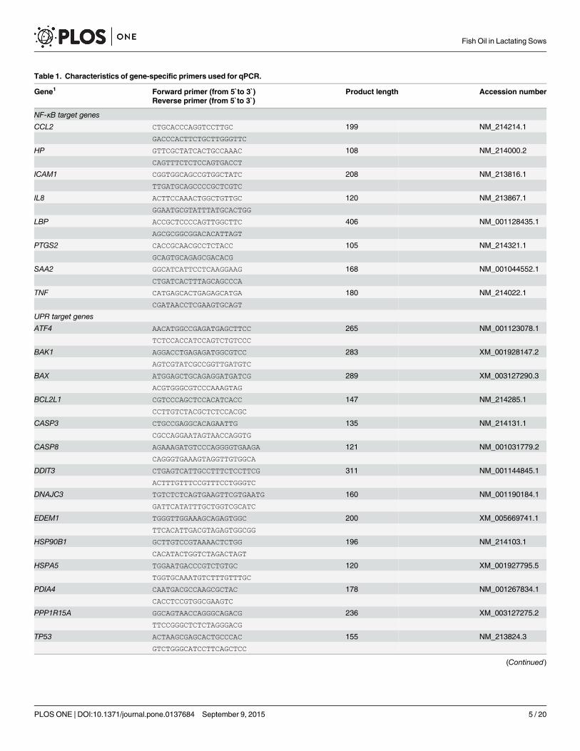
Table 1. Characteristics of gene-specific primers used for qPCR.
Gene1 Forward primer (from 5`to 3`) Product length Accession numberReverse primer (from 5`to 3`)
NF-κB target genes
CCL2 CTGCACCCAGGTCCTTGC 199 NM_214214.1
GACCCACTTCTGCTTGGGTTC
HP GTTCGCTATCACTGCCAAAC 108 NM_214000.2
CAGTTTCTCTCCAGTGACCT
ICAM1 CGGTGGCAGCCGTGGCTATC 208 NM_213816.1
TTGATGCAGCCCCGCTCGTC
IL8 ACTTCCAAACTGGCTGTTGC 120 NM_213867.1
GGAATGCGTATTTATGCACTGG
LBP ACCGCTCCCCAGTTGGCTTC 406 NM_001128435.1
AGCGCGGCGGACACATTAGT
PTGS2 CACCGCAACGCCTCTACC 105 NM_214321.1
GCAGTGCAGAGCGACACG
SAA2 GGCATCATTCCTCAAGGAAG 168 NM_001044552.1
CTGATCACTTTAGCAGCCCA
TNF CATGAGCACTGAGAGCATGA 180 NM_214022.1
CGATAACCTCGAAGTGCAGT
UPR target genes
ATF4 AACATGGCCGAGATGAGCTTCC 265 NM_001123078.1
TCTCCACCATCCAGTCTGTCCC
BAK1 AGGACCTGAGAGATGGCGTCC 283 XM_001928147.2
AGTCGTATCGCCGGTTGATGTC
BAX ATGGAGCTGCAGAGGATGATCG 289 XM_003127290.3
ACGTGGGCGTCCCAAAGTAG
BCL2L1 CGTCCCAGCTCCACATCACC 147 NM_214285.1
CCTTGTCTACGCTCTCCACGC
CASP3 CTGCCGAGGCACAGAATTG 135 NM_214131.1
CGCCAGGAATAGTAACCAGGTG
CASP8 AGAAAGATGTCCCAGGGGTGAAGA 121 NM_001031779.2
CAGGGTGAAAGTAGGTTGTGGCA
DDIT3 CTGAGTCATTGCCTTTCTCCTTCG 311 NM_001144845.1
ACTTTGTTTCCGTTTCCTGGGTC
DNAJC3 TGTCTCTCAGTGAAGTTCGTGAATG 160 NM_001190184.1
GATTCATATTTGCTGGTCGCATC
EDEM1 TGGGTTGGAAAGCAGAGTGGC 200 XM_005669741.1
TTCACATTGACGTAGAGTGGCGG
HSP90B1 GCTTGTCCGTAAAACTCTGG 196 NM_214103.1
CACATACTGGTCTAGACTAGT
HSPA5 TGGAATGACCCGTCTGTGC 120 XM_001927795.5
TGGTGCAAATGTCTTTGTTTGC
PDIA4 CAATGACGCCAAGCGCTAC 178 NM_001267834.1
CACCTCCGTGGCGAAGTC
PPP1R15A GGCAGTAACCAGGGCAGACG 236 XM_003127275.2
TTCCGGGCTCTCTAGGGACG
TP53 ACTAAGCGAGCACTGCCCAC 155 NM_213824.3
GTCTGGGCATCCTTCAGCTCC
(Continued)
Fish Oil in Lactating Sows
PLOSONE | DOI:10.1371/journal.pone.0137684 September 9, 2015 5 / 20
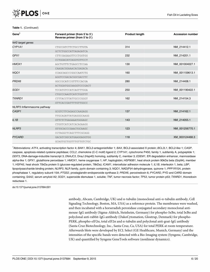
antibody, Abcam, Cambridge, UK) and α-tubulin (monoclonal anti-α-tubulin antibody, CellSignaling Technology, Boston, MA, USA) as a reference protein. The membranes were washed,and then incubated with a horseradish peroxidase conjugated secondary monoclonal anti-mouse-IgG antibody (Sigma-Aldrich, Steinheim, Germany) for phospho-IκBα, total IκBα andpolyclonal anti-rabbit-IgG antibody (DakoCytomation, Glostrup, Denmark) for phospho-PERK, phospho-eIF2α, total eIF2α and α-tubulin and polyclonal anti-goat-IgG antibody(Santa Cruz Biotechnology, Inc., Santa Cruz, Ca, USA) for total PERK at room temperature.Afterwards blots were developed by ECL Select (GE Healthcare, Munich, Germany) and theintensities of the specific bands were detected with a Bio-Imaging system (Syngene, Cambridge,UK) and quantified by Syngene GeneTools software (nonlinear dynamics).
Table 1. (Continued)
Gene1 Forward primer (from 5`to 3`) Product length Accession numberReverse primer (from 5`to 3`)
Nrf2 target genes
CYP1A1 CTGCCATCTTCTGCCTTGTA 314 NM_214412.1
GCTCTGGCCATTAGAGATCA
GPX1 CTTCGAGAAGTTCCTGGTGG 232 NM_214201.1
CCTGGACATCAGGTGTTCCT
HMOX1 AGCTGTTTCTGAGCCTCCAA 130 NM_001004027.1
CAAGACGGAAACACGAGACA
NQO1 CCAGCAGCCCGGCCAATCTG 160 NM_001159613.1
AGGTCCGACACGGCGACCTC
PRDX6 GGCCGCATCCGTTTCCACGA 280 NM_214408.1
ACTGGATGGCAAGGTCCCGACT
SOD1 TCCATGTCCATCAGTTTGGA 250 NM_001190422.1
CTGCCCAAGTCATCTGGTTT
TXNRD1 CTTTACCTTATTGCCCGGGT 162 NM_214154.3
GTTCACCGATTTTGTTGGCC
NLRP3 Inflammasome pathway
CASP1 GCGTCTTCAGAGCCAAGAGG 137 NM_214162.1
TTGCAGATTATGAGGGCAAGG
IL1B GTTCTCTGAGAAATGGGAGC 143 NM_214055.1
CTGGTCATCATCACAGAAGG
NLRP3 GTTGCACCCGAACTGCAAGC 123 NM_001256770.1
CCTAGGCTCAGCTTTCGCAGG
PYCARD GACATCGGCATGAAGGAGGTGG 118 XM_003124468.3
GCAGTGCTGGTTTGTTGTCTGC
1Abbreviations: ATF4, activating transcription factor 4; BAK1, BCL2-antagonist/killer 1; BAX, BCL2-associated X protein; BCL2L1, BCL2-like 1; CASP,
caspase, apoptosis-related cysteine peptidase; CCL2, chemokine (C-C motif) ligand 2; CYP1A1, cytochrome P450, family 1, subfamily A, polypeptide 1;
DDIT3, DNA-damage-inducible transcript 3; DNAJC3, DnaJ (Hsp40) homolog, subfamily C, member 3; EDEM1, ER degradation enhancer, mannosidase
alpha-like 1; GPX1, glutathione peroxidase 1; HMOX1, heme oxygenase 1; HP, haptoglobin; HSP90B1, heat shock protein 90kDa beta (Grp94), member
1; HSPA5, heat shock 70kDa protein 5 (glucose-regulated protein, 78kDa); ICAM1, intercellular adhesion molecule 1; IL1B, interleukin 1, beta; LBP,lipopolysaccharide binding protein; NLRP3, NLR family, pyrin domain containing 3; NQO1, NAD(P)H dehydrogenase, quinone 1; PPP1R15A, protein
phosphatase 1, regulatory subunit 15A; PTGS2, prostaglandin-endoperoxide synthase 2; PRDX6, peroxiredoxin 6; PYCARD, PYD and CARD domain
containing; SAA2, serum amyloid A2; SOD1, superoxide dismutase 1, soluble; TNF, tumor necrosis factor; TP53, tumor protein p53; TXNRD1, thioredoxinreductase 1.
doi:10.1371/journal.pone.0137684.t001
Fish Oil in Lactating Sows
PLOSONE | DOI:10.1371/journal.pone.0137684 September 9, 2015 6 / 20
Determination of plasma concentrations of HP und CRPPlasma concentrations of HP and CRP were determined by Phase Range kits from TrideltaDevelopment Ltd. (Maynooth, Co. Kildare, Ireland; cat. no. TP801 and TA901 for HP kit andCRP kit, respectively).
Statistical analysisAll data were tested for normal distribution by Shapiro-Wilk following testing for detection ofoutliers. Means of the two groups in each of the experiments were compared using student’s t-test for parametric variables, and Kruskal-Wallis test for nonparametric variables. Differencesbetween means were considered statistically significant for p<0.05.
Results
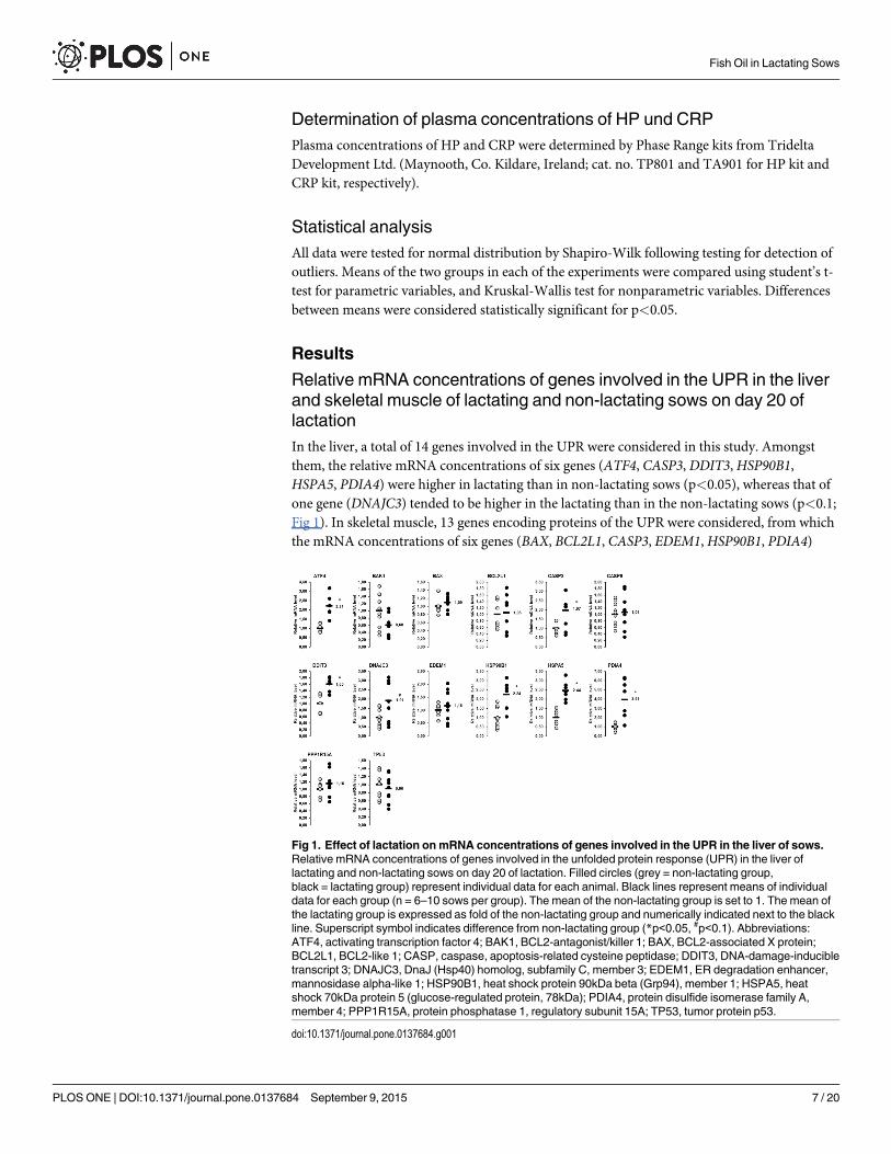
Relative mRNA concentrations of genes involved in the UPR in the liverand skeletal muscle of lactating and non-lactating sows on day 20 oflactationIn the liver, a total of 14 genes involved in the UPR were considered in this study. Amongstthem, the relative mRNA concentrations of six genes (ATF4, CASP3, DDIT3,HSP90B1,HSPA5, PDIA4) were higher in lactating than in non-lactating sows (p<0.05), whereas that ofone gene (DNAJC3) tended to be higher in the lactating than in the non-lactating sows (p<0.1;Fig 1). In skeletal muscle, 13 genes encoding proteins of the UPR were considered, from whichthe mRNA concentrations of six genes (BAX, BCL2L1, CASP3, EDEM1,HSP90B1, PDIA4)
Fig 1. Effect of lactation onmRNA concentrations of genes involved in the UPR in the liver of sows.Relative mRNA concentrations of genes involved in the unfolded protein response (UPR) in the liver oflactating and non-lactating sows on day 20 of lactation. Filled circles (grey = non-lactating group,black = lactating group) represent individual data for each animal. Black lines represent means of individualdata for each group (n = 6–10 sows per group). The mean of the non-lactating group is set to 1. The mean ofthe lactating group is expressed as fold of the non-lactating group and numerically indicated next to the blackline. Superscript symbol indicates difference from non-lactating group (*p<0.05, #p<0.1). Abbreviations:ATF4, activating transcription factor 4; BAK1, BCL2-antagonist/killer 1; BAX, BCL2-associated X protein;BCL2L1, BCL2-like 1; CASP, caspase, apoptosis-related cysteine peptidase; DDIT3, DNA-damage-inducibletranscript 3; DNAJC3, DnaJ (Hsp40) homolog, subfamily C, member 3; EDEM1, ER degradation enhancer,mannosidase alpha-like 1; HSP90B1, heat shock protein 90kDa beta (Grp94), member 1; HSPA5, heatshock 70kDa protein 5 (glucose-regulated protein, 78kDa); PDIA4, protein disulfide isomerase family A,member 4; PPP1R15A, protein phosphatase 1, regulatory subunit 15A; TP53, tumor protein p53.
doi:10.1371/journal.pone.0137684.g001
Fish Oil in Lactating Sows
PLOSONE | DOI:10.1371/journal.pone.0137684 September 9, 2015 7 / 20
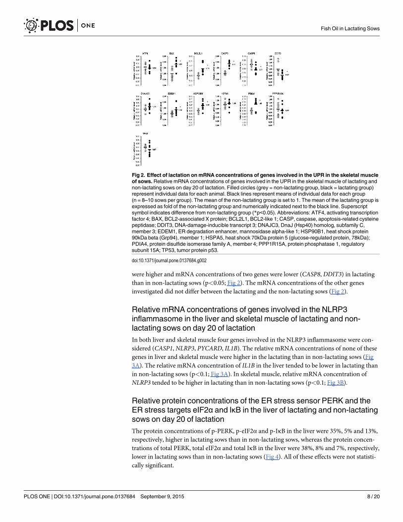
were higher and mRNA concentrations of two genes were lower (CASP8, DDIT3) in lactatingthan in non-lactating sows (p<0.05; Fig 2). The mRNA concentrations of the other genesinvestigated did not differ between the lactating and the non-lactating sows (Fig 2).
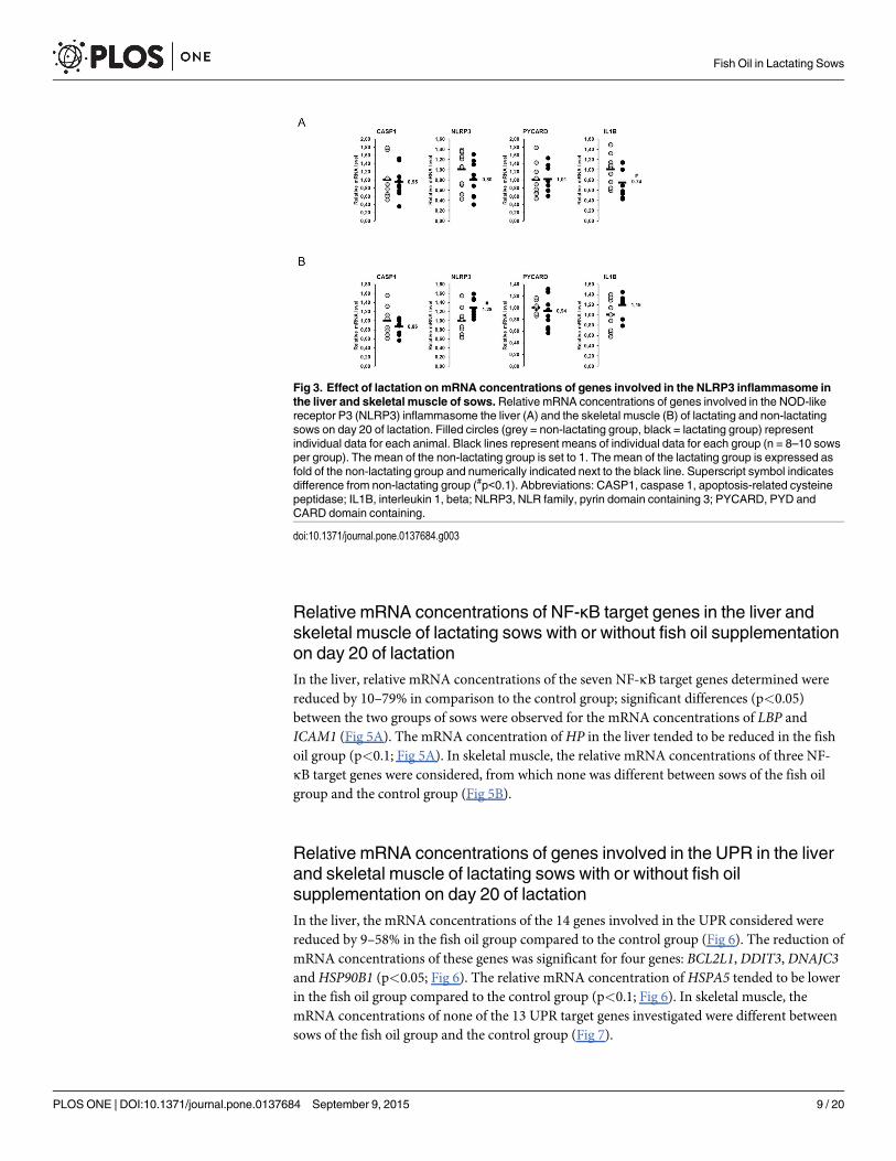
Relative mRNA concentrations of genes involved in the NLRP3inflammasome in the liver and skeletal muscle of lactating and non-lactating sows on day 20 of lactationIn both liver and skeletal muscle four genes involved in the NLRP3 inflammasome were con-sidered (CASP1, NLRP3, PYCARD, IL1B). The relative mRNA concentrations of none of thesegenes in liver and skeletal muscle were higher in the lactating than in non-lactating sows (Fig3A). The relative mRNA concentration of IL1B in the liver tended to be lower in lactating thanin non-lactating sows (p<0.1; Fig 3A). In skeletal muscle, relative mRNA concentration ofNLRP3 tended to be higher in lactating than in non-lactating sows (p<0.1; Fig 3B).
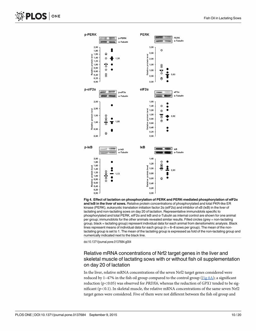
Relative protein concentrations of the ER stress sensor PERK and theER stress targets eIF2α and IκB in the liver of lactating and non-lactatingsows on day 20 of lactationThe protein concentrations of p-PERK, p-eIF2α and p-IκB in the liver were 35%, 5% and 13%,respectively, higher in lactating sows than in non-lactating sows, whereas the protein concen-trations of total PERK, total eIF2α and total IκB in the liver were 38%, 8% and 7%, respectively,lower in lactating sows than in non-lactating sows (Fig 4). All of these effects were not statisti-cally significant.
Fig 2. Effect of lactation onmRNA concentrations of genes involved in the UPR in the skeletal muscleof sows.Relative mRNA concentrations of genes involved in the UPR in the skeletal muscle of lactating andnon-lactating sows on day 20 of lactation. Filled circles (grey = non-lactating group, black = lactating group)represent individual data for each animal. Black lines represent means of individual data for each group(n = 8–10 sows per group). The mean of the non-lactating group is set to 1. The mean of the lactating group isexpressed as fold of the non-lactating group and numerically indicated next to the black line. Superscriptsymbol indicates difference from non-lactating group (*p<0.05). Abbreviations: ATF4, activating transcriptionfactor 4; BAX, BCL2-associated X protein; BCL2L1, BCL2-like 1; CASP, caspase, apoptosis-related cysteinepeptidase; DDIT3, DNA-damage-inducible transcript 3; DNAJC3, DnaJ (Hsp40) homolog, subfamily C,member 3; EDEM1, ER degradation enhancer, mannosidase alpha-like 1; HSP90B1, heat shock protein90kDa beta (Grp94), member 1; HSPA5, heat shock 70kDa protein 5 (glucose-regulated protein, 78kDa);PDIA4, protein disulfide isomerase family A, member 4; PPP1R15A, protein phosphatase 1, regulatorysubunit 15A; TP53, tumor protein p53.
doi:10.1371/journal.pone.0137684.g002
Fish Oil in Lactating Sows
PLOSONE | DOI:10.1371/journal.pone.0137684 September 9, 2015 8 / 20
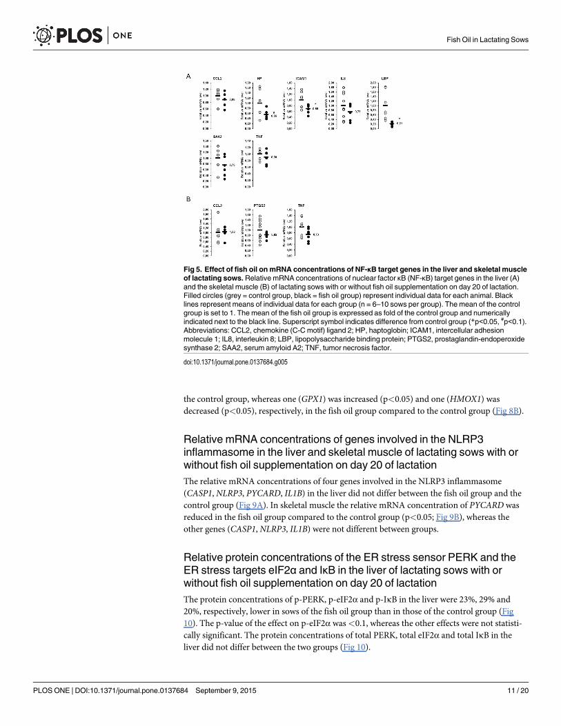
Relative mRNA concentrations of NF-κB target genes in the liver andskeletal muscle of lactating sows with or without fish oil supplementationon day 20 of lactationIn the liver, relative mRNA concentrations of the seven NF-κB target genes determined werereduced by 10–79% in comparison to the control group; significant differences (p<0.05)between the two groups of sows were observed for the mRNA concentrations of LBP andICAM1 (Fig 5A). The mRNA concentration of HP in the liver tended to be reduced in the fishoil group (p<0.1; Fig 5A). In skeletal muscle, the relative mRNA concentrations of three NF-κB target genes were considered, from which none was different between sows of the fish oilgroup and the control group (Fig 5B).
Relative mRNA concentrations of genes involved in the UPR in the liverand skeletal muscle of lactating sows with or without fish oilsupplementation on day 20 of lactationIn the liver, the mRNA concentrations of the 14 genes involved in the UPR considered werereduced by 9–58% in the fish oil group compared to the control group (Fig 6). The reduction ofmRNA concentrations of these genes was significant for four genes: BCL2L1, DDIT3, DNAJC3andHSP90B1 (p<0.05; Fig 6). The relative mRNA concentration of HSPA5 tended to be lowerin the fish oil group compared to the control group (p<0.1; Fig 6). In skeletal muscle, themRNA concentrations of none of the 13 UPR target genes investigated were different betweensows of the fish oil group and the control group (Fig 7).
Fig 3. Effect of lactation onmRNA concentrations of genes involved in the NLRP3 inflammasome inthe liver and skeletal muscle of sows.Relative mRNA concentrations of genes involved in the NOD-likereceptor P3 (NLRP3) inflammasome the liver (A) and the skeletal muscle (B) of lactating and non-lactatingsows on day 20 of lactation. Filled circles (grey = non-lactating group, black = lactating group) representindividual data for each animal. Black lines represent means of individual data for each group (n = 8–10 sowsper group). The mean of the non-lactating group is set to 1. The mean of the lactating group is expressed asfold of the non-lactating group and numerically indicated next to the black line. Superscript symbol indicatesdifference from non-lactating group (#p<0.1). Abbreviations: CASP1, caspase 1, apoptosis-related cysteinepeptidase; IL1B, interleukin 1, beta; NLRP3, NLR family, pyrin domain containing 3; PYCARD, PYD andCARD domain containing.
doi:10.1371/journal.pone.0137684.g003
Fish Oil in Lactating Sows
PLOSONE | DOI:10.1371/journal.pone.0137684 September 9, 2015 9 / 20
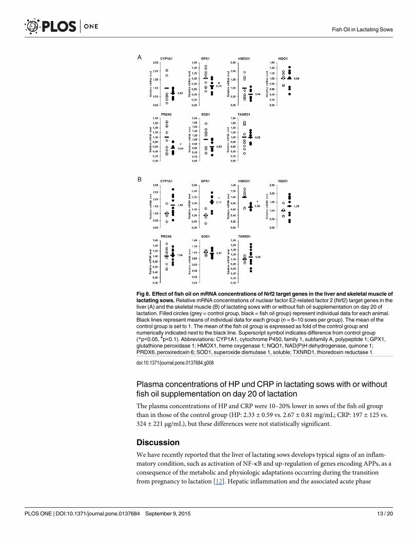
Relative mRNA concentrations of Nrf2 target genes in the liver andskeletal muscle of lactating sows with or without fish oil supplementationon day 20 of lactationIn the liver, relative mRNA concentrations of the seven Nrf2 target genes considered werereduced by 1–47% in the fish oil group compared to the control group (Fig 8A); a significantreduction (p<0.05) was observed for PRDX6, whereas the reduction of GPX1 tended to be sig-nificant (p<0.1). In skeletal muscle, the relative mRNA concentrations of the same seven Nrf2target genes were considered. Five of them were not different between the fish oil group and
Fig 4. Effect of lactation on phosphorylation of PERK and PERK-mediated phosphorylation of eIF2αand IκB in the liver of sows.Relative protein concentrations of phosphorylated and total PKR-like ERkinase (PERK), eukaryotic translation initiation factor 2α (eIF2α) and inhibitor of κB (IκB) in the liver oflactating and non-lactating sows on day 20 of lactation. Representative immunoblots specific tophosphorylated and total PERK, eIF2α and IκB and α-Tubulin as internal control are shown for one animalper group; immunoblots for the other animals revealed similar results. Filled circles (grey = non-lactatinggroup, black = lactating group) represent individual data for each animal from densitometric analysis. Blacklines represent means of individual data for each group (n = 6–8 sows per group). The mean of the non-lactating group is set to 1. The mean of the lactating group is expressed as fold of the non-lactating group andnumerically indicated next to the black line.
doi:10.1371/journal.pone.0137684.g004
Fish Oil in Lactating Sows
PLOSONE | DOI:10.1371/journal.pone.0137684 September 9, 2015 10 / 20
the control group, whereas one (GPX1) was increased (p<0.05) and one (HMOX1) wasdecreased (p<0.05), respectively, in the fish oil group compared to the control group (Fig 8B).
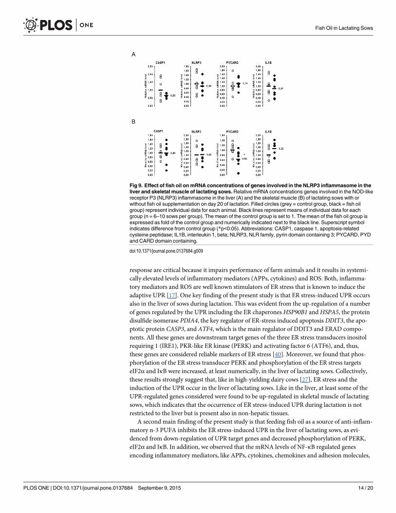
Relative mRNA concentrations of genes involved in the NLRP3inflammasome in the liver and skeletal muscle of lactating sows with orwithout fish oil supplementation on day 20 of lactationThe relative mRNA concentrations of four genes involved in the NLRP3 inflammasome(CASP1, NLRP3, PYCARD, IL1B) in the liver did not differ between the fish oil group and thecontrol group (Fig 9A). In skeletal muscle the relative mRNA concentration of PYCARD wasreduced in the fish oil group compared to the control group (p<0.05; Fig 9B), whereas theother genes (CASP1, NLRP3, IL1B) were not different between groups.
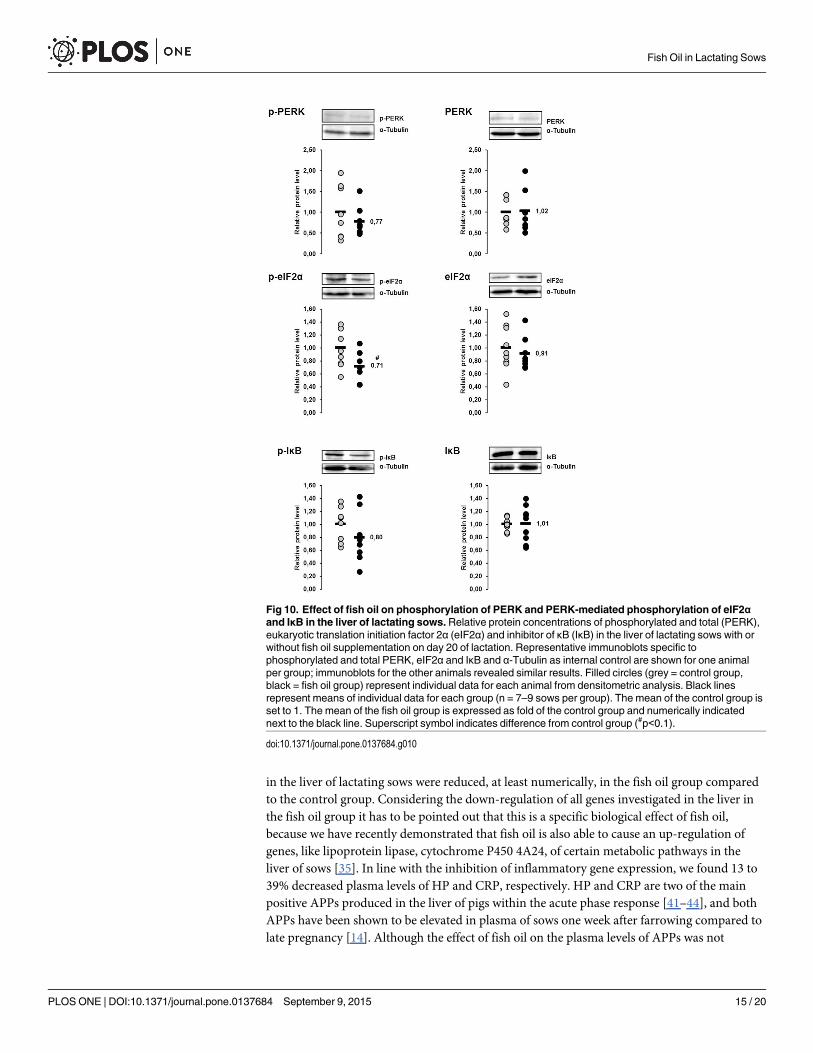
Relative protein concentrations of the ER stress sensor PERK and theER stress targets eIF2α and IκB in the liver of lactating sows with orwithout fish oil supplementation on day 20 of lactationThe protein concentrations of p-PERK, p-eIF2α and p-IκB in the liver were 23%, 29% and20%, respectively, lower in sows of the fish oil group than in those of the control group (Fig10). The p-value of the effect on p-eIF2α was<0.1, whereas the other effects were not statisti-cally significant. The protein concentrations of total PERK, total eIF2α and total IκB in theliver did not differ between the two groups (Fig 10).
Fig 5. Effect of fish oil on mRNA concentrations of NF-κB target genes in the liver and skeletal muscleof lactating sows.Relative mRNA concentrations of nuclear factor κB (NF-κB) target genes in the liver (A)and the skeletal muscle (B) of lactating sows with or without fish oil supplementation on day 20 of lactation.Filled circles (grey = control group, black = fish oil group) represent individual data for each animal. Blacklines represent means of individual data for each group (n = 6–10 sows per group). The mean of the controlgroup is set to 1. The mean of the fish oil group is expressed as fold of the control group and numericallyindicated next to the black line. Superscript symbol indicates difference from control group (*p<0.05, #p<0.1).Abbreviations: CCL2, chemokine (C-C motif) ligand 2; HP, haptoglobin; ICAM1, intercellular adhesionmolecule 1; IL8, interleukin 8; LBP, lipopolysaccharide binding protein; PTGS2, prostaglandin-endoperoxidesynthase 2; SAA2, serum amyloid A2; TNF, tumor necrosis factor.
doi:10.1371/journal.pone.0137684.g005
Fish Oil in Lactating Sows
PLOSONE | DOI:10.1371/journal.pone.0137684 September 9, 2015 11 / 20
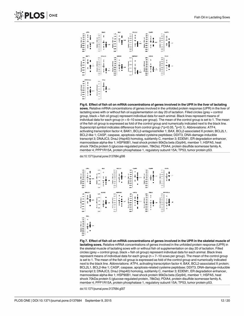
Fig 6. Effect of fish oil on mRNA concentrations of genes involved in the UPR in the liver of lactatingsows.Relative mRNA concentrations of genes involved in the unfolded protein response (UPR) in the liver oflactating sows with or without fish oil supplementation on day 20 of lactation. Filled circles (grey = controlgroup, black = fish oil group) represent individual data for each animal. Black lines represent means ofindividual data for each group (n = 6–10 sows per group). The mean of the control group is set to 1. The meanof the fish oil group is expressed as fold of the control group and numerically indicated next to the black line.Superscript symbol indicates difference from control group (*p<0.05, #p<0.1). Abbreviations: ATF4,activating transcription factor 4; BAK1, BCL2-antagonist/killer 1; BAX, BCL2-associated X protein; BCL2L1,BCL2-like 1; CASP, caspase, apoptosis-related cysteine peptidase; DDIT3, DNA-damage-inducibletranscript 3; DNAJC3, DnaJ (Hsp40) homolog, subfamily C, member 3; EDEM1, ER degradation enhancer,mannosidase alpha-like 1; HSP90B1, heat shock protein 90kDa beta (Grp94), member 1; HSPA5, heatshock 70kDa protein 5 (glucose-regulated protein, 78kDa); PDIA4, protein disulfide isomerase family A,member 4; PPP1R15A, protein phosphatase 1, regulatory subunit 15A; TP53, tumor protein p53.
doi:10.1371/journal.pone.0137684.g006
Fig 7. Effect of fish oil on mRNA concentrations of genes involved in the UPR in the skeletal muscle oflactating sows.Relative mRNA concentrations of genes involved in the unfolded protein response (UPR) inthe skeletal muscle of lactating sows with or without fish oil supplementation on day 20 of lactation. Filledcircles (grey = control group, black = fish oil group) represent individual data for each animal. Black linesrepresent means of individual data for each group (n = 7–10 sows per group). The mean of the control groupis set to 1. The mean of the fish oil group is expressed as fold of the control group and numerically indicatednext to the black line. Abbreviations: ATF4, activating transcription factor 4; BAX, BCL2-associated X protein;BCL2L1, BCL2-like 1; CASP, caspase, apoptosis-related cysteine peptidase; DDIT3, DNA-damage-inducibletranscript 3; DNAJC3, DnaJ (Hsp40) homolog, subfamily C, member 3; EDEM1, ER degradation enhancer,mannosidase alpha-like 1; HSP90B1, heat shock protein 90kDa beta (Grp94), member 1; HSPA5, heatshock 70kDa protein 5 (glucose-regulated protein, 78kDa); PDIA4, protein disulfide isomerase family A,member 4; PPP1R15A, protein phosphatase 1, regulatory subunit 15A; TP53, tumor protein p53.
doi:10.1371/journal.pone.0137684.g007
Fish Oil in Lactating Sows
PLOSONE | DOI:10.1371/journal.pone.0137684 September 9, 2015 12 / 20
Plasma concentrations of HP und CRP in lactating sows with or withoutfish oil supplementation on day 20 of lactationThe plasma concentrations of HP and CRP were 10–20% lower in sows of the fish oil groupthan in those of the control group (HP: 2.33 ± 0.59 vs. 2.67 ± 0.81 mg/mL; CRP: 197 ± 125 vs.324 ± 221 μg/mL), but these differences were not statistically significant.
DiscussionWe have recently reported that the liver of lactating sows develops typical signs of an inflam-matory condition, such as activation of NF-κB and up-regulation of genes encoding APPs, as aconsequence of the metabolic and physiologic adaptations occurring during the transitionfrom pregnancy to lactation [12]. Hepatic inflammation and the associated acute phase
Fig 8. Effect of fish oil on mRNA concentrations of Nrf2 target genes in the liver and skeletal muscle oflactating sows.Relative mRNA concentrations of nuclear factor E2-related factor 2 (Nrf2) target genes in theliver (A) and the skeletal muscle (B) of lactating sows with or without fish oil supplementation on day 20 oflactation. Filled circles (grey = control group, black = fish oil group) represent individual data for each animal.Black lines represent means of individual data for each group (n = 6–10 sows per group). The mean of thecontrol group is set to 1. The mean of the fish oil group is expressed as fold of the control group andnumerically indicated next to the black line. Superscript symbol indicates difference from control group(*p<0.05, #p<0.1). Abbreviations: CYP1A1, cytochrome P450, family 1, subfamily A, polypeptide 1; GPX1,glutathione peroxidase 1; HMOX1, heme oxygenase 1; NQO1, NAD(P)H dehydrogenase, quinone 1;PRDX6, peroxiredoxin 6; SOD1, superoxide dismutase 1, soluble; TXNRD1, thioredoxin reductase 1.
doi:10.1371/journal.pone.0137684.g008
Fish Oil in Lactating Sows
PLOSONE | DOI:10.1371/journal.pone.0137684 September 9, 2015 13 / 20
response are critical because it impairs performance of farm animals and it results in systemi-cally elevated levels of inflammatory mediators (APPs, cytokines) and ROS. Both, inflamma-tory mediators and ROS are well known stimulators of ER stress that is known to induce theadaptive UPR [17]. One key finding of the present study is that ER stress-induced UPR occursalso in the liver of sows during lactation. This was evident from the up-regulation of a numberof genes regulated by the UPR including the ER chaperones HSP90B1 and HSPA5, the proteindisulfide isomerase PDIA4, the key regulator of ER-stress induced apoptosis DDIT3, the apo-ptotic protein CASP3, and ATF4, which is the main regulator of DDIT3 and ERAD compo-nents. All these genes are downstream target genes of the three ER stress transducers inositolrequiring 1 (IRE1), PKR-like ER kinase (PERK) and activating factor 6 (ATF6), and, thus,these genes are considered reliable markers of ER stress [40]. Moreover, we found that phos-phorylation of the ER stress transducer PERK and phosphorylation of the ER stress targetseIF2α and IκB were increased, at least numerically, in the liver of lactating sows. Collectively,these results strongly suggest that, like in high-yielding dairy cows [27], ER stress and theinduction of the UPR occur in the liver of lactating sows. Like in the liver, at least some of theUPR-regulated genes considered were found to be up-regulated in skeletal muscle of lactatingsows, which indicates that the occurrence of ER stress-induced UPR during lactation is notrestricted to the liver but is present also in non-hepatic tissues.
A second main finding of the present study is that feeding fish oil as a source of anti-inflam-matory n-3 PUFA inhibits the ER stress-induced UPR in the liver of lactating sows, as evi-denced from down-regulation of UPR target genes and decreased phosphorylation of PERK,eIF2α and IκB. In addition, we observed that the mRNA levels of NF-κB regulated genesencoding inflammatory mediators, like APPs, cytokines, chemokines and adhesion molecules,
Fig 9. Effect of fish oil on mRNA concentrations of genes involved in the NLRP3 inflammasome in theliver and skeletal muscle of lactating sows.Relative mRNA concentrations genes involved in the NOD-likereceptor P3 (NLRP3) inflammasome in the liver (A) and the skeletal muscle (B) of lactating sows with orwithout fish oil supplementation on day 20 of lactation. Filled circles (grey = control group, black = fish oilgroup) represent individual data for each animal. Black lines represent means of individual data for eachgroup (n = 6–10 sows per group). The mean of the control group is set to 1. The mean of the fish oil group isexpressed as fold of the control group and numerically indicated next to the black line. Superscript symbolindicates difference from control group (*p<0.05). Abbreviations: CASP1, caspase 1, apoptosis-relatedcysteine peptidase; IL1B, interleukin 1, beta; NLRP3, NLR family, pyrin domain containing 3; PYCARD, PYDand CARD domain containing.
doi:10.1371/journal.pone.0137684.g009
Fish Oil in Lactating Sows
PLOSONE | DOI:10.1371/journal.pone.0137684 September 9, 2015 14 / 20
in the liver of lactating sows were reduced, at least numerically, in the fish oil group comparedto the control group. Considering the down-regulation of all genes investigated in the liver inthe fish oil group it has to be pointed out that this is a specific biological effect of fish oil,because we have recently demonstrated that fish oil is also able to cause an up-regulation ofgenes, like lipoprotein lipase, cytochrome P450 4A24, of certain metabolic pathways in theliver of sows [35]. In line with the inhibition of inflammatory gene expression, we found 13 to39% decreased plasma levels of HP and CRP, respectively. HP and CRP are two of the mainpositive APPs produced in the liver of pigs within the acute phase response [41–44], and bothAPPs have been shown to be elevated in plasma of sows one week after farrowing compared tolate pregnancy [14]. Although the effect of fish oil on the plasma levels of APPs was not
Fig 10. Effect of fish oil on phosphorylation of PERK and PERK-mediated phosphorylation of eIF2αand IκB in the liver of lactating sows.Relative protein concentrations of phosphorylated and total (PERK),eukaryotic translation initiation factor 2α (eIF2α) and inhibitor of κB (IκB) in the liver of lactating sows with orwithout fish oil supplementation on day 20 of lactation. Representative immunoblots specific tophosphorylated and total PERK, eIF2α and IκB and α-Tubulin as internal control are shown for one animalper group; immunoblots for the other animals revealed similar results. Filled circles (grey = control group,black = fish oil group) represent individual data for each animal from densitometric analysis. Black linesrepresent means of individual data for each group (n = 7–9 sows per group). The mean of the control group isset to 1. The mean of the fish oil group is expressed as fold of the control group and numerically indicatednext to the black line. Superscript symbol indicates difference from control group (#p<0.1).
doi:10.1371/journal.pone.0137684.g010
Fish Oil in Lactating Sows
PLOSONE | DOI:10.1371/journal.pone.0137684 September 9, 2015 15 / 20
significant due to the large biological variation between individual sows, which has beenreported also from others [45], our observation indicates that fish oil is able to attenuate thepro-inflammatory process associated with lactation. Since inflammatory mediators are impor-tant signals for the induction of ER stress, our findings suggest that fish oil inhibits the ERstress-induced UPR by attenuating the inflammatory condition in the liver of lactating sows.Inhibition of hepatic NF-κB by fish oil in the liver probably also provides the molecular basisfor the observation from Papadopoulos et al. [33] that a diet with a low n-6:n-3 PUFA ratioreduces plasma levels of the APP SAA in lactating sows. The inhibitory effect of n-3 PUFA onNF-κB has long been known and is explained by PPARα-mediated transrepression of NF-κBdue the ability of n-3 PUFA to bind to and activate PPARα [46].
A further interesting finding of the present study is that feeding of fish oil decreases themRNA levels of Nrf2-regulated genes in the liver of lactating sows. This finding is also indica-tive of inhibition of ER stress, because activation of the cytoprotective Nrf2 pathway has beenshown to be the consequence of ER stress and to be mediated through the ER stress inducerPERK [24]. Thus, our recent observation that the Nrf2 pathway is activated in the liver of lac-tating sows compared to non-lactating sows [12] is a further indirect evidence for the occur-rence of ER stress in the liver of sows during lactation. Like in lactating sows, activation of Nrf2was found recently in the liver of high-yielding dairy cows during early lactation [47]. In lactat-ing cows, this effect has been interpreted as a compensatory means to protect the liver againstROS- and inflammation-induced damage [47], because Nrf2 controls the transcription of vari-ous antioxidative and cytoprotective proteins. Thus, the physiologic meaning of Nrf2 activationin the liver of sows during lactation might be the same as in dairy cows.
In contrast to the liver, feeding fish oil failed to inhibit the ER stress-induced UPR in skeletalmuscle of lactating sows as shown by unaltered mRNA levels of UPR-regulated genes. Simi-larly, feeding fish oil largely did not reduce the expression of NF-κB and Nrf2 target genes inskeletal muscle, suggesting that fish oil did not exert an anti-inflammatory and cytoprotectiveaction in skeletal muscle of the lactating sows. Since a pro-inflammatory environment inducesER stress, the observation that fish oil did not attenuate skeletal muscle expression of pro-inflammatory NF-κB target genes is likely responsible for the lack of inhibition of ER stress-induced UPR in skeletal muscle of the lactating sows. We have no true explanation for the lackof anti-inflammatory effect of fish oil in skeletal muscle, but it may be explained by a loweravailability of n-3 PUFA in skeletal muscle than in the liver. It is well known that one impor-tant mechanism explaining the anti-inflammatory effects of n-3 PUFA from fish oil, such asEPA and DHA, is that they compete with arachidonic acid in the membrane phospholipids forcyclooxygenase and lipoxygenase, with the consequent production of less potent inflammatoryeicosanoids and of anti-inflammatory mediators such as resolvins. Although we did not analysethe proportions of n-3 PUFA and arachidonic acid in skeletal muscle and liver lipids due to thelimited amount of liver and skeletal muscle biopsy samples, we postulate that the dietary n-3PUFA taken up from the fish oil were incorporated at greater levels into the liver lipids thaninto the muscle lipids. This assumption is based on several studies in pigs and rats showingthat dietary n-3 PUFA are incorporated to a greater extent into liver lipids than into skeletalmuscle or adipose tissue lipids [48,49].
In the present study, we also considered the NLRP3 inflammasome pathway, a pro-inflam-matory signaling pathway which has not yet been investigated in lactating sows. This pathway isknown to be activated by “danger” signals like saturated fatty acids, but also ROS and pathogen-associated molecular patterns, such as lipopolysaccharides, microbial proteins and double-stranded ribonucleic acids, and to mediate the release of pro-inflammatory cytokines, such asIL-1B and IL-18 [34]. In contrast to the other stress pathways (UPR, NF-κB, Nrf2) considered,we found no evidence for an activation of the NLRP3 inflammasome pathway in neither liver
Fish Oil in Lactating Sows
PLOSONE | DOI:10.1371/journal.pone.0137684 September 9, 2015 16 / 20
nor skeletal muscle of sows during lactation. This was demonstrated by unaltered mRNA con-centrations of four NLRP3 inflammasome-related genes (CASP1, NLRP3, PYCARD, IL1B) in tis-sues of lactating and non-lactating sows. In addition, administration of fish oil failed to reducethe expression of most of the NLRP3 inflammasome-related genes in liver and skeletal muscle oflactating sows, although it has been reported in the literature that n-3 PUFA are able to inhibitNLRP3 inflammasome activation, at least in human THP-1 cells [50]. At the moment, we haveno explanation for the lack of effect of lactation and fish oil treatment on the NLRP3 inflamma-some pathway in sows, but it is possible that further genes involved in this pathway have to beconsidered to obtain a more meaningful picture about regulation of this pathway by lactationand fish oil in sows. Further investigations on this issue are warranted in future studies.
Our study has one limitation: When compared to the typical litter size of high-yielding geno-types in modern pig production, the litter size in the second study (8 piglets/sow) was quitesmall. This indicates that the energy requirement for milk production and consequently the lac-tation-induced metabolic stress and the induction of pro-inflammatory and ER stress signallingpathways was lower in the second than in the first study. Indeed, we have recently reported thatthe lactating sows of the first study were in a strong negative energy balance of approximately-35 MJ ME/day suggesting that the increase of feed intake during lactation was not sufficient tofully compensate the increased energy requirement for milk production [13]. In contrast, thelactating sows of both the control and the fish oil group in the second study had only a slightlynegative energy balance of approximately -5 MJ ME/day [35]. Accordingly, the obviously lowermetabolic stress in sows of the second study may explain that the down-regulation of inflamma-tory and ER stress-related genes in the liver of the fish oil group was only moderate. Neverthe-less, the observation that fish oil was able to inhibit the expression of all of these genes in sowsthat experienced only moderate stress indicates that fish oil is an efficient dietary approach tocombat lactation-induced metabolic and inflammatory stress in sows. Thus, future studies arewarranted investigating the efficacy of dietary fish oil in high-yielding genotypes with markedlygreater litter sizes of 15 and more piglets. In addition, such studies should include several timepoints during lactation for tissue sample collection in order to consider possible dynamicchanges of the inflammatory and ER stress response in sows during lactation.
ConclusionOur study shows for the first time that, like in dairy cows, ER stress-induced UPR is present inthe liver and skeletal muscle of sows during lactation, and dietary fish oil is able to inhibit, atleast in the liver, ER stress-induced UPR and inflammatory and stress signaling pathways,which are involved in the induction of ER stress. The occurrence of ER stress in the liver duringlactation indicates that the metabolic and physiologic changes occurring during the transitionfrom pregnancy to lactation represent cellular stress that might be detrimental to health andperformance of sows. At least in dairy cows it has been suggested that the ER stress-inducedUPR contributes to the pathophysiologic conditions commonly observed in the liver of peri-parturient cows [27], such as fatty liver and ketosis, which are considered critical with regard tomilk and reproductive performance. Although little is known about the occurrence of liver-associated diseases and its relevance for milk and reproductive performance in lactating sows,it is assumed that feeding fish oil is a useful dietary strategy to improve health and performanceof lactating sows.
Author ContributionsConceived and designed the experiments: DKG SR KE. Performed the experiments: DKG BGSR SH SB GE GR AC. Analyzed the data: DKG BG. Wrote the paper: DKG RR KE.
Fish Oil in Lactating Sows
PLOSONE | DOI:10.1371/journal.pone.0137684 September 9, 2015 17 / 20

References1. Aiello RJ, Kenna TM, Herbein JH. Hepatic gluconeogenic and ketogenic interrelationships in the lactat-
ing cow. J Dairy Sci. 1984; 67: 1707–1715. PMID: 6480960
2. Collier RJ, McNamara JP, Wallace CR, Dehoff MH. A review of endocrine regulation of metabolism dur-ing lactation. J Anim Sci. 1984; 59: 498–510. PMID: 6090379
3. Trottier NL, Easter RA. Dietary and plasma branched-chain amino acids in relation to tryptophan: effecton voluntary feed intake and lactation metabolism in the primiparous sow. J Anim Sci. 1995; 73: 1086–1092. PMID: 7628952
4. Trayhurn P, Douglas JB, McGuckin MM. Brown adipose tissue thermogenesis is 'suppressed' duringlactation in mice. Nature. 1982; 298: 59–60. PMID: 6283369
5. Williamson DH. Fuel supply to brown adipose-tissue. Biochem Soc Trans. 1986; 14: 225–227. PMID:3709945
6. Dewey KG. Energy and protein requirements during lactation. Annu Rev Nutr. 1997; 17: 19–36. PMID:9240917
7. Smith MS, Grove KL. Integration of the regulation of reproductive function and energy balance: lactationas a model. Front Neuroendocrinol. 2002; 23: 225–256. PMID: 12127305
8. Gutgesell A, Ringseis R, Brandsch C, Stangl GI, Hirche F, Eder K. Peroxisome proliferator-activatedreceptor α and enzymes of carnitine biosynthesis in the liver are down-regulated during lactation in rats.Metabolism. 2009; 58: 226–232. doi: 10.1016/j.metabol.2008.09.018 PMID: 19154956
9. Gutgesell A, Ringseis R, Schmidt E, Brandsch C, Stangl GI, Eder K. Downregulation of peroxisomeproliferator-activated receptor α and its coactivators in liver and skeletal muscle mediates the metabolicadaptations during lactation in mice. J Mol Endocrinol. 2009; 43: 241–250. doi: 10.1677/JME-09-0064PMID: 19578095
10. Drackley JK. Biology of dairy cows during the transition period: The final frontier? J Dairy Sci. 1999; 82:2259–2273. PMID: 10575597
11. Katoh N. Relevance of apolipoproteins in the development of fatty liver and fatty liver-related peripartumdiseases in dairy cows. J Vet Med Sci. 2002; 64: 293–307. PMID: 12014573
12. Rosenbaum S, Ringseis R, Hillen S, Becker S, Erhardt G, Reiner G, et al. The stress signalling pathwaynuclear factor E2-related factor 2 is activated in the liver of sows during lactation. Acta Vet Scand.2012; 54: 59. doi: 10.1186/1751-0147-54-59 PMID: 23039904
13. Rosenbaum S, Ringseis R, Hillen S, Becker S, Erhardt G, Reiner G, et al. Genome-wide transcript pro-filing indicates induction of energy-generating pathways and an adaptive immune response in the liverof sows during lactation. Comp Biochem Physiol Part D Genomics Proteomics. 2012; 7: 370–381. doi:10.1016/j.cbd.2012.09.001 PMID: 23031603
14. Kovac G, Tothova CS, Nagy O, Seidel H. Acute phase proteins during the reproductive cycle of sows.Acta Vet. 2008; 58: 459–466.
15. Murata H, Shimada N, Yoshioka M. Current research on acute phase proteins in veterinary diagnosis:an overview. Vet J. 2004; 168: 28–40. PMID: 15158206
16. Bossaert P, Trevisi E, Opsomer G, Bertoni G, De Vliegher S, Leroy JL. The association between indica-tors of inflammation and liver variables during the transition period in high-yielding dairy cows: Anobservational study. Vet J. 2012; 192: 222–225. doi: 10.1016/j.tvjl.2011.06.004 PMID: 21742524
17. Cnop M, Foufelle F, Velloso LA. Endoplasmic reticulum stress, obesity and diabetes. Trends Mol Med.2012; 18: 59–68. doi: 10.1016/j.molmed.2011.07.010 PMID: 21889406
18. Fu SN, Watkins SM, Hotamisligil GS. The role of endoplasmic reticulum in hepatic lipid homeostasisand stress signaling. Cell Metab. 2012; 15: 623–634. doi: 10.1016/j.cmet.2012.03.007 PMID:22560215
19. Marciniak SJ, Ron D. Endoplasmic reticulum stress signaling in disease. Physiol Rev. 2006; 86: 1133–1149. PMID: 17015486
20. Ron D, Walter P. Signal integration in the endoplasmic reticulum unfolded protein response. NatureRev Mol Cell Biol. 2007; 8: 519–529.
21. Zhang KZ, Kaufman RJ. From endoplasmic-reticulum stress to the inflammatory response. Nature.2008; 454: 455–462. doi: 10.1038/nature07203 PMID: 18650916
22. Lee JS, Zheng Z, Mendez R, Ha SW, Xie YM, Zhang K. Pharmacologic ER stress induces non-alco-holic steatohepatitis in an animal model. Toxicol Lett. 2012; 211: 29–38. doi: 10.1016/j.toxlet.2012.02.017 PMID: 22414386
23. Schaap FG, Kremer AE, LamersWH, Jansen PLM, Gaemers IC. Fibroblast growth factor 21 is inducedby endoplasmic reticulum stress. Biochimie. 2013; 95: 692–699. doi: 10.1016/j.biochi.2012.10.019PMID: 23123503
Fish Oil in Lactating Sows
PLOSONE | DOI:10.1371/journal.pone.0137684 September 9, 2015 18 / 20
24. Cullinan SB, Zhang D, Hannink M, Arvisais E, Kaufman RJ, Diehl JA. Nrf2 is a direct PERK substrateand effector of PERK-dependent cell survival. Mol Cell Biol. 2003; 23: 7198–7209. PMID: 14517290
25. Breckenridge DG, Germain M, Mathai JP, Nguyen M, Shore GC. Regulation of apoptosis by endoplas-mic reticulum pathways. Oncogene. 2003; 22: 8608–8618. PMID: 14634622
26. Rutkowski DT, Kaufman RJ. A trip to the ER: coping with stress. Trends Cell Biol. 2004; 14: 20–28.PMID: 14729177
27. Gessner DK, Schlegel G, Ringseis R, Schwarz FJ, Eder K. Up-regulation of endoplasmic reticulumstress induced genes of the unfolded protein response in the liver of periparturient dairy cows. BMC VetRes. 2014; 10.
28. Myhrstad MCW, Retterstol K, Telle-Hansen VH, Ottestad I, Halvorsen B, Holven KB, et al. Effect ofmarine n-3 fatty acids on circulating inflammatory markers in healthy subjects and subjects with cardio-vascular risk factors. InflammRes. 2011; 60: 309–319. doi: 10.1007/s00011-010-0302-5 PMID:21229287
29. Miles EA, Calder PC. Influence of marine n-3 polyunsaturated fatty acids on immune function and asystematic review of their effects on clinical outcomes in rheumatoid arthritis. Br J Nutr. 2012; 107:S171–S184. doi: 10.1017/S0007114512001560 PMID: 22591891
30. Rooke JA, Sinclair AG, Edwards SA. Feeding tuna oil to the sow at different times during pregnancyhas different effects on piglet long-chain polyunsaturated fatty acid composition at birth and subsequentgrowth. Br J Nutr. 2001; 86: 21–30. PMID: 11432761
31. Rooke JA, Sinclair AG, Ewen M. Changes in piglet tissue composition at birth in response to increasingmaternal intake of long-chain n-3 polyunsaturated fatty acids are non-linear. Br J Nutr. 2001; 86: 461–470. PMID: 11591233
32. Smits RJ, Luxford BG, Mitchell M, Nottle MB. Sow litter size is increased in the subsequent parity whenlactating sows are fed diets containing n-3 fatty acids from fish oil. J Animal Sci. 2011; 89: 2731–2738.
33. Papadopoulos GA, Maes DGD, VanWeyenberg S, van Kempen TATG, Buyse J, Janssen GP. Peripar-tal feeding strategy with different n-6:n-3 ratios in sows: effects on sows' performance, inflammatoryand periparturient metabolic parameters. Br J Nutr. 2009; 101: 348–357. doi: 10.1017/S0007114508026160 PMID: 18613985
34. Martinon F, Burns K, Tschopp J. The inflammasome: a molecular platform triggering activation ofinflammatory caspases and processing of proIL-beta. Mol Cell. 2002; 10: 417–426. PMID: 12191486
35. Gessner DK, Gröne B, Rosenbaum S, Most E, Hillen S, Becker S, et al. Effect of dietary fish oil on theexpression of genes involved in lipid metabolism in liver and skeletal muscle of lactating sows. J AnimPhysiol Anim Nutr (Berl). doi: 10.1111/jpn.12324 [Epub ahead of print]
36. Gessner DK, Gröne B, Rosenbaum S, Most E, Hillen S, Becker S, et al. Treatment of lactating sowswith clofibrate as a synthetic agonist of PPARα does not influence milk fat content and gains of litters.BMC Vet Res. 2015; 11: 54. doi: 10.1186/s12917-015-0368-y PMID: 25888880
37. Gessner DK, Fiesel A, Most E, Dinges J, Wen G, Ringseis R, et al. Supplementation of a grape seedand grape marc meal extract decreases activities of the oxidative stress-responsive transcription fac-tors NF-κB and Nrf2 in the duodenal mucosa of pigs. Acta Vet Scand. 2013; 55: 18. doi: 10.1186/1751-0147-55-18 PMID: 23453040
38. Vandesompele J, De Preter K, Pattyn F, Poppe B, Van Roy N, De Paepe A, et al. Accurate normaliza-tion of real-time quantitative RT-PCR data by geometric averaging of multiple internal control genes.Genome Biol. 2002; 3.
39. Ringseis R, Mooren FC, Keller J, Couturier A, Wen G, Hirche F, et al. Regular endurance exerciseimproves the diminished hepatic carnitine status in mice fed a high-fat diet. Mol Nutr Food Res. 2011;55 Suppl 2: S193–202. doi: 10.1002/mnfr.201100040 PMID: 21770048
40. Samali A, Fitzgerald U, Deegan S, Gupta S. Methods for monitoring endoplasmic reticulum stress andthe unfolded protein response. Int J Cell Biol. 2010; 2010: 830307. doi: 10.1155/2010/830307 PMID:20169136
41. Eckersall PD, Saini PK, McComb C. The acute phase response of acid soluble glycoprotein, alpha(1)-acid glycoprotein, ceruloplasmin, haptoglobin and C-reactive protein, in the pig. Vet Immunol Immuno-pathol. 1996; 51: 377–385. PMID: 8792574
42. Heegaard PM, Klausen J, Nielsen JP, Gonzalez-Ramon N, Pineiro M, Lampreave F, et al. The porcineacute phase response to infection with Actinobacillus pleuropneumoniae. Haptoglobin, C-reactive pro-tein, major acute phase protein and serum amyloid A protein are sensitive indicators of infection. CompBiochem Physiol B BiochemMol Biol. 1998; 119: 365–373. PMID: 9629669
43. Hultén C, Johansson E, Fossum C, Wallgren P. Interleukin 6, serum amyloid A and haptoglobin asmarkers of treatment efficacy in pigs experimentally infected with Actinobacillus pleuropneumoniae.Vet Microbiol. 2003; 95: 75–89. PMID: 12860078
Fish Oil in Lactating Sows
PLOSONE | DOI:10.1371/journal.pone.0137684 September 9, 2015 19 / 20
44. Cray C, Zaias J, Altman NH. Acute phase response in animals: a review. CompMed. 2009; 59: 517–526. PMID: 20034426
45. Salamano G, Mellia E, Candiani D, Ingravalle F, Bruno R, Ru G, et al. Changes in haptoglobin, C-reac-tive protein and pig-MAP during a housing period following long distance transport in swine. Vet J.2008; 177: 110–115. PMID: 17509918
46. Delerive P, Gervois P, Fruchart JC, Staels B. Induction of IκBα expression as a mechanism contributingto the anti-inflammatory activities of peroxisome proliferator-activated receptor-α activators. J BiolChem. 2000; 275: 36703–36707. PMID: 10980195
47. Gessner DK, Schlegel G, Keller J, Schwarz FJ, Ringseis R, Eder K. Expression of target genes ofnuclear factor E2-related factor 2 in the liver of dairy cows in the transition period and at different stagesof lactation. J Dairy Sci. 2013; 96: 1038–1043. doi: 10.3168/jds.2012-5967 PMID: 23245956
48. Hsu JM, Wang PH, Liu BH, Ding ST. The effect of dietary docosahexaenoic acid on the expression ofporcine lipid metabolism-related genes. J Animal Sci. 2004; 82: 683–689.
49. Feillet-Coudray C, Aoun M, Fouret G, Bonafos B, Ramos J, Casas F, et al. Effects of long-term adminis-tration of saturated and n-3 fatty acid-rich diets on lipid utilisation and oxidative stress in rat liver andmuscle tissues. Br J Nutr. 2013; 110: 1789–1802. doi: 10.1017/S0007114513001311 PMID:23656726
50. Yan YQ, JiangW, Spinetti T, Tardivel A, Castillo R, Bourguin C, et al. Omega-3 fatty acids preventinflammation and metabolic disorder through inhibition of NLRP3 inflammasome activation. Immunity.2013; 38: 1154–1163. doi: 10.1016/j.immuni.2013.05.015 PMID: 23809162
Fish Oil in Lactating Sows
PLOSONE | DOI:10.1371/journal.pone.0137684 September 9, 2015 20 / 20