RESEARCHARTICLE … 2015.pdf · PrincessPaolaHospital,Marche-en-Famenne,Belgium ......
20
RESEARCH ARTICLE Curcuminoids Extract, Hydrolyzed Collagen and Green Tea Extract Synergically Inhibit Inflammatory and Catabolic Mediator’s Synthesis by Normal Bovine and Osteoarthritic Human Chondrocytes in Monolayer Fanny Comblain 1 , Christelle Sanchez 1 , Isabelle Lesponne 2 , Marc Balligand 3 , Samuel Serisier 2 , Yves Henrotin 1,4* 1 Bone and Cartilage Research Unit, Arthropôle Liège, University of Liège, CHU Sart-Tilman, Liège, Belgium, 2 Royal Canin Research Center, Aimargues, France, 3 Department of Clinical Sciences, Faculty of Veterinary Medicine, University of Liège, Liège, Belgium, 4 Physical Therapy and Rehabilitation department, Princess Paola Hospital, Marche-en-Famenne, Belgium * [email protected] Abstract The main objective of this study was to assess the in vitro effects of curcuminoids extract, hydrolyzed collagen and green tea extract in normal bovine chondrocytes and osteoarthritic human chondrocytes cultured in monolayer. This study also investigated the synergic or ad- ditive effects of these compounds. Enzymatically isolated primary bovine or human chon- drocytes were cultured in monolayer until confluence and then incubated for 24 hours or 48 hours in the absence or in the presence of interleukin-1β and with or without curcuminoids extract, hydrolyzed collagen or green tea extract, added alone or in combination, at different concentrations. Cell viability was neither affected by these compounds, nor by interleukin 1β. In the absence of interleukin-1β, compounds did not significantly affect bovine chondro- cytes metabolism. In human chondrocytes and in the absence of interleukin 1β, curcumi- noids extract alone or in combination with hydrolyzed collagen and green tea extract significantly inhibited matrix metalloproteinase-3 production. In interleukin-1β-stimulated bovine chondrocytes, interleukin-6, inducible nitric oxide synthase, cyclooxygenase2, ma- trix metalloproteinase 3, a disintegrin and metalloproteinase with thrombospondin type I mo- tifs 4 and a disintegrin and metalloproteinase with thrombospondin type I motifs 5 expressions were decreased by curcuminoids extract alone or in combination with hydro- lyzed collagen and green tea extract. The combination of the three compounds was signifi- cantly more efficient to inhibit interleukin-1β stimulated matrix metalloproteinase-3 expression than curcuminoids extract alone. In interleukin-1β-stimulated human chondro- cytes, nitric oxide, interleukin-6 and matrix metalloproteinase 3 productions were significant- ly reduced by curcuminoids extract alone or in combination with hydrolyzed collagen and PLOS ONE | DOI:10.1371/journal.pone.0121654 March 23, 2015 1 / 20 a11111 OPEN ACCESS Citation: Comblain F, Sanchez C, Lesponne I, Balligand M, Serisier S, Henrotin Y (2015) Curcuminoids Extract, Hydrolyzed Collagen and Green Tea Extract Synergically Inhibit Inflammatory and Catabolic Mediator’s Synthesis by Normal Bovine and Osteoarthritic Human Chondrocytes in Monolayer. PLoS ONE 10(3): e0121654. doi:10.1371/ journal.pone.0121654 Academic Editor: Christos Chadjichristos, National Institute of Health and Medical Research, FRANCE Received: October 13, 2014 Accepted: February 12, 2015 Published: March 23, 2015 Copyright: © 2015 Comblain et al. This is an open access article distributed under the terms of the Creative Commons Attribution License, which permits unrestricted use, distribution, and reproduction in any medium, provided the original author and source are credited. Data Availability Statement: All relevant data are within the paper. Funding: The research leading to these results was supported by a grant of Royal Canin SAS. FC receives her PhD fellow from Royal Canin SAS. Royal Canin conceived and designed the work, analyzed the data, contributed compounds, revised the article, gave their final approval of the version to be published and agreed to be accountable for all aspects of the work.
Transcript of RESEARCHARTICLE … 2015.pdf · PrincessPaolaHospital,Marche-en-Famenne,Belgium ......
RESEARCH ARTICLE
Curcuminoids Extract, Hydrolyzed Collagenand Green Tea Extract Synergically InhibitInflammatory and Catabolic Mediator’sSynthesis by Normal Bovine andOsteoarthritic Human Chondrocytes inMonolayerFanny Comblain1, Christelle Sanchez1, Isabelle Lesponne2, Marc Balligand3,Samuel Serisier2, Yves Henrotin1,4*
1 Bone and Cartilage Research Unit, Arthropôle Liège, University of Liège, CHU Sart-Tilman, Liège,Belgium, 2 Royal Canin Research Center, Aimargues, France, 3 Department of Clinical Sciences, Faculty ofVeterinary Medicine, University of Liège, Liège, Belgium, 4 Physical Therapy and Rehabilitation department,Princess Paola Hospital, Marche-en-Famenne, Belgium
AbstractThe main objective of this study was to assess the in vitro effects of curcuminoids extract,
hydrolyzed collagen and green tea extract in normal bovine chondrocytes and osteoarthritic
human chondrocytes cultured in monolayer. This study also investigated the synergic or ad-
ditive effects of these compounds. Enzymatically isolated primary bovine or human chon-
drocytes were cultured in monolayer until confluence and then incubated for 24 hours or 48
hours in the absence or in the presence of interleukin-1β and with or without curcuminoids
extract, hydrolyzed collagen or green tea extract, added alone or in combination, at different
concentrations. Cell viability was neither affected by these compounds, nor by interleukin
1β. In the absence of interleukin-1β, compounds did not significantly affect bovine chondro-
cytes metabolism. In human chondrocytes and in the absence of interleukin 1β, curcumi-
noids extract alone or in combination with hydrolyzed collagen and green tea extract
significantly inhibited matrix metalloproteinase-3 production. In interleukin-1β-stimulated
bovine chondrocytes, interleukin-6, inducible nitric oxide synthase, cyclooxygenase2, ma-
trix metalloproteinase 3, a disintegrin and metalloproteinase with thrombospondin type I mo-
tifs 4 and a disintegrin and metalloproteinase with thrombospondin type I motifs 5
expressions were decreased by curcuminoids extract alone or in combination with hydro-
lyzed collagen and green tea extract. The combination of the three compounds was signifi-
cantly more efficient to inhibit interleukin-1β stimulated matrix metalloproteinase-3
expression than curcuminoids extract alone. In interleukin-1β-stimulated human chondro-
cytes, nitric oxide, interleukin-6 and matrix metalloproteinase 3 productions were significant-
ly reduced by curcuminoids extract alone or in combination with hydrolyzed collagen and
PLOSONE | DOI:10.1371/journal.pone.0121654 March 23, 2015 1 / 20
a11111
OPEN ACCESS
Citation: Comblain F, Sanchez C, Lesponne I,Balligand M, Serisier S, Henrotin Y (2015)Curcuminoids Extract, Hydrolyzed Collagen andGreen Tea Extract Synergically Inhibit Inflammatoryand Catabolic Mediator’s Synthesis by NormalBovine and Osteoarthritic Human Chondrocytes inMonolayer. PLoS ONE 10(3): e0121654. doi:10.1371/journal.pone.0121654
Academic Editor: Christos Chadjichristos, NationalInstitute of Health and Medical Research, FRANCE
Received: October 13, 2014
Accepted: February 12, 2015
Published: March 23, 2015
Copyright: © 2015 Comblain et al. This is an openaccess article distributed under the terms of theCreative Commons Attribution License, which permitsunrestricted use, distribution, and reproduction in anymedium, provided the original author and source arecredited.
Data Availability Statement: All relevant data arewithin the paper.
Funding: The research leading to these results wassupported by a grant of Royal Canin SAS. FCreceives her PhD fellow from Royal Canin SAS.Royal Canin conceived and designed the work,analyzed the data, contributed compounds, revisedthe article, gave their final approval of the version tobe published and agreed to be accountable for allaspects of the work.
green tea extract. These findings indicate that a mixture of curcuminoids extract, hydrolyzed
collagen and green tea extract has beneficial effects on chondrocytes culture in inflammato-
ry conditions and provide a preclinical basis for the in vivo testing of this mixture.
IntroductionOsteoarthritis (OA) is a chronic, painful, degenerative and inflammatory condition that affectsthe joints and leads to functional disability. The most prominent feature of OA is the progres-sive degradation of articular cartilage. Chondrocytes play a key role in cartilage degradation inOA by producing matrix metalloproteinases (MMP), free radicals and inflammatory cytokinesin response to mechanical or biochemical stimuli [1, 2]. These mediators, not only are involvedin cartilage matrix degradation but also in cross-talk with synovial and subchondral bone cells.
Nowadays, curative treatments for OA remain missing and the management of OA focuseson the alleviation of symptoms. Current recommendations for the management of OA includea combination of non-pharmacological and pharmacological modalities. Moreover, for pa-tients with severe OA, joint replacement is indicated [3]. Among non-pharmacological recom-mendations, exercise (land-based and water-based), biomechanical interventions, weight loss ifoverweight or obesity, and thermal modalities are widely recommended [3–5]. The most rec-ommended pharmacological treatments include acetaminophen/paracetamol and non-steroidal anti-inflammatory drugs (NSAIDs) (topical or oral). Intra-articular corticosteroidsare generally recommended for hip and knee OA [3–5]. However, the long-term use ofNSAIDs and acetaminophen may be associated with detrimental effects, especially gastrointes-tinal adverse effects [6].
In this context, safer alternative treatments are needed. Such treatments could come fromnutrition and more particularly from nutraceuticals. Some positive beneficial effects arehighlighted with nutraceuticals in the course of OA [7, 8]. Among them, curcumin, which isthe major component of turmeric, a yellow spice derived from the roots of the plant Curcumalonga. Anti-catabolic, anti-apoptotic and anti-inflammatory effects of curcumin are largely de-scribed in vitro. It is demonstrated that curcumin decreases nitric oxide (NO), prostaglandinE2 (PGE2), IL-6, IL-8, cyclooxygenase2 (COX2), inducible nitric oxide synthase (iNOS), MMP-3 and MMP-9 synthesis through the inhibition of nuclear factor κB (NF-κB) translocation andtumor necrosis factor (TNF)-α signaling pathways in chondrocytes [9–13]. Hydrolyzed colla-gen is obtained by the enzymatic hydrolysis of collagenous tissues, like for example, animalbone, and is generally recognized as a safe food ingredient by regulatory agencies [14, 15]. Themain characteristic of hydrolyzed collagen is its amino acid composition, which is identical tocollagen, thus providing high levels of glycine and proline, two amino acids essential for thestability and regeneration of cartilage [16, 17]. Proteoglycans synthesis, aggrecan gene expres-sion and type II collagen synthesis are increased with hydrolyzed collagen in bovine and por-cine articular chondrocytes [18, 19]. Epigallocatechin-3-gallate (EGCG) is a major componentof the polyphenolic fraction of green tea and exhibits anti-oxidant, anti-tumor and anti-mutagenic activities [7, 20]. Advanced glycation end products (AGE)-induced expressions ofTNF-α and MMP-13 are inhibited by EGCG in human OA chondrocytes. Otherwise, AGE-induced mitogen activated protein kinase (MAPK) signaling pathways and NF-κB activationare inhibited in that model [21]. The expression of mediators associated with OA pathogenesisis suppressed in human chondrocytes pre-treated with EGCG and then stimulated with IL-1β[22]. IL-1β induced mRNA expression and protein production of MMP-1 and MMP-13 are
Nutraceuticals Inhibit Inflammation and Catabolism
PLOSONE | DOI:10.1371/journal.pone.0121654 March 23, 2015 2 / 20
Competing Interests: FC receives her PhD fellowfrom Royal Canin SAS. IL and SS are employed byRoyal Canin. YH receives honoraria from Artialis,Bioiberica, Danone, Expanscience, Ibsa, Merck,Pierre Fabre, Synolyne Pharma, Tilman. YH is thefounder and President of Artialis SA, a biomarkermanufacturer and Synolyne Pharma, two spin-offcompanies of the University of Liège. This does notalter the authors' adherence to PLOS ONE policieson sharing data and materials.
inhibited in an EGCG concentration dependant manner in human chondrocytes [23]. Thetranscription activity of NF-κB and the IL-1β-induced glycosaminoglycans (GAG) releasefrom human cartilage explants are also reduced [23].
It has been reported that the majority of the pro-inflammatory and catabolic mediatorslinked to OA are regulated by NF-κB [24]. The subunits of NF-κB, p65 and p50, resides in thecytoplasm as an inactive complex in association with an inhibiting IκBα subunit. When phos-phorylated, IκBα dissociated from the complex, and NF-κB, in response to phosphorylation ofits subunit p65, is translocated to the nucleus, where it induces gene transcription and henceexpression of more than 400 genes some of which are intimately involved in regulating apopto-sis, proliferation and inflammation [25].
For the first time, this paper compares the effects of curcuminoids extract (C), hydrolyzedcollagen (O) and green tea extract (T) in normal bovine and OA human chondrocytes culturedin monolayer. Further, this study investigates the synergic or additive effects of these com-pounds. Finally, we investigated the effects of these compounds on IL-1β induced NF-κBsignaling pathway.
Material and Methods
Origin of chondrocytes and ethics statementNormal bovine articular cartilage was obtained from “Public slaughterhouse of Liege” from themetacarpal/metatarsal-phalangeal joint of 1 to 2 years old steers shortly after death [11]. OAhuman articular cartilage was removed from knee joints of three patients undergoing totalknee-replacement surgery [26]. All subjects provided written informed consent, and ethical ap-proval (ethics committee agreement of Catholic University of Louvain, no. B403201111664)was granted for this study.
Chondrocytes isolationFull-depth articular cartilage was excised and immersed in Dulbecco’s Modified Eagle Medium(DMEM) (with phenol red and 4.5 g/L glucose) supplemented with N-(2-hydroxyethyl)piperazine-N’-(2-ethanesulfonic acid) (HEPES) 10 mM, penicillin (100 U/ml) and streptomy-cin (0.1 mg/ml) (all from Lonza, Verviers, Belgium). After three washings, chondrocytes werereleased from cartilage by sequential enzymatic digestions with 0.5 mg/ml hyaluronidase typeIV S (Sigma-Aldrich, Bornem, Belgium) for 30 min at 37°C, 1 mg/ml pronase E (Merck, Leu-ven, Belgium) for 1 h at 37°C and 0.5 mg/ml clostridial collagenase IA (Sigma-Aldrich, Bor-nem, Belgium) for 16 to 20 h at 37°C. The enzymatically isolated cells were then filteredthrough a nylon mesh (70 μm), washed three times, counted and filled to the density of 0.25 x10⁶ cells/ml (bovine chondrocytes) or 0.1 x 10⁶ cells/ml (human chondrocytes) of DMEM(with phenol red and 4.5 g/L glucose) supplemented with 10% fetal bovine serum, 10 mMHEPES, 100 U/ml penicillin, 0.1 mg/ml streptomycin, 2 mM glutamine (all from Lonza, Ver-viers, Belgium) and 20 μg/ml proline (Sigma-Aldrich, Bornem, Belgium)
Chondrocytes cultureCells were seeded in a 6-well plate at the density of 0.5 x 10⁶ cells/well (bovine chondrocytes)or 0.2 x 10⁶ cells/well (human chondrocytes) and cultured in monolayer for 5 days.
Chondrocytes were then cultured in monolayer until confluence (for about 24 hours) inDMEM (phenol red-free and containing only 1 g/L glucose) (Lonza, Verviers, Belgium) supple-mented with 1% fetal bovine serum, 10 mMHEPES, 100 U/ml penicillin, 0.1 mg/ml streptomy-cin, 2 mM glutamine and 20 μg/ml proline. Only primary cultures were used to ensure the
Nutraceuticals Inhibit Inflammation and Catabolism
PLOSONE | DOI:10.1371/journal.pone.0121654 March 23, 2015 3 / 20
stability of chondrocyte phenotype. When normal bovine chondrocytes achieved confluence,the culture medium was removed and replaced by fresh culture medium containing curcumi-noids extract (Naturex, Avignon, France) or hydrolyzed collagen (Gelita, Eberbach, Germany)or green tea extract (Naturex, Avignon, France) at the final concentration of 0.5 μg/ml, 2.5 μg/ml, 12.5 μg/ml or 62.5 μg/ml, with or without recombinant porcine IL-1β (10–10 M) (R&D Sys-tem, Abingdon, UK). To investigate their potential synergic or additional effects, nutraceuticalswere then tested alone at the final concentration of 12.5 μg/ml or in combination (12.5 μg/mlcurcuminoids extract + 12.5 μg/ml hydrolyzed collagen; 12.5 μg/ml curcuminoids extract +12.5 μg/ml green tea extract; 12.5 μg/ml curcuminoids extract + 12.5 μg/ml hydrolyzedcollagen + 12.5 μg/ml green tea extract) in the absence or in the presence of recombinant por-cine IL-1β (10–10 M). When OA human chondrocytes achieved confluence, the culture medi-um was removed and replaced by fresh culture medium and tested compounds alone or incombination (1–2–4 μg/ml curcuminoids extract + 1–2–4 μg/ml hydrolyzed collagen + 1–2–4 μg/ml green tea extract), at concentrations ranging between 1 to 4 μg/ml, and in the absenceor in the presence of human IL-1β (10–11 M) (R&D System, Abingdon, UK). To investigate theeffects of compounds on IL-1β induced NF-κB signaling pathway, OA human chondrocyteswere pre-incubated with 4 μg/ml curcuminoids extract or with the combination 4 μg/ml curcu-minoids extract + 4 μg/ml hydrolyzed collagen + 4 μg/ml green tea extract for 24 h and thenco-treated with IL-1β (10–11 M) for 5 or 15 minutes or untreated. Curcuma extract is an oleo-resin and is obtained from the roots of the plant Curcuma longa. Curcuminoids extract fromNaturex is composed of natural extract and methylcellulose, and its content in curcuminoids isabout 82% of which 75% are curcumin, 21% are demethoxycurcumin and 4% are bisdemethox-ycurcumin. Hydrolyzed collagen from Gelita is a mix of different peptides. In average the pep-tides are composed by 30 amino acids, meaning a molecular weight of about 3 kDa. Glycineand proline represent more than 35% of total amino acids content. Green tea extract from Nat-urex, obtained from green tea leaves, contained natural extract and maltodextrin. Total poly-phenols content is higher than 25%, catechins content higher than 12.5% and EGCG contenthigher than 9.3%. Curcuma extract was solubilized in tetrahydrofuran (Merck, Leuven, Bel-gium). Tetrahydrofuran was added in each condition to obtain final concentration at 0.1%. Hy-drolyzed collagen and green tea extract were dissolved in water and filtered through a sterilemesh (0.20 μm). The effects of compounds were compared to controls consisting in samemedia without compounds and with or without IL-1β. Each culture condition was tested intriplicate in three independent chondrocytes cultures. Chondrocytes were incubated for 24 or48 hours with the compounds and/or IL-1β. After 24 h of incubation, cells were scrapped, andRNA extraction was performed using RNeasy mini kit (Qiagen, Venlo, Netherlands) for quan-titative real time polymerase chain reaction using the LightCycler 480 (Roche, Vilvoorde,Belgium).
After 48 h of incubation, conditioned culture media were collected for lactate dehydroge-nase (LDH) release assay and then stored at-20°C until additional analysis. Cells were scrappedand homogenized in 500 μl of Tris-HCl buffer by ultrasonic dissociation for 20 s at 4°C, tomeasure desoxyribonucleic acid (DNA) content.
Lactate dehydrogenase release assayCell viability was estimated by quantifying the release of LDH in the culture supernatant as pre-viously described [12]. A sample of the supernatant or dilutions of standard solution (LDHfrom rabbit muscle) was mixed with Tris buffer (10 mM Tris-HCl (pH 8.5), 0.1% bovine serumalbumin) containing 800 mM lactate. Then, colorimetric reagent, 1.6 mg/ml iodonitrotetrazo-lium chloride (Sigma-Aldrich, Bornem, Belgium), 4 mg/ml nicotinamide adenine dinucleotide
Nutraceuticals Inhibit Inflammation and Catabolism
PLOSONE | DOI:10.1371/journal.pone.0121654 March 23, 2015 4 / 20
(Roche Diagnostics, Brussels, Belgium), and 0.4 mg/ml phenazine methosulfate (Sigma-Aldrich, Bornem, Belgium), was added, and the absorbance at 492 nm was read after 10 min ofincubation at room temperature.
DNA assayDNA content was measured in the cell extracts by a fluorimetric method using Hoechst [27].
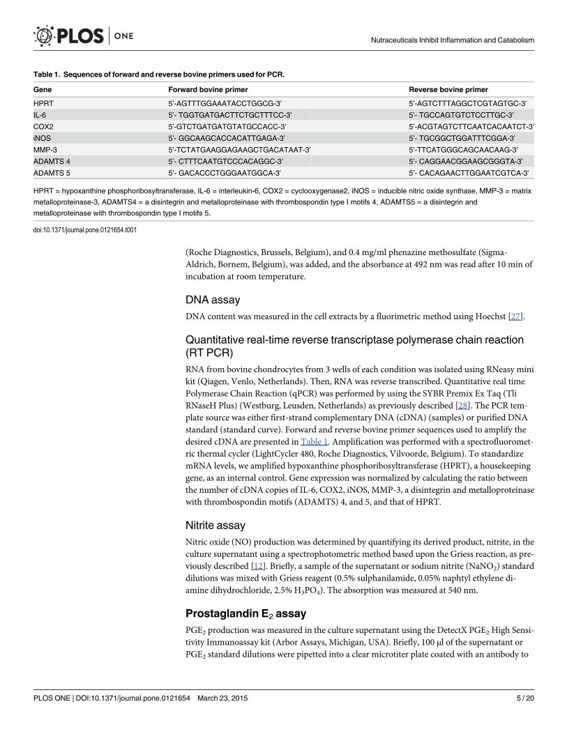
Quantitative real-time reverse transcriptase polymerase chain reaction(RT PCR)RNA from bovine chondrocytes from 3 wells of each condition was isolated using RNeasy minikit (Qiagen, Venlo, Netherlands). Then, RNA was reverse transcribed. Quantitative real timePolymerase Chain Reaction (qPCR) was performed by using the SYBR Premix Ex Taq (TliRNaseH Plus) (Westburg, Leusden, Netherlands) as previously described [28]. The PCR tem-plate source was either first-strand complementary DNA (cDNA) (samples) or purified DNAstandard (standard curve). Forward and reverse bovine primer sequences used to amplify thedesired cDNA are presented in Table 1. Amplification was performed with a spectrofluoromet-ric thermal cycler (LightCycler 480, Roche Diagnostics, Vilvoorde, Belgium). To standardizemRNA levels, we amplified hypoxanthine phosphoribosyltransferase (HPRT), a housekeepinggene, as an internal control. Gene expression was normalized by calculating the ratio betweenthe number of cDNA copies of IL-6, COX2, iNOS, MMP-3, a disintegrin and metalloproteinasewith thrombospondin motifs (ADAMTS) 4, and 5, and that of HPRT.
Nitrite assayNitric oxide (NO) production was determined by quantifying its derived product, nitrite, in theculture supernatant using a spectrophotometric method based upon the Griess reaction, as pre-viously described [12]. Briefly, a sample of the supernatant or sodium nitrite (NaNO2) standarddilutions was mixed with Griess reagent (0.5% sulphanilamide, 0.05% naphtyl ethylene di-amine dihydrochloride, 2.5% H3PO4). The absorption was measured at 540 nm.
Prostaglandin E2 assayPGE2 production was measured in the culture supernatant using the DetectX PGE2 High Sensi-tivity Immunoassay kit (Arbor Assays, Michigan, USA). Briefly, 100 μl of the supernatant orPGE2 standard dilutions were pipetted into a clear microtiter plate coated with an antibody to
Table 1. Sequences of forward and reverse bovine primers used for PCR.
Gene Forward bovine primer Reverse bovine primer
HPRT 5’-AGTTTGGAAATACCTGGCG-3’ 5’-AGTCTTTAGGCTCGTAGTGC-3’
IL-6 5’- TGGTGATGACTTCTGCTTTCC-3’ 5’- TGCCAGTGTCTCCTTGC-3’
COX2 5’-GTCTGATGATGTATGCCACC-3’ 5’-ACGTAGTCTTCAATCACAATCT-3’
iNOS 5’- GGCAAGCACCACATTGAGA-3’ 5’- TGCGGCTGGATTTCGGA-3’
MMP-3 5’-TCTATGAAGGAGAAGCTGACATAAT-3’ 5’-TTCATGGGCAGCAACAAG-3’
ADAMTS 4 5’- CTTTCAATGTCCCACAGGC-3’ 5’- CAGGAACGGAAGCGGGTA-3’
ADAMTS 5 5’- GACACCCTGGGAATGGCA-3’ 5’- CACAGAACTTGGAATCGTCA-3’
HPRT = hypoxanthine phosphoribosyltransferase, IL-6 = interleukin-6, COX2 = cyclooxygenase2, iNOS = inducible nitric oxide synthase, MMP-3 = matrix
metalloproteinase-3, ADAMTS4 = a disintegrin and metalloproteinase with thrombospondin type I motifs 4, ADAMTS5 = a disintegrin and
metalloproteinase with thrombospondin type I motifs 5.
doi:10.1371/journal.pone.0121654.t001
Nutraceuticals Inhibit Inflammation and Catabolism
PLOSONE | DOI:10.1371/journal.pone.0121654 March 23, 2015 5 / 20
capture mouse IgG. A PGE2-peroxidase conjugate (25 μl) is added to the standards and super-natants in the wells. The binding reaction was initiated by the addition of 25 μl of a monoclonalantibody to PGE2. After an overnight incubation at 4°C, the plate was washed and 100 μl ofsubstrate was added. After a short incubation, the reaction was stopped and the intensity of thegenerated colour was detected at 450 nm wavelength.
Immunoassays for interleukin-6, matrix metalloproteinase-3 andaggrecansIL-6, MMP-3, and aggrecans were measured by specific enzyme amplified sensitivity immuno-assays (Invitrogen, Merelbeke, Belgium). IL-6 and MMP-3 productions were measured in cul-ture supernatants, whereas aggrecans production was measured in culture supernatants and inthe cell extracts.
Western blottingCells were collected at 4°C and lysed on ice in 50 μl of buffer (25 mMHepes, 150 mMNaCl,0.5% Triton X-100, 10% glycerol, and 1 mM dithiothreitol) containing protease and phospha-tase inhibitors (Roche, Vilvoorde, Belgium). After incubation at 4°C for 30 minutes, lysateswere then centrifugated at 14,000 g for 30 minutes at 4°C to remove insoluble debris. Proteinconcentrations were determined using the BCA assay (Thermo Fisher Scientific, Erembode-gem, Belgium). Total protein extracts (20 μg) were fractioned by electrophoresis on a polyacryl-amide gel (10%) and transferred onto a PVDF membrane. Membranes were blocked for 1 h inTBS-Tween containing 5% nonfat dried milk (HSC70) or in 2% bovine serum albumin(phospho-NF-κB p65 and phospho-IκBα). Membranes were then incubated overnight at 4°Cwith primary antibodies. Anti-rabbit phospho-NF-κB p65 (1:1000 dilution), anti-rabbit phos-pho-IκBα (1:1000 dilution) and anti-rabbit HSC70 (1:1000 dilution) (Cell signaling, Boston,USA) were used. Horse-radish peroxidase (HRP)-linked anti-rabbit IgG antibody (1:2000 dilu-tion) was used as secondary antibody (Cell signaling, Boston, USA). The reaction was revealedwith the ECLWestern blotting substrate (Thermo Fisher Scientific, Erembodegem, Belgium).
Statistical analysisResults were normalized to the HPRT gene expression or to the DNA content of the cells andexpressed as the mean ± SEM or reported to the control and expressed in percent. Statisticalsignificance is assessed using a parametric one-way ANOVA, followed by Dunnett’s multiplecomparison post-test for statistical significance compared to control, and followed by Tukey’smultiple comparison post-test for statistical significance compared to the mixture. Differenceswere considered statistically significant at p-value< 0.05.
Results
Effects of curcuminoids extract, hydrolyzed collagen and green teaextract on normal bovine chondrocytes in monolayerBovine chondrocytes were cultured for 24 hours (gene expressions analysis) or 48 hours (pro-tein productions analysis) in the absence or in the presence of IL-1β and with or without curcu-minoids extract (C), hydrolyzed collagen (O) or green tea extract (T) added alone or incombination (COT). Cell viability, evaluated by LDH release, and DNA content, was not sig-nificantly modified by compounds alone or in combination, with or without IL-1β (data notshown).
Nutraceuticals Inhibit Inflammation and Catabolism
PLOSONE | DOI:10.1371/journal.pone.0121654 March 23, 2015 6 / 20
In basal conditions, NO and PGE2 productions were undetectable. However, bovine chon-drocytes expressed 0.43 ± 0.23 copies of IL-6 mRNA, 7.83 ± 2.83 copies of iNOS mRNA,1.62 ± 0.93 copies of COX2 mRNA, 3.00 ± 1.00 copies of MMP-3 mRNA, 0.6 ± 0.4 copies ofADAMTS4 mRNA, 13.13 ± 2.64 copies of ADAMTS5 mRNA and 146.9 ± 82.51 copies ofaggrecans mRNA per copy of HPRT. The compounds did not significantly affect the basal ex-pression of these genes.
In the presence of IL-1β, the productions of NO and PGE2 were 5.16 ± 1.42 nmol/μg DNAand 8,372.95 ± 5,187.35 pg/μg DNA, respectively. IL-1β increased the expression of the genescoding for IL-6, iNOS, COX2, MMP-3, ADAMTS4, ADAMTS5 by 178.16 ± 8.04, 17.87 ± 7.72,11.18 ± 2.48, 88.41 ± 45.19, 25.67 ± 14.73, 3.26 ± 1.03-fold, respectively and decreased aggre-cans by 7.00 ± 3.32-fold. In the presence of IL-1β and at the concentrations of 12.5 and 62.5 μg/ml, C significantly and fully inhibited NO production (p = 0.0101), while O and T were withouteffects (Fig. 1a). At the concentrations of 2.5, 12.5 and 62.5 μg/ml, C significantly inhibitedIL-1β stimulated PGE2 production (IL-1β: 8,372.95 ± 5,187.35 pg/μg DNA; 2.5 μg/ml:1,753.93 ± 878.64 pg/μg DNA, p = 0.0478; 12.5 μg/ml: 9.19 ± 9.19 pg/μg DNA, p = 0.0076 and62.5 μg/ml: 9.31 ± 9.31 pg/μg DNA, p = 0.0076) (Fig. 1b). In contrast, O and T had no signifi-cant effect on PGE2 production.
In the presence of IL-1β and at the concentrations of 12.5 and 62.5 μg/ml, C significantly in-hibited IL-6 (IL-1β: 100.23 ± 18.99 copies mRNa per copy of HPRT; 12.5 μg/ml: 3.67 ± 2.52copies mRNA per copy of HPRT, p<0.0001; 62.5 μg/ml: 2.37 ± 1.28 copies mRNA per copy ofHPRT, p<0.0001), COX2 (IL-1β: 13.93 ± 4.52 copies mRNA per copy of HPRT; 12.5 μg/ml:4.79 ± 2.77 copies mRNA per copy of HPRT, p<0.0001; 62.5 μg/ml: 5.01 ± 2.9 copies mRNAper copy of HPRT, p = 0.0001) iNOS (IL-1β: 99.43 ± 1.59 copies mRNA per copy of HPRT;12.5 μg/ml: 5.33 ± 3.26 copies mRNA per copy of HPRT, p<0.0001; 62.5 μg/ml: 5.33 ± 1.41copies mRNA per copy of HPRT p<0.0001), MMP-3 (IL-1β: 220.05 ± 38.5 copies mRNA percopy of HPRT; 12.5 μg/ml: 18.25 ± 8.86 copies mRNA per copy of HPRT, p<0.0001; 62.5 μg/ml: 9.1 ± 2.2 copies mRNA per copy of HPRT, p<0.0001), ADAMTS4 (IL-1β: 7.53 ± 2.24 cop-ies mRNA per copy of HPRT; 12.5 μg/ml: 1.47 ± 0.75 copies mRNA per copy of HPRT,p = 0.0005; 62.5 μg/ml: 1.5 ± 0.85 copies mRNA per copy of HPRT, p = 0.0008) and ADAMTS5(IL-1β: 37.67 ± 4.39 copies mRNA per copy of HPRT; 12.5 μg/ml: 11.87 ± 3.42 copies mRNAper copy of HPRT, p = 0.0073; 62.5 μg/ml: 9.77 ± 3.01 copies mRNA per copy of HPRT,p = 0.0037) gene expressions (Fig. 2a, b, c, d, e, f). In the presence of IL-1β and at the concentra-tion of 62.5 μg/ml, T significantly inhibited IL-6 (IL-1β: 100.23 ± 18.99 copies mRNA per copyof HPRT; 62.5 μg/ml: 35.6 ± 4.92 copies mRNA per copy of HPRT, p = 0.0309) and COX2 (IL-1β: 13.93 ± 4.52 copies mRNA per copy of HPRT; 62.5 μg/ml: 9 ± 4.69 copies mRNA per copyof HPRT, p = 0.0311) gene expressions (Fig. 2a, b). In the presence of IL-1β and at the concen-tration of 12.5 μg/ml, O significantly increased MMP-3 gene expression (IL-1β: 220.05 ± 38.5copies mRNA per copy of HPRT; 12.5 μg/ml: 337.35 ± 67.81 copies mRNA per copy of HPRT,p = 0.0063) (Fig. 2d).
We have also tested two compounds in combination, CO and CT. CO and CT tended to bemore efficient than C on all parameters. The differences between C and CO groups were notstatistically significant. In contrast, CT reduced higher IL-1β-stimulated MMP-3 expressionthan C (p = 0.0386) (Table 2).
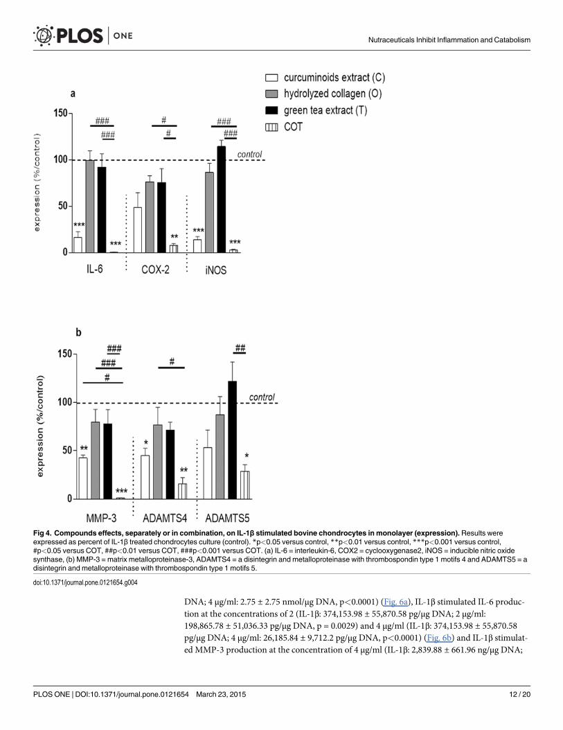
The mixture COT significantly inhibited IL-1β stimulated NO and PGE2 productions(p<0.0001), but the inhibitory effect was not higher than C alone (Fig. 3). The magnitude ofCOT inhibitory effect was higher than C alone on IL-1β-stimulated IL-6 mRNA (52.73 ± 15.64copies mRNA per copy of HPRT for C vs 5.07 ± 4.32 copies mRNA per copy of HPRT forCOT, p<0.0001), COX2 mRNA (6.2 ± 2.04 copies mRNA per copy of HPRT for C vs1.07 ± 0.38 copies mRNA per copy of HPRT for COT, p = 0.0035), iNOS mRNA (11.27 ± 5.04
Nutraceuticals Inhibit Inflammation and Catabolism
PLOSONE | DOI:10.1371/journal.pone.0121654 March 23, 2015 7 / 20
copies mRNA per copy of HPRT for C vs 3.5 ± 2.41 copies mRNA per copy of HPRT for COT,p<0.0001) (Fig.4a), MMP-3 mRNA (85.8 ± 44.88 copies mRNA per copy of HPRT for C vs1.87 ± 0.93 copies mRNA per copy of HPRT for COT, p<0.0001), ADAMTS4 mRNA(4.47 ± 2.09 copies mRNA per copy of HPRT for C vs 1.23 ± 0.48 copies mRNA per copy ofHPRT for COT, p = 0.003) and ADAMTS5 mRNA (50.57 ± 1.62 copies mRNA per copy of
Fig 1. Compounds effects on IL-1β stimulated bovine chondrocytes in monolayer (production).Results were expressed as percent of IL-1β treated chondrocytes culture (control). *p<0.05 versus control,**p<0.01 versus control. (a) NO = nitric oxide, (b) PGE2 = prostaglandin E2.
doi:10.1371/journal.pone.0121654.g001
Nutraceuticals Inhibit Inflammation and Catabolism
PLOSONE | DOI:10.1371/journal.pone.0121654 March 23, 2015 8 / 20
Fig 2. Compounds effects on IL-1β stimulated bovine chondrocytes in monolayer (expression).Results were expressed as percent of IL-1β treated chondrocytes culture (control). *p<0.05 versus control,**p<0.01 versus control, ***p<0.001 versus control. (a) IL-6 = interleukin-6, (b) COX2 = cyclooxygenase2,(c) iNOS = inducible nitric oxide synthase, (d) MMP-3 = matrix metalloproteinase-3, (e) ADAMTS4 = adisintegrin and metalloproteinase with thrombospondin type 1 motifs 4 and (f) ADAMTS5 = a disintegrin andmetalloproteinase with thrombospondin type 1 motifs 5.
doi:10.1371/journal.pone.0121654.g002
Nutraceuticals Inhibit Inflammation and Catabolism
PLOSONE | DOI:10.1371/journal.pone.0121654 March 23, 2015 9 / 20
HPRT for C vs 29.67 ± 4.68 copies mRNA per copy of HPRT for COT, p = 0.0248) (Fig. 4b)gene expressions.
Effects of curcuminoids, hydrolyzed collagen and green tea extract onOA human chondrocytes in monolayerPrimary OA human chondrocytes were cultured in monolayer until confluence and then incu-bated for 48 hours in the absence or in the presence of IL-1β and with or without C, O or Tadded alone or in combination (COT). Cell viability, evaluated by LDH release, and DNA con-tent, was not affected by these compounds added alone or in combination, with or without IL-1β (data not shown).
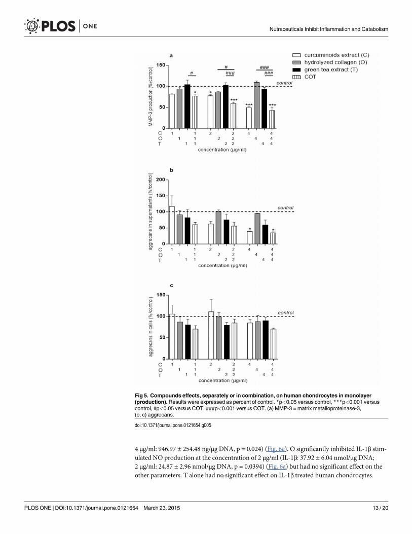
In basal conditions, NO production was undetectable while the content of IL-6, MMP-3and aggrecans in culture supernatants were 232.32 ± 85.3 pg/μg DNA, 207.34 ± 15.83 ng/μgDNA and 538 ± 86.48 ng/μg DNA, respectively. The aggrecans content in chondrocytes extractwas 28.92 ± 4.61 ng/μg DNA. In basal conditions, MMP-3 production was significantly inhib-ited by C at 2 (151.86 ± 19.03 ng/μg DNA, p = 0.0166) and 4 μg/ml (92.84 ± 19.49 ng/μg DNA,p<0.0001) (Fig. 5a). At the concentration of 4 μg/ml, C significantly decreased the aggrecans
Table 2. Compounds effects on IL-1β stimulated bovine chondrocytes in monolayer.
C CO CT
Production NO % of ctrl 3,84 0,000001 0,000001
p-value vs ctrl <0,0001 <0,0001 <0,0001
p-value vs C 0,9996 0,9996
PGE2 % of ctrl 0,9323 0,4982 0,000001
p-value vs ctrl 0,9925 0,9924 0,9922
p-value vs C >0,9999 >0,9999
Gene expression IL-6 % of ctrl 16,37 10,93 0,47
p-value vs ctrl <0,0001 <0,0001 <0,0001
p-value vs C 0,9988 0,7944
COX2 % of ctrl 48,72 42,36 13,28
p-value vs ctrl 0,0719 0,0385 0,0021
p-value vs C 0,9998 0,524
iNOS % of ctrl 14,18 8,671 3,757
p-value vs ctrl <0,0001 <0,0001 <0,0001
p-value vs C 0,9879 0,8005
MMP-3 % of ctrl 42,5 15,41 1,21
p-value vs ctrl 0,0011 <0,0001 <0,0001
p-value vs C 0,2934 0,0386
ADAMTS4 % of ctrl 45,28 31,81 17,16
p-value vs ctrl 0,0279 0,0061 0,0012
p-value vs C 0,9818 0,6516
ADAMTS5 % of ctrl 53,33 48,02 24,32
p-value vs ctrl 0,1905 0,1261 0,0168
p-value vs C >0,9999 0,8233
Ctrl = IL-1β stimulated bovine chondrocytes, C = curcuminoids extract, CO = curcuminoids extract + hydrolyzed collagen, CT = curcuminoids extract +
green tea extract, NO = nitric oxide, PGE2 = prostaglandin E2, IL-6 = interleukin-6, COX2 = cyclooxygenase-2, iNOS = inducible nitric oxide synthase,
MMP-3 = matrix metalloproteinase-3, ADAMTS4 = a disintegrin and metalloproteinase with thrombospondin type I motifs 4, ADAMTS5 = a disintegrin and
metalloproteinase with thrombospondin type I motifs 5.
doi:10.1371/journal.pone.0121654.t002
Nutraceuticals Inhibit Inflammation and Catabolism
PLOSONE | DOI:10.1371/journal.pone.0121654 March 23, 2015 10 / 20
content in culture supernatants (214.87 ± 41.34 ng/μg DNA, p = 0.0232) (Fig. 5b), but had noeffect on aggrecans content in chondrocytes extract (Fig. 5c). None of the compounds, neitherthe combination, showed significant effects on NO and IL-6 productions in the absence of IL-1β on human chondrocytes (data not shown). O and T had no significant effect on humanchondrocytes in basal conditions.
In basal conditions, MMP-3 production was significantly inhibited by the COT mixture at1:1:1 (139.1 ± 30.13 ng/μg DNA, p = 0.0105), 2:2:2 (110.95 ± 20.97 ng/μg DNA, p<0.0001) and4:4:4 μg/ml (78.03 ± 14.85 ng/μg DNA, p<0.0001) (Fig. 5a). At the concentration of 1:1:1 μg/ml, COT inhibited MMP-3 production while C, O or T individually had no effect. This indicat-ed that, at the concentration of 1 μg/ml, C, O and T acted in synergy on this parameter. Fur-ther, COT at 4:4:4 μg/ml significantly decreased the aggrecans content in culture supernatants(192.91 ± 45.98 ng/μg DNA, p = 0.0133) (Fig. 5b), but had no significant effect on aggrecanscontent in chondrocytes extract (Fig. 5c). However, COT had a similar effect on the aggrecansin culture supernatants than C.
In the presence of 10–11 M IL-1β, the production of NO was 37.92 ± 6.04 nmol/μg DNA. IL-1β significantly increased the production of IL-6 and MMP-3 by 2,114.91 ± 789.59 and13.61 ± 2.7-fold, respectively. In contrast, IL-1β inhibited aggrecans content in cells extract andsupernatants by 1.37 ± 0.36 and 4.72 ± 1.83-fold, respectively. C significantly inhibited IL-1βstimulated NO production at the concentration of 4 μg/ml (IL-1β: 37.92 ± 6.04 nmol/μg
Fig 3. Compounds effects, separately or in combination, on IL-1β stimulated bovine chondrocytes in monolayer (production). Results wereexpressed as percent of IL-1β treated chondrocytes culture (control). ***p<0.001 versus control, ###p<0.001 versus COT. NO = nitric oxide, PGE2 =prostaglandin E2.
doi:10.1371/journal.pone.0121654.g003
Nutraceuticals Inhibit Inflammation and Catabolism
PLOSONE | DOI:10.1371/journal.pone.0121654 March 23, 2015 11 / 20
DNA; 4 μg/ml: 2.75 ± 2.75 nmol/μg DNA, p<0.0001) (Fig. 6a), IL-1β stimulated IL-6 produc-tion at the concentrations of 2 (IL-1β: 374,153.98 ± 55,870.58 pg/μg DNA; 2 μg/ml:198,865.78 ± 51,036.33 pg/μg DNA, p = 0.0029) and 4 μg/ml (IL-1β: 374,153.98 ± 55,870.58pg/μg DNA; 4 μg/ml: 26,185.84 ± 9,712.2 pg/μg DNA, p<0.0001) (Fig. 6b) and IL-1β stimulat-ed MMP-3 production at the concentration of 4 μg/ml (IL-1β: 2,839.88 ± 661.96 ng/μg DNA;
Fig 4. Compounds effects, separately or in combination, on IL-1β stimulated bovine chondrocytes in monolayer (expression).Results wereexpressed as percent of IL-1β treated chondrocytes culture (control). *p<0.05 versus control, **p<0.01 versus control, ***p<0.001 versus control,#p<0.05 versus COT, ##p<0.01 versus COT, ###p<0.001 versus COT. (a) IL-6 = interleukin-6, COX2 = cyclooxygenase2, iNOS = inducible nitric oxidesynthase, (b) MMP-3 = matrix metalloproteinase-3, ADAMTS4 = a disintegrin and metalloproteinase with thrombospondin type 1 motifs 4 and ADAMTS5 = adisintegrin and metalloproteinase with thrombospondin type 1 motifs 5.
doi:10.1371/journal.pone.0121654.g004
Nutraceuticals Inhibit Inflammation and Catabolism
PLOSONE | DOI:10.1371/journal.pone.0121654 March 23, 2015 12 / 20
4 μg/ml: 946.97 ± 254.48 ng/μg DNA, p = 0.024) (Fig. 6c). O significantly inhibited IL-1β stim-ulated NO production at the concentration of 2 μg/ml (IL-1β: 37.92 ± 6.04 nmol/μg DNA;2 μg/ml: 24.87 ± 2.96 nmol/μg DNA, p = 0.0394) (Fig. 6a) but had no significant effect on theother parameters. T alone had no significant effect on IL-1β treated human chondrocytes.
Fig 5. Compounds effects, separately or in combination, on human chondrocytes in monolayer(production). Results were expressed as percent of control. *p<0.05 versus control, ***p<0.001 versuscontrol, #p<0.05 versus COT, ###p<0.001 versus COT. (a) MMP-3 = matrix metalloproteinase-3,(b, c) aggrecans.
doi:10.1371/journal.pone.0121654.g005
Nutraceuticals Inhibit Inflammation and Catabolism
PLOSONE | DOI:10.1371/journal.pone.0121654 March 23, 2015 13 / 20

The COT mixture significantly decreased IL-1β-stimulated NO production at the concen-trations of 2:2:2 (IL-1β: 37.92 ± 6.04 nmol/μg DNA; 2:2:2 μg/ml: 17.74 ± 4.72 nmol/μg DNA,p = 0.0003) and 4:4:4 μg/ml (IL-1β: 37.92 ± 6.04 nmol/μg DNA; 4:4:4 μg/ml: 1.65 ± 1.15 nmol/μg DNA, p<0.0001) (Fig. 6a). At the concentration of 4:4:4 μg/ml, COT and C effects on NO
Fig 6. Compounds effects, separately or in combination, on IL-1β stimulated human chondrocytes inmonolayer (production). Results were expressed as percent of IL-1β treated chondrocytes culture (control).*p<0.05 versus control, **p<0.01 versus control, ***p<0.001 versus control, #p<0.05 versus COT,##p<0.01 versus COT, ###p<0.001 versus COT. (a) NO = nitric oxide, (b) IL-6 = interleukin-6,(c) MMP-3 = matrix metalloproteinase-3.
doi:10.1371/journal.pone.0121654.g006
Nutraceuticals Inhibit Inflammation and Catabolism
PLOSONE | DOI:10.1371/journal.pone.0121654 March 23, 2015 14 / 20
were not significantly different whereas, at the concentration of 2:2:2 μg/ml, COT had a higherinhibitory effect than O (Fig. 6a). The COT mixture significantly decreased IL-1β stimulatedIL-6 production at the concentrations of 1:1:1 (IL-1β: 374,153.98 ± 55,870.58 pg/μg DNA;1:1:1 μg/ml: 238,658.42 ± 61,332.79 pg/μg DNA, p = 0.0259), 2:2:2 (IL-1β: 374,153.98 ±55,870.58 pg/μg DNA; 2:2:2 μg/ml: 165,110.45 ± 22,791.39 pg/μg DNA, p = 0.0006) and4:4:4 μg/ml (IL-1β: 374,153.98 ± 55,870.58 pg/μg DNA; 4:4:4 μg/ml: 28,127.75 ± 6,569.56 pg/μgDNA, p<0.0001) (Fig. 6b). C, O and T added separately at the concentration of 1 μg/ml had noeffect on IL-6 production. In contrast, COT mixture (1:1:1 μg/ml) significantly reduced IL-1βstimulated IL-6 production (p = 0.0259). However, at the concentrations of 2:2:2 and4:4:4 μg/ml, there was no additive effect of COT on IL-1β stimulated IL-6 production, com-pared to C (Fig. 6b). The COT mixture, at 4:4:4 μg/ml, decreased IL-1β stimulated MMP-3 pro-duction (IL-1β: 2,839.88 ± 661.96 ng/μg DNA; 4:4:4 μg/ml: 863.56 ± 181.07 ng/μg DNA,p = 0.0119) but this inhibitory effect was similar to that of C (Fig. 6c).
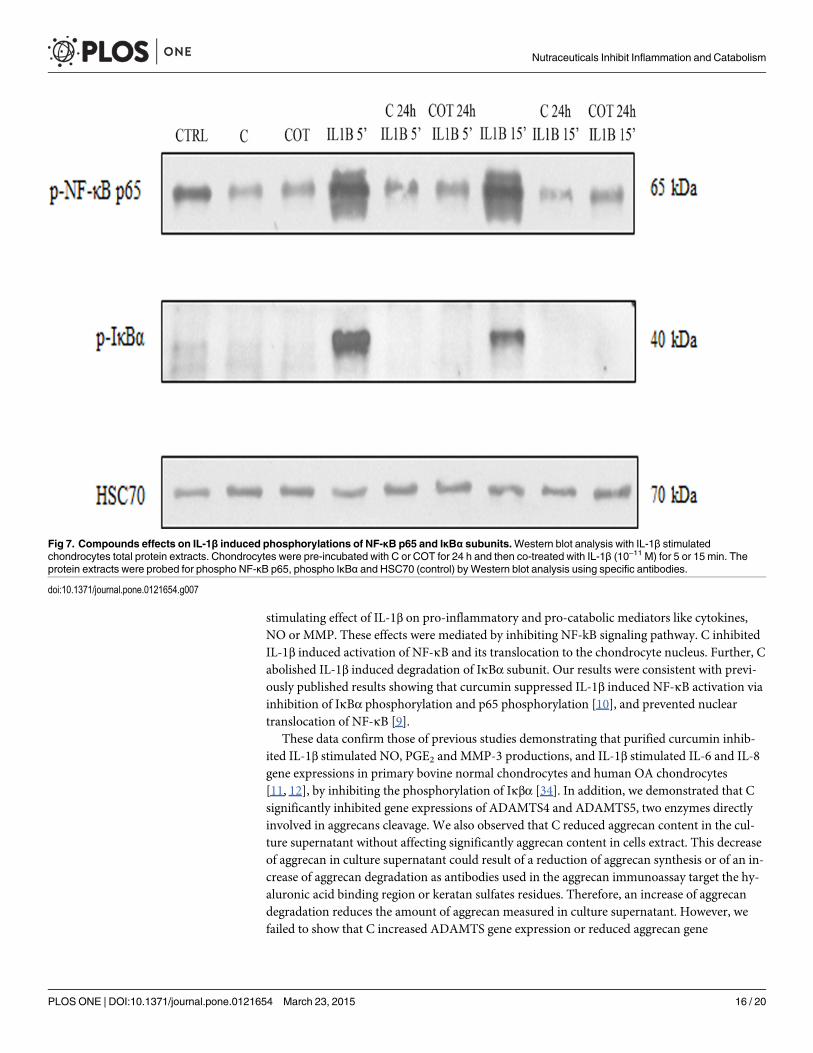
We next investigated the effects of C and COT on human chondrocytes on IL-1β inducedNF-κB activation. Protein extracts of chondrocytes were probed for the phosphorylated p65NF-κB subunit after pre-treatment with C or COT for 24 h followed by co-treatment or notwith IL-1β (10–11 M) for 5 or 15 minutes. C and COT inhibited IL-1β induced phosphorylationand translocation of p65 in nuclear extracts of chondrocytes (Fig. 7). Further, to examinewhether inhibition of IL-1β induced NF-κB activation occurred through inhibition of IκBαdegradation, protein extracts of chondrocytes were probed for the phosphorylated IκBα sub-unit after pre-treatment with C or COT for 24 h followed by co-treatment or not with IL-1β(10–11 M) for 5 or 15 minutes. IL-1β induced IκBα degradation as early as 5 min, but IL-1βcould not induce IκBα subunit degradation in C and COT pre-treated chondrocytes (Fig. 7).
DiscussionThis study aimed 1) to compare the effects of three nutraceuticals, curcuminoids extract, hy-drolyzed collagen and green tea extract on chondrocytes metabolism, 2) to investigate a poten-tial synergic effect between these compounds and 3) to examine whether the effects ofcurcuminoids extract and the combination of the three compounds on human chondrocytesinvolved the NF-κB signaling pathway. Numerous mediators contribute to both degradativeand nociceptive pathways associated with the progression of OA [29]. Among these mediators,some cytokines like IL-1β play a key role in OA, by stimulating cartilage degradation and bytriggering synovial membrane inflammation [30]. In vitro, IL-1β strongly stimulates cartilagedegradation, and inhibits proteoglycan and collagen synthesis [2, 31–33]. IL-1β increases theproduction of a large spectrum of proteolytic enzymes such as ADAMTS4, ADAMTS5, MMP-3 and a variety of other cytokines, including IL-6 and IL-8, by chondrocytes. For these reasons,we treated human primary chondrocytes with IL-1β to mimic OA chondrocyte metabolic re-sponses. Normalizing chondrocyte metabolism and, more particularly, counteracting the dele-terious effects of IL-1β, are targets for pharmaceutical agents in OA. Evidence to date indicatesthat in addition to IL-1β, IL-6 and IL-8 can also promote articular cartilage extracellular matrixdegradation or synergize with other cytokines to amplify and accelerate cartilage destruction[29, 33]. As expected, IL-1β stimulation of chondrocytes significantly induced the expressionof COX2, iNOS, ADAMTS4 and ADAMTS5 and the production of NO and PGE2 in our ex-periments. Moreover, IL-1β treatment significantly induced the mRNA expression as well asprotein production of IL-6 and MMP-3, and significantly reduced the mRNA expression aswell as protein production of aggrecans by chondrocytes.
We have demonstrated that C had beneficial effects on chondrocytes. In fact, C counter-acted the deleterious effects of IL-1β on chondrocytes. More precisely, C reduced the
Nutraceuticals Inhibit Inflammation and Catabolism
PLOSONE | DOI:10.1371/journal.pone.0121654 March 23, 2015 15 / 20
stimulating effect of IL-1β on pro-inflammatory and pro-catabolic mediators like cytokines,NO or MMP. These effects were mediated by inhibiting NF-kB signaling pathway. C inhibitedIL-1β induced activation of NF-κB and its translocation to the chondrocyte nucleus. Further, Cabolished IL-1β induced degradation of IκBα subunit. Our results were consistent with previ-ously published results showing that curcumin suppressed IL-1β induced NF-κB activation viainhibition of IκBα phosphorylation and p65 phosphorylation [10], and prevented nucleartranslocation of NF-κB [9].
These data confirm those of previous studies demonstrating that purified curcumin inhib-ited IL-1β stimulated NO, PGE2 and MMP-3 productions, and IL-1β stimulated IL-6 and IL-8gene expressions in primary bovine normal chondrocytes and human OA chondrocytes[11, 12], by inhibiting the phosphorylation of Iκβα [34]. In addition, we demonstrated that Csignificantly inhibited gene expressions of ADAMTS4 and ADAMTS5, two enzymes directlyinvolved in aggrecans cleavage. We also observed that C reduced aggrecan content in the cul-ture supernatant without affecting significantly aggrecan content in cells extract. This decreaseof aggrecan in culture supernatant could result of a reduction of aggrecan synthesis or of an in-crease of aggrecan degradation as antibodies used in the aggrecan immunoassay target the hy-aluronic acid binding region or keratan sulfates residues. Therefore, an increase of aggrecandegradation reduces the amount of aggrecan measured in culture supernatant. However, wefailed to show that C increased ADAMTS gene expression or reduced aggrecan gene
Fig 7. Compounds effects on IL-1β induced phosphorylations of NF-κB p65 and IκBα subunits.Western blot analysis with IL-1β stimulatedchondrocytes total protein extracts. Chondrocytes were pre-incubated with C or COT for 24 h and then co-treated with IL-1β (10–11 M) for 5 or 15 min. Theprotein extracts were probed for phospho NF-κB p65, phospho IκBα and HSC70 (control) by Western blot analysis using specific antibodies.
doi:10.1371/journal.pone.0121654.g007
Nutraceuticals Inhibit Inflammation and Catabolism
PLOSONE | DOI:10.1371/journal.pone.0121654 March 23, 2015 16 / 20
expression. The monolayer culture model is not the best model to investigate aggrecan produc-tion and accumulation in the extracellular matrix. To verify our hypothesis, it would be inter-esting to test C in chondrocytes cultured in alginate beads. More surprisingly were the feweffects of T and O. T significantly decreased IL-6 and COX-2 gene expressions but had no sig-nificant effects on the other parameters while O had no significant effect. This observation con-trasts with the results of other studies suggesting that O and T could have beneficial effects oncartilage. Indeed, it was demonstrated that Proline-Hydroxyproline (Pro-Hyp), the majorhydroxyproline-containing peptide in human blood after oral ingestion of collagen hydroly-sates [17], increased the aggrecan mRNA levels in murine chondrocytes [35] and collagen hy-drolysates and Pro-Hyp inhibited murine chondrocyte differentiation into mineralizedchondrocytes and increased glycosaminoglycans production [35]. EGCG, the most abundantcatechin in green tea, suppressed the inflammatory response in human chondrocytes [22].Other data proved that EGCG decreased the production of TNFα [21]. These discrepanciescan be explained by the cell type. Indeed, the beneficial effects of hydrolyzed collagen were ob-served on a murine chondrocytic cell line whereas our research was performed on primary bo-vine and human chondrocytes. Moreover, the effects of hydrolyzed collagen may differaccording the origin and formulation of collagen hydrolysates [15]. Furthermore, to show a de-crease in inflammatory response, primary human chondrocytes were pre-treated with differentdoses of EGCG for 1 or 2 hours prior to stimulation (with advanced glycation end products[21] or with 5000 pg/ml IL-1β) while we co-treated our primary human chondrocytes with Tand with IL-1β at a lower concentration (170 pg/ml = 10–11 M) for 48 hours. It was alreadyshown that EGCG individually marginally inhibit COX-2 expression and PGE2 production incytokine-activated equine chondrocytes. Finally, EGCG, in combination with avocado andsoya unsaponifiables, reduced COX2 expression close to non-activated control levels and sig-nificantly inhibit PGE2 production [36].
For the first time, this paper described the effects of C, O and T in combination on chondro-cyte metabolism. In bovine chondrocytes, the three compounds showed additive inhibitory ef-fects on IL-1β stimulated IL-6, iNOS and ADAMTS4 gene expressions. Moreover, the threecompounds acted synergically to inhibit IL-1β stimulated COX2, MMP-3 and ADAMTS5 geneexpressions. COT showed a higher effect on these parameters than C alone. Further, the inhibi-tory effect of COT on IL-1β-induced COX-2, iNOS, MMP-3 and ADAMTS4 expressionstended to be greater than that of CO or CT. In human chondrocytes, COT had an additive in-hibitory effect on MMP-3 and IL-1β stimulated NO production and acted synergically on IL-1β stimulated IL-6 production.
Altogether, these data gave a rationale for combining these compounds in the managementof OA.
One major limitation of our study is that compounds have been tested on a limited numberof cartilage specimens. Additional experiments are needed, for example in order to reach thesignificance of the effects and identify the best responders among a large panel of specimens.
Of course, before recommending these natural products in the management of OA, someclinical trials are required to demonstrate the beneficial effects of these nutraceuticals on OAsymptoms and structural changes. Another major concern is the absence of information aboutdietary supplements bioavailability. This point is particularly true for polyphenol like curcu-min. In fact, natural curcumin is known for its very low bioavailability. This point is crucial toenvisage its administration by oral route. For example, the average peak serum concentrationsafter oral intake of 4, 6 and 8 g of curcumin per day were 0.51 ± 0.11, 0.63 ± 0.06 and1.77 ± 1.87 μM, respectively [37]. The serum concentration of curcumin peaked at 1–2 h afteroral intake of curcumin and gradually declined within 12 hours. Urinary excretion of curcuminwas undetectable [37]. This mean that the concentrations of curcuminoids extract tested in this
Nutraceuticals Inhibit Inflammation and Catabolism
PLOSONE | DOI:10.1371/journal.pone.0121654 March 23, 2015 17 / 20
in vitro study (4 μg/ml ~ 10 μM) are superior to those found in plasma after oral administrationof high doses of natural curcumin. Therefore, the extrapolation of our in vitro data to humannutrition must be done with caution. However, many effort have been made to increase curcu-min bioavaibility. Recently, we performed a Phase I pharmacokinetics study on Flexofytol, ahigh bioavailable turmeric extract with a water solubility increased 4000 times, that was run on2 groups of 12 healthy individuals. Each group received orally 1 (42 mg curcumin) or 2 cap-sules (84 mg of curcumin) of Flexofytol respectively. With 2 capsules administered orally, themean of Cmax on 12 individuals was 0.9 μM, with a statistical extrapolation at 1.6 μMwith 4capsules (administering 84 mg and 168 mg of curcumin respectively). These values are closerwith those used in our in vitro study.
The challenge is to develop a curcumin with an increased bioavailability, with the aim ofreaching plasma concentrations that have demonstrated biological activity. In this purpose,curcumin has been dissolved in oil and then absorbed into chylomicrons, to propose a surface-controlled water-dispersible curcumin, with no modification of its structure [38, 39]. Curcu-min was also co-administered with piperine, an inhibitor of hepatic and intestinal glucuronida-tion [40] or included in a phosphatidylcholine phytosome complex [41, 42]. The clinicalrelevance of these new formulations compared to the native curcumin should be demonstratedin good clinical trials.
Altogether, these in vitro results indicate that the mixture COT might reduce inflammationand pain in OA by reducing the synthesis of inflammatory and catabolic mediators by chon-drocytes, probably by inhibiting NF-κB activation. These findings provide a preclinical basisfor the in vivo testing of the combinations and suggest that these natural compounds could behelpful to alleviate symptoms in OA patients. These results support firstly the developmentand use of the mixture COT as an anti-inflammatory / anti-osteoarthritic agent for treatmentof OA.
AcknowledgmentsWe are grateful to public slaughterhouse in Liege for supplying bovine articular cartilage andto Catholic University of Louvain, especially Dr Jean-Emile Dubuc, for supplying OA humanarticular cartilage samples. We thank the Laboratory of Tumor and Development Biology atthe University of Liege for the supply of the LightCycler 480. We also are thankful to Prof. Bel-lahcène (Metastasis Research Laboratory, GIGA-Cancer, University of Liege, Belgium) fortechnical support with the western blotting.
Author ContributionsConceived and designed the experiments: FC CS SS YH. Performed the experiments: FC. Ana-lyzed the data: FC CS IL MB SS YH. Contributed reagents/materials/analysis tools: FC IL SSYH. Wrote the paper: FC YH.
References1. Xu J, Zhang C. In vitro isolation and cultivation of human chondrocytes for osteoarthritis renovation. In
Vitro Cell Dev Biol Anim. 2014.
2. Mathy-Hartert M, Hogge L, Sanchez C, Deby-Dupont G, Crielaard JM, Henrotin Y. Interleukin-1betaand interleukin-6 disturb the antioxidant enzyme system in bovine chondrocytes: a possible explanationfor oxidative stress generation. Osteoarthritis Cartilage. 2008; 16(7):756–63. doi: 10.1016/j.joca.2007.10.009 PMID: 18291685
3. Nelson AE, Allen KD, Golightly YM, Goode AP, Jordan JM. A systematic review of recommendationsand guidelines for the management of osteoarthritis: The Chronic Osteoarthritis Management Initiativeof the U.S. Bone and Joint Initiative. Semin Arthritis Rheum. 2014; 43(6):701–12. doi: 10.1016/j.semarthrit.2013.11.012 PMID: 24387819
Nutraceuticals Inhibit Inflammation and Catabolism
PLOSONE | DOI:10.1371/journal.pone.0121654 March 23, 2015 18 / 20
4. McAlindon TE, Bannuru RR, Sullivan MC, Arden NK, Berenbaum F, Bierma-Zeinstra SM, et al. OARSIguidelines for the non-surgical management of knee osteoarthritis. Osteoarthritis Cartilage. 2014; 22(3):363–88. doi: 10.1016/j.joca.2014.01.003 PMID: 24462672
5. Hochberg MC. Osteoarthritis year 2012 in review: clinical. Osteoarthritis Cartilage. 2012; 20(12):1465–9. doi: 10.1016/j.joca.2012.07.022 PMID: 22885568
6. Carter GT, Duong V, Ho S, Ngo KC, Greer CL, Weeks DL. Side effects of commonly prescribed analge-sic medications. Phys Med Rehabil Clin N Am. 2014; 25(2):457–70. doi: 10.1016/j.pmr.2014.01.007PMID: 24787343
7. Henrotin Y, Lambert C, Couchourel D, Ripoll C, Chiotelli E. Nutraceuticals: do they represent a new erain the management of osteoarthritis?—a narrative review from the lessons taken with five products. Os-teoarthritis Cartilage. 2011; 19(1):1–21. doi: 10.1016/j.joca.2010.10.017 PMID: 21035558
8. Ameye LG, CheeWS. Osteoarthritis and nutrition. From nutraceuticals to functional foods: a systematicreview of the scientific evidence. Arthritis Res Ther. 2006; 8(4):R127. PMID: 16859534
9. Schulze-Tanzil G, Mobasheri A, Sendzik J, John T, Shakibaei M. Effects of curcumin (diferuloyl-methane) on nuclear factor kappaB signaling in interleukin-1beta-stimulated chondrocytes. Ann N YAcad Sci. 2004; 1030:578–86. PMID: 15659840
10. Shakibaei M, John T, Schulze-Tanzil G, Lehmann I, Mobasheri A. Suppression of NF-kappaB activa-tion by curcumin leads to inhibition of expression of cyclo-oxygenase-2 and matrix metalloproteinase-9in human articular chondrocytes: Implications for the treatment of osteoarthritis. Biochem Pharmacol.2007; 73(9):1434–45. PMID: 17291458
11. Mathy M, Sanchez C, Priem F, Henrotin Y. Curcumin inhibits interleukin-6, -8, nitric oxide and prosta-glandin E2 synthesis by bovine chondrocytes. Osteoarthritis Cartilage. 2007; 15:C115 (Abstract).
12. Mathy-Hartert M, Jacquemond-Collet I, Priem F, Sanchez C, Lambert C, Henrotin Y. Curcumin inhibitspro-inflammatory mediators and metalloproteinase-3 production by chondrocytes. InflammRes. 2009;58(12):899–908. doi: 10.1007/s00011-009-0063-1 PMID: 19579007
13. Henrotin Y, Clutterbuck AL, Allaway D, Lodwig EM, Harris P, Mathy-Hartert M, et al. Biological actionsof curcumin on articular chondrocytes. Osteoarthritis Cartilage. 2010; 18(2):141–9. doi: 10.1016/j.joca.2009.10.002 PMID: 19836480
14. EFSA. Opinion of the Food Safety Authority on safety of collagen and a processing method for the pro-duction of collagen. European Food Safety Authority Journal. 2005; 174:1–9.
15. Schadow S, Siebert HC, Lochnit G, Kordelle J, Rickert M, Steinmeyer J. Collagen metabolism ofhuman osteoarthritic articular cartilage as modulated by bovine collagen hydrolysates. PLoS One.2013; 8(1):e53955. doi: 10.1371/journal.pone.0053955 PMID: 23342047
16. Walrand S, Chiotelli E, Noirt F, Mwewa S, Lassel T. Consumption of a functional fermented milk con-taining collagen hydrolysate improves the concentration of collagen-specific amino acids in plasma.J Agric Food Chem. 2008; 56(17):7790–5. doi: 10.1021/jf800691f PMID: 18707117
17. Ohara H, Iida H, Ito K, Takeuchi Y, Nomura Y. Effects of Pro-Hyp, a collagen hydrolysate-derived pep-tide, on hyaluronic acid synthesis using in vitro cultured synovium cells and oral ingestion of collagenhydrolysates in a guinea pig model of osteoarthritis. Biosci Biotechnol Biochem. 2010; 74(10):2096–9.PMID: 20944430
18. Schunck M, Schulze C, Oesser S. Disparate efficacy of collagen hydrolysate and glucosamine on theextracellular matrix metabolism of articular chondrocytes. Osteoarthritis Cartilage. 2006; 14:S114(Abstract).
19. Oesser S, Seifert J. Stimulation of type II collagen biosynthesis and secretion in bovine chondrocytescultured with degraded collagen. Cell Tissue Res. 2003; 311(3):393–9. PMID: 12658447
20. Ahmed S. Green tea polyphenol epigallocatechin 3-gallate in arthritis: progress and promise. ArthritisRes Ther. 2010; 12(2):208. doi: 10.1186/ar2982 PMID: 20447316
21. Rasheed Z, Anbazhagan AN, Akhtar N, Ramamurthy S, Voss FR, Haqqi TM. Green tea polyphenolepigallocatechin-3-gallate inhibits advanced glycation end product-induced expression of tumor necro-sis factor-alpha and matrix metalloproteinase-13 in human chondrocytes. Arthritis Res Ther. 2009; 11(3):R71. doi: 10.1186/ar2700 PMID: 19445683
22. Akhtar N, Haqqi TM. Epigallocatechin-3-gallate suppresses the global interleukin-1beta-induced in-flammatory response in human chondrocytes. Arthritis Res Ther. 2011; 13(3):R93. doi: 10.1186/ar3368PMID: 21682898
23. Ahmed S, Wang N, Lalonde M, Goldberg VM, Haqqi TM. Green tea polyphenol epigallocatechin-3-gallate (EGCG) differentially inhibits interleukin-1 beta-induced expression of matrix metalloproteinase-1 and-13 in human chondrocytes. J Pharmacol Exp Ther. 2004; 308(2):767–73. PMID: 14600251
24. Kumar A, Takada Y, Boriek AM, Aggarwal BB. Nuclear factor-kappaB: its role in health and disease.J Mol Med (Berl). 2004; 82(7):434–48. PMID: 15175863
Nutraceuticals Inhibit Inflammation and Catabolism
PLOSONE | DOI:10.1371/journal.pone.0121654 March 23, 2015 19 / 20
25. Aggarwal BB. Nuclear factor-kappa B: a transcription factor for all seasons. Expert Opin Ther Targets.2007; 11(2):109–10. PMID: 17227227
26. Pesesse L, Sanchez C, Delcour JP, Bellahcene A, Baudouin C, Msika P, et al. Consequences of chon-drocyte hypertrophy on osteoarthritic cartilage: potential effect on angiogenesis. Osteoarthritis Carti-lage. 2013; 21(12):1913–23. doi: 10.1016/j.joca.2013.08.018 PMID: 23973427
27. Labarca C, Paigen K. A simple, rapid, and sensitive DNA assay procedure. Anal Biochem. 1980; 102(2):344–52. PMID: 6158890
28. Sanchez C, Pesesse L, Gabay O, Delcour JP, Msika P, Baudouin C, et al. Regulation of subchondralbone osteoblast metabolism by cyclic compression. Arthritis Rheum. 2012; 64(4):1193–203. doi: 10.1002/art.33445 PMID: 22034083
29. Lee AS, Ellman MB, Yan D, Kroin JS, Cole BJ, vanWijnen AJ, et al. A current review of molecularmechanisms regarding osteoarthritis and pain. Gene. 2013; 527(2):440–7. doi: 10.1016/j.gene.2013.05.069 PMID: 23830938
30. Malemud CJ. Anticytokine therapy for osteoarthritis: evidence to date. Drugs Aging. 2010; 27(2):95–115. doi: 10.2165/11319950-000000000-00000 PMID: 20104937
31. Kobayashi M, Squires GR, Mousa A, Tanzer M, Zukor DJ, Antoniou J, et al. Role of interleukin-1 andtumor necrosis factor alpha in matrix degradation of human osteoarthritic cartilage. Arthritis Rheum.2005; 52(1):128–35. PMID: 15641080
32. Daheshia M, Yao JQ. The interleukin 1beta pathway in the pathogenesis of osteoarthritis. J Rheumatol.2008; 35(12):2306–12. PMID: 18925684
33. Kapoor M, Martel-Pelletier J, Lajeunesse D, Pelletier JP, Fahmi H. Role of proinflammatory cytokines inthe pathophysiology of osteoarthritis. Nat Rev Rheumatol. 2011; 7(1):33–42. doi: 10.1038/nrrheum.2010.196 PMID: 21119608
34. Csaki C, Mobasheri A, Shakibaei M. Synergistic chondroprotective effects of curcumin and resveratrolin human articular chondrocytes: inhibition of IL-1beta-induced NF-kappaB-mediated inflammation andapoptosis. Arthritis Res Ther. 2009; 11(6):R165. doi: 10.1186/ar2850 PMID: 19889203
35. Nakatani S, Mano H, Sampei C, Shimizu J, Wada M. Chondroprotective effect of the bioactive peptideprolyl-hydroxyproline in mouse articular cartilage in vitro and in vivo. Osteoarthritis Cartilage. 2009; 17(12):1620–7. doi: 10.1016/j.joca.2009.07.001 PMID: 19615963
36. Heinecke LF, Grzanna MW, Au AY, Mochal CA, Rashmir-Raven A, Frondoza CG. Inhibition of cycloox-ygenase-2 expression and prostaglandin E2 production in chondrocytes by avocado soybean unsapo-nifiables and epigallocatechin gallate. Osteoarthritis Cartilage. 2010; 18(2):220–7. doi: 10.1016/j.joca.2009.08.015 PMID: 19748608
37. Cheng AL, Hsu CH, Lin JK, Hsu MM, Ho YF, Shen TS, et al. Phase I clinical trial of curcumin, a chemo-preventive agent, in patients with high-risk or pre-malignant lesions. Anticancer Res. 2001; 21(4B):2895–900. PMID: 11712783
38. Henrotin Y, Priem F, Mobasheri A. Curcumin: a new paradigm and therapeutic opportunity for the treat-ment of osteoarthritis: curcumin for osteoarthritis management. Springerplus. 2013; 2(1):56. doi: 10.1186/2193-1801-2-56 PMID: 23487030
39. Nakagawa Y, Mukai S, Yamada S, MatsuokaM, Tarumi E, Hashimoto T, et al. Short-term effects ofhighly-bioavailable curcumin for treating knee osteoarthritis: a randomized, double-blind, placebo-controlled prospective study. J Orthop Sci. 2014; 19(6):933–9. doi: 10.1007/s00776-014-0633-0 PMID:25308211
40. Shoba G, Joy D, Joseph T, Majeed M, Rajendran R, Srinivas PS. Influence of piperine on the pharma-cokinetics of curcumin in animals and human volunteers. Planta Med. 1998; 64(4):353–6. PMID:9619120
41. Belcaro G, Cesarone MR, Dugall M, Pellegrini L, Ledda A, Grossi MG, et al. Efficacy and safety of Mer-iva(R), a curcumin-phosphatidylcholine complex, during extended administration in osteoarthritis pa-tients. Altern Med Rev. 2010; 15(4):337–44. PMID: 21194249
42. Belcaro G, Cesarone MR, Dugall M, Pellegrini L, Ledda A, Grossi MG, et al. Product-evaluation registryof Meriva(R), a curcumin-phosphatidylcholine complex, for the complementary management of osteo-arthritis. Panminerva Med. 2010; 52(2 Suppl 1):55–62. PMID: 20657536
Nutraceuticals Inhibit Inflammation and Catabolism
PLOSONE | DOI:10.1371/journal.pone.0121654 March 23, 2015 20 / 20