Research Article - downloads.hindawi.comdownloads.hindawi.com/journals/mi/2018/6029135.pdf · are...
11
Research Article Anti-Inflammatory Effects of an Extract of Polygonum hydropiper Stalks on 2,4,6-Trinitrobenzenesulphonic Acid-Induced Intestinal Inflammation in Rats by Inhibiting the NF-κB Pathway Wei Zhang, 1 Yingni Pan, 1 Shouhe Qu, 1 Dongmei Wang, 2 Song Cheng, 1 and Xiaoqiu Liu 1 1 School of Traditional Chinese Materia Medica, Shenyang Pharmaceutical University, 103 Wenhua Road, Shenyang, Liaoning 110016, China 2 School of Pharmacy, Shenyang Pharmaceutical University, 103 Wenhua Road, Shenyang, Liaoning 110016, China Correspondence should be addressed to Xiaoqiu Liu; [email protected] Received 16 October 2017; Revised 6 February 2018; Accepted 11 March 2018; Published 7 May 2018 Academic Editor: Julio Galvez Copyright © 2018 Wei Zhang et al. This is an open access article distributed under the Creative Commons Attribution License, which permits unrestricted use, distribution, and reproduction in any medium, provided the original work is properly cited. The stalks of Polygonum hydropiper L. (PHL) have been traditionally used in clinical practice for thousands of years in China to treat various inflammatory diseases. However, little research has been conducted to investigate the anti-inflammatory effects of PHL on TNBS-induced intestinal inflammation in rats. The aim of the present study was to investigate the anti-inflammatory effects and to explain the underlying mechanism of PHL on TNBS-induced intestinal inflammation in rats. PHL (125, 250, and 500 mg/kg) was given for 7 consecutive days to rats with intestinal inflammation induced by TNBS. Oral administration of an aqueous extract of a high dose of PHL (H-PHL) significantly improved TNBS-induced symptoms such as the macroscopic score and histological examination. H-PHL treatment significantly ameliorated the activity of MPO and improved the GSH content. In addition, there was a downregulation of the TNBS-induced increase in the activity of iNOS and levels of Cox-2, TNF-α, and IL-1β while the protein expression of NF-κB was significantly unregulated after administration of H-PHL. The present findings suggested that H-PHL has a protective effect on experimental intestinal inflammation in rats and its anti-inflammatory effects are closely related to inhibition of NF-κB signal pathways. 1. Introduction Inflammatory bowel disease (IBD) is a chronic digestive tract disease, mainly Crohn’s disease and ulcerative colitis (UC), which is characterized by continuous or recurrent diffuse inflammation of the colon mucosa, and submucosa, and its clinical manifestations are diarrhea, blood in the stool, and abdominal pain [1, 2]. To date, the abnormal exacerbated immune response and overexpression of proinflammatory mediators are recognized as major risk factors for the devel- opment of IBD. Currently, the main therapeutic strategy for human IBD is to interfere with the multiple stages of the inflammatory cascade to downregulate the exacerbated immune response by treatment with anti-inflammatory med- ication (5-aminosalicylic acid and glucocorticosteroids) and immunomodulators (azathioprine, mercaptopurines, and cyclosporine) [3, 4]. Several investigations have demon- strated the efficacy of these treatments which have made a substantial contribution to the treatment of the disease. However, these drugs have many serious side effects and there is a high recurrence of IBD, and no definitive therapies are available for this disorder [5, 6]. Therefore, seeking assis- tance from complementary and alternative therapies is of great interest. Chinese herbal medicine, an important cate- gory of complementary and alternative medicine, has been proven to be an efficacious and safe treatment option for patients with IBD [7–9]. The stalks of Polygonum hydropiper L. (PHL), commonly used as a traditional Chinese medicine, have been widely used in the treatment of inflammatory dis- eases. The compounds present in PHL have been reported to Hindawi Mediators of Inflammation Volume 2018, Article ID 6029135, 10 pages https://doi.org/10.1155/2018/6029135
-
Upload
nguyentuyen -
Category
Documents
-
view
215 -
download
0
Transcript of Research Article - downloads.hindawi.comdownloads.hindawi.com/journals/mi/2018/6029135.pdf · are...
Research ArticleAnti-Inflammatory Effects of an Extract of Polygonumhydropiper Stalks on 2,4,6-TrinitrobenzenesulphonicAcid-Induced Intestinal Inflammation in Rats by Inhibiting theNF-κB Pathway
Wei Zhang,1 Yingni Pan,1 Shouhe Qu,1 Dongmei Wang,2 Song Cheng,1 and Xiaoqiu Liu 1
1School of Traditional Chinese Materia Medica, Shenyang Pharmaceutical University, 103 Wenhua Road, Shenyang,Liaoning 110016, China2School of Pharmacy, Shenyang Pharmaceutical University, 103 Wenhua Road, Shenyang, Liaoning 110016, China
Correspondence should be addressed to Xiaoqiu Liu; [email protected]
Received 16 October 2017; Revised 6 February 2018; Accepted 11 March 2018; Published 7 May 2018
Academic Editor: Julio Galvez
Copyright © 2018 Wei Zhang et al. This is an open access article distributed under the Creative Commons Attribution License,which permits unrestricted use, distribution, and reproduction in any medium, provided the original work is properly cited.
The stalks of Polygonum hydropiper L. (PHL) have been traditionally used in clinical practice for thousands of years in China totreat various inflammatory diseases. However, little research has been conducted to investigate the anti-inflammatory effects ofPHL on TNBS-induced intestinal inflammation in rats. The aim of the present study was to investigate the anti-inflammatoryeffects and to explain the underlying mechanism of PHL on TNBS-induced intestinal inflammation in rats. PHL (125, 250, and500mg/kg) was given for 7 consecutive days to rats with intestinal inflammation induced by TNBS. Oral administration of anaqueous extract of a high dose of PHL (H-PHL) significantly improved TNBS-induced symptoms such as the macroscopic scoreand histological examination. H-PHL treatment significantly ameliorated the activity of MPO and improved the GSH content.In addition, there was a downregulation of the TNBS-induced increase in the activity of iNOS and levels of Cox-2, TNF-α, andIL-1β while the protein expression of NF-κB was significantly unregulated after administration of H-PHL. The present findingssuggested that H-PHL has a protective effect on experimental intestinal inflammation in rats and its anti-inflammatory effectsare closely related to inhibition of NF-κB signal pathways.
1. Introduction
Inflammatory bowel disease (IBD) is a chronic digestive tractdisease, mainly Crohn’s disease and ulcerative colitis (UC),which is characterized by continuous or recurrent diffuseinflammation of the colon mucosa, and submucosa, and itsclinical manifestations are diarrhea, blood in the stool, andabdominal pain [1, 2]. To date, the abnormal exacerbatedimmune response and overexpression of proinflammatorymediators are recognized as major risk factors for the devel-opment of IBD. Currently, the main therapeutic strategy forhuman IBD is to interfere with the multiple stages of theinflammatory cascade to downregulate the exacerbatedimmune response by treatment with anti-inflammatory med-ication (5-aminosalicylic acid and glucocorticosteroids) and
immunomodulators (azathioprine, mercaptopurines, andcyclosporine) [3, 4]. Several investigations have demon-strated the efficacy of these treatments which have made asubstantial contribution to the treatment of the disease.However, these drugs have many serious side effects andthere is a high recurrence of IBD, and no definitive therapiesare available for this disorder [5, 6]. Therefore, seeking assis-tance from complementary and alternative therapies is ofgreat interest. Chinese herbal medicine, an important cate-gory of complementary and alternative medicine, has beenproven to be an efficacious and safe treatment option forpatients with IBD [7–9]. The stalks of Polygonum hydropiperL. (PHL), commonly used as a traditional Chinese medicine,have been widely used in the treatment of inflammatory dis-eases. The compounds present in PHL have been reported to
HindawiMediators of InflammationVolume 2018, Article ID 6029135, 10 pageshttps://doi.org/10.1155/2018/6029135
have multiple effects and involve antifungal agents (polygo-dial and warburganal), anti-inflammatory agents (polygono-lide), and antioxidants (hydropiperoside, rhamnazin,persicarin) [10–14]. The PHL agents extracted with differentsolvents, such as methanol, hexane, and ethyl acetate, allexhibited significant activity on acetic acid-induced writhing[15]. In addition, the PHL methanol extract has a potentialanti-inflammatory effect [16]. PHL aqueous extract has beenwidely used as a herbal medicine; however, its anti-inflammatory effects on IBD have not been studied untilnow. In addition, the mechanism involved in the protectiveeffect against intestinal inflammation remains unknown.
Accordingly, in the present study, we examined the pro-tective effects of a PHL aqueous extract on TNBS-inducedintestinal inflammation using a macroscopic score and histo-logical analysis as well as by determination of the MPO activ-ity and GSH content. In order to investigate the underlyingmechanisms of a water decoction of PHL in treating inflam-matory injury in experimental intestinal inflammation, theproduction of proinflammatory cytokines, such as the pro-duction of TNF-α, IL-1β, and Cox-2; the activity of iNOS;and activation of the NF-κB signaling pathway were alsoevaluated by enzyme-linked immunosorbent assay (ELISA)and Western blotting.
2. Materials and Methods
2.1. Chemicals and Reagents. TNBS was obtained fromSigma Chemical Co. Ltd. (St. Louis, MO, USA). Sulphasala-zine (SAZ) was obtained from Sine Pharmaceutical Co. Ltd.(Shanghai, China). Sodium carboxymethyl cellulose (CMC-Na) was obtained from Sanchine Pharmaceutical Co. Ltd.(Harbin, China). The kits for biochemical analysis ofMPO, GSH, LDH, iNOS, Cox-2, TNF-α, and IL-1β wereobtained from Nanjing Jiancheng Bioengineering Institute(Nanjing, China).
2.2. Preparation of Aqueous Extract of Polygonum hydropiperL. (PHL). Stalks of Polygonum hydropiper L. were obtainedfrom Hainan Huigu Pharmaceutical Company (Hainan,China) and authenticated by Dr. Xiaoqiu Liu of ShenyangPharmaceutical University, and a voucher specimen wasdeposited in the School of Traditional Chinese MateriaMedica of Shenyang Pharmaceutical University. Then, 30 gof stalks of PHL was immersed in 360mL distilled water for30min and then extracted at 100°C for 1.5 h. The supernatantwas collected by filtering, and the residue was mixed with300mL distilled water and boiled again for another 60min.The supernatants were combined, filtered, and concentratedto a volume equivalent to 3 g/mL plant material. The concen-trated solution was stored at 4°C.
2.3. Animals. Specific pathogen-free- (SPF-) grade maleSprague Dawley (SD) rats (200± 20 g) were obtained fromthe Experimental Animal Center of Shenyang Pharmaceuti-cal University and maintained at a temperature of 22–24°Cand a relative humidity of 45–65%. The rats were housed inpolypropylene cages with ad libitum access to food and waterin a room with a 12 : 12 light/dark cycle. All animal care was
in accordance with the Guidelines for Animal Experimenta-tion of Shenyang Pharmaceutical University, and the proto-col was approved by the Animal Ethics Committee of theinstitution. All animals were acclimatized for 1 week beforethe experiment was started.
2.4. Induction of Intestinal Inflammation.Rats were randomlydivided into6groups, anonintestinal inflammation (NII) con-trol group, a TNBS control group, a SAZ group, a low-dosePolygonum hydropiper L.(L-PHL) group, a medium-dosePolygonum hydropiper L. (M-PHL) group, and a high-dosePolygonum hydropiper L. (H-PHL) group (5–8 animals/group). Intestinal inflammation was induced with TNBS aspreviously described. Animals were fasted overnight andanesthetized by ether inhalation before induction of intesti-nal inflammation. The model group and each treatmentgroup were given 10mg TNBS dissolved in 0.25mL 50%ethanol (v/v) via a 2mm diameter Teflon cannula inserted8 cm into the anus. An equal volume of 50% ethanol wasgiven to the rats in the normal control group by intracolonicinjection. During and after TNBS administration, the ratsremained in a head-down position to prevent liquid outflowuntil they recovered from the anesthesia [17–19]. The ratswere then treated with PHL aqueous extracts (125, 250,and 500mg/kg) or with SAZ (120mg/kg) as a positive con-trol for 7 consecutive days.
2.5. Macroscopic Assessment. Rats were monitored daily forchanges in body weights for a period of 8 days. Once the ani-mals were killed, the abdominal cavity of each animal wasopened along the middle line, the colon was drawn out andplaced on an ice-cold plate, and fat and mesentery wereremoved. After opening longitudinally, the colon was washedwith cold saline to remove fecal residues. Then the weightand length of the colon were recorded. Each colon was imme-diately examined visually, and the damage to the colon wasassessed using scores from 0–10 by three observers who wereblinded to the group, according to the criteria described byBell et al. [20]. Representative whole gut specimens takenfrom a region of the inflamed colon corresponding to the seg-ment adjacent to the gross macroscopic damage were usedfor the histological examination. The left material of the dis-tal part (8 cm from the anus) divided into different pieces wasstored at −80°C and subsequently used for the followingbiochemical assays.
2.6. Histological Examination. Colonic tissues were fixed in10% formalin in phosphate-buffered saline for 24h, dehy-drated in increasing concentrations of ethanol, and subse-quently embedded in paraffin. Sections of 4μm thick colonwere stained with hematoxylin and eosin (H&E), and a histo-logical damage score was obtained based on three parametersaccording to the criteria previously described by Stucchiet al. [21].
2.7. Assessment of the GSH Content and Activity of MPO andiNOS. The colon segments were homogenized in 1mL coldsaline, the homogenates were then centrifuged at 3500g for15min at 4°C, and the supernatant was used for themeasurement of MPO, GSH, and iNOS according to the
2 Mediators of Inflammation
manufacturer’s instructions (Nanjing Jiancheng Bioengineer-ing Institute, China).
2.8. Determination of Cox-2, TNF-α, and IL-1β Using ELISAKits. The production of inflammatory cytokines, TNF-α,IL-1β, and Cox-2, in the rat colon, were determined bystandard enzyme-linked immunosorbent assay (ELISA) kitsspecific for murine cytokines according to the manufacturer’sinstructions (Nanjing Jiancheng Biotech Co. Ltd., China).
2.9. Western Blot Analysis. Colon tissues were homogenizedwith a handheld homogenizer on ice and centrifuged at 4°C(3500g, 15min), and the supernatants were collected. Theprotein concentration in the colon samples was determinedby BCA. Equal amounts of protein (40μg) were subjectedto electrophoresis on 12% SDS-PAGE at 80–120 V for120min, and then the target protein at the appropriate placeon the gel compared with the marker was transferred to aPVDF membrane. Blots were blocked with 5% (w/v)skimmed milk and incubated in primary antibodies. Subse-quently, the blots were washed with 0.01M Tris-bufferedsaline containing 0.05 Tween-20 (TBST) for 10–15min 3times. The blots were then incubated with horseradishperoxidase-conjugated secondary antibodies (Cell SignalingTechnology Inc.) and developed by enhanced chemilumines-cence (ECL) Western blotting detection reagents (ThermoScientific, Waltham, MA). Protein expression quantificationwas performed by densitometric analysis of the blots.
2.10. Statistical Analysis. All data in this study were takenfrom three independent experiments and then expressed asmeans± standard deviation (SD) or means± standard errorof mean (SEM). The statistical significance was analyzedusing one-way analysis of variance (ANOVA) with the Statis-tical Package for the Social Sciences (SPSS, 19.0) software.Data as a score were evaluated by nonparametric statisticalanalysis, and results were analyzed using analysis of theKruskal-Wallis test. P values less than 0.05 were consideredstatistically significant.
3. Results
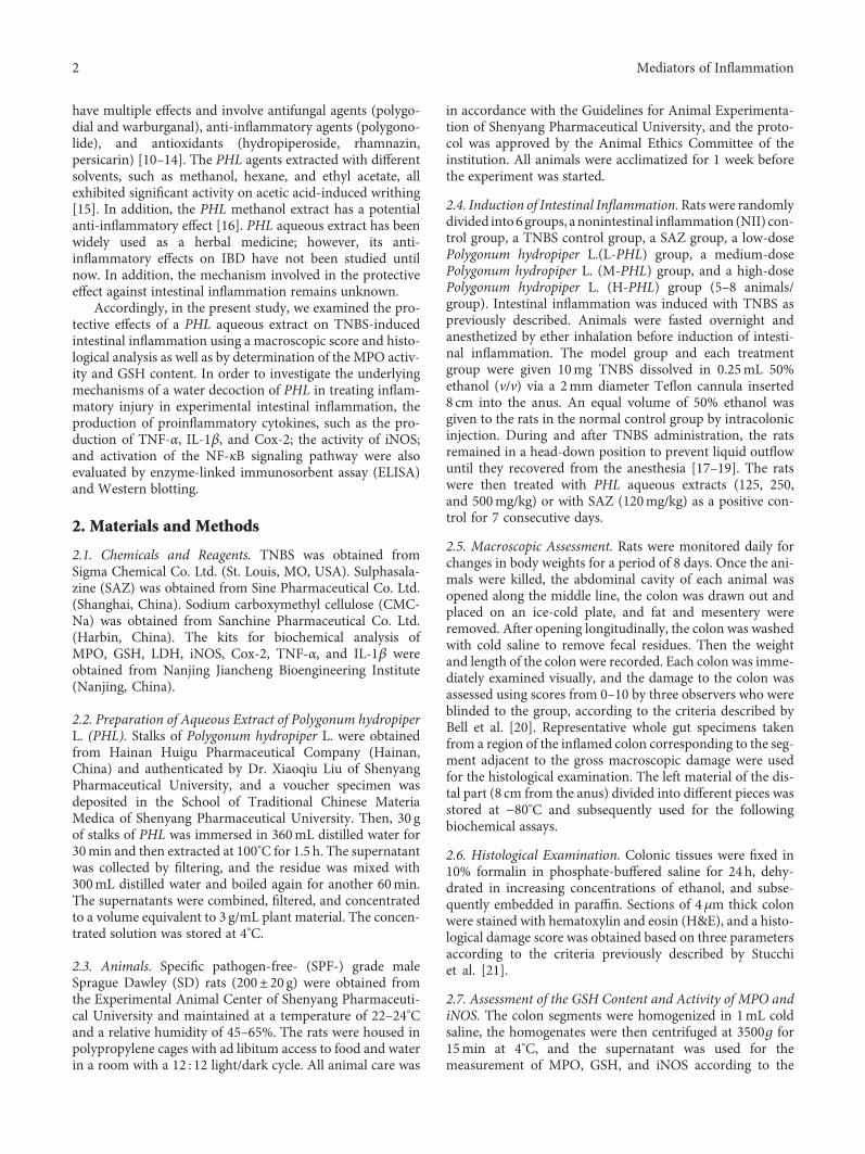
3.1. A Beneficial Effect of PHL on TNBS-Induced MacroscopicAssessment and Weight Loss. Rats treated with TNBS showedprostration, hypomotility, diarrhea, and a significant bodyweight loss. By contrast, a significant reduction in bodyweight loss was observed in rats given H-PHL, on days 6, 7,and 8. Macroscopic inspection of the colon showed evidenceof severe colonic mucosal damage with obvious hyperemia,bowel wall thickening, ulceration, necrosis of the mucosa,and presence of adhesions to adjacent organs. Accordingly,the TNBS control had a significantly shorter colon lengthcompared with the nonintestinal inflammation (NII) control.Rats given oral H-PHL had a significantly extended colonlength compared with the TNBS control. Similarly, H-PHLtreatment significantly combatted severity of the colonicinjury, reducing the macroscopic damage score, and attenu-ated the weight/length ratio of the rat colon (P < 0 01)(Figures 1(a)–1(d)).
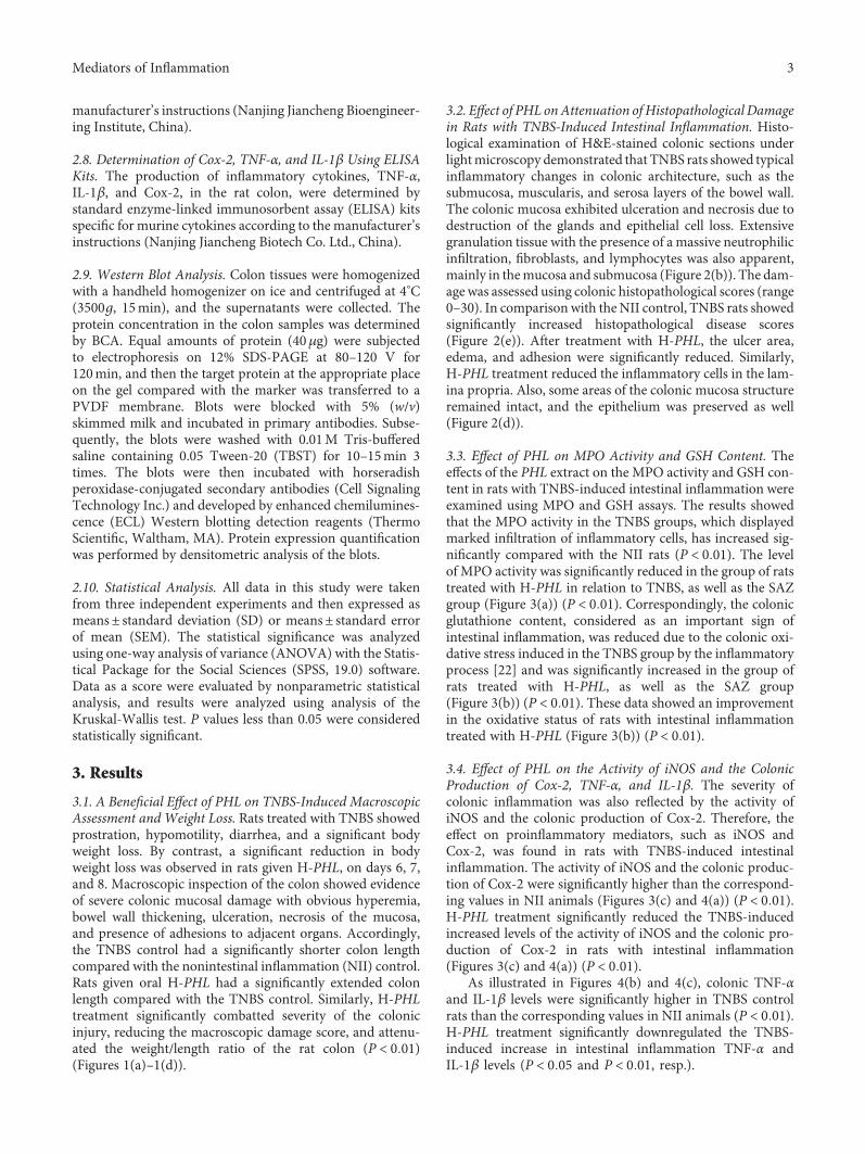
3.2. Effect of PHL onAttenuation of Histopathological Damagein Rats with TNBS-Induced Intestinal Inflammation. Histo-logical examination of H&E-stained colonic sections underlightmicroscopy demonstrated that TNBS rats showed typicalinflammatory changes in colonic architecture, such as thesubmucosa, muscularis, and serosa layers of the bowel wall.The colonic mucosa exhibited ulceration and necrosis due todestruction of the glands and epithelial cell loss. Extensivegranulation tissue with the presence of a massive neutrophilicinfiltration, fibroblasts, and lymphocytes was also apparent,mainly in themucosa and submucosa (Figure 2(b)). The dam-age was assessed using colonic histopathological scores (range0–30). In comparison with theNII control, TNBS rats showedsignificantly increased histopathological disease scores(Figure 2(e)). After treatment with H-PHL, the ulcer area,edema, and adhesion were significantly reduced. Similarly,H-PHL treatment reduced the inflammatory cells in the lam-ina propria. Also, some areas of the colonic mucosa structureremained intact, and the epithelium was preserved as well(Figure 2(d)).
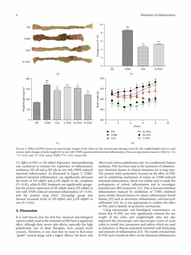
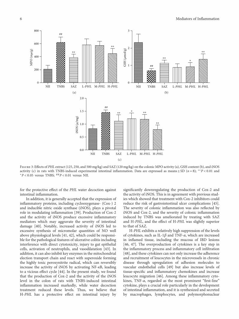
3.3. Effect of PHL on MPO Activity and GSH Content. Theeffects of the PHL extract on the MPO activity and GSH con-tent in rats with TNBS-induced intestinal inflammation wereexamined using MPO and GSH assays. The results showedthat the MPO activity in the TNBS groups, which displayedmarked infiltration of inflammatory cells, has increased sig-nificantly compared with the NII rats (P < 0 01). The levelof MPO activity was significantly reduced in the group of ratstreated with H-PHL in relation to TNBS, as well as the SAZgroup (Figure 3(a)) (P < 0 01). Correspondingly, the colonicglutathione content, considered as an important sign ofintestinal inflammation, was reduced due to the colonic oxi-dative stress induced in the TNBS group by the inflammatoryprocess [22] and was significantly increased in the group ofrats treated with H-PHL, as well as the SAZ group(Figure 3(b)) (P < 0 01). These data showed an improvementin the oxidative status of rats with intestinal inflammationtreated with H-PHL (Figure 3(b)) (P < 0 01).
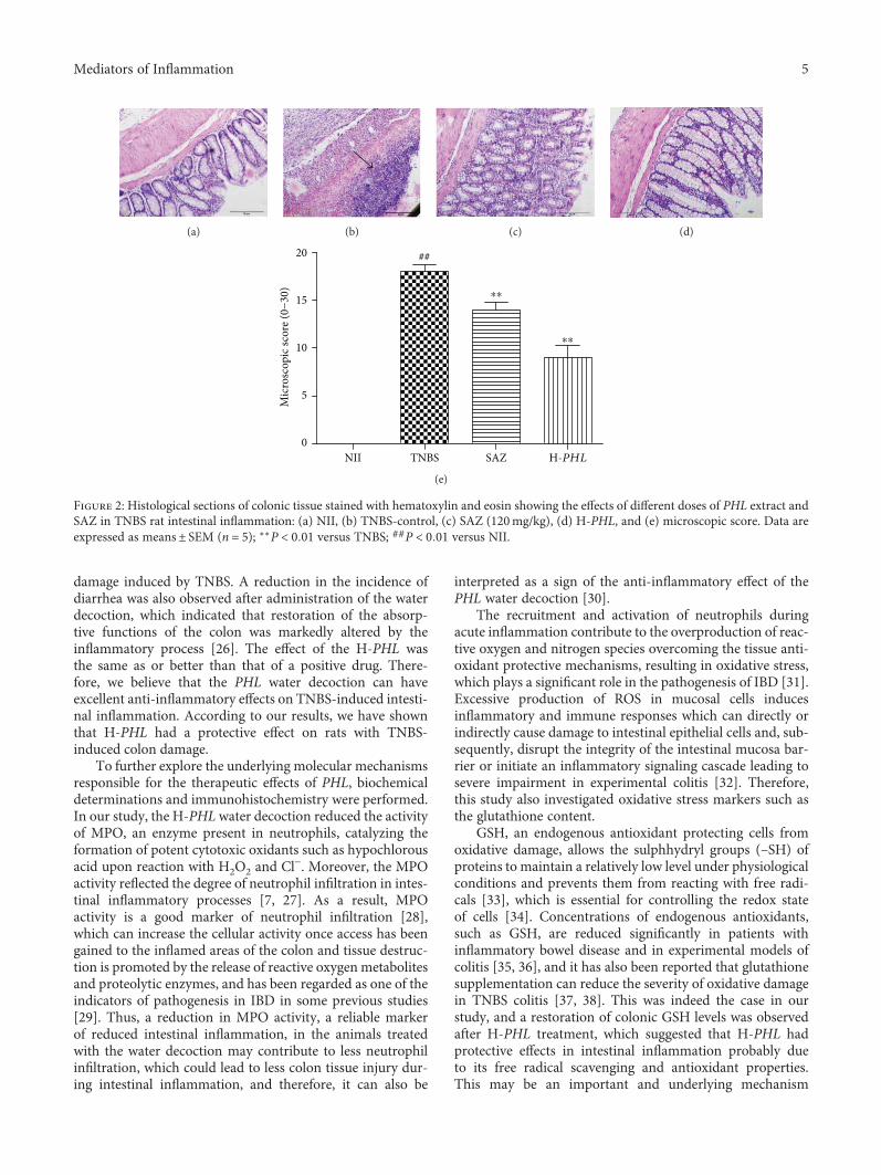
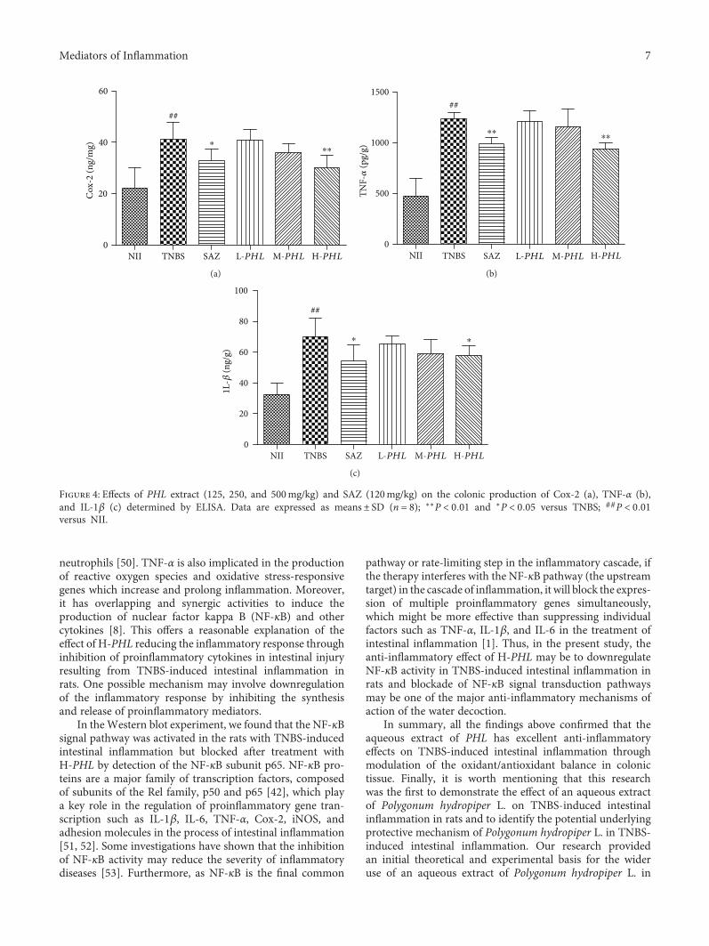
3.4. Effect of PHL on the Activity of iNOS and the ColonicProduction of Cox-2, TNF-α, and IL-1β. The severity ofcolonic inflammation was also reflected by the activity ofiNOS and the colonic production of Cox-2. Therefore, theeffect on proinflammatory mediators, such as iNOS andCox-2, was found in rats with TNBS-induced intestinalinflammation. The activity of iNOS and the colonic produc-tion of Cox-2 were significantly higher than the correspond-ing values in NII animals (Figures 3(c) and 4(a)) (P < 0 01).H-PHL treatment significantly reduced the TNBS-inducedincreased levels of the activity of iNOS and the colonic pro-duction of Cox-2 in rats with intestinal inflammation(Figures 3(c) and 4(a)) (P < 0 01).
As illustrated in Figures 4(b) and 4(c), colonic TNF-αand IL-1β levels were significantly higher in TNBS controlrats than the corresponding values in NII animals (P < 0 01).H-PHL treatment significantly downregulated the TNBS-induced increase in intestinal inflammation TNF-α andIL-1β levels (P < 0 05 and P < 0 01, resp.).
3Mediators of Inflammation
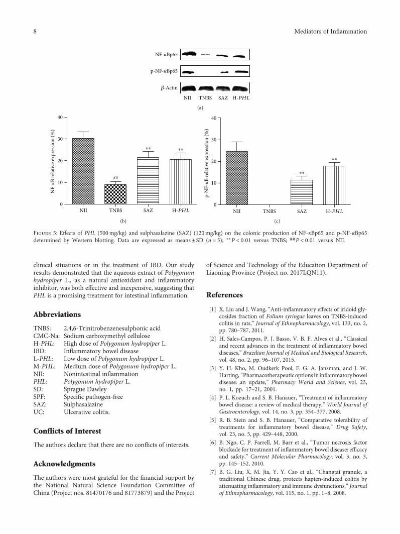
3.5. Effect of PHL on NF-κBp65 Expression. Immunoblottingwas conducted to evaluate the expression of inflammatorymediators, NF-κB and p-NF-κB, in rats with TBNS-inducedintestinal inflammation. As illustrated in Figure 5, TNBS-induced intestinal inflammation can significantly attenuatethe levels of NF-κBp65 and p-NF-κBp65 in the cytoplasm(P < 0 01), while H-PHL treatment can significantly upregu-late the protein expression of NF-κBp65 and p-NF-κBp65 inrats with TNBS-induced intestinal inflammation (P < 0 01),and the positive drug (SAZ 120mg/kg) group alsoshowed increased levels of NF-κBp65 and p-NF-κBp65 inrats (P < 0 01).
4. Discussion
It is well known that the first-line chemical and biologicaloptions widely used in the treatment of IBD have a significanteffect, although their severe side effects, especially the highpalindromic rate of these therapies, have caused muchconcern. Therefore, it was clear that we need to find some“gentle” natural drugs, with a higher efficacy but fewer side
effects and a lower palindromic rate. As a traditional Chinesemedicine, PHL has been used in the treatment of inflamma-tory intestinal disease in clinical situations for a long time.The present study particularly focused on the effect of PHLand its underlying mechanism of action on TNBS-inducedintestinal inflammation, which was widely used to study thepathogenesis of colonic inflammation and to investigatepotential new IBD treatments [23]. This is because intestinalinflammation induced by instillation of TNBS exhibitedmany similar clinical features to colonic inflammatory boweldisease [22] such as ulceration, inflammation, and leucocyteinfiltration [24]. So, it was appropriate to confirm the effectof PHL and to identify its protective mechanism.
Using macroscopic and histological examinations, wefound that H-PHL not only significantly reduced the wetweight of the colon and weight/length ratio but alsoimproved the microscopic and macroscopic scores of acutecolitis in model rats, parameters and indices which are usedas indicators of disease-associated intestinal wall thickeningand intensity of inflammation [25]. The results revealed thatH-PHL had a beneficial effect on the intestinal inflammation
NII
TNBS
SAZ
H-PHL
(a)
10
8
6
4
2
0NII TNBS SAZ H-PHL
Dam
age s
core
(0−
10)
⁎⁎⁎⁎
##
(b)
NII TNBS SAZ L-PHL M-PHL H-PHL
200
150
100
50
0
Wei
ght/l
engt
h (m
g/cm
)
⁎⁎
⁎⁎⁎
##
(c)
0 2 4 6
⁎
8 10(Days)
240
220
200
180
160
Body
wei
ght (
g)
NIITNBSSAZ
H-PHL
M-PHL
L-PHL
(d)
Figure 1: Effects of PHL extract on macroscopic images of the colon (a), the macroscopic damage score (b), the weight/length ratio (c), andcolonic daily changes in body weight (d) in rats with TNBS experimental intestinal inflammation. Data are expressed as means± SEM (n = 5);∗∗P < 0 01 and ∗P < 0 05 versus TNBS; ##P < 0 01 versus NII.
4 Mediators of Inflammation
damage induced by TNBS. A reduction in the incidence ofdiarrhea was also observed after administration of the waterdecoction, which indicated that restoration of the absorp-tive functions of the colon was markedly altered by theinflammatory process [26]. The effect of the H-PHL wasthe same as or better than that of a positive drug. There-fore, we believe that the PHL water decoction can haveexcellent anti-inflammatory effects on TNBS-induced intesti-nal inflammation. According to our results, we have shownthat H-PHL had a protective effect on rats with TNBS-induced colon damage.
To further explore the underlying molecular mechanismsresponsible for the therapeutic effects of PHL, biochemicaldeterminations and immunohistochemistry were performed.In our study, the H-PHL water decoction reduced the activityof MPO, an enzyme present in neutrophils, catalyzing theformation of potent cytotoxic oxidants such as hypochlorousacid upon reaction with H2O2 and Cl−. Moreover, the MPOactivity reflected the degree of neutrophil infiltration in intes-tinal inflammatory processes [7, 27]. As a result, MPOactivity is a good marker of neutrophil infiltration [28],which can increase the cellular activity once access has beengained to the inflamed areas of the colon and tissue destruc-tion is promoted by the release of reactive oxygen metabolitesand proteolytic enzymes, and has been regarded as one of theindicators of pathogenesis in IBD in some previous studies[29]. Thus, a reduction in MPO activity, a reliable markerof reduced intestinal inflammation, in the animals treatedwith the water decoction may contribute to less neutrophilinfiltration, which could lead to less colon tissue injury dur-ing intestinal inflammation, and therefore, it can also be
interpreted as a sign of the anti-inflammatory effect of thePHL water decoction [30].
The recruitment and activation of neutrophils duringacute inflammation contribute to the overproduction of reac-tive oxygen and nitrogen species overcoming the tissue anti-oxidant protective mechanisms, resulting in oxidative stress,which plays a significant role in the pathogenesis of IBD [31].Excessive production of ROS in mucosal cells inducesinflammatory and immune responses which can directly orindirectly cause damage to intestinal epithelial cells and, sub-sequently, disrupt the integrity of the intestinal mucosa bar-rier or initiate an inflammatory signaling cascade leading tosevere impairment in experimental colitis [32]. Therefore,this study also investigated oxidative stress markers such asthe glutathione content.
GSH, an endogenous antioxidant protecting cells fromoxidative damage, allows the sulphhydryl groups (–SH) ofproteins to maintain a relatively low level under physiologicalconditions and prevents them from reacting with free radi-cals [33], which is essential for controlling the redox stateof cells [34]. Concentrations of endogenous antioxidants,such as GSH, are reduced significantly in patients withinflammatory bowel disease and in experimental models ofcolitis [35, 36], and it has also been reported that glutathionesupplementation can reduce the severity of oxidative damagein TNBS colitis [37, 38]. This was indeed the case in ourstudy, and a restoration of colonic GSH levels was observedafter H-PHL treatment, which suggested that H-PHL hadprotective effects in intestinal inflammation probably dueto its free radical scavenging and antioxidant properties.This may be an important and underlying mechanism
(a) (b) (c) (d)
NII TNBS SAZ H-PHL
20
15
10
5
0
Mic
rosc
opic
scor
e (0−
30)
⁎⁎
⁎⁎
##
(e)
Figure 2: Histological sections of colonic tissue stained with hematoxylin and eosin showing the effects of different doses of PHL extract andSAZ in TNBS rat intestinal inflammation: (a) NII, (b) TNBS-control, (c) SAZ (120mg/kg), (d) H-PHL, and (e) microscopic score. Data areexpressed as means± SEM (n = 5); ∗∗P < 0 01 versus TNBS; ##P < 0 01 versus NII.
5Mediators of Inflammation
for the protective effect of the PHL water decoction againstintestinal inflammation.
In addition, it is generally accepted that the expression ofinflammatory proteins, including cyclooxygenase- (Cox-) 2and inducible nitric oxide synthase (iNOS), plays a pivotalrole in modulating inflammation [39]. Production of Cox-2and the activity of iNOS produce excessive inflammatorymediators which may aggravate the severity of intestinaldamage [40]. Notably, increased activity of iNOS led toexcessive synthesis of micromolar quantities of NO wellabove physiological levels [41, 42], which could be responsi-ble for the pathological features of ulcerative colitis includinginterference with direct cytotoxicity, injury to gut epithelialcells, activation of neutrophils, and vasodilatation [43]. Inaddition, it can also inhibit key enzymes in the mitochondrialelection transport chain and react with superoxide formingthe highly toxic peroxynitrite radical, which can reversiblyincrease the activity of iNOS by activating NF-κB, leadingto a vicious effect cycle [44]. In the present study, we foundthat the production of Cox-2 and the activity of the iNOSlevel in the colon of rats with TNBS-induced intestinalinflammation increased markedly, while water decoctiontreatment reduced these levels. Thus, we believe thatH-PHL has a protective effect on intestinal injury by
significantly downregulating the production of Cox-2 andthe activity of iNOS. This is in agreement with previous stud-ies which showed that treatment with Cox-2 inhibitors couldreduce the risk of gastrointestinal ulcer complications [45].The severity of colonic inflammation was also reflected byiNOS and Cox-2, and the severity of colonic inflammationinduced by TNBS was ameliorated by treating with SAZand H-PHL, and the effect of H-PHL was slightly superiorto that of SAZ.
H-PHL exhibits a relatively high suppression of the levelsof cytokines, such as IL-1β and TNF-α, which are increasedin inflamed tissue, including the mucosa of IBD lesions[46, 47]. The overproduction of cytokines is a key step inthe inflammatory process and inflammatory cell infiltration[48], and these cytokines can not only increase the adherenceand recruitment of leucocytes in the microvessels in chronicdisease through upregulation of adhesion molecules tovascular endothelial cells [49] but also increase levels oftissue-specific and inflammatory chemokines and increaseleucocyte migration [46]. Among these inflammatory cyto-kines, TNF-α, regarded as the most prominent “first-line”cytokine, plays a crucial role particularly in the developmentof intestinal inflammation, and it is synthesized and secretedby macrophages, lymphocytes, and polymorphonuclear
##
⁎⁎
⁎⁎
800
600
400
200
0
MPO
(m𝜇
/g)
NII TNBS SAZ L-PHL M-PHL H-PHL
(a)
⁎⁎⁎
⁎⁎
##
3
2
1
0
GSH
(𝜇m
ol/g
)
NII TNBS SAZ L-PHL M-PHL H-PHL
(b)
##⁎⁎
⁎ ⁎⁎
⁎⁎
NII TNBS SAZ L-PHL M-PHL H-PHL
2.0
1.5
1.0
0.5
0.0
iNO
S (𝜇/m
g)
(c)
Figure 3: Effects of PHL extract (125, 250, and 500mg/kg) and SAZ (120mg/kg) on the colonic MPO activity (a), GSH content (b), and iNOSactivity (c) in rats with TNBS-induced experimental intestinal inflammation. Data are expressed as means± SD (n = 8); ∗∗P < 0 01 and∗P < 0 05 versus TNBS; ##P < 0 01 versus NII.
6 Mediators of Inflammation
neutrophils [50]. TNF-α is also implicated in the productionof reactive oxygen species and oxidative stress-responsivegenes which increase and prolong inflammation. Moreover,it has overlapping and synergic activities to induce theproduction of nuclear factor kappa B (NF-κB) and othercytokines [8]. This offers a reasonable explanation of theeffect of H-PHL reducing the inflammatory response throughinhibition of proinflammatory cytokines in intestinal injuryresulting from TNBS-induced intestinal inflammation inrats. One possible mechanism may involve downregulationof the inflammatory response by inhibiting the synthesisand release of proinflammatory mediators.
In theWestern blot experiment, we found that the NF-κBsignal pathway was activated in the rats with TNBS-inducedintestinal inflammation but blocked after treatment withH-PHL by detection of the NF-κB subunit p65. NF-κB pro-teins are a major family of transcription factors, composedof subunits of the Rel family, p50 and p65 [42], which playa key role in the regulation of proinflammatory gene tran-scription such as IL-1β, IL-6, TNF-α, Cox-2, iNOS, andadhesion molecules in the process of intestinal inflammation[51, 52]. Some investigations have shown that the inhibitionof NF-κB activity may reduce the severity of inflammatorydiseases [53]. Furthermore, as NF-κB is the final common
pathway or rate-limiting step in the inflammatory cascade, ifthe therapy interferes with the NF-κB pathway (the upstreamtarget) in the cascade of inflammation, it will block the expres-sion of multiple proinflammatory genes simultaneously,which might be more effective than suppressing individualfactors such as TNF-α, IL-1β, and IL-6 in the treatment ofintestinal inflammation [1]. Thus, in the present study, theanti-inflammatory effect of H-PHL may be to downregulateNF-κB activity in TNBS-induced intestinal inflammation inrats and blockade of NF-κB signal transduction pathwaysmay be one of the major anti-inflammatory mechanisms ofaction of the water decoction.
In summary, all the findings above confirmed that theaqueous extract of PHL has excellent anti-inflammatoryeffects on TNBS-induced intestinal inflammation throughmodulation of the oxidant/antioxidant balance in colonictissue. Finally, it is worth mentioning that this researchwas the first to demonstrate the effect of an aqueous extractof Polygonum hydropiper L. on TNBS-induced intestinalinflammation in rats and to identify the potential underlyingprotective mechanism of Polygonum hydropiper L. in TNBS-induced intestinal inflammation. Our research providedan initial theoretical and experimental basis for the wideruse of an aqueous extract of Polygonum hydropiper L. in
60
40
20
0
Cox
-2 (n
g/m
g)
NII TNBS SAZ L-PHL M-PHL H-PHL
⁎⁎⁎
##
(a)
##
⁎⁎⁎⁎
1500
1000
500
0
TNF-𝛼
(pg/
g)
NII TNBS SAZ L-PHL M-PHL H-PHL
(b)
##
⁎ ⁎
100
80
60
40
20
0
1L-𝛽
(ng/
g)
NII TNBS SAZ L-PHL M-PHL H-PHL
(c)
Figure 4: Effects of PHL extract (125, 250, and 500mg/kg) and SAZ (120mg/kg) on the colonic production of Cox-2 (a), TNF-α (b),and IL-1β (c) determined by ELISA. Data are expressed as means± SD (n = 8); ∗∗P < 0 01 and ∗P < 0 05 versus TNBS; ##P < 0 01versus NII.
7Mediators of Inflammation
clinical situations or in the treatment of IBD. Our studyresults demonstrated that the aqueous extract of Polygonumhydropiper L., as a natural antioxidant and inflammatoryinhibitor, was both effective and inexpensive, suggesting thatPHL is a promising treatment for intestinal inflammation.
Abbreviations
TNBS: 2,4,6-Trinitrobenzenesulphonic acidCMC-Na: Sodium carboxymethyl celluloseH-PHL: High dose of Polygonum hydropiper L.IBD: Inflammatory bowel diseaseL-PHL: Low dose of Polygonum hydropiper L.M-PHL: Medium dose of Polygonum hydropiper L.NII: Nonintestinal inflammationPHL: Polygonum hydropiper L.SD: Sprague DawleySPF: Specific pathogen-freeSAZ: SulphasalazineUC: Ulcerative colitis.
Conflicts of Interest
The authors declare that there are no conflicts of interests.
Acknowledgments
The authors were most grateful for the financial support bythe National Natural Science Foundation Committee ofChina (Project nos. 81470176 and 81773879) and the Project
of Science and Technology of the Education Department ofLiaoning Province (Project no. 2017LQN11).
References
[1] X. Liu and J. Wang, “Anti-inflammatory effects of iridoid gly-cosides fraction of Folium syringae leaves on TNBS-inducedcolitis in rats,” Journal of Ethnopharmacology, vol. 133, no. 2,pp. 780–787, 2011.
[2] H. Sales-Campos, P. J. Basso, V. B. F. Alves et al., “Classicaland recent advances in the treatment of inflammatory boweldiseases,” Brazilian Journal of Medical and Biological Research,vol. 48, no. 2, pp. 96–107, 2015.
[3] Y. H. Kho, M. Oudkerk Pool, F. G. A. Jansman, and J. W.Harting, “Pharmacotherapeutic options in inflammatoryboweldisease: an update,” Pharmacy World and Science, vol. 23,no. 1, pp. 17–21, 2001.
[4] P. L. Kozuch and S. B. Hanauer, “Treatment of inflammatorybowel disease: a review of medical therapy,” World Journal ofGastroenterology, vol. 14, no. 3, pp. 354–377, 2008.
[5] R. B. Stein and S. B. Hanauer, “Comparative tolerability oftreatments for inflammatory bowel disease,” Drug Safety,vol. 23, no. 5, pp. 429–448, 2000.
[6] B. Ngo, C. P. Farrell, M. Barr et al., “Tumor necrosis factorblockade for treatment of inflammatory bowel disease: efficacyand safety,” Current Molecular Pharmacology, vol. 3, no. 3,pp. 145–152, 2010.
[7] B. G. Liu, X. M. Jia, Y. Y. Cao et al., “Changtai granule, atraditional Chinese drug, protects hapten-induced colitis byattenuating inflammatory and immune dysfunctions,” Journalof Ethnopharmacology, vol. 115, no. 1, pp. 1–8, 2008.
NF-𝜅Bp65
p-NF-𝜅Bp65
𝛽-Actin
NII TNBS SAZ H-PHL
(a)
⁎⁎⁎⁎
##
40
30
20
10
0NII TNBS SAZ H-PHL
NF-𝜅
B re
lativ
e exp
ress
ion
(%)
(b)
⁎⁎
⁎⁎
NII TNBS SAZ H-PHL
40
30
20
10
0p-
NF-𝜅
B re
lativ
e exp
ress
ion
(%)
(c)
Figure 5: Effects of PHL (500mg/kg) and sulphasalazine (SAZ) (120mg/kg) on the colonic production of NF-κBp65 and p-NF-κBp65determined by Western blotting. Data are expressed as means± SD (n = 5); ∗∗P < 0 01 versus TNBS; ##P < 0 01 versus NII.
8 Mediators of Inflammation
[8] Y.-H. Zhou, J.-P. Yu, Y.-F. Liu et al., “Effects of Ginkgo bilobaextract on inflammatory mediators (SOD, MDA, TNF-α,NF-κBp65, IL-6) in TNBS-induced colitis in rats,” Mediatorsof Inflammation, vol. 2006, Article ID 92642, 9 pages, 2006.
[9] A. B. A. de Almeida, M. Sánchez-Hidalgo, A. R. Martín et al.,“Anti-inflammatory intestinal activity of Arctium lappa L.(Asteraceae) in TNBS colitis model,” Journal of Ethnopharma-cology, vol. 146, no. 1, pp. 300–310, 2013.
[10] S. V. Ley, “Synthesis of antifeedants for insects: novelbehaviour-modifying chemicals from plants,” Ciba Founda-tion Symposium, vol. 154, pp. 80–87, 1990.
[11] J.-C. Park, Y.-B. Yu, J.-H. Lee, M. Hattori, C.-K. Lee, andJ.-W. Choi, “Protective effect of Oenanthe javanica on thehepatic lipid peroxidation in bromobenzene-treated rats andits bioactive component,” Planta Medica, vol. 62, no. 6,pp. 488–490, 1996.
[12] K. Takeda and S. Akira, “Roles of Toll-like receptors in innateimmune responses,” Genes to Cells, vol. 6, no. 9, pp. 733–742,2001.
[13] Z. F. Peng, D. Strack, A. Baumert et al., “Antioxidant flavonoidsfrom leaves of Polygonum hydropiper L,” Phytochemistry,vol. 62, no. 2, pp. 219–228, 2003.
[14] P. V. Kiem, N. X. Nhiem, N. X. Cuong et al., “New phenylpro-panoid esters of sucrose from Polygonum hydropiper and theirantioxidant activity,” Archives of Pharmacal Research, vol. 31,no. 11, pp. 1477–1482, 2008.
[15] E. Rahman, S. A. Goni, M. T. Rahman, and M. Ahmed, “Anti-nociceptive activity of Polygonum hydropiper,” Fitoterapia,vol. 73, no. 7-8, pp. 704–706, 2002.
[16] Y. Yang, T. Yu, H. J. Jang et al., “In vitro and in vivo anti-inflammatory activities of Polygonum hydropiper methanolextract,” Journal of Ethnopharmacology, vol. 139, no. 2,pp. 616–625, 2012.
[17] M. S. da Silva, S. Sánchez-Fidalgo, E. Talero et al., “Anti-inflammatory intestinal activity of Abarema cochliacarpos(Gomes) Barneby & Grimes in TNBS colitis model,” Journalof Ethnopharmacology, vol. 128, no. 2, pp. 467–475, 2010.
[18] J. Bilski, A. I. Mazur-Bialy, B. Brzozowski et al., “Moderateexercise training attenuates the severity of experimental rodentcolitis: the importance of crosstalk between adipose tissueand skeletal muscles,” Mediators of Inflammation, vol. 2015,Article ID 605071, 12 pages, 2015.
[19] K. Siczek, H. Zatorski, A. Chmielowiec-Korzeniowska,R. Kordek, L. Tymczyna, and J. Fichna, “Evaluation of anti-inflammatory effect of silver-coated glass beads in mice withexperimentally induced colitis as a new type of treatment ininflammatory bowel disease,” Pharmacological Reports,vol. 69, no. 3, pp. 386–392, 2017.
[20] C. J. Bell, D. G. Gall, and J. L. Wallace, “Disruption of colonicelectrolyte transport in experimental colitis,”American Journalof Physiology-Gastrointestinal and Liver Physiology, vol. 268,no. 4, pp. G622–G630, 1995.
[21] A. F. Stucchi, S. Shofer, S. Leeman et al., “NK-1 antagonistreduces colonic inflammation and oxidative stress in dex-tran sulfate-induced colitis in rats,” American Journal ofPhysiology-Gastrointestinal and Liver Physiology, vol. 279,no. 6, pp. G1298–G1306, 2000.
[22] G. P. Morris, P. L. Beck, M. S. Herridge, W. T. Depew, M. R.Szewczuk, and J. L. Wallace, “Hapten-induced model ofchronic inflammation and ulceration in the rat colon,” Gastro-enterology, vol. 96, no. 2, pp. 795–803, 1989.
[23] S. Wirtz and M. F. Neurath, “Animal models of intestinalinflammation: new insights into the molecular pathogenesisand immunotherapy of inflammatory bowel disease,” Interna-tional Journal of Colorectal Disease, vol. 15, no. 3, pp. 144–160,2000.
[24] T. V. Brito, J. P. R. P. Neto, R. S. Prudêncio et al., “Sulfated-polysaccharide fraction extracted from red algae Gracilariabirdiae ameliorates trinitrobenzenesulfonic acid-inducedcolitis in rats,” Journal of Pharmacy and Pharmacology,vol. 66, no. 8, pp. 1161–1170, 2014.
[25] J. Y. Lee, H. S. Kang, B. E. Park, H. J. Moon, S. S. Sim, and C. J.Kim, “Inhibitory effects of Geijigajakyak-Tang on trinitroben-zene sulfonic acid-induced colitis,” Journal of Ethnopharma-cology, vol. 126, no. 2, pp. 244–251, 2009.
[26] F. Sánchez de Medina, B. Vera, J. Gálvez, and A. Zarzuelo,“Effect of quercitrin on the early stages of hapten inducedcolonic inflammation in the rat,” Life Sciences, vol. 70,no. 26, pp. 3097–3108, 2002.
[27] M. S. Islam, T. Murata, M. Fujisawa et al., “Anti-inflammatoryeffects of phytosteryl ferulates in colitis induced by dextransulphate sodium in mice,” British Journal of Pharmacology,vol. 154, no. 4, pp. 812–824, 2008.
[28] I. Villegas, C. la Casa, A. Orjales, and C. Alarcón de la Lastra,“Effects of dosmalfate, a new cytoprotective agent, on acuteand chronic trinitrobenzene sulphonic acid-induced colitis inrats,” European Journal of Pharmacology, vol. 460, no. 2-3,pp. 209–218, 2003.
[29] F. Algieri, P. Zorrilla, A. Rodriguez-Nogales et al., “Intestinalanti-inflammatory activity of hydroalcoholic extracts ofPhlomis purpurea L. and Phlomis lychnitis L. in the trinitro-benzenesulphonic acid model of rat colitis,” Journal of Ethno-pharmacology, vol. 146, no. 3, pp. 750–759, 2013.
[30] M. Veljaca, C. A. Lesch, R. Pllana, B. Sanchez, K. Chan, andA. Guglietta, “BPC-15 reduces trinitrobenzene sulfonic acid-induced colonic damage in rats,” Journal of Pharmacologyand Experimental Therapeutics, vol. 272, no. 1, pp. 417–422,1995.
[31] E. Talero, S. Sánchez-Fidalgo, C. A. de la Lastra, M. Illanes,J. R. Calvo, and V. Motilva, “Acute and chronic responsesassociated with adrenomedullin administration in experimen-tal colitis,” Peptides, vol. 29, no. 11, pp. 2001–2012, 2008.
[32] E. B. Kurutas, A. Cetinkaya, E. Bulbuloglu, and B. Kantarceken,“Effects of antioxidant therapy on leukocyte myeloperoxidaseand Cu/Zn-superoxide dismutase and plasma malondialde-hyde levels in experimental colitis,”Mediators of Inflammation,vol. 2005, no. 6, 394 pages, 2005.
[33] K. Amirshahrokhi, S. Bohlooli, and M. M. Chinifroush, “Theeffect of methylsulfonylmethane on the experimental colitisin the rat,” Toxicology and Applied Pharmacology, vol. 253,no. 3, pp. 197–202, 2011.
[34] H. Sies, “Glutathione and its role in cellular functions,” FreeRadical Biology and Medicine, vol. 27, no. 9-10, pp. 916–921,1999.
[35] G. Tahan, R. Gramignoli, F. Marongiu et al., “Melatoninexpresses powerful anti-inflammatory and antioxidant activi-ties resulting in complete improvement of acetic-acid-inducedcolitis in rats,” Digestive Diseases and Sciences, vol. 56, no. 3,pp. 715–720, 2011.
[36] D. R. Spitz, E. I. Azzam, J. Jian Li, and D. Gius, “Metabolic oxi-dation/reduction reactions and cellular responses to ionizingradiation: a unifying concept in stress response biology,” Can-cer and Metastasis Reviews, vol. 23, no. 3/4, pp. 311–322, 2004.
9Mediators of Inflammation
[37] C. Loguercio, G. D’Argenio, M. Delle Cave et al., “Glutathionesupplementation improves oxidative damage in experimentalcolitis,” Digestive and Liver Disease, vol. 35, no. 9, pp. 635–641, 2003.
[38] D. Camuesco, L. Peran, M. Comalada et al., “Preventativeeffects of lactulose in the trinitrobenzenesulphonic acid modelof rat colitis,” Inflammatory Bowel Diseases, vol. 11, no. 3,pp. 265–271, 2005.
[39] S. Ardizzone and G. Bianchi Porro, “Biologic therapy forinflammatory bowel disease,” Drugs, vol. 65, no. 16,pp. 2253–2286, 2005.
[40] S. H. Itzkowitz, “Molecular biology of dysplasia and cancer ininflammatory bowel disease,” Gastroenterology Clinics ofNorth America, vol. 35, no. 3, pp. 553–571, 2006.
[41] H. H. Hagar, A. el Medany, E. el Eter, and M. Arafa, “Amelio-rative effect of pyrrolidinedithiocarbamate on acetic acid-induced colitis in rats,” European Journal of Pharmacology,vol. 554, no. 1, pp. 69–77, 2007.
[42] K. M. Sakthivel and C. Guruvayoorappan, “Amentoflavoneinhibits iNOS, COX-2 expression andmodulates cytokine pro-file, NF-κB signal transduction pathways in rats with ulcerativecolitis,” International Immunopharmacology, vol. 17, no. 3,pp. 907–916, 2013.
[43] S. J. McKenzie, M. S. Baker, G. D. Buffinton, and W. F. Doe,“Evidence of oxidant-induced injury to epithelial cells duringinflammatory bowel disease,” Journal of Clinical Investigation,vol. 98, no. 1, pp. 136–141, 1996.
[44] C.-L. M. Cooke and S. T. Davidge, “Peroxynitrite increasesiNOS through NF-κB and decreases prostacyclin synthase inendothelial cells,” American Journal of Physiology-Cell Physiol-ogy, vol. 282, no. 2, pp. C395–C402, 2002.
[45] J. L. Goldstein, F. E. Silverstein, N. M. Agrawal et al., “Reducedrisk of upper gastrointestinal ulcer complications with cele-coxib, a novel COX-2 inhibitor,” The American Journal ofGastroenterology, vol. 95, no. 7, pp. 1681–1690, 2000.
[46] C. Fiocchi, “Lymphokines and the intestinal immune response.Role in inflammatory bowel disease,” Immunological Investi-gations, vol. 18, no. 1–4, pp. 91–102, 1989.
[47] O. A. Hatoum, J. Heidemann, and D. G. Binion, “The intesti-nal microvasculature as a therapeutic target in inflammatorybowel disease,” Annals of the New York Academy of Sciences,vol. 1072, no. 1, pp. 78–97, 2006.
[48] J. A. Katz, J. Itoh, and C. Fiocchi, “Pathogenesis of inflamma-tory bowel disease,” Current Opinion in Gastroenterology,vol. 15, no. 4, pp. 291–297, 1999.
[49] J. R. Mora and U. H. von Andrian, “T-cell homing specificityand plasticity: new concepts and future challenges,” Trendsin Immunology, vol. 27, no. 5, pp. 235–243, 2006.
[50] M. F. Neurath, I. Fuss, M. Pasparakis et al., “Predominantpathogenic role of tumor necrosis factor in experimental colitisin mice,” European Journal of Immunology, vol. 27, no. 7,pp. 1743–1750, 1997.
[51] D. K. Zhang, L. N. Cheng, X. L. Huang, W. Shi, J. Y. Xiang, andH. T. Gan, “Tetrandrine ameliorates dextran-sulfate-sodium-induced colitis in mice through inhibition of nuclear factor-κB activation,” International Journal of Colorectal Disease,vol. 24, no. 1, pp. 5–12, 2009.
[52] J. K. Kundu and Y. J. Surh, “Molecular basis of chemopreven-tion by resveratrol: NF-κB and AP-1 as potential targets,”Mutation Research/Fundamental and Molecular Mechanismsof Mutagenesis, vol. 555, no. 1-2, pp. 65–80, 2004.
[53] I. Atreya, R. Atreya, and M. F. Neurath, “NF-κB in inflamma-tory bowel disease,” Journal of Internal Medicine, vol. 263,no. 6, pp. 591–596, 2008.
10 Mediators of Inflammation

Stem Cells International
Hindawiwww.hindawi.com Volume 2018
Hindawiwww.hindawi.com Volume 2018
MEDIATORSINFLAMMATION
of
EndocrinologyInternational Journal of
Hindawiwww.hindawi.com Volume 2018
Hindawiwww.hindawi.com Volume 2018
Disease Markers
Hindawiwww.hindawi.com Volume 2018
BioMed Research International
OncologyJournal of
Hindawiwww.hindawi.com Volume 2013
Hindawiwww.hindawi.com Volume 2018
Oxidative Medicine and Cellular Longevity
Hindawiwww.hindawi.com Volume 2018
PPAR Research
Hindawi Publishing Corporation http://www.hindawi.com Volume 2013Hindawiwww.hindawi.com
The Scientific World Journal
Volume 2018
Immunology ResearchHindawiwww.hindawi.com Volume 2018
Journal of
ObesityJournal of
Hindawiwww.hindawi.com Volume 2018
Hindawiwww.hindawi.com Volume 2018
Computational and Mathematical Methods in Medicine
Hindawiwww.hindawi.com Volume 2018
Behavioural Neurology
OphthalmologyJournal of
Hindawiwww.hindawi.com Volume 2018
Diabetes ResearchJournal of
Hindawiwww.hindawi.com Volume 2018
Hindawiwww.hindawi.com Volume 2018
Research and TreatmentAIDS
Hindawiwww.hindawi.com Volume 2018
Gastroenterology Research and Practice
Hindawiwww.hindawi.com Volume 2018
Parkinson’s Disease
Evidence-Based Complementary andAlternative Medicine
Volume 2018Hindawiwww.hindawi.com
Submit your manuscripts atwww.hindawi.com


![Mycobacteria-specific CD4IFN- cell expresses naïve-surface … · SCM) [15, 16]. These cells have been detected in BCG vaccinated infected subjects [17]. We had previously identified](https://static.fdocument.org/doc/165x107/5fa54c277baf7c74b671181f/mycobacteria-specific-cd4ifn-cell-expresses-nave-surface-scm-15-16-these.jpg)