Recent topics in toxinology 2019 · pathway, among them factor VIIIa (FVIIIa), have been recognized...
65
BOOK OF ABSTRACTS Recent topics in toxinology 7. 6. 2019 Ljubljana τοξικόν
Transcript of Recent topics in toxinology 2019 · pathway, among them factor VIIIa (FVIIIa), have been recognized...
BOOK OF ABSTRACTS
Recent topics in toxinology
7. 6. 2019Ljubljana
τοξικόν
Recent topics in toxinology
τοξικόν
[gr. toxicon] "poison for use on arrows"
7. 6. 2019
Ljubljana
2
RECENT TOPICS IN TOXINOLOGY 2019
Organised by
Department of Molecular Biology and Nanobiotechnology, National Institute of Chemistry
Chair of Biochemistry, Department of Biology, Biotechnical Faculty, University of
Ljubljana
Programme Committee
Gregor
Organising committee
Editors
Gregor Anderluh, Katja Pirc,
Technical editors
Gregor Anderluh, Matic Kisovec, Katja Pirc
Credits for presentation of toxic organisms to
Gregor Anderluh, , Mojca Mally, Katja Pirc, , Tom Turk for text
and , Denis Kunkel, Pixabay, Tom Turk, Bruce Watt (University of Maine) for
images
Issued by
Department of Molecular Biology and Nanobiotechnology, National Institute of Chemistry
@NanoSciNIC @kemijski http://bit.do/toxin2019
Printed by
Infokart d.o.o., Ljubljana
Circulation
75 issues
Complimentary publication
CIP -
615.9(082)
RECENT Topics in Toxinology (simpozij) (2019 ; Ljubljana)
Recent topics in toxinology : 7. 6. 2019 Ljubljana / [Recent topics in toxinology 2019 ; organized by Department of
Molecular Biology and Nanobiotechnology, National Institute of Chemistry [and] Chair of Biochemistry, Department
of Biology, Biotechnical Faculty, University of Ljubljana ; editors Gregor Anderluh, Katja Pirc -
Ljubljana : Department of Molecular Biology and Nanobiotechnology, National Institute of Chemistry, 2019
ISBN 978-961-6104-44-9
1. Gl. stv. nasl. 2. Anderluh, Gregor, 1969-
COBISS.SI-ID 300251904
3
Welcome
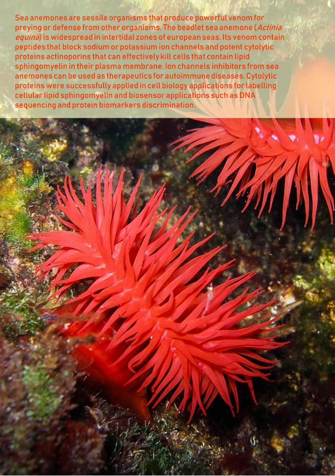
Venoms are a fascinating and rich source of molecules with interesting properties
and mechanisms of action. The aim of this symposium is to bring an overview of
current efforts on various toxinological topics, spanning from the evolution and
structure of toxin molecules, through their physiological effects, to their use in
medical and biotechnological applications. Renowned scientists working in the field
of natural toxins from Slovenia and abroad will present these exciting topics. This
University of Ljubljana, who is one of the doyens of toxinology in Slovenia, and
whose research interest was oriented mainly towards membrane-active proteins from
Cnidaria.
Historically, modern toxinology in Slovenia was initiated in fifties by a biochemist
Prof. Drago Lebez from Ljubljana in collaboration with a Croatian physician, Dr.
nt publication was a monography,
his younger collaborators started biochemical characterization of some snake
venoms, in particular Vipera ammodytes, and cnidarian neurotoxins and cytolytic
toxins. So, it is allowed to say that research of venoms and toxins here in Ljubljana
is rather traditional and continuous for more than sixty years.
We are particularly happy that many young researchers will attend the symposium.
We hope that a full day of interesting presentation and stimulating discussions will
raise an interest in various toxinological topics in young generation of scientists to
continue with excellent stories of toxic molecules that will lead to basic knowledge,
as well as useful applications in medicine and biotechnology.
Prof. dr. Gregor Anderluh, P
4
5
Contents
Welcome 3
Sponsors 7
Programme 9
Abstracts - Invited Lectures 13
Abstracts - Oral Presentations 29
Abstracts - Posters 39
List of Participants 55
Author Index 61
6

7
Sponsors
Thank you for your support!
8
9
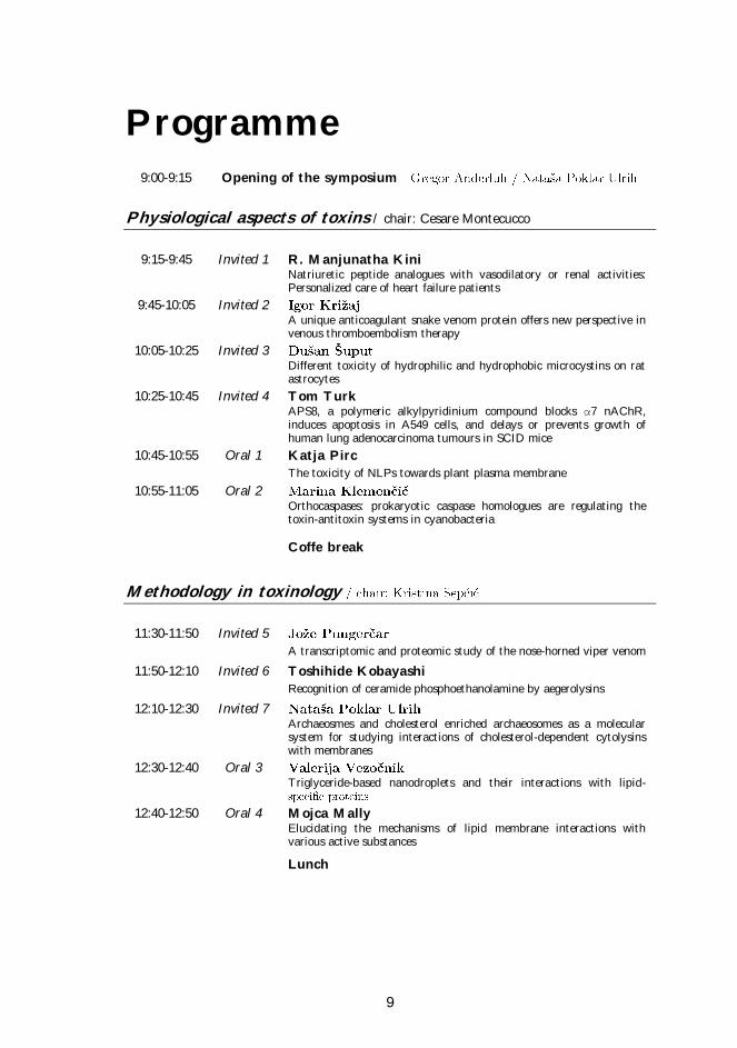
Programme
9:00-9:15 Opening of the symposium
Physiological aspects of toxins / chair: Cesare Montecucco
9:15-9:45 Invited 1 R. Manjunatha Kini
Natriuretic peptide analogues with vasodilatory or renal activities: Personalized care of heart failure patients
9:45-10:05 Invited 2
A unique anticoagulant snake venom protein offers new perspective in venous thromboembolism therapy
10:05-10:25 Invited 3
Different toxicity of hydrophilic and hydrophobic microcystins on rat astrocytes
10:25-10:45 Invited 4 Tom Turk
APS8, a polymeric alkylpyridinium compound blocks α7 nAChR, induces apoptosis in A549 cells, and delays or prevents growth of human lung adenocarcinoma tumours in SCID mice
10:45-10:55 Oral 1 Katja Pirc
The toxicity of NLPs towards plant plasma membrane
10:55-11:05 Oral 2
Orthocaspases: prokaryotic caspase homologues are regulating the toxin-antitoxin systems in cyanobacteria
Coffe break
Methodology in toxinology
11:30-11:50 Invited 5
A transcriptomic and proteomic study of the nose-horned viper venom
11:50-12:10 Invited 6 Toshihide Kobayashi
Recognition of ceramide phosphoethanolamine by aegerolysins
12:10-12:30 Invited 7
Archaeosmes and cholesterol enriched archaeosomes as a molecular system for studying interactions of cholesterol-dependent cytolysins with membranes
12:30-12:40 Oral 3
Triglyceride-based nanodroplets and their interactions with lipid-
12:40-12:50 Oral 4 Mojca Mally
Elucidating the mechanisms of lipid membrane interactions with various active substances
Lunch
10
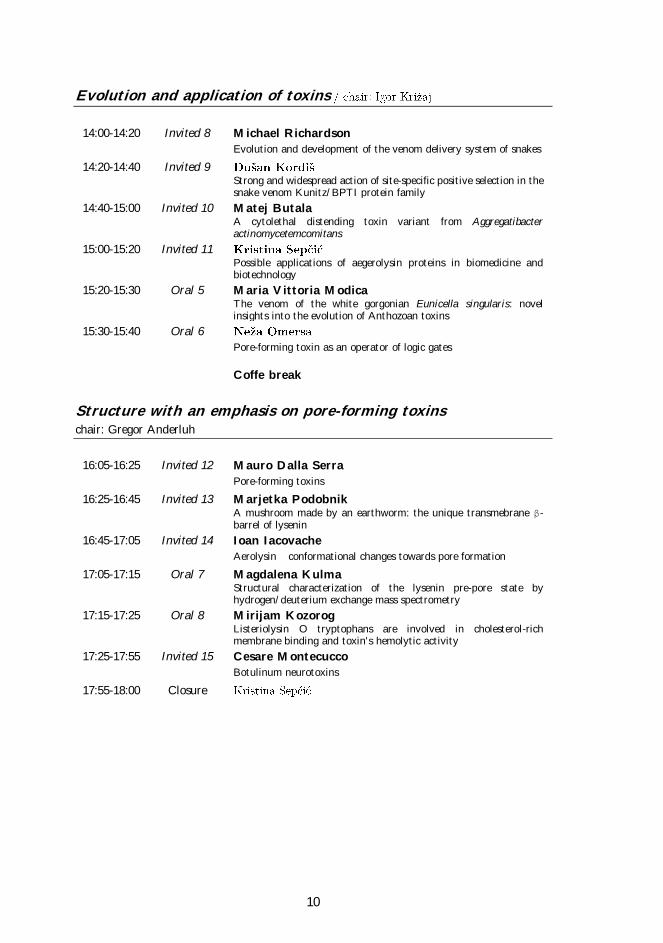
Evolution and application of toxins
14:00-14:20 Invited 8 Michael Richardson
Evolution and development of the venom delivery system of snakes
14:20-14:40 Invited 9
Strong and widespread action of site-specific positive selection in the snake venom Kunitz/BPTI protein family
14:40-15:00 Invited 10 Matej Butala
A cytolethal distending toxin variant from Aggregatibacter actinomycetemcomitans
15:00-15:20 Invited 11
Possible applications of aegerolysin proteins in biomedicine and biotechnology
15:20-15:30 Oral 5 Maria Vittoria Modica
The venom of the white gorgonian Eunicella singularis: novel insights into the evolution of Anthozoan toxins
15:30-15:40 Oral 6
Pore-forming toxin as an operator of logic gates
Coffe break
Structure with an emphasis on pore-forming toxins chair: Gregor Anderluh
16:05-16:25 Invited 12 Mauro Dalla Serra
Pore-forming toxins
16:25-16:45 Invited 13 Marjetka Podobnik
A mushroom made by an earthworm: the unique transmebrane β-barrel of lysenin
16:45-17:05 Invited 14 Ioan Iacovache
Aerolysin conformational changes towards pore formation
17:05-17:15 Oral 7 Magdalena Kulma
Structural characterization of the lysenin pre-pore state by hydrogen/deuterium exchange mass spectrometry
17:15-17:25 Oral 8 Mirijam Kozorog
Listeriolysin O tryptophans are involved in cholesterol-rich membrane binding and toxin's hemolytic activity
17:25-17:55 Invited 15 Cesare Montecucco
Botulinum neurotoxins
17:55-18:00 Closure
11
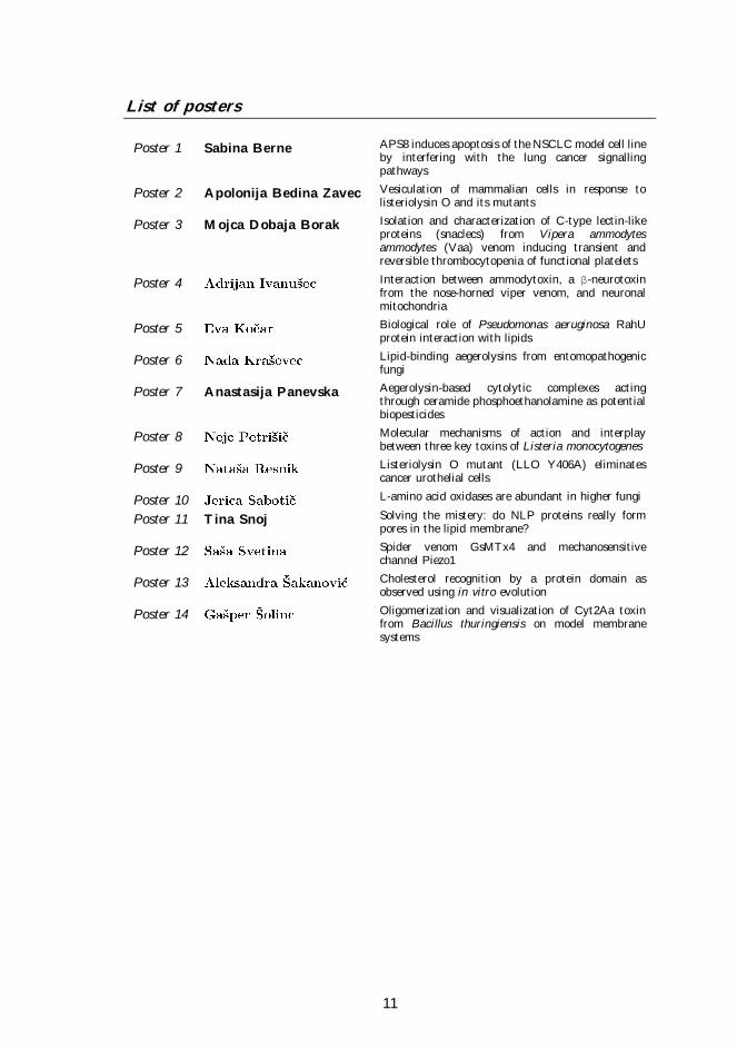
List of posters
Poster 1 Sabina Berne APS8 induces apoptosis of the NSCLC model cell line by interfering with the lung cancer signalling pathways
Poster 2 Apolonija Bedina Zavec Vesiculation of mammalian cells in response to listeriolysin O and its mutants
Poster 3 Mojca Dobaja Borak Isolation and characterization of C-type lectin-like proteins (snaclecs) from Vipera ammodytes ammodytes (Vaa) venom inducing transient and reversible thrombocytopenia of functional platelets
Poster 4 Interaction between ammodytoxin, a β-neurotoxin from the nose-horned viper venom, and neuronal mitochondria
Poster 5 Biological role of Pseudomonas aeruginosa RahU protein interaction with lipids
Poster 6 Lipid-binding aegerolysins from entomopathogenic fungi
Poster 7 Anastasija Panevska Aegerolysin-based cytolytic complexes acting through ceramide phosphoethanolamine as potential biopesticides
Poster 8 Molecular mechanisms of action and interplay between three key toxins of Listeria monocytogenes
Poster 9 Listeriolysin O mutant (LLO Y406A) eliminates cancer urothelial cells
Poster 10 L-amino acid oxidases are abundant in higher fungi
Poster 11 Tina Snoj Solving the mistery: do NLP proteins really form pores in the lipid membrane?
Poster 12 Spider venom GsMTx4 and mechanosensitive channel Piezo1
Poster 13 Cholesterol recognition by a protein domain as observed using in vitro evolution
Poster 14 Oligomerization and visualization of Cyt2Aa toxin from Bacillus thuringiensis on model membrane systems

13
Abstracts - Invited Lectures
9:15 / Invited 1
14
Natriuretic peptide analogues with vasodilatory or renal
activities: Personalized care of heart failure patients
R. Manjunatha Kini
Department of Biological Sciences, Faculty of Science, National University of Singapore (Singapore)
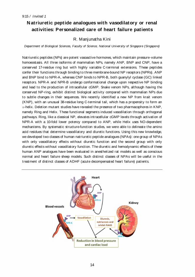
Natriuretic peptides (NPs) are potent vasoactive hormones, which maintain pressure volume
homoeostasis. All three isoforms of mammalian NPs, namely ANP, BNP and CNP, have a
conserved 17-residue ring but with highly variable C-terminal extensions. These peptides
confer their functions through binding to three membrane-bound NP receptors (NPRs). ANP
and BNP bind to NPR-A, whereas CNP binds to NPR-B, both guanylyl cyclase (GC) linked
receptors. NPR-A and NPR-B undergo conformational change upon respective NP binding
and lead to the production of intracellular cGMP. Snake venom NPs, although having the
conserved NP-ring, exhibit distinct biological activity compared with mammalian NPs due
to subtle changes in their sequences. We recently identified a new NP from krait venom
(KNP), with an unusual 38-residue long C-terminal tail, which has a propensity to form an
α-helix. Deletion mutant studies have revealed the presence of two pharmacophores in KNP,
namely Ring and Helix. These functional segments induced vasodilation through orthogonal
pathways. Ring, like a classical NP, elevates intracellular cGMP levels through activation of
NPR-A with a 10-fold lower potency compared to ANP, while Helix uses NO-dependent
mechanisms. By systematic structure-function studies, we were able to delineate the amino
acid residues that determine vasodilatory and diuretic functions. Using this new knowledge,
we developed two classes of human natriuretic peptide analogues (NPAs): one group of NPAs
with only vasodilatory effects without diuretic function and the second group with only
diuretic effects without vasodilatory function. The diuretic and hemodynamic effects of these
human ANP analogues have been evaluated in anesthetized rat models as well as conscious
normal and heart failure sheep models. Such distinct classes of NPAs will be useful in the
treatment of distinct classes of ADHF (acute decompensated heart failure) patients.
9:45 / Invited 2
15
A unique anticoagulant snake venom protein offers new
perspective in venous thromboembolism therapy
(Slovenia)
Venous thromboembolism (VTE) is the pathological process behind two very serious
cardiovascular diseases (CVD), deep vein thrombosis and pulmonary embolism. VTE is the
third leading cause of CVD-related mortality, also because no adequate therapy is available.
Consequently, there is an enormous need for new anticoagulants to cure VTE, which would
not impose high risk of bleeding on patients. Components of the intrinsic blood coagulation
pathway, among them factor VIIIa (FVIIIa), have been recognized as the most suitable
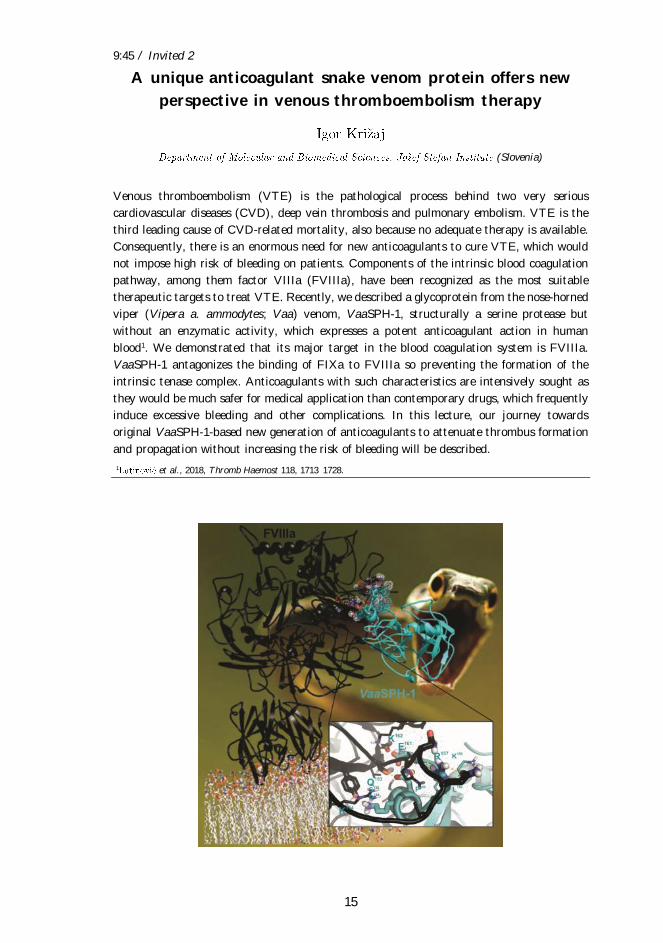
therapeutic targets to treat VTE. Recently, we described a glycoprotein from the nose-horned
viper (Vipera a. ammodytes; Vaa) venom, VaaSPH-1, structurally a serine protease but
without an enzymatic activity, which expresses a potent anticoagulant action in human
blood1. We demonstrated that its major target in the blood coagulation system is FVIIIa.
VaaSPH-1 antagonizes the binding of FIXa to FVIIIa so preventing the formation of the
intrinsic tenase complex. Anticoagulants with such characteristics are intensively sought as
they would be much safer for medical application than contemporary drugs, which frequently
induce excessive bleeding and other complications. In this lecture, our journey towards
original VaaSPH-1-based new generation of anticoagulants to attenuate thrombus formation
and propagation without increasing the risk of bleeding will be described.
1 et al., 2018, Thromb Haemost 118, 1713 1728.
10:05 / Invited 3
16
Different toxicity of hydrophilic and hydrophobic
microcystins on rat astrocytes
, Klara Bulc Rozman, Damijana Mojca
Institute of Pathophysiology, Faculty of Medicine, University of Ljubljana (Slovenia)
Microcystins (MCs) are toxic cyanobacterial peptides. Their potent hepatotoxic action mainly
causes acute lethality. Chronic intoxication may result in hepatotoxic, nephrotoxic,
cardiotoxic, and neurotoxic effects in mammals, including humans. MCs enter cells via multi-
specific organic anion-transporting polypeptides (Oatp). Several Oatps have been located in
the mammalian blood-brain-barrier (BBB), suggesting that glial and neuronal cells can be
exposed to MCs. It is reasonable to expect that hydrophobic MCs could pass cellular
membranes more easily then hydrophilic MCs that can cross cellular membranes only by use
of Oatps. Astrocytes are crucially involved in the homeostasis of the central nervous system,
and our goal was to assess microcystin toxicity on astrocytes in primary cell culture. MCLR
was used as a standard hydrophilic MC. MCLW and MCLF were chosen as representatives
of hydrophobic microcystins.
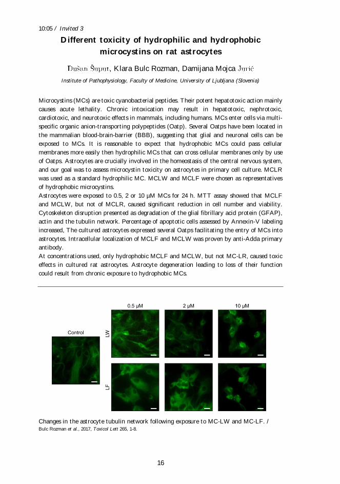
Astrocytes were exposed to 0.5, 2 or 10 µM MCs for 24 h. MTT assay showed that MCLF
and MCLW, but not of MCLR, caused significant reduction in cell number and viability.
Cytoskeleton disruption presented as degradation of the glial fibrillary acid protein (GFAP),
actin and the tubulin network. Percentage of apoptotic cells assessed by Annexin-V labeling
increased, The cultured astrocytes expressed several Oatps facilitating the entry of MCs into
astrocytes. Intracellular localization of MCLF and MCLW was proven by anti-Adda primary
antibody.
At concentrations used, only hydrophobic MCLF and MCLW, but not MC-LR, caused toxic
effects in cultured rat astrocytes. Astrocyte degeneration leading to loss of their function
could result from chronic exposure to hydrophobic MCs.
Changes in the astrocyte tubulin network following exposure to MC-LW and MC-LF. / Bulc Rozman et al., 2017, Toxicol Lett 265, 1-8.
10:25 / Invited 4
17
APS8, a polymeric alkylpyridinium compound blocks α7
nAChR, induces apoptosis in A549 cells, and delays or
prevents growth of human lung adenocarcinoma tumours in
SCID mice
Tom Turk
Department of Biology, Biotechnical Faculty, University of Ljubljana (Slovenia)
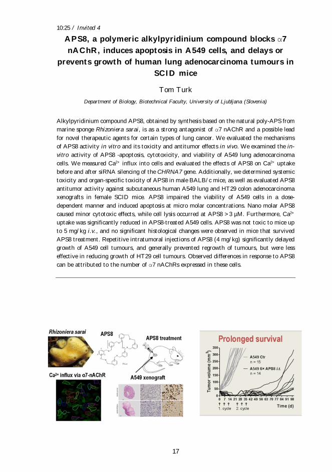
Alkylpyridinium compound APS8, obtained by synthesis based on the natural poly-APS from
marine sponge Rhizoniera sarai, is as a strong antagonist of α7 nAChR and a possible lead
for novel therapeutic agents for certain types of lung cancer. We evaluated the mechanisms
of APS8 activity in vitro and its toxicity and antitumor effects in vivo. We examined the in-
vitro activity of APS8 -apoptosis, cytotoxicity, and viability of A549 lung adenocarcinoma
cells. We measured Ca2+ influx into cells and evaluated the effects of APS8 on Ca2+ uptake
before and after siRNA silencing of the CHRNA7 gene. Additionally, we determined systemic
toxicity and organ-specific toxicity of APS8 in male BALB/c mice, as well as evaluated APS8
antitumor activity against subcutaneous human A549 lung and HT29 colon adenocarcinoma
xenografts in female SCID mice. APS8 impaired the viability of A549 cells in a dose-
dependent manner and induced apoptosis at micro molar concentrations. Nano molar APS8
caused minor cytotoxic effects, while cell lysis occurred at APS8 >3 µM. Furthermore, Ca2+
uptake was significantly reduced in APS8-treated A549 cells. APS8 was not toxic to mice up
to 5 mg/kg i.v., and no significant histological changes were observed in mice that survived
APS8 treatment. Repetitive intratumoral injections of APS8 (4 mg/kg) significantly delayed
growth of A549 cell tumours, and generally prevented regrowth of tumours, but were less
effective in reducing growth of HT29 cell tumours. Observed differences in response to APS8
can be attributed to the number of α7 nAChRs expressed in these cells.
11:30 / Invited 5
18
A transcriptomic and proteomic study of the nose-horned
viper venom
, Adrijana Leonardi, Tamara Sajevic,
nstitute (Slovenia)
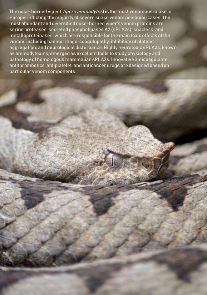
The nose-horned viper (Vipera ammodytes) is the most venomous, and thus medically most
important, snake species in Europe. At least four subspecies - ammodytes (Vaa), meridionalis,
montandoni and transcaucasiana - have been recognized and found mainly in southern
Europe and partly in western Asia, spreading from the northwest to the southeast. The main
aim of our study was to identify a complete arsenal of Vaa venom proteins and peptides, to
guide the production of a more specific and effective antivenom, and promote structure-based
drug design for the treatment of certain cardiovascular, neurological and cancer disorders.
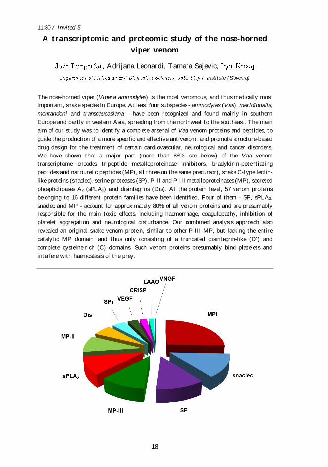
We have shown that a major part (more than 88%, see below) of the Vaa venom
transcriptome encodes tripeptide metalloproteinase inhibitors, bradykinin-potentiating
peptides and natriuretic peptides (MPi, all three on the same precursor), snake C-type lectin-
like proteins (snaclec), serine proteases (SP), P-II and P-III metalloproteinases (MP), secreted
phospholipases A2 (sPLA2) and disintegrins (Dis). At the protein level, 57 venom proteins
belonging to 16 different protein families have been identified. Four of them - SP, sPLA2,
snaclec and MP - account for approximately 80% of all venom proteins and are presumably
responsible for the main toxic effects, including haemorrhage, coagulopathy, inhibition of
platelet aggregation and neurological disturbance. Our combined analysis approach also
revealed an original snake venom protein, similar to other P-III MP, but lacking the entire
catalytic MP domain, and thus only consisting of a truncated disintegrin-like (D') and
complete cysteine-rich (C) domains. Such venom proteins presumably bind platelets and
interfere with haemostasis of the prey.
11:50 / Invited 6
19
Recognition of ceramide phosphoethanolamine by
aegerolysins
Toshihide Kobayashi
Laboratory of Bioimaging and Pathologies, CNRS University of Strasbourg (France)
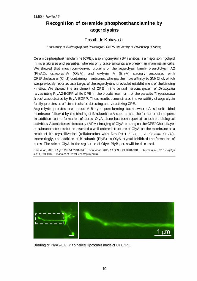
Ceramide phosphoethanolamine (CPE), a sphingomyelin (SM) analog, is a major sphingolipid
in invertebrates and parasites, whereas only trace amounts are present in mammalian cells.
We showed that mushroom-derived proteins of the aegerolysin family pleurotolysin A2
(PlyA2), ostreolysinA (OlyA), and erylysin A (EryA) strongly associated with
CPE/cholesterol (Chol)-containing membranes, whereas their low affinity to SM/Chol, which
was previously reported as a target of the aegerolysins, precluded establishment of the binding
kinetics. We showed the enrichment of CPE in the central nervous system of Drosophila
larvae using PlyA2-EGFP while CPE in the bloodstream form of the parasite Trypanosoma
brucei was detected by EryA-EGFP. These results demonstrated the versatility of aegerolysin
family proteins as efficient tools for detecting and visualizing CPE.
Aegerolysin proteins are unique A-B type pore-forming toxins where A subunits bind
membrane, followed by the binding of B subunit to A subunit and the formation of the pore.
In addition to the formation of pores, OlyA alone has been reported to exhibit biological
activities. Atomic force microscopy (AFM) imaging of OlyA binding on the CPE/Chol bilayer
at subnanometer resolution revealed a well-ordered structure of OlyA on the membrane as a
result of its crystallization (collaboration with Drs Peter ).
Interestingly, the addition of B subunit (PlyB) to OlyA crystal inhibited the formation of
pores. The role of OlyA in the regulation of OlyA-PlyB pores will be discussed.
Bhat et al., 2013, J Lipid Res 54, 2933-2943. / Bhat et al., 2015, FASEB J 29, 3920-3934. / Shirota et al., 2016, Biophys
J 111, 999-1007. / Inaba et al., 2019, Sci Rep in press.
Binding of PlyA2-EGFP to helical liposomes made of CPE/PC.
12:10 / Invited 7
20
Archaeosmes and cholesterol enriched archaeosomes as a
molecular system for studying interactions of cholesterol-
dependent cytolysins with membranes
Na 1, Aden2, Mirijam Kozorog2, 2, Nada 3, Marjetka Podobnik2, Gregor Anderluh2
1Department of Food Science and Technology, Biotechnical Faculty, University of Ljubljana (Slovenia)
/ 2Department of Molecular Biology and Nanobiotechnology, National Institute of Chemistry (Slovenia)
/ 3Department of Biology, Biotechnical Faculty, University of Ljubljana (Slovenia)
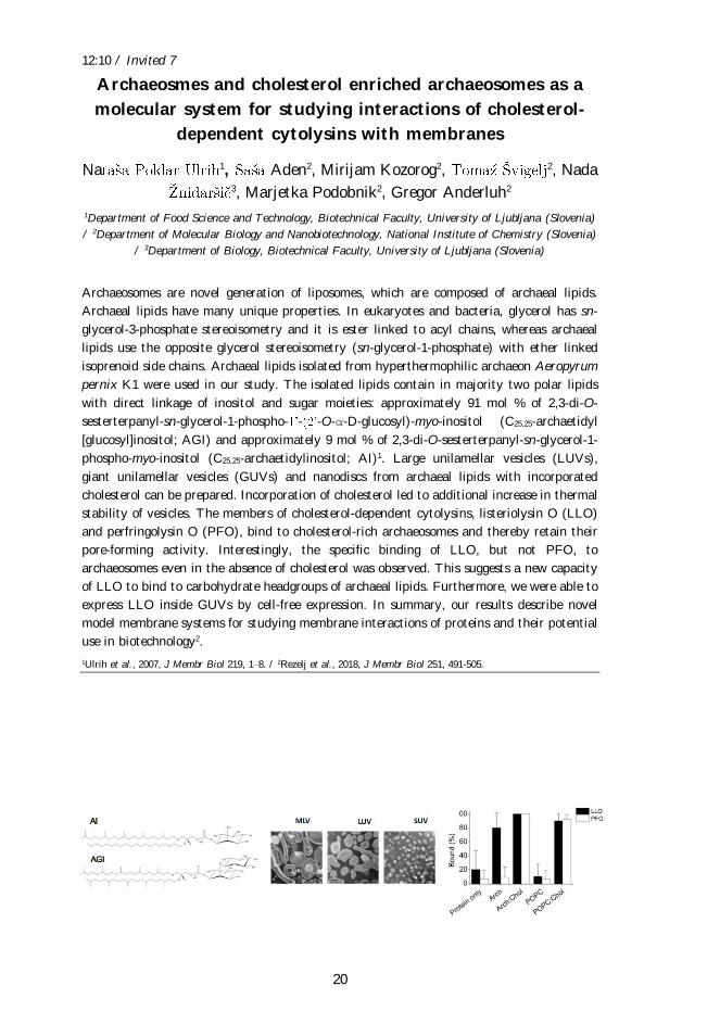
Archaeosomes are novel generation of liposomes, which are composed of archaeal lipids.
Archaeal lipids have many unique properties. In eukaryotes and bacteria, glycerol has sn-
glycerol-3-phosphate stereoisometry and it is ester linked to acyl chains, whereas archaeal
lipids use the opposite glycerol stereoisometry (sn-glycerol-1-phosphate) with ether linked
isoprenoid side chains. Archaeal lipids isolated from hyperthermophilic archaeon Aeropyrum
pernix K1 were used in our study. The isolated lipids contain in majority two polar lipids
with direct linkage of inositol and sugar moieties: approximately 91 mol % of 2,3-di-O-
sesterterpanyl-sn-glycerol-1-phospho- - -O-α-D-glucosyl)-myo-inositol (C25,25-archaetidyl
[glucosyl]inositol; AGI) and approximately 9 mol % of 2,3-di-O-sesterterpanyl-sn-glycerol-1-
phospho-myo-inositol (C25,25-archaetidylinositol; AI)1. Large unilamellar vesicles (LUVs),
giant unilamellar vesicles (GUVs) and nanodiscs from archaeal lipids with incorporated
cholesterol can be prepared. Incorporation of cholesterol led to additional increase in thermal
stability of vesicles. The members of cholesterol-dependent cytolysins, listeriolysin O (LLO)
and perfringolysin O (PFO), bind to cholesterol-rich archaeosomes and thereby retain their
pore-forming activity. Interestingly, the specific binding of LLO, but not PFO, to
archaeosomes even in the absence of cholesterol was observed. This suggests a new capacity
of LLO to bind to carbohydrate headgroups of archaeal lipids. Furthermore, we were able to
express LLO inside GUVs by cell-free expression. In summary, our results describe novel
model membrane systems for studying membrane interactions of proteins and their potential
use in biotechnology2.
1Ulrih et al., 2007, J Membr Biol 219, 1 8. / 2Rezelj et al., 2018, J Membr Biol 251, 491-505.
14:00 / Invited 8
21
Evolution and development of the venom delivery system of
snakes
Michael Richardson
Institute of Biology, Leiden University (Netherlands)
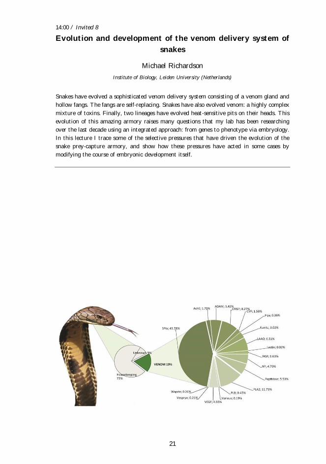
Snakes have evolved a sophisticated venom delivery system consisting of a venom gland and
hollow fangs. The fangs are self-replacing. Snakes have also evolved venom: a highly complex
mixture of toxins. Finally, two lineages have evolved heat-sensitive pits on their heads. This
evolution of this amazing armory raises many questions that my lab has been researching
over the last decade using an integrated approach: from genes to phenotype via embryology.
In this lecture I trace some of the selective pressures that have driven the evolution of the
snake prey-capture armory, and show how these pressures have acted in some cases by
modifying the course of embryonic development itself.
14:20 / Invited 9
22
Strong and widespread action of site-specific positive
selection in the snake venom Kunitz/BPTI protein family
1, 2
1Department of Chemistry and Biochemistry, Faculty of Chemistry and Chemical Technology,
University of Ljubljana (Slovenia) / 2Department of Molecular and Biomedical Sciences, Stefan
Institute (Slovenia)
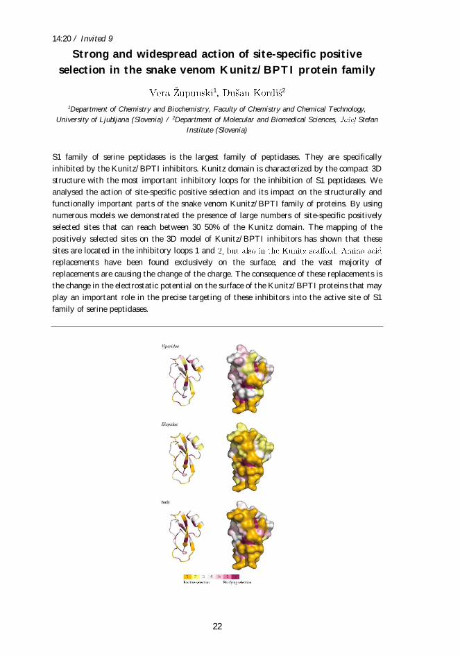
S1 family of serine peptidases is the largest family of peptidases. They are specifically
inhibited by the Kunitz/BPTI inhibitors. Kunitz domain is characterized by the compact 3D
structure with the most important inhibitory loops for the inhibition of S1 peptidases. We
analysed the action of site-specific positive selection and its impact on the structurally and
functionally important parts of the snake venom Kunitz/BPTI family of proteins. By using
numerous models we demonstrated the presence of large numbers of site-specific positively
selected sites that can reach between 30 50% of the Kunitz domain. The mapping of the
positively selected sites on the 3D model of Kunitz/BPTI inhibitors has shown that these
sites are located in the inhibitory loops 1 and
replacements have been found exclusively on the surface, and the vast majority of
replacements are causing the change of the charge. The consequence of these replacements is
the change in the electrostatic potential on the surface of the Kunitz/BPTI proteins that may
play an important role in the precise targeting of these inhibitors into the active site of S1
family of serine peptidases.
14:40 / Invited 10
23
A cytolethal distending toxin variant from Aggregatibacter
actinomycetemcomitans
Davor 1, Rok 2, Simon Caserman3, Adrijana Leonardi4,
Maja Jamnik3, Zdravko Podlesek1, Katja Seme5, Gregor Anderluh3, Igor 4,6, Peter 1, Matej Butala1
1Department of Biology, Biotechnical Faculty, University of Ljubljana (Slovenia) / 2Department of
Oral Medicine and Periodontology, Faculty of Medicine, University of Ljubljana (Slovenia) / 3Department of Molecular Biology and Nanobiotechnology, National Institute of Chemistry (Slovenia) /
4 (Slovenia) / 5Institute of
Microbiology and Immunology, Faculty of Medicine, University of Ljubljana (Slovenia) / 6Department
of Chemistry and Biochemistry, Faculty of Chemistry and Chemical Technology, University of
Ljubljana (Slovenia)
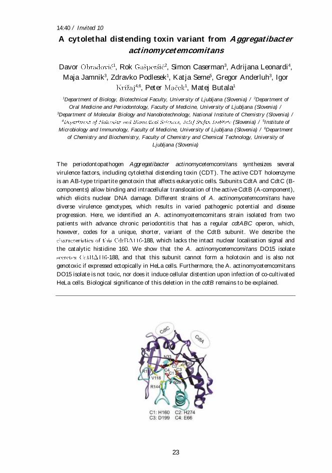
The periodontopathogen Aggregatibacter actinomycetemcomitans synthesizes several
virulence factors, including cytolethal distending toxin (CDT). The active CDT holoenzyme
is an AB-type tripartite genotoxin that affects eukaryotic cells. Subunits CdtA and CdtC (B-
components) allow binding and intracellular translocation of the active CdtB (A-component),
which elicits nuclear DNA damage. Different strains of A. actinomycetemcomitans have
diverse virulence genotypes, which results in varied pathogenic potential and disease
progression. Here, we identified an A. actinomycetemcomitans strain isolated from two
patients with advance chronic periodontitis that has a regular cdtABC operon, which,
however, codes for a unique, shorter, variant of the CdtB subunit. We describe the
-188, which lacks the intact nuclear localisation signal and
the catalytic histidine 160. We show that the A. actinomycetemcomitans DO15 isolate
-188, and that this subunit cannot form a holotoxin and is also not
genotoxic if expressed ectopically in HeLa cells. Furthermore, the A. actinomycetemcomitans
DO15 isolate is not toxic, nor does it induce cellular distention upon infection of co-cultivated
HeLa cells. Biological significance of this deletion in the cdtB remains to be explained.
https://www.ncbi.nlm.nih.gov/pubmed/?term=Caserman%20S%5BAuthor%5D&cauthor=true&cauthor_uid=27414641
https://www.ncbi.nlm.nih.gov/pubmed/?term=Leonardi%20A%5BAuthor%5D&cauthor=true&cauthor_uid=27414641

15:00 / Invited 11
24
Possible applications of aegerolysin proteins in biomedicine
and biotechnology
Department of Biology, Biotechnical Faculty, University of Ljubljana (Slovenia)
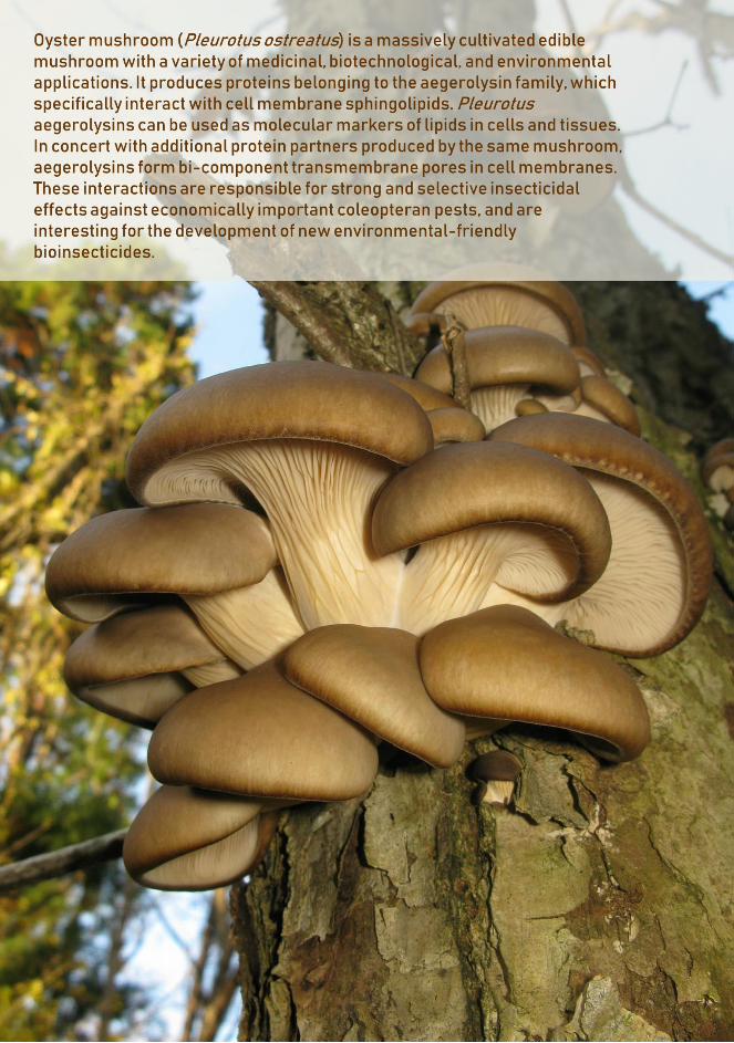
Aegerolysin protein family (Pfam 06355, InterPro IPR009413) comprises low molecular (15-
20 kDa), acidic, beta-structured proteins, found in several eukaryotic and bacterial taxa.
Although they appear to be among major proteins secreted by the organisms that produce
them, their functions and biological roles remain poorly understood.
The common feature of the aegerolysins is their ability to bind different lipids and lipid
membranes. Some aegerolysins can target sphingomyelin/ cholesterol membrane
nanodomains, while aegerolysins from the fungal genus Pleurotus preferentially bind to
ceramide phosphoethanolamine (CPE), which is the major membrane sphingolipid in
invertebrates (particularly insects). Moreover, the genomes of some aegerolysin-producing
fungi have nucleotide sequences that encode proteins with membrane-attack complex/
perforin (MACPF) domain. In the presence of a protein with a MACPF domain, fungal
aegerolysins can function as bi-component lytic complexes for target cell membranes.
Selected fluorescent fusion derivatives of fungal aegerolysins could be used as useful tools to
track raft-like membrane nanodomains composed of sphingomyelin and cholesterol. Moreover,
the selectivity of some aegerolysin-based cytolytic complexes for increased membrane
sphingomyelin/ cholesterol contents can be exploited for selective killing of urothelial
carcinoma cells. Finally, due to their specific interaction with CPE, some cytolytic complexes
based on Pleurotus-derived aegerolysins could represent a novel promising class of
biopesticides for controlling plant pests.
16:05 / Invited 12
25
Pore-forming toxins
Mauro Dalla Serra
Institute of Biophysics, Italian National Research Council (Italy)
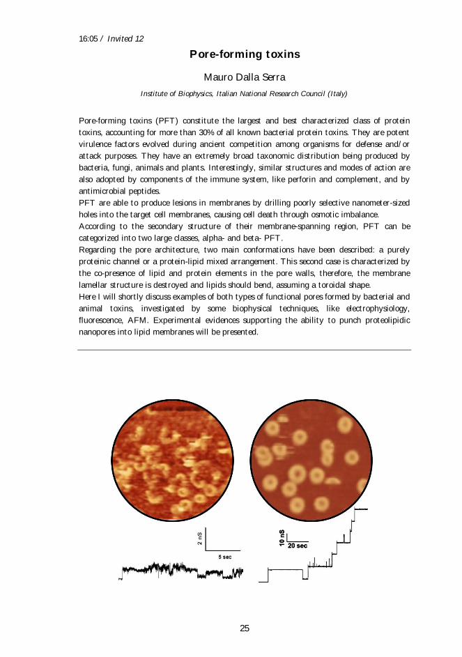
Pore-forming toxins (PFT) constitute the largest and best characterized class of protein
toxins, accounting for more than 30% of all known bacterial protein toxins. They are potent
virulence factors evolved during ancient competition among organisms for defense and/or
attack purposes. They have an extremely broad taxonomic distribution being produced by
bacteria, fungi, animals and plants. Interestingly, similar structures and modes of action are
also adopted by components of the immune system, like perforin and complement, and by
antimicrobial peptides.
PFT are able to produce lesions in membranes by drilling poorly selective nanometer-sized
holes into the target cell membranes, causing cell death through osmotic imbalance.
According to the secondary structure of their membrane-spanning region, PFT can be
categorized into two large classes, alpha- and beta- PFT.
Regarding the pore architecture, two main conformations have been described: a purely
proteinic channel or a protein-lipid mixed arrangement. This second case is characterized by
the co-presence of lipid and protein elements in the pore walls, therefore, the membrane
lamellar structure is destroyed and lipids should bend, assuming a toroidal shape.
Here I will shortly discuss examples of both types of functional pores formed by bacterial and
animal toxins, investigated by some biophysical techniques, like electrophysiology,
fluorescence, AFM. Experimental evidences supporting the ability to punch proteolipidic
nanopores into lipid membranes will be presented.
16:25 / Invited 13
26
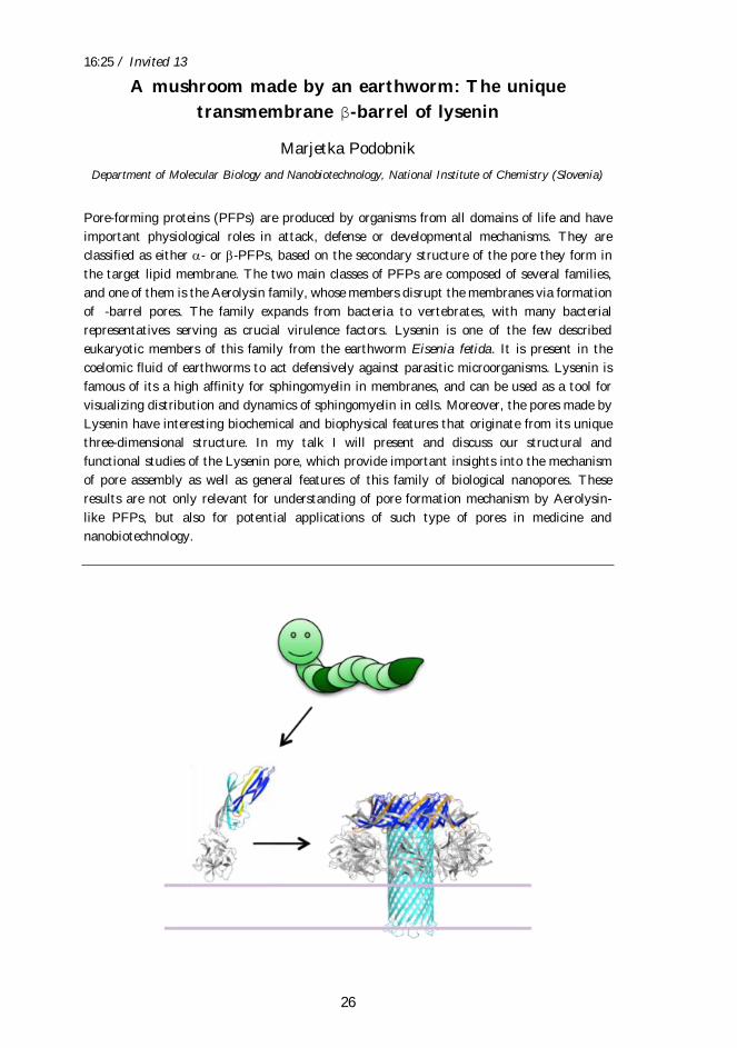
A mushroom made by an earthworm: The unique
transmembrane β-barrel of lysenin
Marjetka Podobnik
Department of Molecular Biology and Nanobiotechnology, National Institute of Chemistry (Slovenia)
Pore-forming proteins (PFPs) are produced by organisms from all domains of life and have
important physiological roles in attack, defense or developmental mechanisms. They are
classified as either - or -PFPs, based on the secondary structure of the pore they form in
the target lipid membrane. The two main classes of PFPs are composed of several families,
and one of them is the Aerolysin family, whose members disrupt the membranes via formation
of -barrel pores. The family expands from bacteria to vertebrates, with many bacterial
representatives serving as crucial virulence factors. Lysenin is one of the few described
eukaryotic members of this family from the earthworm Eisenia fetida. It is present in the
coelomic fluid of earthworms to act defensively against parasitic microorganisms. Lysenin is
famous of its a high affinity for sphingomyelin in membranes, and can be used as a tool for
visualizing distribution and dynamics of sphingomyelin in cells. Moreover, the pores made by
Lysenin have interesting biochemical and biophysical features that originate from its unique
three-dimensional structure. In my talk I will present and discuss our structural and
functional studies of the Lysenin pore, which provide important insights into the mechanism
of pore assembly as well as general features of this family of biological nanopores. These
results are not only relevant for understanding of pore formation mechanism by Aerolysin-
like PFPs, but also for potential applications of such type of pores in medicine and
nanobiotechnology.
16:45 / Invited 14
27
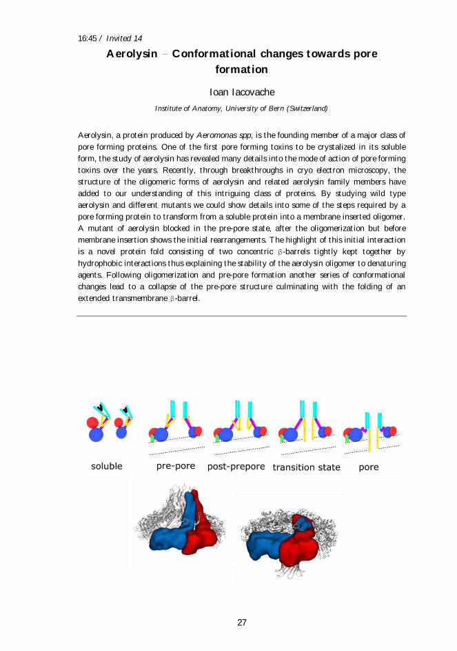
Aerolysin Conformational changes towards pore
formation
Ioan Iacovache
Institute of Anatomy, University of Bern (Switzerland)
Aerolysin, a protein produced by Aeromonas spp, is the founding member of a major class of
pore forming proteins. One of the first pore forming toxins to be crystalized in its soluble
form, the study of aerolysin has revealed many details into the mode of action of pore forming
toxins over the years. Recently, through breakthroughs in cryo electron microscopy, the
structure of the oligomeric forms of aerolysin and related aerolysin family members have
added to our understanding of this intriguing class of proteins. By studying wild type
aerolysin and different mutants we could show details into some of the steps required by a
pore forming protein to transform from a soluble protein into a membrane inserted oligomer.
A mutant of aerolysin blocked in the pre-pore state, after the oligomerization but before
membrane insertion shows the initial rearrangements. The highlight of this initial interaction
is a novel protein fold consisting of two concentric β-barrels tightly kept together by
hydrophobic interactions thus explaining the stability of the aerolysin oligomer to denaturing
agents. Following oligomerization and pre-pore formation another series of conformational
changes lead to a collapse of the pre-pore structure culminating with the folding of an
extended transmembrane β-barrel.
17:25 / Invited 15
28
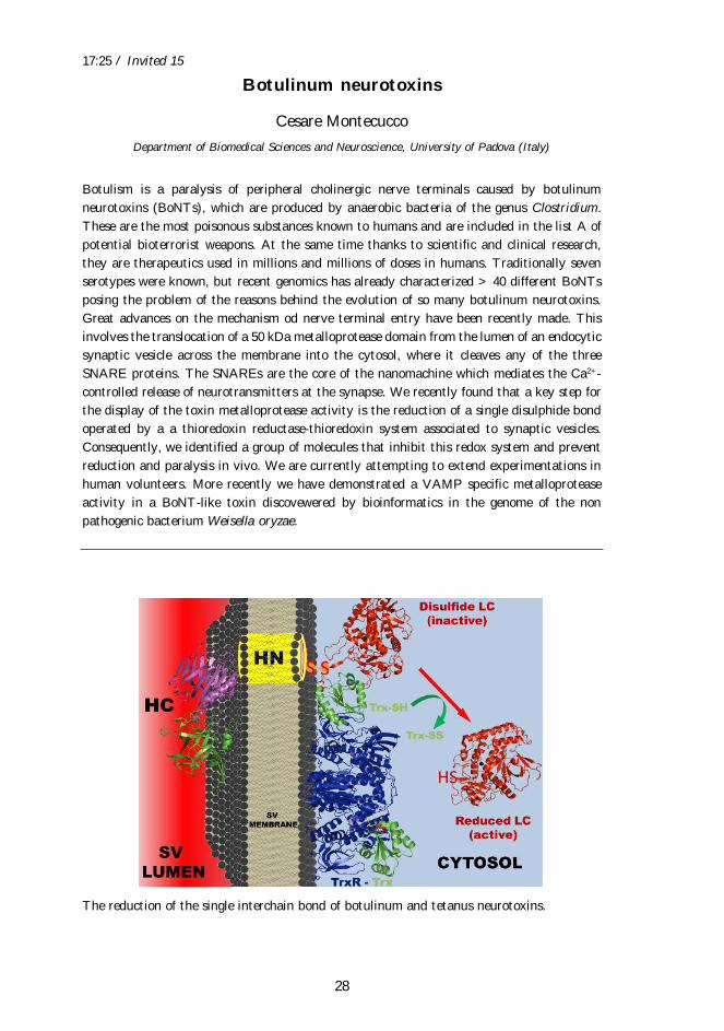
Botulinum neurotoxins
Cesare Montecucco
Department of Biomedical Sciences and Neuroscience, University of Padova (Italy)
Botulism is a paralysis of peripheral cholinergic nerve terminals caused by botulinum
neurotoxins (BoNTs), which are produced by anaerobic bacteria of the genus Clostridium.
These are the most poisonous substances known to humans and are included in the list A of
potential bioterrorist weapons. At the same time thanks to scientific and clinical research,
they are therapeutics used in millions and millions of doses in humans. Traditionally seven
serotypes were known, but recent genomics has already characterized > 40 different BoNTs
posing the problem of the reasons behind the evolution of so many botulinum neurotoxins.
Great advances on the mechanism od nerve terminal entry have been recently made. This
involves the translocation of a 50 kDa metalloprotease domain from the lumen of an endocytic
synaptic vesicle across the membrane into the cytosol, where it cleaves any of the three
SNARE proteins. The SNAREs are the core of the nanomachine which mediates the Ca2+-
controlled release of neurotransmitters at the synapse. We recently found that a key step for
the display of the toxin metalloprotease activity is the reduction of a single disulphide bond
operated by a a thioredoxin reductase-thioredoxin system associated to synaptic vesicles.
Consequently, we identified a group of molecules that inhibit this redox system and prevent
reduction and paralysis in vivo. We are currently attempting to extend experimentations in
human volunteers. More recently we have demonstrated a VAMP specific metalloprotease
activity in a BoNT-like toxin discovewered by bioinformatics in the genome of the non
pathogenic bacterium Weisella oryzae.
The reduction of the single interchain bond of botulinum and tetanus neurotoxins.

Abstracts - Oral Presentations
30
10:45 / Oral 1
31
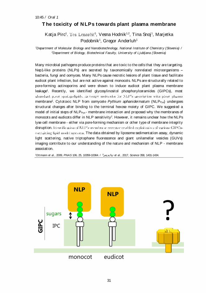
The toxicity of NLPs towards plant plasma membrane
Katja Pirc1 1, Vesna Hodnik1,2, Tina Snoj1, Marjetka
Podobnik1, Gregor Anderluh1
1Department of Molecular Biology and Nanobiotechnology, National Institute of Chemistry (Slovenia) / 2Department of Biology, Biotechnical Faculty, University of Ljubljana (Slovenia)
Many microbial pathogens produce proteins that are toxic to the cells that they are targeting.
Nep1-like proteins (NLPs) are secreted by taxonomically nonrelated microorganisms
bacteria, fungi and oomyces. Many NLPs cause necrotic lesions of plant tissue and facilitate
eudicot plant infection, but are not active against monocots. NLPs are structurally related to
pore-forming actinoporins and were shown to induce eudicot plant plasma membrane
leakage1. Recently, we identified glycosylinositol phosphorylceramides (GIPCs), most
membrane2. Cytotoxic NLP from oomycete Pythium aphanidermatum (NLPPya) undergoes
structural changes after binding to the terminal hexose moiety of GIPC. We suggested a
model of initial steps of NLPPya - membrane interaction and proposed why the membranes of
monocots and eudicots differ in NLP sensitivity2. However, it remains unclear how the NLPs
lyse cell membrane - either via pore-forming mechanism or other type of membrane integrity
disruption.
The data obtained by liposome sedimentation assay, dynamic
light scattering, native triptophane fluorescence and giant unilamellar vesicles (GUVs)
imaging contribute to our understanding of the nature and mechanism of NLP - membrane
association.
1Ottmann et al., 2009, PNAS 106, 25, 10359-10364. / 2 et al., 2017, Science 358, 1431-1434.
10:55 / Oral 2
32
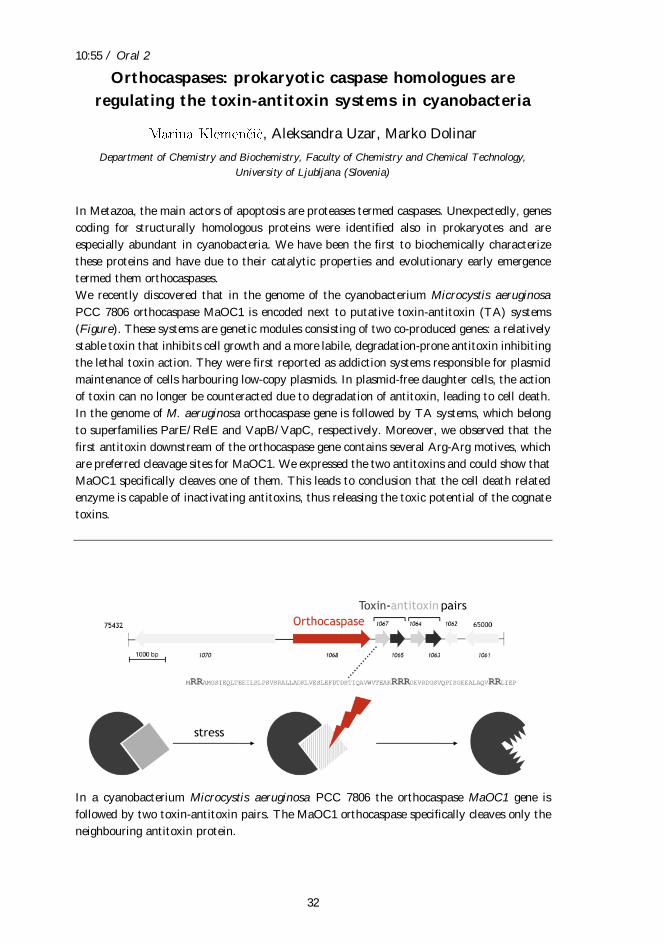
Orthocaspases: prokaryotic caspase homologues are
regulating the toxin-antitoxin systems in cyanobacteria
, Aleksandra Uzar, Marko Dolinar
Department of Chemistry and Biochemistry, Faculty of Chemistry and Chemical Technology,
University of Ljubljana (Slovenia)
In Metazoa, the main actors of apoptosis are proteases termed caspases. Unexpectedly, genes
coding for structurally homologous proteins were identified also in prokaryotes and are
especially abundant in cyanobacteria. We have been the first to biochemically characterize
these proteins and have due to their catalytic properties and evolutionary early emergence
termed them orthocaspases.
We recently discovered that in the genome of the cyanobacterium Microcystis aeruginosa
PCC 7806 orthocaspase MaOC1 is encoded next to putative toxin-antitoxin (TA) systems
(Figure). These systems are genetic modules consisting of two co-produced genes: a relatively
stable toxin that inhibits cell growth and a more labile, degradation-prone antitoxin inhibiting
the lethal toxin action. They were first reported as addiction systems responsible for plasmid
maintenance of cells harbouring low-copy plasmids. In plasmid-free daughter cells, the action
of toxin can no longer be counteracted due to degradation of antitoxin, leading to cell death.
In the genome of M. aeruginosa orthocaspase gene is followed by TA systems, which belong
to superfamilies ParE/RelE and VapB/VapC, respectively. Moreover, we observed that the
first antitoxin downstream of the orthocaspase gene contains several Arg-Arg motives, which
are preferred cleavage sites for MaOC1. We expressed the two antitoxins and could show that
MaOC1 specifically cleaves one of them. This leads to conclusion that the cell death related
enzyme is capable of inactivating antitoxins, thus releasing the toxic potential of the cognate
toxins.
In a cyanobacterium Microcystis aeruginosa PCC 7806 the orthocaspase MaOC1 gene is
followed by two toxin-antitoxin pairs. The MaOC1 orthocaspase specifically cleaves only the
neighbouring antitoxin protein.
12:30 / Oral 3
33
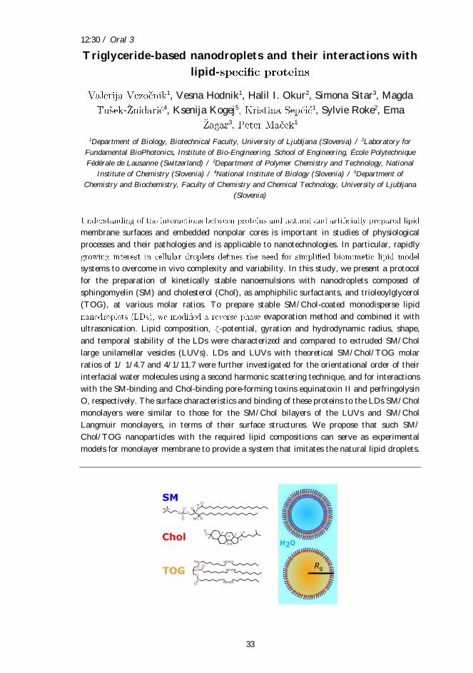
Triglyceride-based nanodroplets and their interactions with
lipid-
1, Vesna Hodnik1, Halil I. Okur2, Simona Sitar3, Magda
- 4, Ksenija Kogej5 1, Sylvie Roke2, Ema 3 1
1Department of Biology, Biotechnical Faculty, University of Ljubljana (Slovenia) / 2Laboratory for
Fundamental BioPhotonics, Institute of Bio-Engineering, School of Engineering, École Polytechnique
Fédérale de Lausanne (Switzerland) / 3Department of Polymer Chemistry and Technology, National
Institute of Chemistry (Slovenia) / 4National Institute of Biology (Slovenia) / 5Department of
Chemistry and Biochemistry, Faculty of Chemistry and Chemical Technology, University of Ljubljana
(Slovenia)
membrane surfaces and embedded nonpolar cores is important in studies of physiological
processes and their pathologies and is applicable to nanotechnologies. In particular, rapidly
systems to overcome in vivo complexity and variability. In this study, we present a protocol
for the preparation of kinetically stable nanoemulsions with nanodroplets composed of
sphingomyelin (SM) and cholesterol (Chol), as amphiphilic surfactants, and trioleoylglycerol
(TOG), at various molar ratios. To prepare stable SM/Chol-coated monodisperse lipid
evaporation method and combined it with
ultrasonication. Lipid composition, ζ-potential, gyration and hydrodynamic radius, shape,
and temporal stability of the LDs were characterized and compared to extruded SM/Chol
large unilamellar vesicles (LUVs). LDs and LUVs with theoretical SM/Chol/TOG molar
ratios of 1/ 1/4.7 and 4/1/11.7 were further investigated for the orientational order of their
interfacial water molecules using a second harmonic scattering technique, and for interactions
with the SM-binding and Chol-binding pore-forming toxins equinatoxin II and perfringolysin
O, respectively. The surface characteristics and binding of these proteins to the LDs SM/Chol
monolayers were similar to those for the SM/Chol bilayers of the LUVs and SM/Chol
Langmuir monolayers, in terms of their surface structures. We propose that such SM/
Chol/TOG nanoparticles with the required lipid compositions can serve as experimental
models for monolayer membrane to provide a system that imitates the natural lipid droplets.
12:40 / Oral 4
34
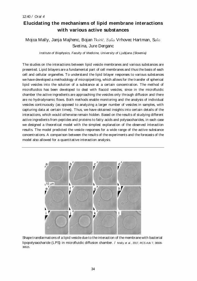
Elucidating the mechanisms of lipid membrane interactions
with various active substances
Mojca Mally, Janja Majhenc, Bojan Vrhovec Hartman, S
Svetina, Jure Derganc
Institute of Biophysics, Faculty of Medicine, University of Ljubljana (Slovenia)
The studies on the interactions between lipid vesicle membranes and various substances are
presented. Lipid bilayers are a fundamental part of cell membranes and thus the basis of each
cell and cellular organelles. To understand the lipid bilayer responses to various substances
we have developed a methodology of micropipetting, which allows for the transfer of spherical
lipid vesicles into the solution of a substance at a certain concentration. The method of
microfluidics has been developed to deal with flaccid vesicles, since in the microfluidic
chamber the active ingredients are approaching the vesicles only through diffusion and there
are no hydrodynamic flows. Both methods enable monitoring and the analysis of individual
vesicles continuously (as opposed to analyzing a larger number of vesicles in samples, with
capturing data at certain times). Thus, we have obtained insights into certain details of the
interactions, which would otherwise remain hidden. Based on the results of studying different
active ingredients from peptides and proteins to fatty acids and polysaccharides, in each case
we designed a theoretical model with the simplest explanation of the observed interaction
results. The model predicted the vesicle responses for a wide range of the active substance
concentrations. A comparison between the results of the experiments and the forecasts of the
model also allowed for a quantitative interaction analysis.
Shape transfarmations of a lipid vesicle due to the interaction of the membrane with bacterial
lipopolysaccharide (LPS) in microfluidic diffusion chamber. / Mally et al., 2017, RCS Adv 7, 36506-
36515.
15:20 / Oral 5
35
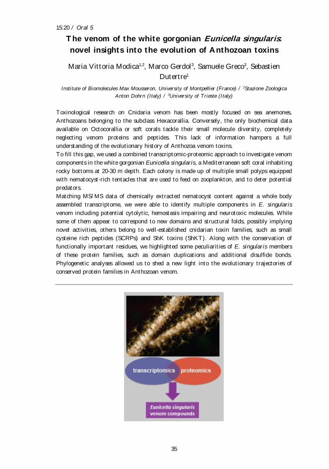
The venom of the white gorgonian Eunicella singularis:
novel insights into the evolution of Anthozoan toxins
Maria Vittoria Modica1,2, Marco Gerdol3, Samuele Greco3, Sebastien
Dutertre1
Institute of Biomolecules Max Mousseron, University of Montpellier (France) / 2Stazione Zoologica
Anton Dohrn (Italy) / 3University of Trieste (Italy)
Toxinological research on Cnidaria venom has been mostly focused on sea anemones,
Anthozoans belonging to the subclass Hexacorallia. Conversely, the only biochemical data
available on Octocorallia or soft corals tackle their small molecule diversity, completely
neglecting venom proteins and peptides. This lack of information hampers a full
understanding of the evolutionary history of Anthozoa venom toxins.
To fill this gap, we used a combined transcriptomic-proteomic approach to investigate venom
components in the white gorgonian Eunicella singularis, a Mediterranean soft coral inhabiting
rocky bottoms at 20-30 m depth. Each colony is made up of multiple small polyps equipped
with nematocyst-rich tentacles that are used to feed on zooplankton, and to deter potential
predators.
Matching MS/MS data of chemically extracted nematocyst content against a whole body
assembled transcriptome, we were able to identify multiple components in E. singularis
venom including potential cytolytic, hemostasis impairing and neurotoxic molecules. While
some of them appear to correspond to new domains and structural folds, possibly implying
novel activities, others belong to well-established cnidarian toxin families, such as small
cysteine rich peptides (SCRPs) and ShK toxins (ShKT). Along with the conservation of
functionally important residues, we highlighted some peculiarities of E. singularis members
of these protein families, such as domain duplications and additional disulfide bonds.
Phylogenetic analyses allowed us to shed a new light into the evolutionary trajectories of
conserved protein families in Anthozoan venom.
15:30 / Oral 6
36
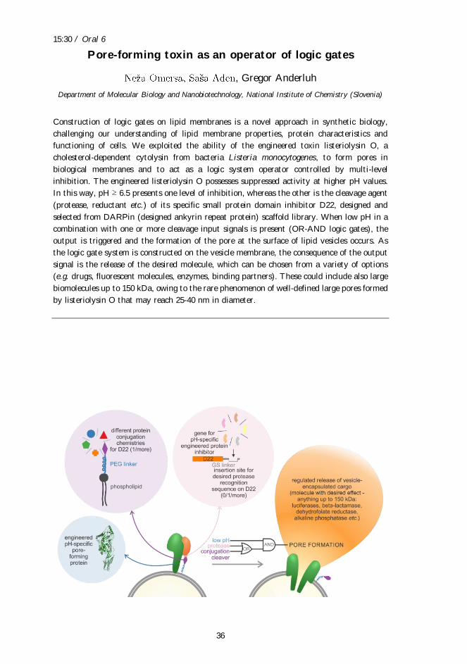
Pore-forming toxin as an operator of logic gates
, Gregor Anderluh
Department of Molecular Biology and Nanobiotechnology, National Institute of Chemistry (Slovenia)
Construction of logic gates on lipid membranes is a novel approach in synthetic biology,
challenging our understanding of lipid membrane properties, protein characteristics and
functioning of cells. We exploited the ability of the engineered toxin listeriolysin O, a
cholesterol-dependent cytolysin from bacteria Listeria monocytogenes, to form pores in
biological membranes and to act as a logic system operator controlled by multi-level
inhibition. The engineered listeriolysin O possesses suppressed activity at higher pH values.
In this way, pH ≥ 6.5 presents one level of inhibition, whereas the other is the cleavage agent
(protease, reductant etc.) of its specific small protein domain inhibitor D22, designed and
selected from DARPin (designed ankyrin repeat protein) scaffold library. When low pH in a
combination with one or more cleavage input signals is present (OR-AND logic gates), the
output is triggered and the formation of the pore at the surface of lipid vesicles occurs. As
the logic gate system is constructed on the vesicle membrane, the consequence of the output
signal is the release of the desired molecule, which can be chosen from a variety of options
(e.g. drugs, fluorescent molecules, enzymes, binding partners). These could include also large
biomolecules up to 150 kDa, owing to the rare phenomenon of well-defined large pores formed
by listeriolysin O that may reach 25-40 nm in diameter.
17:05 / Oral 7
37
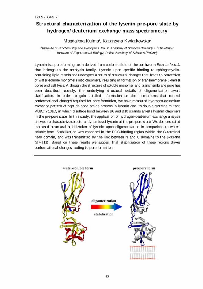
Structural characterization of the lysenin pre-pore state by
hydrogen/deuterium exchange mass spectrometry
Magdalena Kulma1, Katarzyna Kwiatkowska2
1Institute of Biochemistry and Biophysics, Polish Academy of Sciences (Poland) / 2The Nencki
Institute of Experimental Biology, Polish Academy of Sciences (Poland)
Lysenin is a pore-forming toxin derived from coelomic fluid of the earthworm Eisenia foetida
that belongs to the aerolysin family. Lysenin upon specific binding to sphingomyelin-
containing lipid membrane undergoes a series of structural changes that leads to conversion
of water-soluble monomers into oligomers, resulting in formation of transmembrane β-barrel
pores and cell lysis. Although the structure of soluble monomer and transmembrane pore has
been described recently, the underlying structural details of oligomerization await
clarification. In order to gain detailed information on the mechanisms that control
conformational changes required for pore formation, we have measured hydrogen-deuterium
exchange pattern of peptide bond amide protons in lysenin and its double cysteine mutant
V88C/Y131C, in which disulfide bond between β6 and β10 strands arrests lysenin oligomers
in the pre-pore state. In this study, the application of hydrogen-deuterium exchange analysis
allowed to characterize structural dynamics of lysenin at the pre-pore state. We demonstrated
increased structural stabilization of lysenin upon oligomerization in comparison to water-
soluble form. Stabilization was enhanced in the POC-binding region within the C-terminal
head domain, and was transmitted by the link between N and C domains to the β-strand
(β7-β11). Based on these results we suggest that stabilization of these regions drives
conformational changes leading to pore formation.
17:15 / Oral 8
38
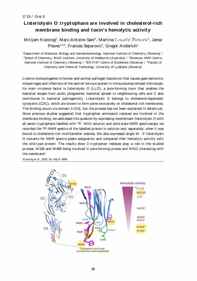
Listeriolysin O tryptophans are involved in cholesterol-rich
membrane binding and toxin's hemolytic activity
Mirijam Kozorog1, Marc-Antoine Sani2, Martina 1, Janez
Plavec1,3,4, Frances Separovic2, Gregor Anderluh1
1Department of Molecular Biology and Nanobiotechnology, National Institute of Chemistry (Slovenia) / 2School of Chemistry, Bio21 Institute, University of Melbourne (Australia) / 3Slovenian NMR Centre,
National Institute of Chemistry (Slovenia) / 5EN-FIST Centre of Excellence (Slovenia) / 4Faculty of
Chemistry and Chemical Technology, University of Ljubljana (Slovenia)
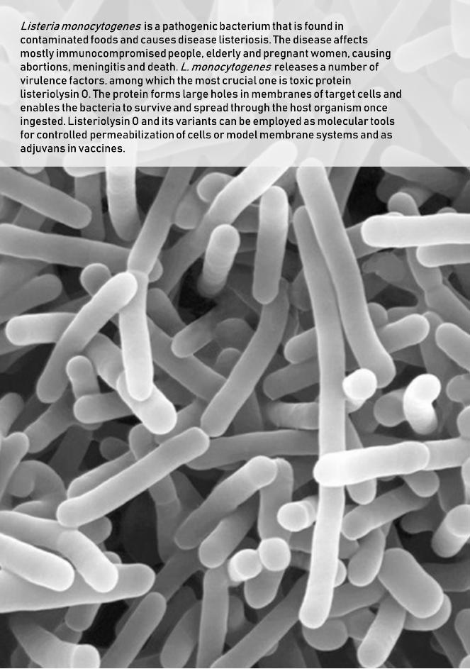
Listeria monocytogenes is human and animal pathogen bacterium that causes gastroenteritis,
miscarriages and infections of the central nervous system in immunocompromised individuals.
Its main virulence factor is listeriolysin O (LLO), a pore-forming toxin that enables the
bacterial escape from acidic phagosome, bacterial spread to neighbouring cells and it also
contributes to bacterial pathogenicity. Listeriolysin O belongs to cholesterol-dependent
cytolysins (CDC), which are known to form pores exclusively on cholesterol-rich membranes.
The binding occurs via domain 4 (D4), but the process has not been explained in details yet.
Since previous studies suggested that tryptophan aminoacid residues are involved in the
membrane binding, we addressed this question by expressing recombinant listeriolysin O with
all seven tryptophans labelled with 19F. With solution and solid-state NMR spectroscopy we
recorded the 19F-NMR spectra of the labelled protein in solution and, separately, when it was
bound to cholesterol-rich multilamellar vesicles. We also expressed single W F listeriolysin
O mutants for NMR spectra peaks assignation and compared their hemolytic activity with
the wild-type protein. The results show 3 tryptophan residues play a role in the studied
process; W189 and W489 being involved in pore-forming proces and W512 interacting with
the membrane1.
1Kozorog et al., 2018, Sci Rep 8, 6894.

Abstracts - Posters
40
Poster 1
41
APS8 induces apoptosis of the NSCLC model cell line by
interfering with the lung cancer signalling pathways
Sabina Berne1, Simona Kranjc2 2,3, Tom Turk4
1Department of Agronomy, Biotechnical faculty, University of Ljubljana (Slovenia) / 2Institute of
Oncology Ljubljana (Slovenia) / 3Faculty for Health Sciences, University of Primorska (Slovenia) / 4Department of Biology, Biotechnical faculty, University of Ljubljana (Slovenia)
The poly-APS synthetic analogue, APS8, is a potent antagonist of α7 nicotinic acetylcholine
receptors (nAChRs), which are emerging as a potential target for lung cancer therapy.
Recently, the therapeutic value of APS8 has been demonstrated in human adenocarcinoma
xenograft models. APS8 was not toxic to mice up to 5 mg/kg i.v. and induced no significant
histological changes but has significantly delayed tumour growth and generally prevented
regrowth of tumours.
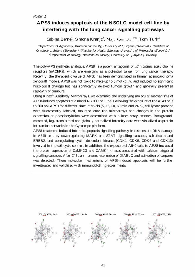
Using Kinex Antibody Microarrays, we examined the underlying molecular mechanisms of
APS8-induced apoptosis of a model NSCLC cell line. Following the exposure of the A549 cells
to 500 nM APS8 for different time intervals (5, 15, 30, 60 min and 24 h), cell lysate proteins
were fluorescently labelled, mounted onto the microarrays and changes in the protein
expression or phosphorylation were determined with a laser array scanner. Background-
corrected, log2 transformed and globally normalized intensity data were visualized as protein
interaction networks in the Cytoscape platform.
APS8 treatment induced intrinsic apoptosis signalling pathway in response to DNA damage
in A549 cells by downregulating MAPK and STAT signalling cascades, calreticulin and
ERBB2, and upregulating cyclin dependent kinases (CDK1, CDK5, CDK6 and CDK13)
involved in the cell cycle control. In addition, the exposure of A549 cells to APS8 increased
the protein expression of CaMK2G and CAMK4 kinases associated with calcium triggered
signalling cascades. After 24 h, an increased expression of DIABLO and activation of caspases
was detected. These molecular mechanisms of APS8-induced apoptosis will be further
investigated and validated with immunoblotting experiments
Poster 2
42
Vesiculation of mammalian cells in response to listeriolysin
O and its mutants
Apolonija Bedina Zavec1 1, Matic Kisovec1, Maja Jamnik1,
Veronika Kralj- 2, Gregor Anderluh1, Marjetka Podobnik1
1Department of Molecular Biology and Nanobiotechnology, National Institute of Chemistry (Slovenia) / 2Biomedical Research Group, Faculty of Health Sciences, University of Ljubljana (Slovenia)
Listeriolysin O (LLO) is a toxin from the intracellular pathogen Listeria monocytogenes,
which forms pores in cholesterol-rich lipid membranes of host cells. Besides its biological
relevance, LLO and its pore forming ability is also interesting for the applications in medicine
and biotechnology. Two mutant proteins were generated by our group, Y406A and
A318C+L334C. The single mutant Y406A is able to bound to membranes and oligomerizes
similarly to the wild-type LLO (wtLLO), but in contrast to wtLLO, the final membrane
insertion step of Y406A mutant is accomplished only under acidic pH. The double cysteine
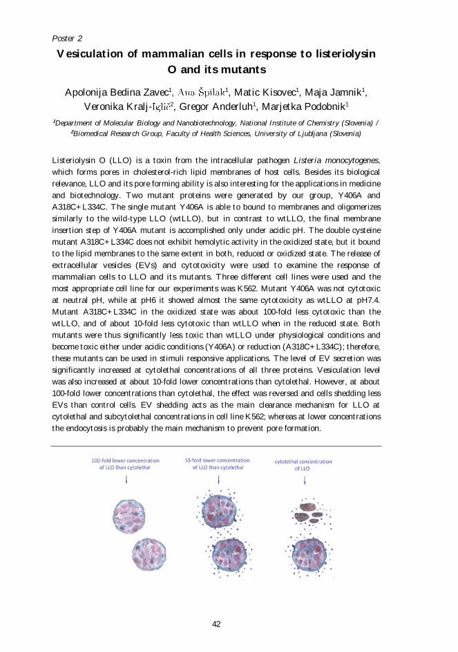
mutant A318C+L334C does not exhibit hemolytic activity in the oxidized state, but it bound
to the lipid membranes to the same extent in both, reduced or oxidized state. The release of
extracellular vesicles (EVs) and cytotoxicity were used to examine the response of
mammalian cells to LLO and its mutants. Three different cell lines were used and the
most appropriate cell line for our experiments was K562. Mutant Y406A was not cytotoxic
at neutral pH, while at pH6 it showed almost the same cytotoxicity as wtLLO at pH7.4.
Mutant A318C+L334C in the oxidized state was about 100-fold less cytotoxic than the
wtLLO, and of about 10-fold less cytotoxic than wtLLO when in the reduced state. Both
mutants were thus significantly less toxic than wtLLO under physiological conditions and
become toxic either under acidic conditions (Y406A) or reduction (A318C+L334C); therefore,
these mutants can be used in stimuli responsive applications. The level of EV secretion was
significantly increased at cytolethal concentrations of all three proteins. Vesiculation level
was also increased at about 10-fold lower concentrations than cytolethal. However, at about
100-fold lower concentrations than cytolethal, the effect was reversed and cells shedding less
EVs than control cells. EV shedding acts as the main clearance mechanism for LLO at
cytolethal and subcytolethal concentrations in cell line K562; whereas at lower concentrations
the endocytosis is probably the main mechanism to prevent pore formation.
Poster 3
43
Isolation and characterization of C-type lectin-like proteins
(snaclecs) from Vipera ammodytes ammodytes (Vaa)
venom inducing transient and reversible thrombocytopenia
of functional platelets
Mojca Dobaja Borak1, Adrijana Leonardi2, Miran Brvar1, Vid Leban1, Igor 2
1Centre for Clinical Toxicology and Pharmacology, University Medical Centre Ljubljana (Slovenia) / 2Department of (Slovenia)
Platelets play an essential role in the initial response to vascular injury. In thromboembolic
diseases like myocardial infarction and ischemic stroke they participate in early stage of the
pathophysiological process. There are already some antiplatelet drugs existing that are used
to prevent and reverse platelet aggregation in acute coronary disease and they work as GP
IIb/IIIa receptor antagonists of irreversibly activated platelets. However, in interventional
angiology and cardiology, an agent is needed with a transient and reversible antithrombotic
effect without impairment of primary hemostasis (e.g. platelet activation). Profound and
transient thrombocytopenia of functional platelets without bleeding was observed in patients
bitten by Vaa at the University Medical Centre Ljubljana and thrombocytopenia could be
rapidly reversed with antivenom application. So far different components were found in Vaa
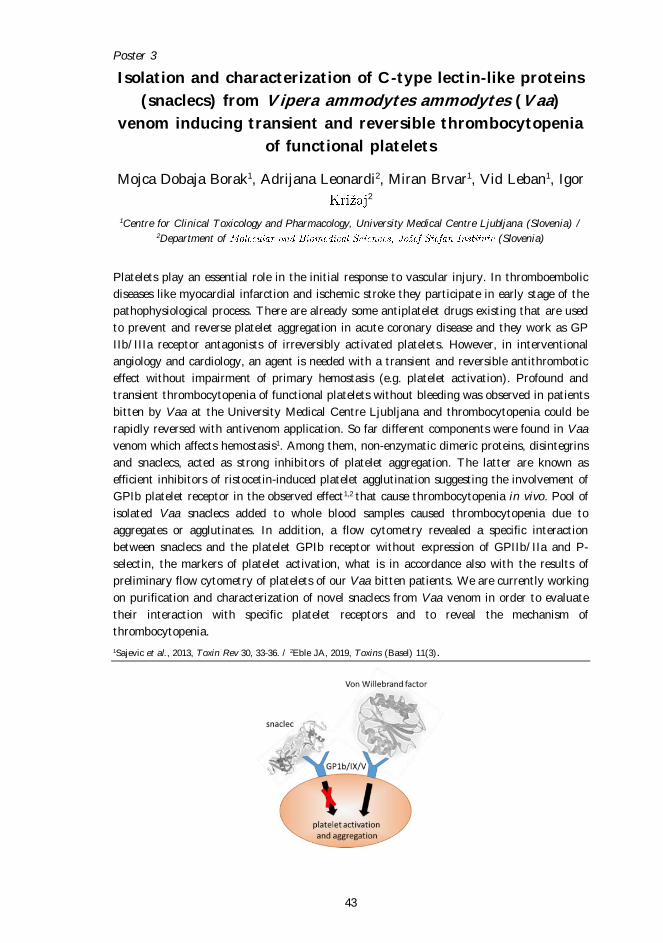
venom which affects hemostasis1. Among them, non-enzymatic dimeric proteins, disintegrins
and snaclecs, acted as strong inhibitors of platelet aggregation. The latter are known as
efficient inhibitors of ristocetin-induced platelet agglutination suggesting the involvement of
GPIb platelet receptor in the observed effect1,2 that cause thrombocytopenia in vivo. Pool of
isolated Vaa snaclecs added to whole blood samples caused thrombocytopenia due to
aggregates or agglutinates. In addition, a flow cytometry revealed a specific interaction
between snaclecs and the platelet GPIb receptor without expression of GPIIb/IIa and P-
selectin, the markers of platelet activation, what is in accordance also with the results of
preliminary flow cytometry of platelets of our Vaa bitten patients. We are currently working
on purification and characterization of novel snaclecs from Vaa venom in order to evaluate
their interaction with specific platelet receptors and to reveal the mechanism of
thrombocytopenia.
1Sajevic et al., 2013, Toxin Rev 30, 33-36. / 2Eble JA, 2019, Toxins (Basel) 11(3).
Poster 4
44
Interaction between ammodytoxin, a β-neurotoxin from the
nose-horned viper venom, and neuronal mitochondria
1,2 1 2 1
1 (Slovenia) / 2Faculty of
Medicine, University of Ljubljana (Slovenia)
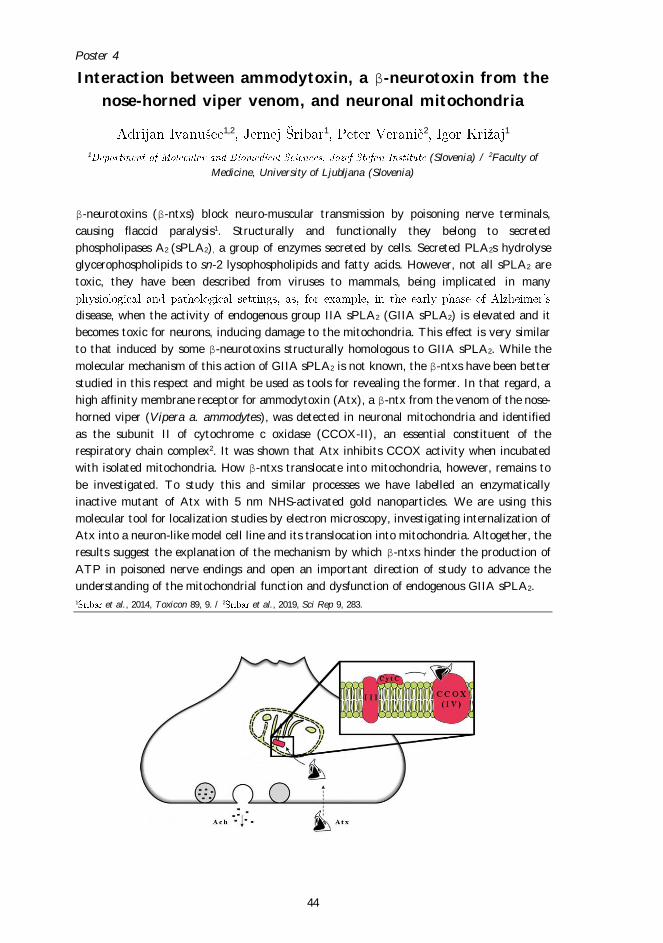
β-neurotoxins (β-ntxs) block neuro-muscular transmission by poisoning nerve terminals,
causing flaccid paralysis1. Structurally and functionally they belong to secreted
phospholipases A2 (sPLA2), a group of enzymes secreted by cells. Secreted PLA2s hydrolyse
glycerophospholipids to sn-2 lysophospholipids and fatty acids. However, not all sPLA2 are
toxic, they have been described from viruses to mammals, being implicated in many
disease, when the activity of endogenous group IIA sPLA2 (GIIA sPLA2) is elevated and it
becomes toxic for neurons, inducing damage to the mitochondria. This effect is very similar
to that induced by some β-neurotoxins structurally homologous to GIIA sPLA2. While the
molecular mechanism of this action of GIIA sPLA2 is not known, the β-ntxs have been better
studied in this respect and might be used as tools for revealing the former. In that regard, a
high affinity membrane receptor for ammodytoxin (Atx), a β-ntx from the venom of the nose-
horned viper (Vipera a. ammodytes), was detected in neuronal mitochondria and identified
as the subunit II of cytochrome c oxidase (CCOX-II), an essential constituent of the
respiratory chain complex2. It was shown that Atx inhibits CCOX activity when incubated
with isolated mitochondria. How β-ntxs translocate into mitochondria, however, remains to
be investigated. To study this and similar processes we have labelled an enzymatically
inactive mutant of Atx with 5 nm NHS-activated gold nanoparticles. We are using this
molecular tool for localization studies by electron microscopy, investigating internalization of
Atx into a neuron-like model cell line and its translocation into mitochondria. Altogether, the
results suggest the explanation of the mechanism by which β-ntxs hinder the production of
ATP in poisoned nerve endings and open an important direction of study to advance the
understanding of the mitochondrial function and dysfunction of endogenous GIIA sPLA2.
1 et al., 2014, Toxicon 89, 9. / 2 et al., 2019, Sci Rep 9, 283.
Poster 5
45
Biological role of Pseudomonas aeruginosa RahU protein
interaction with lipids
1 2, Vesna Hodnik1, Marjetka Podobnik2,
Gregor Anderluh2 1, 1, Matej Butala1
1Department of Biology, Biotechnical faculty, University of Ljubljana (Slovenia) / 2Department of
Molecular Biology and Nanobiotechnology, National Institute of Chemistry (Slovenia)
Aegerolysin RahU of the opportunistic pathogen Pseudomonas aeruginosa is a bacterial
protein belonging to the aegerolysin protein family (Pfam 06355), mainly composed of
proteins originating from fungi and bacteria. The common property of fungal and bacterial
aegerolysins is their ability to interact with membrane lipids. Some of them also possess
hemolytic and toxic activity against eukaryotic cells. It has been shown that RahU interacts
with oxidized low-density lipoprotein, lysophosphatidylcholine and bacterial biosurfactants
rhamnolipids. To elucidate the biological importance of the interaction of aegerolysin RahU
with selected lipids, we successfully purified single alanine substitution RahU mutants in the
Escherichia coli heterologous expression system. We proved that single amino acid
substitutions did not affect the structure and the stability of the RahU derivatives. With
sedimentation assay and surface plasmon resonance spectrometry, we showed that RahU
specifically binds ceramide phosphoethanolamine, the main sphingolipid in the membranes of
invertebrates, and identified crucial residue for this interaction. Therefore, this is the first
report of a bacterial aegerolysin interaction with the aforementioned lipid. We have also
shown the binding of RahU to polar head of ceramid phosphoethanolamine, O-
phosphorylethanolamine, and the neuroendocrinic hormone noradrenaline. In addition, by
using the swarming tests on Pseudomonas aeruginosa PA01, PW1205 in PW1206 strains, we
suggest that aegerolysin RahU affects the swarming of Pseudomonas aeruginosa. Our results
add to the growing knowledge on virulence potential of aegerolysin RahU and starting point
for developing new antimicrobial agents.
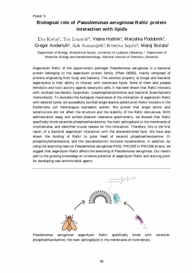
Pseudomonas aeruginosa aegerolysin RahU specifically binds with ceramide
phosphoethanolamine, the main sphingolipid in the membranes of invertebrats.
Poster 6
46
Lipid-binding aegerolysins from entomopathogenic fungi
1,2, Anastasija Panevska3, Gregor Anderluh1, Marjetka
Podobnik1 3 1
1Department of Molecular Biology and Nanobiotechnology, National Institute of Chemistry (Slovenia) / 2Biotechnical Faculty, University of Ljubljana (Slovenia) / 3Department of Biology, Biotechnical
Faculty, University of Ljubljana (Slovenia)
Fungi have a remarkable influence on ecosystems including human beings; they act as
disintegrators, symbionts and pathogens. Among others, fungi are the most common
pathogens of insects and thus represent key regulators of their populations. The rapid
advancement of nucleotide sequencing technologies has accelerated our understanding of
entomopathogenic fungi by defining genomic sequences of several species from the genus
Metarhizium, Beauveria bassiana, Cordyceps militaris and Ophiocordyceps sinensis. We are
interested in proteins from the aegerolysin family, which have characteristic compact beta-
sandwich folds, low isoelectric points, low molecular weights and are stable in a wide pH
range. Aegerolysins are encoded in Dikarya genomes but absent in some of the clades. To
function, some of aegerolysins need interaction to MACPF-like proteins, which we also
identify in entomopathogenic fungi. For some aegerolysins it has been shown they interact
with biological and artificial lipid membranes either alone or in interaction of MACPF-like
protein to form pores with cytolytic or haemolytic action. Although, their roles still need
detailed clarification, different processes dependent on lifestyle of the producing organisms
may be involved: attack or defence against predators, ontogenic development or cell cycle
regulation. The goal of the presented work is preparation of recombinant aegerolysin from
the entomopathogenic fungus B. bassiana in bacteria Escherichia coli, determination of
characteristics of this protein including lipid binding capability and comparison to related
proteins.
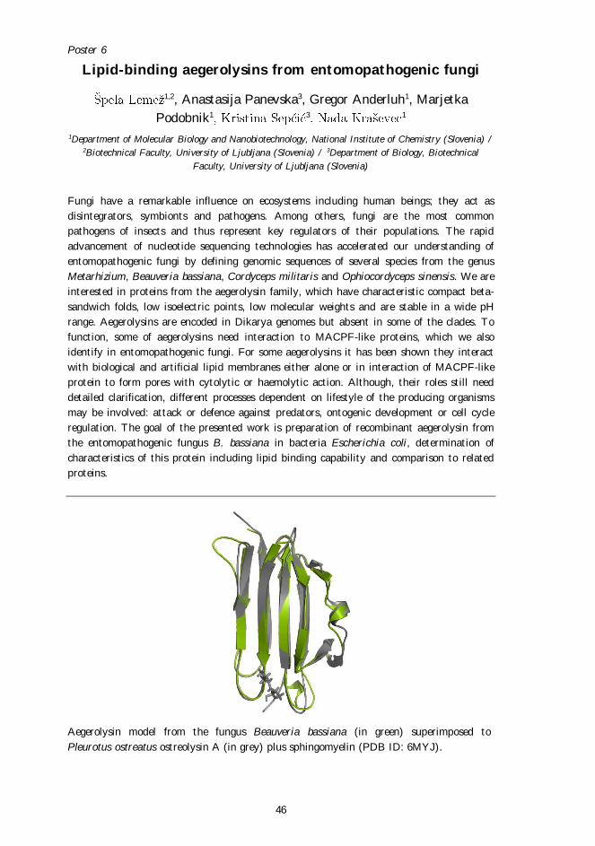
Aegerolysin model from the fungus Beauveria bassiana (in green) superimposed to
Pleurotus ostreatus ostreolysin A (in grey) plus sphingomyelin (PDB ID: 6MYJ).
Poster 7
47
Aegerolysin-based cytolytic complexes acting through
ceramide phosphoethanolamine as potential biopesticides
Anastasija Panevska1, Jaka Razinger2 2, Zoran Arsov3, 1, 1
1Department of Biology, Biotechnical Faculty, University of Ljubljana (Slovenia) / 2Agricultural
Institute of Slovenia (Slovenia) / 3Department of Condensed Matter Physics,
(Slovenia)
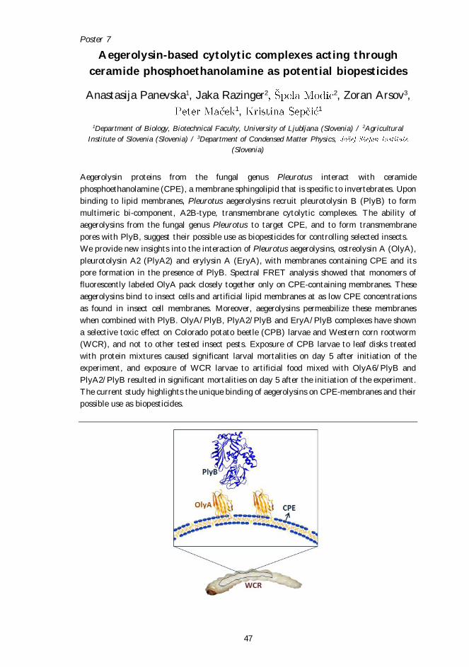
Aegerolysin proteins from the fungal genus Pleurotus interact with ceramide
phosphoethanolamine (CPE), a membrane sphingolipid that is specific to invertebrates. Upon
binding to lipid membranes, Pleurotus aegerolysins recruit pleurotolysin B (PlyB) to form
multimeric bi-component, A2B-type, transmembrane cytolytic complexes. The ability of
aegerolysins from the fungal genus Pleurotus to target CPE, and to form transmembrane
pores with PlyB, suggest their possible use as biopesticides for controlling selected insects.
We provide new insights into the interaction of Pleurotus aegerolysins, ostreolysin A (OlyA),
pleurotolysin A2 (PlyA2) and erylysin A (EryA), with membranes containing CPE and its
pore formation in the presence of PlyB. Spectral FRET analysis showed that monomers of
fluorescently labeled OlyA pack closely together only on CPE-containing membranes. These
aegerolysins bind to insect cells and artificial lipid membranes at as low CPE concentrations
as found in insect cell membranes. Moreover, aegerolysins permeabilize these membranes
when combined with PlyB. OlyA/PlyB, PlyA2/PlyB and EryA/PlyB complexes have shown
a selective toxic effect on Colorado potato beetle (CPB) larvae and Western corn rootworm
(WCR), and not to other tested insect pests. Exposure of CPB larvae to leaf disks treated
with protein mixtures caused significant larval mortalities on day 5 after initiation of the
experiment, and exposure of WCR larvae to artificial food mixed with OlyA6/PlyB and
PlyA2/PlyB resulted in significant mortalities on day 5 after the initiation of the experiment.
The current study highlights the unique binding of aegerolysins on CPE-membranes and their
possible use as biopesticides.
Poster 8
48
Molecular mechanisms of action and interplay between
three key toxins of Listeria monocytogenes
Nejc P , Gregor Anderluh, Marjetka Podobnik
Department of Molecular Biology and Nanobiotechnology, National Institute of Chemistry (Slovenia)
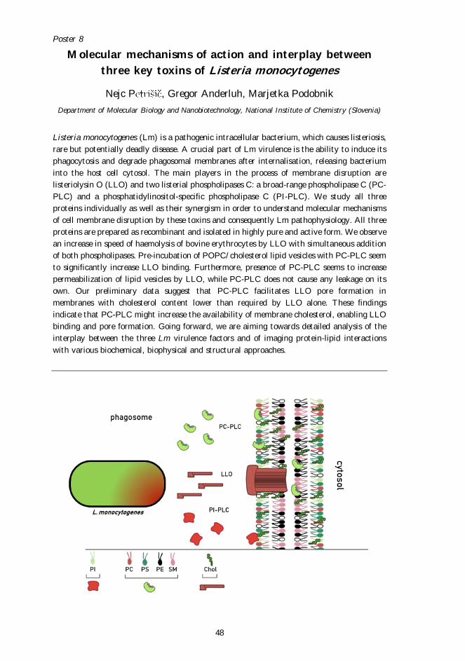
Listeria monocytogenes (Lm) is a pathogenic intracellular bacterium, which causes listeriosis,
rare but potentially deadly disease. A crucial part of Lm virulence is the ability to induce its
phagocytosis and degrade phagosomal membranes after internalisation, releasing bacterium
into the host cell cytosol. The main players in the process of membrane disruption are
listeriolysin O (LLO) and two listerial phospholipases C: a broad-range phospholipase C (PC-
PLC) and a phosphatidylinositol-specific phospholipase C (PI-PLC). We study all three
proteins individually as well as their synergism in order to understand molecular mechanisms
of cell membrane disruption by these toxins and consequently Lm pathophysiology. All three
proteins are prepared as recombinant and isolated in highly pure and active form. We observe
an increase in speed of haemolysis of bovine erythrocytes by LLO with simultaneous addition
of both phospholipases. Pre-incubation of POPC/cholesterol lipid vesicles with PC-PLC seem
to significantly increase LLO binding. Furthermore, presence of PC-PLC seems to increase
permeabilization of lipid vesicles by LLO, while PC-PLC does not cause any leakage on its
own. Our preliminary data suggest that PC-PLC facilitates LLO pore formation in
membranes with cholesterol content lower than required by LLO alone. These findings
indicate that PC-PLC might increase the availability of membrane cholesterol, enabling LLO
binding and pore formation. Going forward, we are aiming towards detailed analysis of the
interplay between the three Lm virulence factors and of imaging protein-lipid interactions
with various biochemical, biophysical and structural approaches.
Poster 9
49
Listeriolysin O mutant (LLO Y406A) eliminates cancer
urothelial cells
1, Dominik Dekleva1, Matic Kisovec2 2, Marjetka
Podobnik2, Gregor Anderluh2, Larisa Tratnjek1, Mateja Erdani Kreft1, 1
1Institute of Cell Biology, Faculty of Medicine, University of Ljubljana (Slovenia) / 2Department of
Molecular Biology and Nanobiotechnology, National Institute of Chemistry (Slovenia)
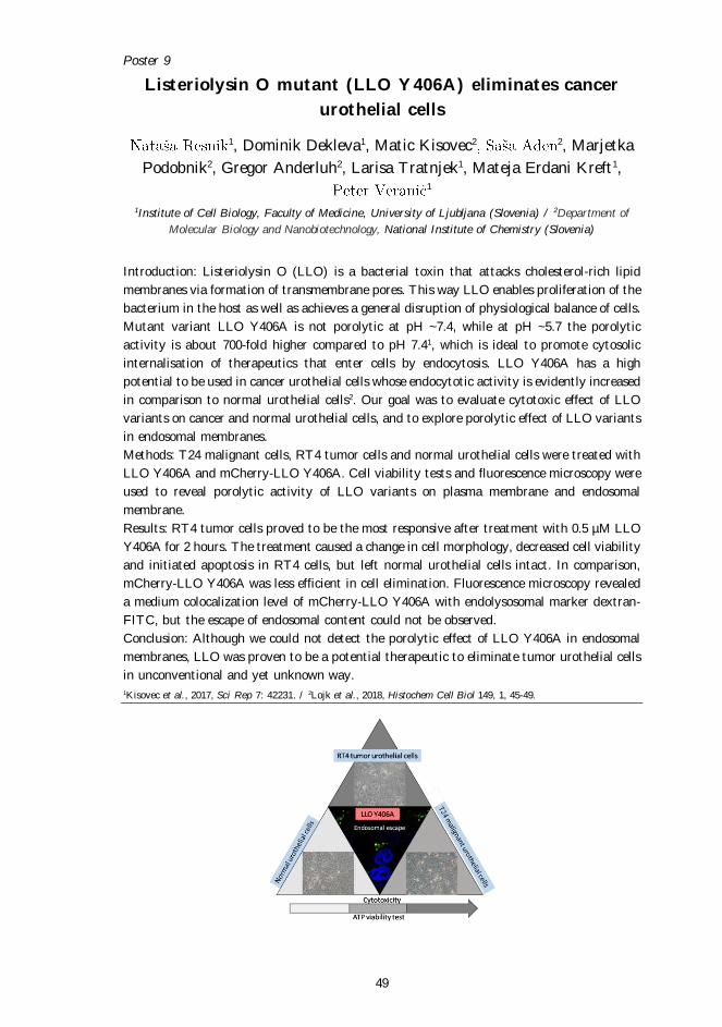
Introduction: Listeriolysin O (LLO) is a bacterial toxin that attacks cholesterol-rich lipid
membranes via formation of transmembrane pores. This way LLO enables proliferation of the
bacterium in the host as well as achieves a general disruption of physiological balance of cells.
Mutant variant LLO Y406A is not porolytic at pH ~7.4, while at pH ~5.7 the porolytic
activity is about 700-fold higher compared to pH 7.41, which is ideal to promote cytosolic
internalisation of therapeutics that enter cells by endocytosis. LLO Y406A has a high
potential to be used in cancer urothelial cells whose endocytotic activity is evidently increased
in comparison to normal urothelial cells2. Our goal was to evaluate cytotoxic effect of LLO
variants on cancer and normal urothelial cells, and to explore porolytic effect of LLO variants
in endosomal membranes.
Methods: T24 malignant cells, RT4 tumor cells and normal urothelial cells were treated with
LLO Y406A and mCherry-LLO Y406A. Cell viability tests and fluorescence microscopy were
used to reveal porolytic activity of LLO variants on plasma membrane and endosomal
membrane.
Results: RT4 tumor cells proved to be the most responsive after treatment with 0.5 µM LLO
Y406A for 2 hours. The treatment caused a change in cell morphology, decreased cell viability
and initiated apoptosis in RT4 cells, but left normal urothelial cells intact. In comparison,
mCherry-LLO Y406A was less efficient in cell elimination. Fluorescence microscopy revealed
a medium colocalization level of mCherry-LLO Y406A with endolysosomal marker dextran-
FITC, but the escape of endosomal content could not be observed.
Conclusion: Although we could not detect the porolytic effect of LLO Y406A in endosomal
membranes, LLO was proven to be a potential therapeutic to eliminate tumor urothelial cells
in unconventional and yet unknown way.
1Kisovec et al., 2017, Sci Rep 7: 42231. / 2Lojk et al., 2018, Histochem Cell Biol 149, 1, 45-49.
Poster 10
50
L-amino acid oxidases are abundant in higher fungi
, Janko Kos
(Slovenia)
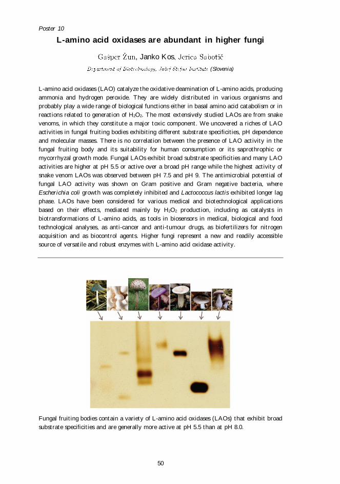
L-amino acid oxidases (LAO) catalyze the oxidative deamination of L-amino acids, producing
ammonia and hydrogen peroxide. They are widely distributed in various organisms and
probably play a wide range of biological functions either in basal amino acid catabolism or in
reactions related to generation of H2O2. The most extensively studied LAOs are from snake
venoms, in which they constitute a major toxic component. We uncovered a riches of LAO
activities in fungal fruiting bodies exhibiting different substrate specificities, pH dependence
and molecular masses. There is no correlation between the presence of LAO activity in the
fungal fruiting body and its suitability for human consumption or its saprothrophic or
mycorrhyzal growth mode. Fungal LAOs exhibit broad substrate specificities and many LAO
activities are higher at pH 5.5 or active over a broad pH range while the highest activity of
snake venom LAOs was observed between pH 7.5 and pH 9. The antimicrobial potential of
fungal LAO activity was shown on Gram positive and Gram negative bacteria, where
Escherichia coli growth was completely inhibited and Lactococcus lactis exhibited longer lag
phase. LAOs have been considered for various medical and biotechnological applications
based on their effects, mediated mainly by H2O2 production, including as catalysts in
biotransformations of L-amino acids, as tools in biosensors in medical, biological and food
technological analyses, as anti-cancer and anti-tumour drugs, as biofertilizers for nitrogen
acquisition and as biocontrol agents. Higher fungi represent a new and readily accessible
source of versatile and robust enzymes with L-amino acid oxidase activity.
Fungal fruiting bodies contain a variety of L-amino acid oxidases (LAOs) that exhibit broad
substrate specificities and are generally more active at pH 5.5 than at pH 8.0.
Poster 11
51
Solving the mistery: Do NLP proteins really form pores in
the lipid membrane?
Tina Snoj, Katja Pirc, Gregor Anderluh
Department of Molecular Biology and Nanobiotechnology, National Institute of Chemistry (Slovenia)
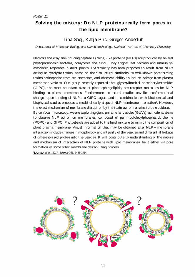
Necrosis and ethylene-inducing peptide 1 (Nep1)-like proteins (NLPs) are produced by several
phytopathogenic bacteria, oomycetes and fungi. They trigger leaf necrosis and immunity-
associated responses in dicot plants. Cytotoxicity has been proposed to result from NLPs
acting as cytolytic toxins, based on their structural similarity to well-known pore-forming
toxins actinoporins from sea anemones, and observed ability to induce leakage from plasma
membrane vesicles. Our group recently reported that glycosylinositol phosphorylceramides
(GIPC), the most abundant class of plant sphingolipids, are receptor molecules for NLP
binding to plasma membranes. Furthermore, structural studies unveiled conformational
changes upon binding of NLPs to GIPC sugars and in combination with biochemical and
biophyscal studies proposed a model of early steps of NLP membrane interaction1. However,
the exact mechanism of membrane disruption by the toxin action remains to be elucidated.
By confocal microscopy, we are exploiting giant unilamellar vesicles (GUVs) as model systems
to observe NLP action on membranes, composed of palmitoyloleoylphosphatidylcholine
(POPC) and GIPC. Phytosterols are added to the lipid mixture to mimic the composition of
plant plasma membrane. Visual information that may be obtained after NLP membrane
interaction include changes in morphology and integrity of the vesicles and differential leakage
of different-sized probes into the vesicles. It will contribute to understanding of the nature
and mechanism of interaction of NLP proteins with lipid membranes, be it either via pore
formation or some other membrane destabilizing process.
1 et al., 2017, Science 358, 1431-1434.
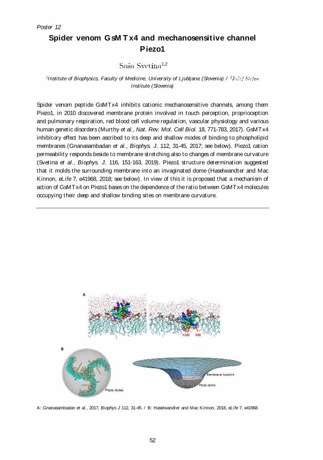
Poster 12
52
Spider venom GsMTx4 and mechanosensitive channel
Piezo1
1,2
1Institute of Biophysics, Faculty of Medicine, University of Ljubljana (Slovenia) / 2J
Institute (Slovenia)
Spider venom peptide GsMTx4 inhibits cationic mechanosensitive channels, among them
Piezo1, in 2010 discovered membrane protein involved in touch perception, proprioception
and pulmonary respiration, red blood cell volume regulation, vascular physiology and various
human genetic disorders (Murthy et al., Nat. Rev. Mol. Cell Biol. 18, 771-783, 2017). GsMTx4
inhibitory effect has been ascribed to its deep and shallow modes of binding to phospholipid
membranes (Gnanasambadan et al., Biophys. J. 112, 31-45, 2017; see below). Piezo1 cation
permeability responds beside to membrane stretching also to changes of membrane curvature
(Svetina et al., Biophys. J. 116, 151-163, 2019). Piezo1 structure determination suggested
that it molds the surrounding membrane into an invaginated dome (Haselwandter and Mac
Kinnon, eLife 7, e41968, 2018; see below). In view of this it is proposed that a mechanism of
action of GsMTx4 on Piezo1 bases on the dependence of the ratio between GsMTx4 molecules
occupying their deep and shallow binding sites on membrane curvature.
A: Gnanasambadan et al., 2017, Biophys J 112, 31-45. / B: Haselwandter and Mac Kinnon, 2018, eLife 7, e41968.
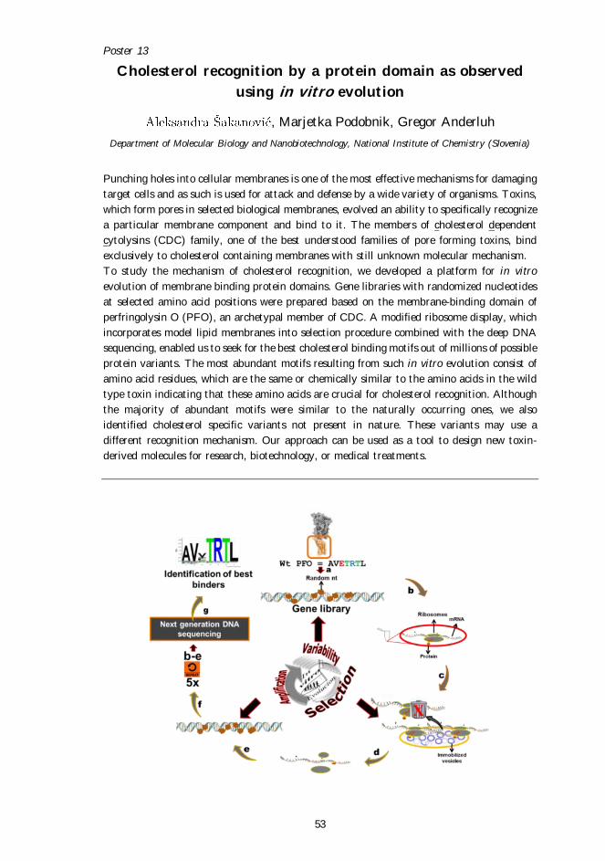
Poster 13
53
Cholesterol recognition by a protein domain as observed
using in vitro evolution
, Marjetka Podobnik, Gregor Anderluh
Department of Molecular Biology and Nanobiotechnology, National Institute of Chemistry (Slovenia)
Punching holes into cellular membranes is one of the most effective mechanisms for damaging
target cells and as such is used for attack and defense by a wide variety of organisms. Toxins,
which form pores in selected biological membranes, evolved an ability to specifically recognize
a particular membrane component and bind to it. The members of cholesterol dependent
cytolysins (CDC) family, one of the best understood families of pore forming toxins, bind
exclusively to cholesterol containing membranes with still unknown molecular mechanism.
To study the mechanism of cholesterol recognition, we developed a platform for in vitro
evolution of membrane binding protein domains. Gene libraries with randomized nucleotides
at selected amino acid positions were prepared based on the membrane-binding domain of
perfringolysin O (PFO), an archetypal member of CDC. A modified ribosome display, which
incorporates model lipid membranes into selection procedure combined with the deep DNA
sequencing, enabled us to seek for the best cholesterol binding motifs out of millions of possible
protein variants. The most abundant motifs resulting from such in vitro evolution consist of
amino acid residues, which are the same or chemically similar to the amino acids in the wild
type toxin indicating that these amino acids are crucial for cholesterol recognition. Although
the majority of abundant motifs were similar to the naturally occurring ones, we also
identified cholesterol specific variants not present in nature. These variants may use a
different recognition mechanism. Our approach can be used as a tool to design new toxin-
derived molecules for research, biotechnology, or medical treatments.
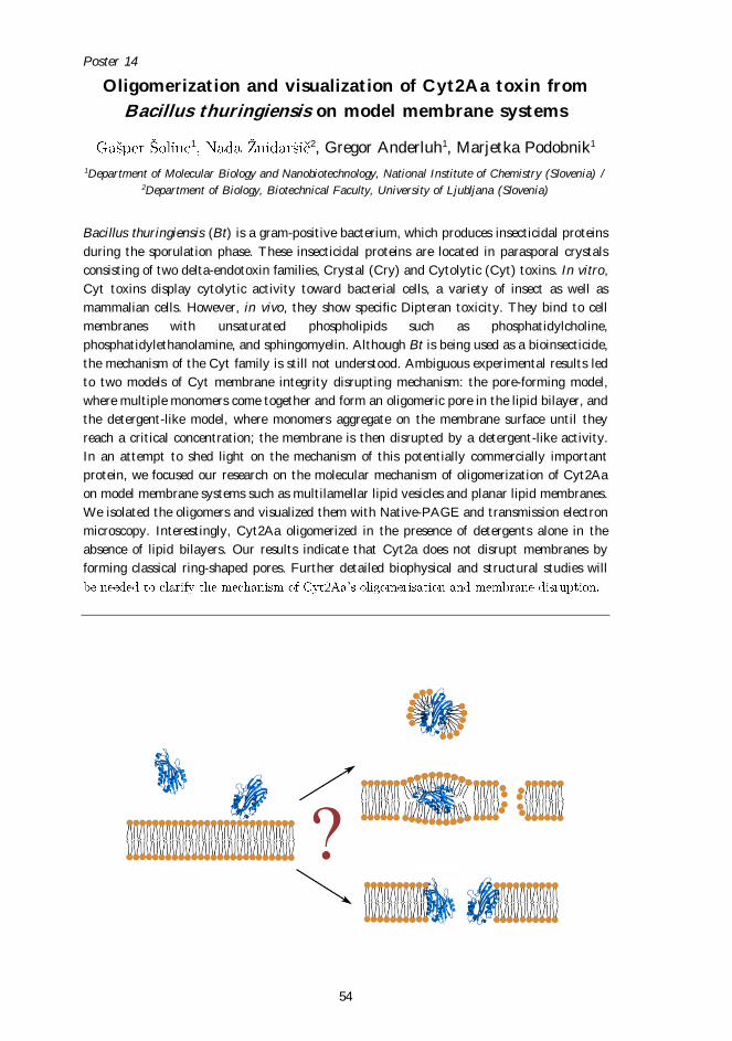
Poster 14
54
Oligomerization and visualization of Cyt2Aa toxin from
Bacillus thuringiensis on model membrane systems
1 2, Gregor Anderluh1, Marjetka Podobnik1
1Department of Molecular Biology and Nanobiotechnology, National Institute of Chemistry (Slovenia) / 2Department of Biology, Biotechnical Faculty, University of Ljubljana (Slovenia)
Bacillus thuringiensis (Bt) is a gram-positive bacterium, which produces insecticidal proteins
during the sporulation phase. These insecticidal proteins are located in parasporal crystals
consisting of two delta-endotoxin families, Crystal (Cry) and Cytolytic (Cyt) toxins. In vitro,
Cyt toxins display cytolytic activity toward bacterial cells, a variety of insect as well as
mammalian cells. However, in vivo, they show specific Dipteran toxicity. They bind to cell
membranes with unsaturated phospholipids such as phosphatidylcholine,
phosphatidylethanolamine, and sphingomyelin. Although Bt is being used as a bioinsecticide,
the mechanism of the Cyt family is still not understood. Ambiguous experimental results led
to two models of Cyt membrane integrity disrupting mechanism: the pore-forming model,
where multiple monomers come together and form an oligomeric pore in the lipid bilayer, and
the detergent-like model, where monomers aggregate on the membrane surface until they
reach a critical concentration; the membrane is then disrupted by a detergent-like activity.
In an attempt to shed light on the mechanism of this potentially commercially important
protein, we focused our research on the molecular mechanism of oligomerization of Cyt2Aa
on model membrane systems such as multilamellar lipid vesicles and planar lipid membranes.
We isolated the oligomers and visualized them with Native-PAGE and transmission electron
microscopy. Interestingly, Cyt2Aa oligomerized in the presence of detergents alone in the
absence of lipid bilayers. Our results indicate that Cyt2a does not disrupt membranes by
forming classical ring-shaped pores. Further detailed biophysical and structural studies will

List of Participants
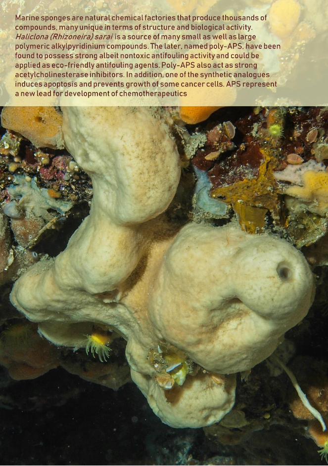
57
Gregor Anderluh Department of Molecular Biology and Nanobiotechnology National Institute of Chemistry Hajdrihova 19 1000 Ljubljana, Slovenia [email protected]
Gregor Bajc Department of Biology Biotechnical Faculty, University of Ljubljana
1000 Ljubljana, Slovenia [email protected] - l j .s i Apolonija Bedina Zavec Department of Molecular Biology and Nanobiotechnology National Institute of Chemistry Hajdrihova 19 1000 Ljubljana, Slovenia [email protected] Sabina Berne Department of Agronomy Biotechnical Faculty, University of Ljubljana Jamnikarjeva 101 1000 Ljubljana, Slovenia [email protected] - lj .s i Matej Butala Department of Biology Biotechnical Faculty, University of Ljubljana
1000 Ljubljana, Slovenia matej [email protected] - lj .s i Klara Bulc Rozman Institute of Pathophysiology Faculty of Medicine, University of Ljubljana Vrazov trg 2 1000 Ljubljana, Slovenia [email protected] Mauro Dalla Serra Institute of Biophysics Ital ian National Research Council , CNR via Sommarive 18 38123 Trento, Italy [email protected]
Mojca Dobaja Borak Center for Clinical Toxicology and Pharmacology University Medical Centre Ljubljana
1000 Ljubljana, Slovenia mojca.dobaja@kclj .s i
Maja Grundner Department of Biology Biotechnical Faculty, University of Ljubljana
pot 111 1000 Ljubljana, Slovenia [email protected] - lj .s i Ioan Iacovache Institute of Anatomy University of Bern Baltzerstrasse 2 3012 Bern, Switzerland [email protected]
Department of Molecular and Biomedical Sciences
Jamova cesta 39 1000 Ljubljana, Slovenia [email protected]
Department of Molecular Biology and Nanobiotechnology National Institute of Chemistry Hajdrihova 19 1000 Ljubljana, Slovenia [email protected] R. Manjunatha Kini Department of Biological Sciences Faculty of Science, National University of Singapore Science Drive 14 117543 Singapore [email protected] Matic Kisovec Department of Molecular Biology and Nanobiotechnology National Institute of Chemistry Hajdrihova 19 1000 Ljubljana, Slovenia [email protected]
Department of Chemistry and Biochemistry Faculty of Chemistry and Chemical Technology, University of Ljubljana
1000 Ljubljana, Slovenia [email protected] - lj .s i
Toshihide Kobayashi Laboratory of Bioimaging and Pathologies CNRS, University of Strasbourg Strasbourg, France [email protected]
58
Department of Biology Biotechnical Faculty, University of Ljubljana
1000 Ljubljana, Slovenia [email protected] - l j .si
Department of Molecular and Biomedical Sciences
Jamova cesta 39 1000 Ljubljana, Slovenia [email protected] i Mirijam Kozorog Department of Molecular Biology and Nanobiotechnology National Institute of Chemistry Hajdrihova 19 1000 Ljubljana, Slovenia [email protected]
Department of Molecular Biology and Nanobiotechnology National Institute of Chemistry Hajdrihova 19 1000 Ljubljana, Slovenia [email protected]
Department of Molecular and Biomedical Sciences
Jamova cesta 39 1000 Ljubljana, Slovenia [email protected] i Magdalena Kulma Institute of Biochemistry and Biophysics Polish Academy of Sciences
05-077 Warsaw, Poland magdakulma@gmail .com Adrijana Leonardi Department of Molecular and Biomedical Sciences
Jamova cesta 39 1000 Ljubljana, Slovenia [email protected] i
Department of Biology Biotechnical Faculty, University of Ljubljana
1000 Ljubljana, Slovenia [email protected] - lj .s i
Mojca Mally Institute of Biophysics Faculty of Medicine, University of Ljubljana Vrazov trg 2 1000 Ljubljana , Slovenia [email protected] - lj .s i Maria Vittoria Modica Institute of Biomolecules Max Mousseron University of Montpell ier CC 1703, Place Eugène Batail lon 34095 Montpell ier , France maria-vittoria.modica@umontpell ier .fr Cesare Montecucco Department of Biomedical Sciences and Neuroscience University of Padova Via Ugo Bassi 58/B 3512 Padova, Italy [email protected]
Department of Molecular and Biomedical Sciences
Jamova cesta 39 1000 Ljubljana, Slovenia [email protected] i
Department of Molecular Biology and Nanobiotechnology National Institute of Chemistry Hajdrihova 19 1000 Ljubljana, Slovenia [email protected] Nina Orehar Department of Biology Biotechnical Faculty, University of Ljubljana
a pot 111 1000 Ljubljana, Slovenia [email protected] - lj .s i
Department of Biology Biotechnical Faculty, University of Ljubljana
1000 Ljubljana, Slovenia [email protected]
Anastasija Panevska Department of Biology Biotechnical Faculty, University of Ljubljana
1000 Ljubljana, Slovenia [email protected] - lj .s i
59
Anja Pavlin Department of Biology Biotechnical Faculty, University of Ljubljana
1000 Ljubljana, Slovenia [email protected] - l j .s i
Department of Molecular Biology and Nanobiotechnology National Institute of Chemistry Hajdrihova 19 1000 Ljubljana, Slovenia [email protected] Katja Pirc Department of Molecular Biology and Nanobiotechnology National Institute of Chemistry Hajdrihova 19 1000 Ljubljana, Slovenia [email protected] Marjetka Podobnik Department of Molecular Biology and Nanobiotechnology National Institute of Chemistry Hajdrihova 19 1000 Ljubljana, Slovenia [email protected]
Department of Food Science and Technology Biotechnical Faculty, University of Ljubljana Jamnikarjeva 101 1000 Ljubljana, Slovenia [email protected] - lj .s i
Department of Molecular and Biomedical Sciences
Jamova cesta 39 1000 Ljubljana, Slovenia kity.pozek@gmail .com
Institute of Pathophysiology Faculty of Medicine, University of Ljubljana
1000 Ljubljana, Slovenia [email protected] - lj .s i
Department of Molecular and Biomedical Sciences
Jamova cesta 39 1000 Ljubljana, Slovenia [email protected]
Institute of Cell biology Faculty of Medicine, University of Ljubljana Vrazov trg 2 1000 Ljubljana, Slovenia [email protected] - lj .s i Michael Richardson Institute of Biology Leiden Leiden University Rapenburg 70 2311 EZ Leiden, The Netherlands [email protected] Samuel Ristovski Department of Biology Biotechnical Faculty, University of Ljubljana
1000 Ljubljana, Slovenia samuel.r istovski@gmail .com
Department of Biotechnology
Jamova cesta 39 1000 Ljubljana, Slovenia jer [email protected] i Kri Department of Biology Biotechnical Faculty, University of Ljubljana
1000 Ljubljana, Slovenia krist [email protected] - lj .s i Tina Snoj Department of Molecular Biology and Nanobiotechnology National Institute of Chemistry Hajdrihova 19 1000 Ljubljana, Slovenia t [email protected]
Department of Molecular Biology and Nanobiotechnology National Institute of Chemistry Hajdrihova 19 1000 Ljubljana, Slovenia [email protected]
Department of Molecular Biology and Nanobiotechnology National Institute of Chemistry Hajdrihova 19 1000 Ljubljana, Slovenia gasper.sol [email protected]
60
Department of Molecular and Biomedical Sciences
Jamova cesta 39 1000 Ljubljana, Slovenia jernej .sr [email protected]
Department of Biotechnology Biotechnical Faculty, University of Ljubljana Jamnikarjeva 101 1000 Ljubljana, Slovenia [email protected]
Institute of Pathophysiology, Faculty of Medicine, University of Ljubljana
1000 Ljubljana, Slovenia [email protected] - lj .s i
Institute of Biophysics Faculty of Medicine, University of Ljubljana Vrazov trg 2 1000 Ljubljana, Slovenia [email protected] - lj .s i Tom Turk Department of Biology Biotechnical Faculty, University of Ljubljana
1000 Ljubljana, Slovenia [email protected] - l j .si
Department of Biology Biotechnical Faculty, University of Ljubljana
1000 Ljubljana, Slovenia valerija.vezocnik@gmail .com Benoît Zuber Institute of Anatomy University of Bern Baltzerstrasse 2 3012 Bern, Switzerland benoit [email protected]
Department of Molecular and Biomedical Sciences
Jamova cesta 39 1000 Ljubljana, Slovenia [email protected]
61
Author Index
Gregor Anderluh, 20, 23, 31, 36, 38, 42, 45, 46,
48, 49, 51, 53, 54
Zoran Arsov, 47 Apolonija Bedina Zavec, 42
Sabina Berne, 41
Miran Brvar, 43
Klara Bulc Rozman, 16
Matej Butala, 23, 45 Simon Caserman, 23
Mauro Dalla Serra, 25 Dominik Dekleva, 49
Jure Derganc, 34
Mojca Dobaja Borak, 43 Marko Dolinar, 32
Mateja Erdani Kreft, 49
Marco Gerdol, 35
Samuele Greco, 35
Vesna Hodnik, 31, 33, 45 Halil I. Okur, 33
Ioan Iacovache, 27
, 44 Maja Jamnik, 23, 42
Matic Kisovec, 42, 49
32 Toshihide Kobayashi, 19
, 45
Ksenija Kogej, 33 22
Janko Kos, 50
, 45 Mirijam Kozorog, 20, 38
Veronika Kralj- , 42
Simona Kranjc, 41 46
, 15, 18, 23, 43, 44
Magdalena Kulma, 37 Katarzyna Kwiatkowska, 37
Vid Leban, 43
, 31, 45
Adrijana Leonardi, 18, 23, 43
Janja Majhenc, 34
Mojca Mally, 34 R. Manjunatha Kini, 14
, 47
Damijana Mojca, 16
Cesare Montecucco, 28
36
Anastasija Panevska, 46, 47
48 Katja Pirc, 31, 51
Janez Plavec, 38
Zdravko Podlesek, 23 Marjetka Podobnik, 20, 26, 31, 42, 45, 46, 48,
49, 53, 54
20 18
Jaka Razinger, 47
49 Michael Richardson, 21
Sylvie Roke, 33
50 Tamara Sajevic, 18
Marc-Antoine Sani, 38
Katja Seme, 23 Frances Separovic, 38
24, 33, 45, 46, 47
Simona Sitar, 33 Tina Snoj, 31, 51
52
53 54
, 42
, 44 , 16
Larisa Tratnjek, 49 Tom Turk, 17, 41
-
Aleksandra Uzar, 32 , 44, 49
33
Maria Vittoria Modica, 35
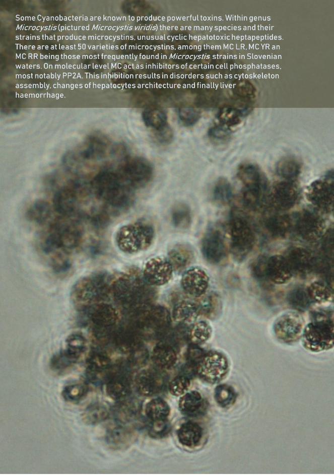
62
63

64


![Υψίσυχνος Αερισμός [High Frequency Oscillation (HFO)] · Targets ARDS • Gas Exchange • Lung but also RV PROTECTION . Traumatic Brain Injury (TBI) • Maintain](https://static.fdocument.org/doc/165x107/5e9abd7ece9fb21b0444bac5/f-foe-high-frequency-oscillation-hfo-targets-ards.jpg)