E3 ubiquitin ligase Mule targets β-catenin under …E3 ubiquitin ligase Mule targets β-catenin...
10
E3 ubiquitin ligase Mule targets β-catenin under conditions of hyperactive Wnt signaling Carmen Dominguez-Brauer a , Rahima Khatun b , Andrew J. Elia a , Kelsie L. Thu a , Parameswaran Ramachandran a , Shakiba P. Baniasadi a , Zhenyue Hao a , Lisa D. Jones a , Jillian Haight a , Yi Sheng b,1 , and Tak W. Mak a,1 a The Campbell Family Institute for Breast Cancer Research, Ontario Cancer Institute, University Health Network, Toronto, Ontario M5G 2C1, Canada; and b Department of Biology, York University, Toronto, Ontario M3J 1P3, Canada Contributed by Tak W. Mak, December 29, 2016 (sent for review December 2, 2016, reviewed by Christian Rask-Madsen and Natalie Rivard) Wnt signaling, named after the secreted proteins that bind to cell surface receptors to activate the pathway, plays critical roles both in embryonic development and the maintenance of homeostasis in many adult tissues. Two particularly important cellular programs orchestrated by Wnt signaling are proliferation and stem cell self- renewal. Constitutive activation of the Wnt pathway resulting from mutation or improper modulation of pathway components contrib- utes to cancer development in various tissues. Colon cancers fre- quently bear inactivating mutations of the adenomatous polyposis coli (APC) gene, whose product is an important component of the destruction complex that regulates β-catenin levels. Stabilization and nuclear localization of β-catenin result in the expression of a panel of Wnt target genes. We previously showed that Mule/ Huwe1/Arf-BP1 (Mule) controls murine intestinal stem and progen- itor cell proliferation by modulating the Wnt pathway via c-Myc. Here we extend our investigation of Mule’s influence on oncogen- esis by showing that Mule interacts directly with β-catenin and targets it for degradation under conditions of hyperactive Wnt signaling. Our findings suggest that Mule uses various mechanisms to fine- tune the Wnt pathway and provides multiple safeguards against tumorigenesis. β-catenin | Mule | Wnt signaling | stem cells | colorectal cancer A ctivation of the Wnt pathway, named after the secreted proteins that bind to cell surface receptors to activate the pathway, plays a critical role in stem cell self-renewal, and thus the homeostasis of normal mammalian tissues. However, consti- tutive activation of Wnt signaling has been implicated in a broad range of cancers, including melanoma, hepatocellular carcinoma, and cancers of the prostate, thyroid, ovary, and breast (1–3). Perhaps the best-studied malignancy arising from the loss of reg- ulation of the Wnt signaling pathway is colorectal cancer, which develops through a multistage process driven by the progressive accumulation of genetic mutations (4). The key initial activating mutation occurs in components of the Wnt pathway, but most commonly in APC. Canonical Wnt signaling involves a relay of protein interactions that serve to transmit a signal from the extracellular space to the plasma membrane. This signal is amplified in the cytoplasm and then directed to the nucleus to culminate in the activation of the Wnt target gene program. In molecular terms, when the glycoprotein Wnt is secreted into the extracellular space, it binds to a cell’ s Frizzled receptors in conjunction with the low-density lipoprotein receptor- related proteins 5 and 6 (LRP5/6). The engagement of these re- ceptors recruits the scaffolding protein Dishevelled (Dvl), leading to LRP5/6 phosphorylation and consequent recruitment of the scaffolding protein, Axin2 and glycogen synthase kinase 3 beta (GSK-3β). This recruitment results in disruption of the “destruction complex” that contains APC and normally binds to the signal transduction protein β-catenin to trigger its degradation. On Wnt signaling, however, β-catenin is released from the destruction complex, accumulates in the cytoplasm, and then translocates into the nucleus, where it associates with the T-cell factor/lymphoid enhancing factor transcription factors to activate the transcription of the battery of genes composing the Wnt program (5, 6). These genes influence cellular processes such as proliferation, differenti- ation, migration, and adhesion (7). Conversely, in the absence of Wnt, the destruction complex binding to β-catenin phosphorylates it and marks it for ubiquitination by the E3 ubiquitin (Ub) ligase β-TRCP, encoded by the beta-transducin repeat-containing gene. The ubiquitinated β-catenin is then targeted to the proteasome for degradation (5). Numerous mechanisms of positive and negative regulation have evolved to fine-tune the Wnt pathway. In a previous study (8), we showed that the E3 Ub ligase Mule (Huwe1/Arf-BP1) is critical for regulating the Wnt pathway in the intestine. By studying the effects of Mule deficiency in mice of an Apc competent (Mule conditional knockout, herein referred to as cKO) or Apc min back- ground (Mule cKO Apc min ), we demonstrated that a lack of Mule alone could promote tumorigenesis in the intestine, and that Mule deficiency accelerated adenoma development caused by the Apc min mutation. Our data established that Mule is a bona fide tumor suppressor in the gut. Our previous work also established that, in the normal in- testine, Mule regulates the protein levels of the receptor tyrosine kinase EphB3 by targeting it for proteasomal and lysosomal degradation. EphB/ephrinB interactions position cells along the intestinal crypt/villus axis and can compartmentalize incipient colorectal tumors. We further demonstrated that Mule controls murine intestinal stem and progenitor cell proliferation via its effects on c-Myc, which is a Mule substrate and a Wnt target. We found that c-Myc was up-regulated in Mule cKO mice, not only Significance Wnt signaling, named after the secreted proteins that bind to cell surface receptors to activate the pathway, is crucial for normal cell functions, and its deregulation can culminate in cancer. Several surveillance mechanisms have evolved as a precautionary measure to ensure proper development and the avoidance of cancer. Here we reveal that the E3 ubiquitin ligase Mule can target β-catenin for degradation to stop the Wnt signal during constitutive activation. Significantly, our data indicate that a combined loss of Mule and Apc accelerates the conversion of normal intestinal stem cells into cancer stem cells, paving the way to colorectal cancer develop- ment. It may be possible to use this knowledge to manipulate Mule, β-catenin, or Wnt pathway functions to reduce cancer initiation. Author contributions: C.D.-B. and Y.S. designed research; C.D.-B., R.K., A.J.E., K.L.T., P.R., S.P.B., Z.H., L.D.J., J.H., and Y.S. performed research; C.D.-B., Y.S., and T.W.M. analyzed data; and C.D.-B., Y.S., and T.W.M. wrote the paper. Reviewers: C.R.-M., Joslin Diabetes Center and Harvard Medical School; and N.R., Univer- sity of Sherbrooke. The authors declare no conflict of interest. Freely available online through the PNAS open access option. 1 To whom correspondence may be addressed. Email: [email protected] or yisheng@ yorku.ca. This article contains supporting information online at www.pnas.org/lookup/suppl/doi:10. 1073/pnas.1621355114/-/DCSupplemental. E1148–E1157 | PNAS | Published online January 30, 2017 www.pnas.org/cgi/doi/10.1073/pnas.1621355114 Downloaded by guest on February 28, 2020
Transcript of E3 ubiquitin ligase Mule targets β-catenin under …E3 ubiquitin ligase Mule targets β-catenin...
E3 ubiquitin ligase Mule targets β-catenin underconditions of hyperactive Wnt signalingCarmen Dominguez-Brauera, Rahima Khatunb, Andrew J. Eliaa, Kelsie L. Thua, Parameswaran Ramachandrana,Shakiba P. Baniasadia, Zhenyue Haoa, Lisa D. Jonesa, Jillian Haighta, Yi Shengb,1, and Tak W. Maka,1
aThe Campbell Family Institute for Breast Cancer Research, Ontario Cancer Institute, University Health Network, Toronto, Ontario M5G 2C1, Canada;and bDepartment of Biology, York University, Toronto, Ontario M3J 1P3, Canada
Contributed by Tak W. Mak, December 29, 2016 (sent for review December 2, 2016, reviewed by Christian Rask-Madsen and Natalie Rivard)
Wnt signaling, named after the secreted proteins that bind to cellsurface receptors to activate the pathway, plays critical roles both inembryonic development and the maintenance of homeostasis inmany adult tissues. Two particularly important cellular programsorchestrated by Wnt signaling are proliferation and stem cell self-renewal. Constitutive activation of the Wnt pathway resulting frommutation or improper modulation of pathway components contrib-utes to cancer development in various tissues. Colon cancers fre-quently bear inactivating mutations of the adenomatous polyposiscoli (APC) gene, whose product is an important component of thedestruction complex that regulates β-catenin levels. Stabilizationand nuclear localization of β-catenin result in the expression of apanel of Wnt target genes. We previously showed that Mule/Huwe1/Arf-BP1 (Mule) controls murine intestinal stem and progen-itor cell proliferation by modulating the Wnt pathway via c-Myc.Here we extend our investigation of Mule’s influence on oncogen-esis by showing that Mule interacts directly with β-catenin and targetsit for degradation under conditions of hyperactive Wnt signaling.Our findings suggest that Mule uses various mechanisms to fine-tune the Wnt pathway and provides multiple safeguards againsttumorigenesis.
β-catenin | Mule | Wnt signaling | stem cells | colorectal cancer
Activation of the Wnt pathway, named after the secretedproteins that bind to cell surface receptors to activate the
pathway, plays a critical role in stem cell self-renewal, and thusthe homeostasis of normal mammalian tissues. However, consti-tutive activation of Wnt signaling has been implicated in a broadrange of cancers, including melanoma, hepatocellular carcinoma,and cancers of the prostate, thyroid, ovary, and breast (1–3).Perhaps the best-studied malignancy arising from the loss of reg-ulation of the Wnt signaling pathway is colorectal cancer, whichdevelops through a multistage process driven by the progressiveaccumulation of genetic mutations (4). The key initial activatingmutation occurs in components of the Wnt pathway, but mostcommonly in APC.Canonical Wnt signaling involves a relay of protein interactions
that serve to transmit a signal from the extracellular space to theplasma membrane. This signal is amplified in the cytoplasm and thendirected to the nucleus to culminate in the activation of the Wnttarget gene program. In molecular terms, when the glycoproteinWntis secreted into the extracellular space, it binds to a cell’s Frizzledreceptors in conjunction with the low-density lipoprotein receptor-related proteins 5 and 6 (LRP5/6). The engagement of these re-ceptors recruits the scaffolding protein Dishevelled (Dvl), leadingto LRP5/6 phosphorylation and consequent recruitment of thescaffolding protein, Axin2 and glycogen synthase kinase 3 beta(GSK-3β). This recruitment results in disruption of the “destructioncomplex” that contains APC and normally binds to the signaltransduction protein β-catenin to trigger its degradation. On Wntsignaling, however, β-catenin is released from the destructioncomplex, accumulates in the cytoplasm, and then translocates intothe nucleus, where it associates with the T-cell factor/lymphoidenhancing factor transcription factors to activate the transcription
of the battery of genes composing the Wnt program (5, 6). Thesegenes influence cellular processes such as proliferation, differenti-ation, migration, and adhesion (7). Conversely, in the absence ofWnt, the destruction complex binding to β-catenin phosphorylatesit and marks it for ubiquitination by the E3 ubiquitin (Ub) ligaseβ-TRCP, encoded by the beta-transducin repeat-containing gene.The ubiquitinated β-catenin is then targeted to the proteasome fordegradation (5).Numerous mechanisms of positive and negative regulation have
evolved to fine-tune the Wnt pathway. In a previous study (8), weshowed that the E3 Ub ligase Mule (Huwe1/Arf-BP1) is criticalfor regulating the Wnt pathway in the intestine. By studying theeffects of Mule deficiency in mice of an Apc competent (Muleconditional knockout, herein referred to as cKO) or Apcmin back-ground (Mule cKO Apcmin), we demonstrated that a lack of Mulealone could promote tumorigenesis in the intestine, and that Muledeficiency accelerated adenoma development caused by the Apcmin
mutation. Our data established that Mule is a bona fide tumorsuppressor in the gut.Our previous work also established that, in the normal in-
testine, Mule regulates the protein levels of the receptor tyrosinekinase EphB3 by targeting it for proteasomal and lysosomaldegradation. EphB/ephrinB interactions position cells along theintestinal crypt/villus axis and can compartmentalize incipientcolorectal tumors. We further demonstrated that Mule controlsmurine intestinal stem and progenitor cell proliferation via itseffects on c-Myc, which is a Mule substrate and a Wnt target. Wefound that c-Myc was up-regulated in Mule cKO mice, not only
Significance
Wnt signaling, named after the secreted proteins that bind to cellsurface receptors to activate the pathway, is crucial for normal cellfunctions, and its deregulation can culminate in cancer. Severalsurveillance mechanisms have evolved as a precautionary measureto ensure proper development and the avoidance of cancer. Herewe reveal that the E3 ubiquitin ligaseMule can target β-catenin fordegradation to stop the Wnt signal during constitutive activation.Significantly, our data indicate that a combined loss of Mule andApc accelerates the conversion of normal intestinal stem cells intocancer stem cells, paving the way to colorectal cancer develop-ment. It may be possible to use this knowledge to manipulateMule, β-catenin, or Wnt pathway functions to reduce cancerinitiation.
Author contributions: C.D.-B. and Y.S. designed research; C.D.-B., R.K., A.J.E., K.L.T., P.R., S.P.B.,Z.H., L.D.J., J.H., and Y.S. performed research; C.D.-B., Y.S., and T.W.M. analyzed data; andC.D.-B., Y.S., and T.W.M. wrote the paper.
Reviewers: C.R.-M., Joslin Diabetes Center and Harvard Medical School; and N.R., Univer-sity of Sherbrooke.
The authors declare no conflict of interest.
Freely available online through the PNAS open access option.1To whom correspondence may be addressed. Email: [email protected] or [email protected].
This article contains supporting information online at www.pnas.org/lookup/suppl/doi:10.1073/pnas.1621355114/-/DCSupplemental.
E1148–E1157 | PNAS | Published online January 30, 2017 www.pnas.org/cgi/doi/10.1073/pnas.1621355114
Dow
nloa
ded
by g
uest
on
Feb
ruar
y 28
, 202
0
because of the absence of Mule-mediated c-Myc degradation butalso because of hyperactivated Wnt signaling.Previous work has shown that Mule regulates the Wnt pathway
in a negative feedback loop by ubiquitinating Dvl in a Wntligand-dependent manner (9). However, our previous examina-tion of Mule cKO Apcmin mice suggested that Mule targets othercomponents of the Wnt pathway. Here we demonstrate that Mulecan bind directly to β-catenin, the Wnt signal transducer. Mule-mediated β-catenin degradation occurs only under conditions ofcellular hyperproliferation, as would arise when APC mutationspermanently inactivate the destruction complex and allow β-cat-enin to stabilize and promote constitutive Wnt signaling. Ourfindings indicate that β-catenin degradation is an importantmechanism used by Mule under conditions of Wnt pathwayhyperactivation to execute its function as a tumor suppressor toprevent colon cancer.
ResultsLoss of Mule Accelerates Morphological Alterations in Apcmin Organoids.Previous in vitro work has shown that intestinal organoids estab-lished from tissue lacking a functional APC lose their crypt villusarchitecture and adopt an abnormal spheroid cyst-like morphology
(10), and that this altered morphology is a result of up-regulatedWnt signaling instructing the cells to adopt a proliferative pro-genitor phenotype (11). Our earlier study of intestinal adenomadevelopment in mice showed that Mule ablation on the Apcmin
background further increased Wnt signaling over that induced byApc mutation alone (8). Moreover, single cells isolated from ade-nomas that developed in Mule cKO Apcmin mice formed spheroidcysts more efficiently than cells isolated from Apcmin adenomas (8).These cystic organoids are reminiscent of the organoids grownfrom Lgr5-GFP-ires-CreERT2 X Apc fl/fl mice. The Lgr5-EGFP-IRES cre ERT2 “knock-in” allele ablates Lgr5 (leucine-rich repeat-containing G-coupled receptor 5) gene function and expresses GFPand the Cre ERT2 fusion protein. APCfl/fl are mice having anAPC-floxed allele. Tamoxifen-mediated cre recombinase activationresults in the deletion of Apc specifically in the Lgr5 stem cells (11).Loss of Apc in Lgr5 stem cells has been reported to be the cell oforigin of intestinal cancer (12). These findings suggested that loss ofMule under conditions of Wnt hyperactivity promotes stem cell pro-liferation and expansion. This prompted us to generate and furtherstudy intestinal organoids from Apcmin and Mule cKO Apcmin mice.Organoids were cultured from crypts isolated from Apcmin and
Mule cKO Apcmin mice. After 7 d in culture and 1 d after passaging,
Fig. 1. Mule-deficient Apcmin organoids instantly take on a cystic morphology. Bright-field microscopy of intestinal organoids that were derived from Apcmin
(A, B, F, and G) or Mule cKO Apcmin (C, D, H, and I) mice and cultured for 7 d (A–D) or 17 d (F–I). Low-magnification images (A, C, F, and H) and high-magnification images of the boxed areas (B, D, G, and I) are shown. (Scale bars, 100 μm.) (E) Quantification for Apcmin and Mule cKO Apcmin organoidsdisplaying a budded or cystic morphology on day 7 was obtained by counting four fields in three wells per sample (n = 2/genotype). (J) Quantification forApcmin and Mule cKO Apcmin organoids displaying a budded or cystic morphology on day 17. Data are representative of two independent experiments in3-mo-old mice. The data were also reproduced in a pair of 2-mo-old mice, albeit at different kinetics.
Dominguez-Brauer et al. PNAS | Published online January 30, 2017 | E1149
CELL
BIOLO
GY
PNASPL
US
Dow
nloa
ded
by g
uest
on
Feb
ruar
y 28
, 202
0
Apcmin organoids showed essentially normal morphology (Fig. 1 Aand B). At this point, only 1% of the organoids displayed the cysticmorphology (Fig. 1E). It was not until after 2 wk in culture that 99%of the organoids developed the cyst-like spheroid morphology (Fig.1 F, G, and J). In sharp contrast, after 7 d in culture, all the MulecKO Apcmin organoids already displayed the cyst-like morphology(Fig. 1 C–E). This altered morphology was retained by Mule cKOApcmin organoids for the full 2 wk of culture (Fig. 1 H–J). Inter-estingly, there were some Mule cKO Apcmin organoids that weresignificantly larger than average cystic organoids, demonstratingthat the loss of Mule further enhances proliferation (Fig. 1H and I).The fact that the concurrent loss of Mule caused Apcmin organoidsto display the morphology defect so quickly after establishmentsuggests that under conditions of Wnt hyperactivation, Mule has adirect regulatory effect on Wnt signaling. Further, the fact thatthese observations were made on an Apcmin background suggestsMule’s regulatory role is downstream of the destruction complex.
Loss of Mule in Addition to APC Results in Elevated β-Catenin Protein.Our previous immunohistochemical examinations of WT andMule cKO intestines revealed no differences in β-catenin staining(8). However, we suspected that Mule might regulate a Wntpathway element upstream of β-catenin because Wnt target geneswere up-regulated in Mule cKO organoids (8). Indeed, de Grootet al. reported that Mule-mediated K63-linked ubiquitination ofDvl normally inhibits Wnt pathway activation (9). Consistent withthis observation, we previously identified accumulation of nuclear
β-catenin in our Mule cKO adenomas (8), which could accountfor deregulation of the Wnt pathway in Mule-deficient cells atsteady-state. Because the Apcmin background precludes any studyof Mule-mediated regulation of Dvl, we focused on the onlyelement downstream of the destruction complex: β-catenin. Wefirst carried out immunoblotting analyses of WT (Mulefl/fl(y)) andMule cKO organoids, but found no differences in levels ofβ-catenin protein or Wnt target genes Axin2 and Cyclin D1 at theprotein level (Fig. 2A, quantification shown in the bar graph).Mule cKO Apcmin organoids displayed an increased level ofβ-catenin, Axin2, and Cyclin D1 protein compared with Apcmin
organoids (Fig. 2B, quantification shown in the bar graph). Thus,loss of Mule leads to excessive β-catenin accumulation in theabsence of a functional APC.
The Change in β-Catenin Protein Levels Translates into an ElevatedWnt Target Gene Expression. To determine the influence the in-creased levels of nuclear β-catenin have on global gene expression,we subjected RNA isolated from WT, Mule cKO, Apcmin, and MulecKO Apcmin organoids to comparative gene expression profiling.The heat map generated in Fig. 3A shows that Wnt target genesLgr5, Tnfrsf19, Mmp7 (metalloproteinase 7), and Rnf43 are amongthe genes most highly enriched in the Mule cKO Apcmin conditionwhen normalized to WT. Pathway analysis of the most highlyexpressed genes reported by our profiling was used to enlist the top10 enriched pathways from the KEGG (Kyoto Encyclopedia ofGenes and Genomes) Database (Fig. 3B). The width of the bar and
Fig. 2. Loss of Mule increases the β-catenin protein. (A and B) Immunoblots to detect the indicated proteins in lysates of intestinal organoids generated fromthe indicated mouse strains (n = 3 mice/group). β-actin, loading control. Relative protein fold change is shown in the bar graphs below each immunoblot.Values were determined by quantitation of bands by Image J, followed by normalization first to the loading control and then to Apcmin. n.s., not significant.*P < 0.05; **P < 0.01.
E1150 | www.pnas.org/cgi/doi/10.1073/pnas.1621355114 Dominguez-Brauer et al.
Dow
nloa
ded
by g
uest
on
Feb
ruar
y 28
, 202
0
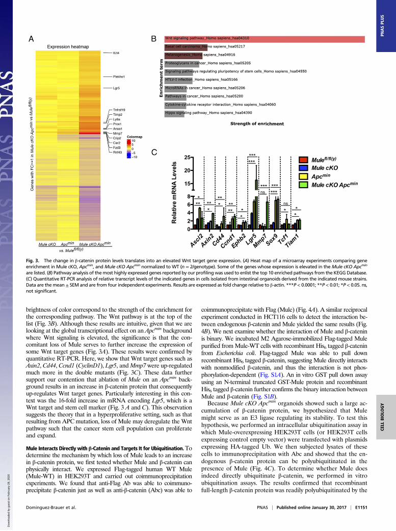
brightness of color correspond to the strength of the enrichment forthe corresponding pathway. The Wnt pathway is at the top of thelist (Fig. 3B). Although these results are intuitive, given that we arelooking at the global transcriptional effect on an Apcmin backgroundwhere Wnt signaling is elevated, the significance is that the con-comitant loss of Mule serves to further increase the expression ofsome Wnt target genes (Fig. 3A). These results were confirmed byquantitative RT-PCR. Here, we show that Wnt target genes such asAxin2, Cd44, Ccnd1 (CyclinD1), Lgr5, andMmp7 were up-regulatedmuch more in the double mutants (Fig. 3C). These data furthersupport our contention that ablation of Mule on an Apcmin back-ground results in an increase in β-catenin protein that consequentlyup-regulates Wnt target genes. Particularly interesting in this con-text was the 16-fold increase in mRNA encoding Lgr5, which is aWnt target and stem cell marker (Fig. 3 A and C). This observationsuggests the theory that in a hyperproliferative setting, such as thatresulting from APC mutation, loss of Mule may deregulate the Wntpathway such that the cancer stem cell population can proliferateand expand.
Mule Interacts Directly with β-Catenin and Targets It for Ubiquitination.Todetermine the mechanism by which loss of Mule leads to an increasein β-catenin protein, we first tested whether Mule and β-catenin canphysically interact. We expressed Flag-tagged human WT Mule(Mule-WT) in HEK293T and carried out coimmunoprecipitationexperiments. We found that anti-Flag Ab was able to coimmuno-precipitate β-catenin just as well as anti-β-catenin (Abc) was able to
coimmunoprecipitate with Flag (Mule) (Fig. 4A). A similar reciprocalexperiment conducted in HCT116 cells to detect the interaction be-tween endogenous β-catenin and Mule yielded the same results (Fig.4B). We next examine whether the interaction of Mule and β-cateninis binary. We incubated M2 Agarose-immobilized Flag-tagged Mulepurified fromMule-WT cells with recombinant His6 tagged β-cateninfrom Escherichia coli. Flag-tagged Mule was able to pull downrecombinant His6 tagged β-catenin, suggesting Mule directly interactswith nonmodified β-catenin, and thus the interaction is not phos-phorylation-dependent (Fig. S1A). An in vitro GST pull down assayusing an N-terminal truncated GST-Mule protein and recombinantHis6 tagged β-catenin further confirms the binary interaction betweenMule and β-catenin (Fig. S1B).Because Mule cKO Apcmin organoids showed such a large ac-
cumulation of β-catenin protein, we hypothesized that Mulemight serve as an E3 ligase regulating its stability. To test thishypothesis, we performed an intracellular ubiquitination assay inwhich Mule-overexpressing HEK293T cells (or HEK293T cellsexpressing control empty vector) were transfected with plasmidsexpressing HA-tagged Ub. We then subjected lysates of thesecells to immunoprecipitation with Abc and showed that the en-dogenous β-catenin protein can be polyubiquitinated in thepresence of Mule (Fig. 4C). To determine whether Mule doesindeed directly ubiquitinate β-catenin, we performed in vitroubiquitination assays. The results confirmed that recombinantfull-length β-catenin protein was readily polyubiquitinated by the
Fig. 3. The change in β-catenin protein levels translates into an elevated Wnt target gene expression. (A) Heat map of a microarray experiments comparing geneenrichment in Mule cKO, Apcmin, and Mule cKO Apcmin normalized to WT (n = 2/genotype). Some of the genes whose expression is elevated in theMule cKO Apcmin
are listed. (B) Pathway analysis of themost highly expressed genes reported by our profiling was used to enlist the top 10 enriched pathways from the KEGGDatabase.(C) Quantitative RT-PCR analysis of relative transcript levels of the indicated genes in cells isolated from intestinal organoids derived from the indicated mouse strains.Data are themean ± SEM and are from four independent experiments. Results are expressed as fold change relative to β-actin. ***P < 0.0001; **P < 0.01; *P < 0.05. ns,not significant.
Dominguez-Brauer et al. PNAS | Published online January 30, 2017 | E1151
CELL
BIOLO
GY
PNASPL
US
Dow
nloa
ded
by g
uest
on
Feb
ruar
y 28
, 202
0
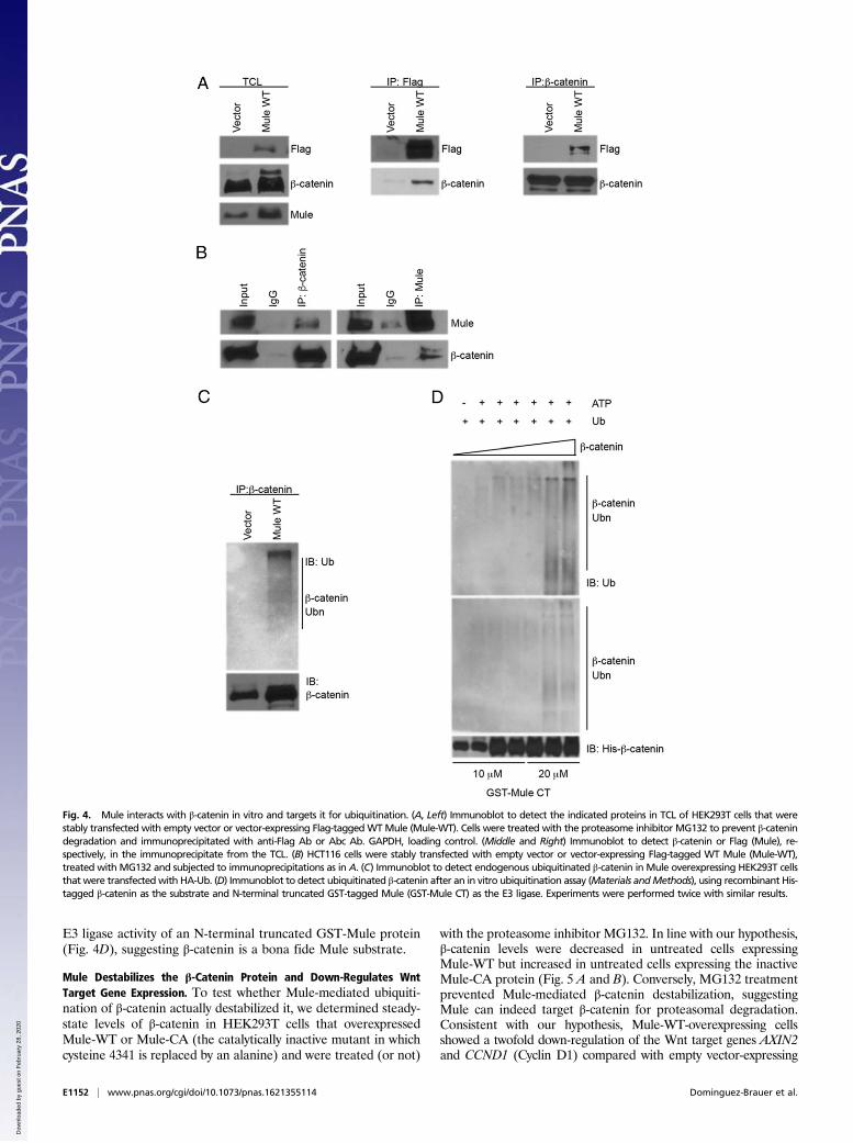
E3 ligase activity of an N-terminal truncated GST-Mule protein(Fig. 4D), suggesting β-catenin is a bona fide Mule substrate.
Mule Destabilizes the β-Catenin Protein and Down-Regulates WntTarget Gene Expression. To test whether Mule-mediated ubiquiti-nation of β-catenin actually destabilized it, we determined steady-state levels of β-catenin in HEK293T cells that overexpressedMule-WT or Mule-CA (the catalytically inactive mutant in whichcysteine 4341 is replaced by an alanine) and were treated (or not)
with the proteasome inhibitor MG132. In line with our hypothesis,β-catenin levels were decreased in untreated cells expressingMule-WT but increased in untreated cells expressing the inactiveMule-CA protein (Fig. 5 A and B). Conversely, MG132 treatmentprevented Mule-mediated β-catenin destabilization, suggestingMule can indeed target β-catenin for proteasomal degradation.Consistent with our hypothesis, Mule-WT-overexpressing cellsshowed a twofold down-regulation of the Wnt target genes AXIN2and CCND1 (Cyclin D1) compared with empty vector-expressing
Fig. 4. Mule interacts with β-catenin in vitro and targets it for ubiquitination. (A, Left) Immunoblot to detect the indicated proteins in TCL of HEK293T cells that werestably transfected with empty vector or vector-expressing Flag-taggedWTMule (Mule-WT). Cells were treated with the proteasome inhibitor MG132 to prevent β-catenindegradation and immunoprecipitated with anti-Flag Ab or Abc Ab. GAPDH, loading control. (Middle and Right) Immunoblot to detect β-catenin or Flag (Mule), re-spectively, in the immunoprecipitate from the TCL. (B) HCT116 cells were stably transfected with empty vector or vector-expressing Flag-tagged WT Mule (Mule-WT),treated with MG132 and subjected to immunoprecipitations as in A. (C) Immunoblot to detect endogenous ubiquitinated β-catenin in Mule overexpressing HEK293T cellsthat were transfectedwith HA-Ub. (D) Immunoblot to detect ubiquitinated β-catenin after an in vitro ubiquitination assay (Materials andMethods), using recombinant His-tagged β-catenin as the substrate and N-terminal truncated GST-tagged Mule (GST-Mule CT) as the E3 ligase. Experiments were performed twice with similar results.
E1152 | www.pnas.org/cgi/doi/10.1073/pnas.1621355114 Dominguez-Brauer et al.
Dow
nloa
ded
by g
uest
on
Feb
ruar
y 28
, 202
0
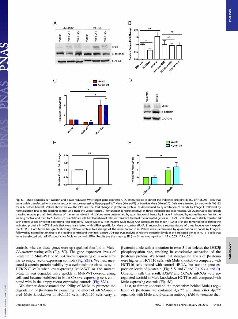
controls, whereas these genes were up-regulated fourfold in Mule-CA-overexpressing cells (Fig. 5C). The gene expression levels ofβ-catenin in Mule-WT or Mule-CA-overexpressing cells were sim-ilar to empty vector-expressing controls (Fig. S2A). We next mea-sured β-catenin protein stability by a cycloheximide chase assay inHEK293T cells when overexpressing Mule-WT or the mutant.β-catenin was degraded more quickly in Mule-WT-overexpressingcells and became stabilized in Mule-CA-overexpressing cells com-pared with in the empty vector-expressing controls (Fig. S2B).We further demonstrated the ability of Mule to promote the
degradation of β-catenin by examining the effect of siRNA-medi-ated Mule knockdown in HCT116 cells. HCT116 cells carry a
β-catenin allele with a mutation in exon 3 that deletes the GSK3βphosphorylation site, resulting in constitutive activation of theβ-catenin protein. We found that steady-state levels of β-cateninwere higher in HCT116 cells with Mule knockdown compared withHCT116 cells treated with control siRNA, but not the gene ex-pression levels of β-catenin (Fig. 5 D and E and Fig. S3 A and B).Consistent with this result, AXIN2 and CCND1 mRNAs were up-regulated twofold in Mule knockdown HCT116 cells compared withMule-expressing controls (Fig. 5F).Last, to further understand the mechanism behind Mule’s regu-
lation of β-catenin, we costained Apcmin and Mule cKO Apcmin
organoids with Mule and β-catenin antibody (Ab) to visualize their
Fig. 5. Mule destabilizes β-catenin and down-regulates Wnt target gene expression. (A) Immunoblot to detect the indicated proteins in TCL of HEK293T cells thatwere stably transfected with empty vector or vector-expressing Flag-tagged WT Mule (Mule-WT) or inactive Mule (Mule-CA). Cells were treated (or not) with MG132for 6 h before harvest. Values shown below the blot are the fold change in β-catenin protein, as determined by quantitation of bands by Image J, followed bynormalization first to the loading control and then the vector control. Immunoblot is representative of three independent experiments. (B) Quantitative bar graphshowing relative protein fold change of the immunoblot in A. Values were determined by quantitation of bands by Image J, followed by normalization first to theloading control and then to 293-Vec. (C) quantitative (q)RT-PCR analysis of relative transcript levels of the indicated genes in HEK293T cells that were stably transfectedwith empty vector or vector-expressing Flag-taggedWTMule (Mule-WT) or inactive Mule (Mule-CA). Results are the mean ± SD (n = 4). (D) Immunoblot to detect theindicated proteins in HCT116 cells that were transfected with siRNA specific for Mule or control siRNA. Immunoblot is representative of three independent experi-ments. (E) Quantitative bar graph showing relative protein fold change of the immunoblot in D. Values were determined by quantitation of bands by Image J,followed by normalization first to the loading control and then to si-Control. (F) qRT-PCR analysis of relative transcript levels of the indicated genes in HCT116 cells thatwere transfected with siRNA specific for Mule or control siRNA. Results are the mean ± SD (n = 3). ns, not significant. *P < 0.05; **P < 0.01.
Dominguez-Brauer et al. PNAS | Published online January 30, 2017 | E1153
CELL
BIOLO
GY
PNASPL
US
Dow
nloa
ded
by g
uest
on
Feb
ruar
y 28
, 202
0
colocalization. We were able to capture their colocalization in thenucleus (Fig. S4). The merged image for Apcmin appears purple,which is clearly absent in the Mule cKO Apcmin representativeorganoid shown. Collectively, these results establish that Mule canregulate the stability of β-catenin via ubiquitination, leading toproteasomal degradation.
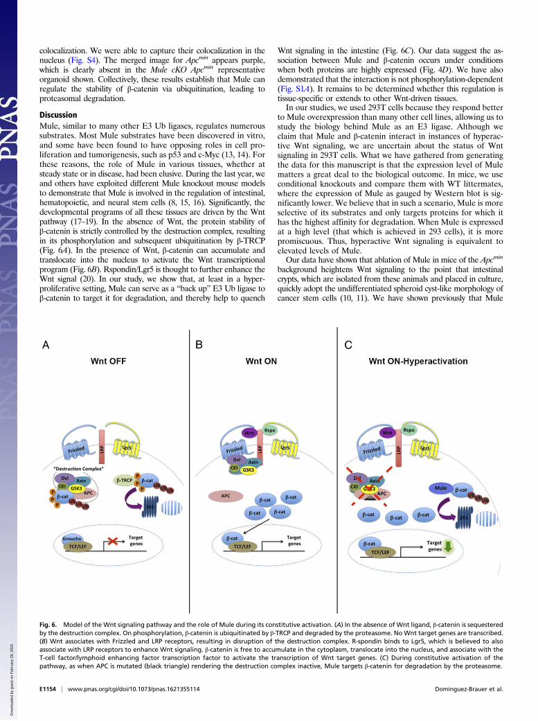
DiscussionMule, similar to many other E3 Ub ligases, regulates numeroussubstrates. Most Mule substrates have been discovered in vitro,and some have been found to have opposing roles in cell pro-liferation and tumorigenesis, such as p53 and c-Myc (13, 14). Forthese reasons, the role of Mule in various tissues, whether atsteady state or in disease, had been elusive. During the last year, weand others have exploited different Mule knockout mouse modelsto demonstrate that Mule is involved in the regulation of intestinal,hematopoietic, and neural stem cells (8, 15, 16). Significantly, thedevelopmental programs of all these tissues are driven by the Wntpathway (17–19). In the absence of Wnt, the protein stability ofβ-catenin is strictly controlled by the destruction complex, resultingin its phosphorylation and subsequent ubiquitination by β-TRCP(Fig. 6A). In the presence of Wnt, β-catenin can accumulate andtranslocate into the nucleus to activate the Wnt transcriptionalprogram (Fig. 6B). Rspondin/Lgr5 is thought to further enhance theWnt signal (20). In our study, we show that, at least in a hyper-proliferative setting, Mule can serve as a “back up” E3 Ub ligase toβ-catenin to target it for degradation, and thereby help to quench
Wnt signaling in the intestine (Fig. 6C). Our data suggest the as-sociation between Mule and β-catenin occurs under conditionswhen both proteins are highly expressed (Fig. 4D). We have alsodemonstrated that the interaction is not phosphorylation-dependent(Fig. S1A). It remains to be determined whether this regulation istissue-specific or extends to other Wnt-driven tissues.In our studies, we used 293T cells because they respond better
to Mule overexpression than many other cell lines, allowing us tostudy the biology behind Mule as an E3 ligase. Although weclaim that Mule and β-catenin interact in instances of hyperac-tive Wnt signaling, we are uncertain about the status of Wntsignaling in 293T cells. What we have gathered from generatingthe data for this manuscript is that the expression level of Mulematters a great deal to the biological outcome. In mice, we useconditional knockouts and compare them with WT littermates,where the expression of Mule as gauged by Western blot is sig-nificantly lower. We believe that in such a scenario, Mule is moreselective of its substrates and only targets proteins for which ithas the highest affinity for degradation. When Mule is expressedat a high level (that which is achieved in 293 cells), it is morepromiscuous. Thus, hyperactive Wnt signaling is equivalent toelevated levels of Mule.Our data have shown that ablation of Mule in mice of the Apcmin
background heightens Wnt signaling to the point that intestinalcrypts, which are isolated from these animals and placed in culture,quickly adopt the undifferentiated spheroid cyst-like morphology ofcancer stem cells (10, 11). We have shown previously that Mule
Fig. 6. Model of the Wnt signaling pathway and the role of Mule during its constitutive activation. (A) In the absence of Wnt ligand, β-catenin is sequesteredby the destruction complex. On phosphorylation, β-catenin is ubiquitinated by β-TRCP and degraded by the proteasome. NoWnt target genes are transcribed.(B) Wnt associates with Frizzled and LRP receptors, resulting in disruption of the destruction complex. R-spondin binds to Lgr5, which is believed to alsoassociate with LRP receptors to enhance Wnt signaling. β-catenin is free to accumulate in the cytoplasm, translocate into the nucleus, and associate with theT-cell factor/lymphoid enhancing factor transcription factor to activate the transcription of Wnt target genes. (C) During constitutive activation of thepathway, as when APC is mutated (black triangle) rendering the destruction complex inactive, Mule targets β-catenin for degradation by the proteasome.
E1154 | www.pnas.org/cgi/doi/10.1073/pnas.1621355114 Dominguez-Brauer et al.
Dow
nloa
ded
by g
uest
on
Feb
ruar
y 28
, 202
0
cKO organoids alone also adopt the same undifferentiated spheroidcyst-like morphology after 66 d in culture (8). We believe that thedifference in latency is the increasedWnt feedback that is present inthe Mule cKO Apcmin organoids. Although it would be interestingto speculate that the loss of Mule contributes to the acquisition ofmutations in Apc or other Wnt components to drive this phenotype,our previous RNA sequencing ofMule cKO adenomas suggests thisis not the case but, rather, the loss of Mule may function as the Wntinitiating event. Should this hold to be true, ourMule cKO organoidshould grow independent of exogenous R-spondin. This is a keyexperiment that cannot be carried out in organoids derived frommice on an Apcmin background. We have never conducted this ex-periment in our Mule cKO organoids, but expect to do so in thefuture. For now, we acknowledge that this phenotype may not beentirely a result of loss of Mule-mediated regulation of β-catenin,but we can conclude that Mule plays an important part in fine-tuning Wnt signaling in both the normal and diseased intestine.These findings are significant because Wnt signaling is the mainproliferative pathway in the gut that controls cell fate along thecrypt/villus axis (21). Thus, in the absence ofMule, constitutive Wntsignaling can drive stem cell expansion and the conversion of anormal stem cell into a cancer stem cell.Mule has been shown to be overexpressed in cancers of the
breast, lung, colon, liver, pancreas, thyroid, prostate, and larynx (14,22). It is also subject to somatic mutations, homozygous deletion,or amplification (www.cbioportal.org). In respect to colon cancer,Mule is overexpressed in 50% of all cases; moreover, its expressionis higher in tumor samples compared with nonmalignant tissue, andincreases with tumor grade (Fig. S5A) (14) (cancergenome.nih.gov/publications/publicationguidelines). Mutations within Mule’s HECT(homologous to the E6-AP carboxyl terminus) domain have beenidentified in patients with colon and rectal adenocarcinomas(sanger.ac.uk). We searched available databases to specifically de-termine whether mutations within Mule, whether in its functional(HECT) domain or elsewhere, would correlate differentially withexpression. Although we found no significant difference in expres-sion with mutational site, no conclusion can be made, given thesmall number of tumors reported (Fig. S5B). At this point, we favorthe hypothesis that a mutation within Mule, regardless of location,would alter its structure in such a way that it either will not effec-tively bind its substrates or will render it inactive.Further mutational analyses of available databases show that the
mutations that arise within Mule are classified as putative passengermutations. Although the label undermines the potential significanceof Mule in colon cancer, only about a dozen alterations are consid-ered functionally important and positively selected for during co-lorectal cancer (“putative driver”) (23). The fact that most tumorcohorts reported show that mutations within Mule occur in con-junction with mutations in APC would classify it as a redundant “notsignificant” hit on the Wnt pathway (Fig. S5C). Our data show,however, that loss of function of both proteins leads to rapid con-version of stem cells into cancer stem cells and consequent stem cellexpansion (Fig. 1 C and D) (8). Interestingly, the oncoprints gener-ated from cBioPortal show that some tumors only have mutationswithin Mule, not APC or β-catenin; we speculate that in these cases,Mule serves as the activating mutation within the Wnt pathway (Fig.S5C; www.cbioportal.org).Our genetic mouse knockout data thus far strongly support the
role of Mule being a tumor suppressor in the gut. We do ac-knowledge, however, that there are conflicting data published (14,24), and further acknowledge the possibility that Mule may functiondifferently if it is overexpressed. Thus, we are attempting to un-derstand whether the role of Mule in an established adenoma ortumor would differ from its role in adenoma initiation, and whetherMule overexpression in colon cancer results because of its signalingfeedback that may occur if mutations render it inactive, as is clas-sically the case for p53 in cancer (25).
In conclusion, our in vitro and in vivo efforts have shown thatMule’s regulation of the Wnt pathway extends to include its mainsignal transducer, β-catenin. We have demonstrated that Mule playsa vital role in fine-tuning the Wnt pathway in the normal intestine,as well as in helping extinguish hyperactive Wnt signaling in thediseased intestine. Thus, Mule has an indirect but important influ-ence on intestinal stem cell self-renewal and cell fate.
Materials and MethodsMice. Mulefl/fl(y) mice were as described (26) and were crossed to Villin-Cre (27)and Apcmin (28) mice to generate Mulefl/fl(y) Villin-Cre (Mule cKO) mice andMulefl/fl(y) Villin-Cre Apcmin (Mule cKO Apcmin) mice, respectively. All animal ex-periments were approved by the University Health Network Animal CareCommittee (AUP985).
Organoids.Mouse intestinal organoidswere established fromcrypts isolated fromthe proximal small intestine of 2–4-mo-old WT andMule cKO littermate mice, orApcmin and Mule cKO Apcmin littermate mice, as described (29). Organoids weremaintained in Advanced Dulbecco’s Modified Eagle’s medium/F12 supplementedwith 100 U/mL penicillin and streptomycin, 10 mM Hepes, 2 mM Glutamax, 1 ×B27, and 1 × N2 (all from Life Technologies). This base medium was furthersupplemented with 1 mM N-acetylcysteine (Sigma), 50 ng/mL murine recombi-nant EGF (Invitrogen), 100 ng/mL Noggin (Peprotech), and 5% (vol/vol) R-spon-din-1 conditioned medium (C. Kuo, Stanford University).
Cell Lines. HEK293T cells (ATCC) were cultured in DMEM H21 (Invitrogen).HCT116 cells (ATCC) were cultured in McCoy’s medium. Media were sup-plemented with 10% (vol/vol) FBS (Sigma) and penicillin/streptomycin.
Immunoblotting. Organoids were prepared for immunoblotting according topreviously reported protocols (30). Pelleted organoids were lysed in RIPAbuffer (150 mM NaCl, 1% Triton X-100, 0.5% sodium deoxycholate, 0.1%SDS, 50 mM Tris at pH 8.0) on ice for 30 min and centrifuged at 13,000 rpm(Eppendorf centrifuge 5417R, rotor 45-30-11) for 10 min. The protein con-tent of the supernatants was determined by BCA assay (Pierce). Proteinsamples (50 μg) were fractionated by 3–8% or 4–12% (vol/vol) reducing SDS/PAGE and transferred to PVDF membranes. Blots were incubated overnightwith the following primary Abs: anti-Huwe1 (Bethyl Laboratories), anti-Abc(Cell Signaling, #9562), anti-Cyclin D1 (Cell Signaling, #2978), anti-Axin2(abcam, ab32197), and anti-β-actin (Abcam). Primary Abs were detected withHRP-conjugated secondary Ab against rabbit IgG (GE Healthcare). Proteinswere visualized using enhanced luminol-based chemiluminescence (GEHealthcare). As appropriate, bands on immunoblots were quantified withImage J densitometric software.
Microarray: Analysis Procedure. Themicroarray gene-expression data from theAffymetrix scanner were read using the Oligo R package and then processeddownstream using the Limma R package from the Bioconductor suite of tools.The background-corrected and RMA-normalized intensity values were fittedinto linear models, after which fold changes of differential gene expressionbetween selected contrasts were computed [Mule cKO Apcmin, Apcmin, andMule cKO, all vs. Mulefl/fl(y) (WT)].
Heat Map. The gene expression fold change values from the reduced list of383 genes were also plotted as a heat map, with three columns showing thedifferent comparisons (Fig. 3A). The rows of the heat map are hierarchicallyclustered, using Euclidean distance and complete linkage. The most inter-esting genes, which are clearly up-regulated in Mule cKO Apcmin comparedwith Mulefl/fl(y), are labeled.
Pathway Analysis. The gene symbols of the most interesting genes that aremarked in the heat map were fed into the pathway analysis tool Enrichr (nar.oxfordjournals.org/content/early/2016/05/03/nar.gkw377.full.pdf). This toolcompares the input gene list against preannotated gene sets in its databaseand outputs ranked lists of enrichment terms (pathways, cell lines, or dis-eases), one per gene-set library. The most highly ranked enrichment termsfor the input gene list provide knowledge about the list. In our case, the toolwas used to enlist the top 10 enriched pathways in the KEGG Database.
qRT-PCR. RNA was isolated from organoids and cells with TRIzol (Invitrogen),according to the manufacturer’s instructions. RNA was processed by RT-PCRas described (8). Primer sequences are available on request.
Dominguez-Brauer et al. PNAS | Published online January 30, 2017 | E1155
CELL
BIOLO
GY
PNASPL
US
Dow
nloa
ded
by g
uest
on
Feb
ruar
y 28
, 202
0
Immunoprecipitation. Cellular interaction of Mule and β-catenin was carriedout in HEK293T stable cell lines overexpressing Flag-tagged Mule or vector.Total cell lysates (TCL) were prepared in lysis buffer [50 mM Tri at pH 8.0,150 mM NaCl, 0.6% Nonidet P-40, 1× protease inhibitor mixture (Roche)].For immunoprecipitation studies, TCL were incubated at 4 °C overnight withanti-Flag M2 Ab (Sigma #F3165) to immunoprecipitate Flag-tagged Mule orincubated with an Abc Ab (CST #8480) to immunoprecipitate endogenousβ-catenin. Cellular interaction of endogenous Mule and β-catenin was car-ried out in HCT116 cell lines. TCL were incubated at 4 °C overnight with anti-Mule Ab (monoclone Ab 7BF12) to immunoprecipitate endogenous Mule orincubated with an Abc Ab (CST #8480) to immunoprecipitate endogenousβ-catenin. IgG was used as a negative control. In both cases, the immunecomplexes bound to Protein A/G resin were washed 5× in lysis buffer, de-tached from the agarose using SDS/PAGE loading buffer, and detected byimmunoblotting using anti-Flag (or anti-Mule) and Abc Abs.
In Vitro and GST Pull-Down Assays. Cell lysates of HEK293T overexpressingMule-WTor empty vector (3 mg) were mixed with 20 μL Anti-Flag M2 agarose (Sigma#M8823) precleared with 5% (wt/vol) BSA at 4 °C in 1 × PBS buffer for 2 h. Afterincubation, the Anti-Flag M2 agarose resin was washed extensively with a bufferof 50 mM Tris at pH 7.6, 500 mM NaCl, 10% (vol/vol) glycerol, 0.1% Triton X-100,and protease inhibitor mixture (Roche) and mixed with recombinant His6 taggedβ-catenin (20 μg) in 500 μL binding buffer containing 50 mM Tris at pH 7.6,150 mM NaCl, 10% (vol/vol) glycerol, 0.1% Triton X-100, and protease inhibitormixture at 4 °C for 2 h. After second incubation, Anti-FlagM2 agarose was washedextensively with the same buffer, and the resulting immobilized proteins weredetected using immunoblotting. GST pull-down assay was carried out by mixingan equal amount of recombinant GST-tagged N-terminal truncated Mule (3760–4374) and His6 tagged β-catenin (20 μg) in the binding buffer at 4 °C for 2 h. Afterextensive wash with the binding buffer, the immobilized proteins were elutedusing reduced glutathione and detected by immunoblotting using anti-His(Novagen #71841) or GST Abs (Thermo #8–326). Recombinant GST tagged N-ter-minal truncated Mule (3760-4374) and His6 tagged β-catenin were expressed inE. coli BL21 cells and purified using standard GST- and Ni-affinity chromatographymethods.
Protein Stability: Cycloheximide Chase Assay. Cells were cultured with cyclo-heximide at a concentration of 100 μg/mL and incubated for various hours. Cellswere harvested at the indicated points. Lysates were prepared and subjected toimmunoblotting analysis. The protein stability was determined by percentage ofβ-catenin remaining at an indicated point compared with the initial point. Datawere plotted from three independent trials.In vivo ubiquitination. HEK293T stable cell lines overexpressing FLAG-tagged WTMule or inactive Mule C4341A were cultured as described earlier. Near-confluentcells were transfected with pCDNA 3.1-HA-Ub, using PolyJet transfection reagent(FraggaBio), recovered after 6 h, and grown for a total of 48 h. Cells were treatedwith 25 μM MG132 for 4 h before harvesting, and TCL were prepared in lysisbuffer of 50 mM Tris at pH 8.0, 150 mM NaCl, 0.6% Nonidet P-40, 1 × proteaseinhibitor mixture (Roche) with 10 mM freshly prepared N-ethylmaleimide. β-cat-enin was immunoprecipitated from TCL by incubating with Abc Ab (CST #8480)and Protein A/G resin. Ubiquitinated β-catenin was detected on immunoblotsusing anti-Ub Ab (P4G7, Covance MMS-258R) and Abc Ab (CST #8480).In vitro ubiquitination. Recombinant His-tagged β-catenin (full-length), GST-Mule (3760-4374) were expressed in E. coli BL-21 DE3 strain and purified
using standard affinity purification methods. Ubiquitination was performedas previously described (22496338) by mixing 10 μM each E1, E2, and Ub andvarious amounts of GST-Mule as the E3 ligase and His-tagged β-catenin asthe substrate in reaction buffer (50 mM Tris at pH 7.6, 2 mM ATP, 5 mMMgCl2, 2.5 mM DTT). Reactions were carried out for 90 min at 30 °C andterminated by adding SDS/PAGE loading buffer. Ubiquinated β-catenin wasdetected by immunoblotting using anti-Ub (P4G7, Covance MMS-258R) andAbc (CST #8480) Abs.siRNA knockdown. HCT116 cells were transfected with siRNA against Mule(AAUUGCUAUGUCU CUGGGACA) (13) or negative control siRNA (both fromGenepharma). siRNAs were transfected into cells using Lipojet (SignaGen)according to the manufacturer’s protocol, and cells were harvested at 48 hposttransfection. Lysates were resolved by 7.5% (vol/vol) SDS/PAGE, followedby immunoblotting, as described earlier.
Immunostaining. Organoids were collected in cold medium, pelleted andresuspended in coldCell Recovery Solution (BD).After incubationon ice for 20minwith gentle mixing, organoids were washed in PBS and were allowed to settle bygravity to a pellet in the bottom of the tubes. The pellet was resuspended in4% (vol/vol) paraformaldehyde and incubated for 20 min with gentle mixing.Organoids were washed twice with 1 × PBS, resuspended in Histogel (ThermoScientific) according to manufacturer’s instructions, processed, and embedded inparaffin. Sections (4.5 μm) were rehydrated and incubated with blocking solu-tion for 30 min, then with β-catenin (CST #8480) and Mule (homemade mouse,clone 7B15) Abs overnight at 4 °C. Slides were washed with PBS and incubatedwith secondary Abs (Jackson Donkeyxmouse-Cy5 #715–175-150 and goatxrabbit-Cy3 #111–165-144) for 35 min at room temperature. After washing with PBS,they were treated with DAPI and mounted in Entellan mounting medium.Fluorescent images were made from an Olympus FV1000 confocal microscopeand Zeiss AxioImager wide-field fluorescence microscope.
Clinical Data Profiling. Normalized RNA-seq derived gene expression data weredownloaded from the University of California, Santa Cruz’s Cancer GenomicsBrowser (https://genome-cancer.ucsc.edu/proj/site/hgHeatmap/) (31). Genomicalterations affecting APC, CTNNB1, and HUWE1 were queried in publiclyavailable TCGA (The Cancer Genome Atlas), DFCI (Dana-Farber Cancer Insti-tute), and Genentech colorectal adenocarcinoma datasets using cBioPortal(cbioportal.org) (32–36). HUWE1 (Mule) mutation and microarray Z-score nor-malized expression data were downloaded from cBioPortal. Statistical analyseswere performed using GraphPad Prism software.
Statistical Analyses. P values for intergroup comparisons were determinedusing the unpaired Student t test. P < 0.05 was considered significant. Unlessotherwise indicated, all experiments contained 3 replicates per condition,and all experiments were repeated at least 3 times.
ACKNOWLEDGMENTS. We are grateful to K. Gill of the Genotyping Facility,Dr. Chao Lu of the Center for Applied Genomics at the Hospital for Sick Children,and the staff of the Animal Resource Centre at the Princess Margaret Hospital(Toronto, Ontario, Canada). We thank M. Saunders for scientific editing of themanuscript. We thank the TCGA Research Network for sharing their compre-hensive genomic data for colorectal adenocarcinoma tumors (cancergenome.nih.gov/publications/publicationguidelines). This research was supported by grantsfrom the Canadian Institutes of Health Research (to T.W.M.).
1. Polakis P (2000) Wnt signaling and cancer. Genes Dev 14(15):1837–1851.2. Brown AM (2001) Wnt signaling in breast cancer: Have we come full circle? Breast
Cancer Res 3(6):351–355.3. Rubinfeld B, et al. (1997) Stabilization of beta-catenin by genetic defects in melanoma
cell lines. Science 275(5307):1790–1792.4. Fearon ER, Vogelstein B (1990) A genetic model for colorectal tumorigenesis. Cell
61(5):759–767.5. Clevers H, Nusse R (2012) Wnt/β-catenin signaling and disease. Cell 149(6):1192–1205.6. Angers S, Moon RT (2009) Proximal events in Wnt signal transduction. Nat Rev Mol
Cell Biol 10(7):468–477.7. Colussi D, Brandi G, Bazzoli F, Ricciardiello L (2013) Molecular pathways involved in
colorectal cancer: Implications for disease behavior and prevention. Int J Mol Sci 14(8):
16365–16385.8. Dominguez-Brauer C, et al. (2016) Mule regulates the intestinal stem cell niche via the
Wnt pathway and targets EphB3 for proteasomal and lysosomal degradation. Cell
Stem Cell 19(2):205–216.9. de Groot RE, et al. (2014) Huwe1-mediated ubiquitylation of dishevelled defines a
negative feedback loop in the Wnt signaling pathway. Sci Signal 7(317):ra26.10. Fatehullah A, Appleton PL, Näthke IS (2013) Cell and tissue polarity in the intestinal
tract during tumourigenesis: Cells still know the right way up, but tissue organization
is lost. Philos Trans R Soc Lond B Biol Sci 368(1629):20130014.
11. Sato T, et al. (2011) Paneth cells constitute the niche for Lgr5 stem cells in intestinalcrypts. Nature 469(7330):415–418.
12. Barker N, et al. (2009) Crypt stem cells as the cells-of-origin of intestinal cancer.Nature 457(7229):608–611.
13. Chen D, et al. (2005) ARF-BP1/Mule is a critical mediator of the ARF tumor suppressor.Cell 121(7):1071–1083.
14. Adhikary S, et al. (2005) The ubiquitin ligase HectH9 regulates transcriptional acti-vation by Myc and is essential for tumor cell proliferation. Cell 123(3):409–421.
15. King B, et al. (2016) The ubiquitin ligase Huwe1 regulates the maintenance andlymphoid commitment of hematopoietic stem cells. Nat Immunol 17(11):1312–1321.
16. Urbán N, et al. (2016) Return to quiescence of mouse neural stem cells by degradationof a proactivation protein. Science 353(6296):292–295.
17. Gregorieff A, Clevers H (2005) Wnt signaling in the intestinal epithelium: From endodermto cancer. Genes Dev 19(8):877–890.
18. Lento W, Congdon K, Voermans C, Kritzik M, Reya T (2013) Wnt signaling in normaland malignant hematopoiesis. Cold Spring Harb Perspect Biol 5(2):a008011.
19. Lie DC, et al. (2005) Wnt signalling regulates adult hippocampal neurogenesis. Nature437(7063):1370–1375.
20. Schuijers J, Clevers H (2012) Adult mammalian stem cells: The role ofWnt, Lgr5 and R-spondins.EMBO J 31(12):2685–2696.
21. Radtke F, Clevers H (2005) Self-renewal and cancer of the gut: Two sides of a coin.Science 307(5717):1904–1909.
E1156 | www.pnas.org/cgi/doi/10.1073/pnas.1621355114 Dominguez-Brauer et al.
Dow
nloa
ded
by g
uest
on
Feb
ruar
y 28
, 202
0
22. Confalonieri S, et al. (2009) Alterations of ubiquitin ligases in human cancer and their
association with the natural history of the tumor. Oncogene 28(33):2959–2968.23. Wood LD, et al. (2007) The genomic landscapes of human breast and colorectal
cancers. Science 318(5853):1108–1113.24. Peter S, et al. (2014) Tumor cell-specific inhibition of MYC function using small mol-
ecule inhibitors of the HUWE1 ubiquitin ligase. EMBO Mol Med 6(12):1525–1541.25. Rodrigues NR, et al. (1990) p53 mutations in colorectal cancer. Proc Natl Acad Sci USA
87(19):7555–7559.26. Hao Z, et al. (2012) The E3 ubiquitin ligase Mule acts through the ATM-p53 axis to
maintain B lymphocyte homeostasis. J Exp Med 209(1):173–186.27. Madison BB, et al. (2002) Cis elements of the villin gene control expression in re-
stricted domains of the vertical (crypt) and horizontal (duodenum, cecum) axes of the
intestine. J Biol Chem 277(36):33275–33283.28. Su LK, et al. (1992) Multiple intestinal neoplasia caused by a mutation in the murine
homolog of the APC gene. Science 256(5057):668–670.
29. Sato T, et al. (2009) Single Lgr5 stem cells build crypt-villus structures in vitro withouta mesenchymal niche. Nature 459(7244):262–265.
30. Matano M, et al. (2015) Modeling colorectal cancer using CRISPR-Cas9-mediated en-gineering of human intestinal organoids. Nat Med 21(3):256–262.
31. Cline MS, et al. (2013) Exploring TCGA Pan-Cancer data at the UCSC Cancer GenomicsBrowser. Sci Rep 3:2652.
32. Cancer Genome Atlas Network (2012) Comprehensive molecular characterization ofhuman colon and rectal cancer. Nature 487(7407):330–337.
33. Giannakis M, et al. (2016) Genomic correlates of immune-cell infiltrates in colorectalcarcinoma. Cell Rep 17(4):1206.
34. Seshagiri S, et al. (2012) Recurrent R-spondin fusions in colon cancer. Nature 488(7413):660–664.
35. Cerami E, et al. (2012) The cBio cancer genomics portal: An open platform for ex-ploring multidimensional cancer genomics data. Cancer Discov 2(5):401–404.
36. Gao J, et al. (2013) Integrative analysis of complex cancer genomics and clinicalprofiles using the cBioPortal. Sci Signal 6(269):pl1.
Dominguez-Brauer et al. PNAS | Published online January 30, 2017 | E1157
CELL
BIOLO
GY
PNASPL
US
Dow
nloa
ded
by g
uest
on
Feb
ruar
y 28
, 202
0