ReBOSSIS : The First Cotton-Like Synthetic Biologic...
4
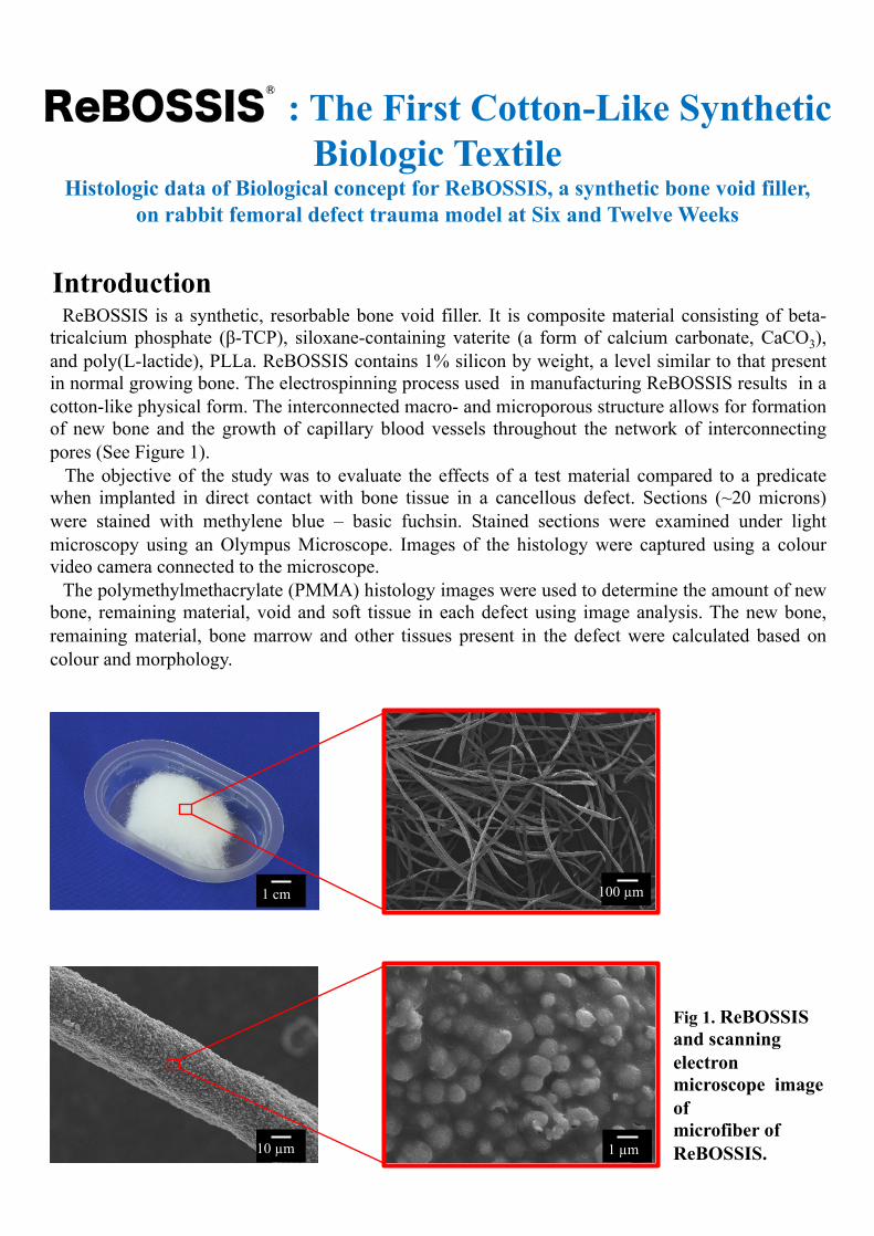
Introduction ReBOSSIS is a synthetic, resorbable bone void filler. It is composite material consisting of beta- tricalcium phosphate (β-TCP), siloxane-containing vaterite (a form of calcium carbonate, CaCO 3 ), and poly(L-lactide), PLLa. ReBOSSIS contains 1% silicon by weight, a level similar to that present in normal growing bone. The electrospinning process used in manufacturing ReBOSSIS results in a cotton-like physical form. The interconnected macro- and microporous structure allows for formation of new bone and the growth of capillary blood vessels throughout the network of interconnecting pores (See Figure 1). The objective of the study was to evaluate the effects of a test material compared to a predicate when implanted in direct contact with bone tissue in a cancellous defect. Sections (~20 microns) were stained with methylene blue – basic fuchsin. Stained sections were examined under light microscopy using an Olympus Microscope. Images of the histology were captured using a colour video camera connected to the microscope. The polymethylmethacrylate (PMMA) histology images were used to determine the amount of new bone, remaining material, void and soft tissue in each defect using image analysis. The new bone, remaining material, bone marrow and other tissues present in the defect were calculated based on colour and morphology. ReBOSSIS : The First Cotton-Like Synthetic Biologic Textile Histologic data of Biological concept for ReBOSSIS, a synthetic bone void filler, on rabbit femoral defect trauma model at Six and Twelve Weeks Fig 1. ReBOSSIS and scanning electron microscope image of microfiber of ReBOSSIS. 1 cm 100 µm 1 µm 10 µm ®
Transcript of ReBOSSIS : The First Cotton-Like Synthetic Biologic...
Introduction ReBOSSIS is a synthetic, resorbable bone void filler. It is composite material consisting of beta-tricalcium phosphate (β-TCP), siloxane-containing vaterite (a form of calcium carbonate, CaCO3), and poly(L-lactide), PLLa. ReBOSSIS contains 1% silicon by weight, a level similar to that present in normal growing bone. The electrospinning process used in manufacturing ReBOSSIS results in a cotton-like physical form. The interconnected macro- and microporous structure allows for formation of new bone and the growth of capillary blood vessels throughout the network of interconnecting pores (See Figure 1). The objective of the study was to evaluate the effects of a test material compared to a predicate when implanted in direct contact with bone tissue in a cancellous defect. Sections (~20 microns) were stained with methylene blue – basic fuchsin. Stained sections were examined under light microscopy using an Olympus Microscope. Images of the histology were captured using a colour video camera connected to the microscope. The polymethylmethacrylate (PMMA) histology images were used to determine the amount of new bone, remaining material, void and soft tissue in each defect using image analysis. The new bone, remaining material, bone marrow and other tissues present in the defect were calculated based on colour and morphology.
ReBOSSIS : The First Cotton-Like Synthetic Biologic Textile
Histologic data of Biological concept for ReBOSSIS, a synthetic bone void filler, on rabbit femoral defect trauma model at Six and Twelve Weeks
Fig 1. ReBOSSIS and scanning electron microscope image of microfiber of ReBOSSIS.
1 cm 100 µm
1 µm 10 µm
®
Actifuse Shape +
Blood 12 weeks
ReBOSSIS + Blood 6 weeks
ReBOSSIS + Blood 12 weeks
Actifuse Shape + Blood 6 weeks
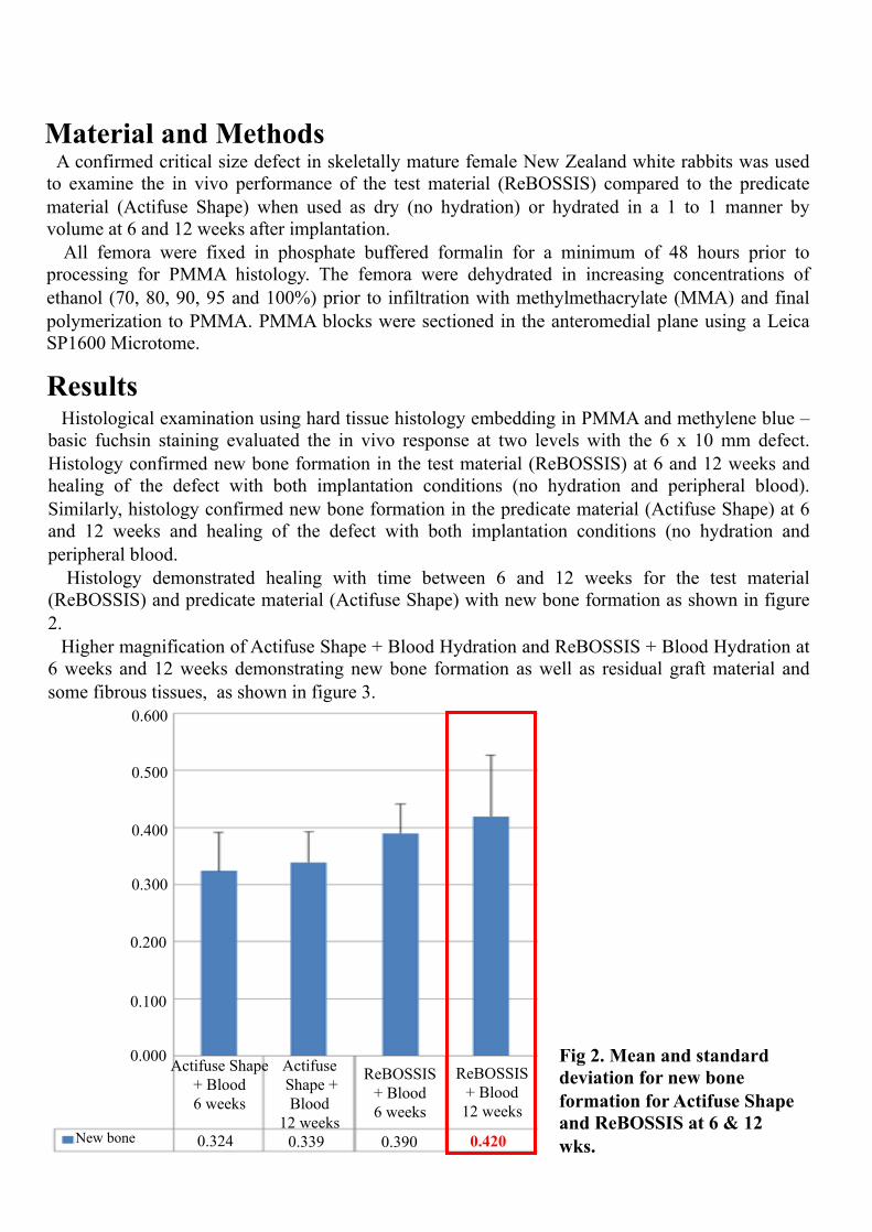
0.324 0.339 0.390 0.420 New bone
0.000
0.100
0.400
0.300
0.200
0.500
0.600
Material and Methods A confirmed critical size defect in skeletally mature female New Zealand white rabbits was used to examine the in vivo performance of the test material (ReBOSSIS) compared to the predicate material (Actifuse Shape) when used as dry (no hydration) or hydrated in a 1 to 1 manner by volume at 6 and 12 weeks after implantation. All femora were fixed in phosphate buffered formalin for a minimum of 48 hours prior to processing for PMMA histology. The femora were dehydrated in increasing concentrations of ethanol (70, 80, 90, 95 and 100%) prior to infiltration with methylmethacrylate (MMA) and final polymerization to PMMA. PMMA blocks were sectioned in the anteromedial plane using a Leica SP1600 Microtome.
Results
Fig 2. Mean and standard deviation for new bone formation for Actifuse Shape and ReBOSSIS at 6 & 12 wks.
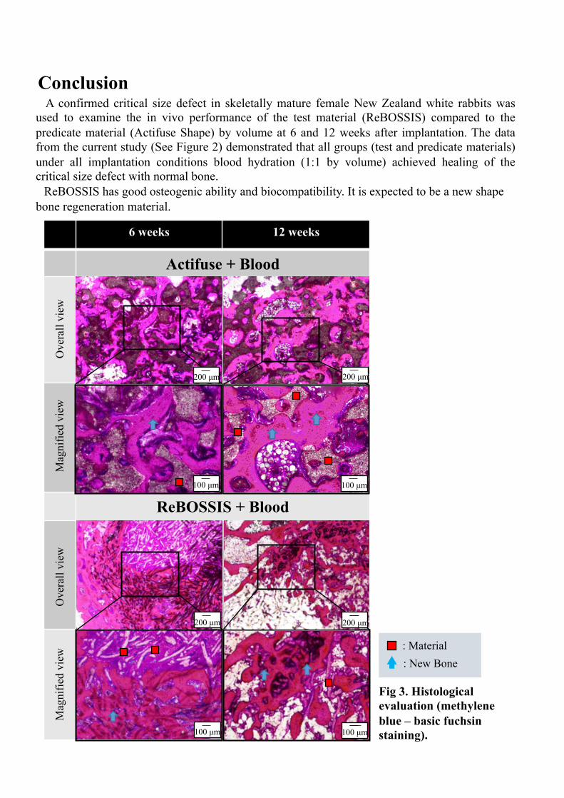
Histological examination using hard tissue histology embedding in PMMA and methylene blue – basic fuchsin staining evaluated the in vivo response at two levels with the 6 x 10 mm defect. Histology confirmed new bone formation in the test material (ReBOSSIS) at 6 and 12 weeks and healing of the defect with both implantation conditions (no hydration and peripheral blood). Similarly, histology confirmed new bone formation in the predicate material (Actifuse Shape) at 6 and 12 weeks and healing of the defect with both implantation conditions (no hydration and peripheral blood. Histology demonstrated healing with time between 6 and 12 weeks for the test material (ReBOSSIS) and predicate material (Actifuse Shape) with new bone formation as shown in figure 2. Higher magnification of Actifuse Shape + Blood Hydration and ReBOSSIS + Blood Hydration at 6 weeks and 12 weeks demonstrating new bone formation as well as residual graft material and some fibrous tissues, as shown in figure 3.
Conclusion A confirmed critical size defect in skeletally mature female New Zealand white rabbits was used to examine the in vivo performance of the test material (ReBOSSIS) compared to the predicate material (Actifuse Shape) by volume at 6 and 12 weeks after implantation. The data from the current study (See Figure 2) demonstrated that all groups (test and predicate materials) under all implantation conditions blood hydration (1:1 by volume) achieved healing of the critical size defect with normal bone. ReBOSSIS has good osteogenic ability and biocompatibility. It is expected to be a new shape bone regeneration material.
6 weeks 12 weeks
Actifuse + Blood
ReBOSSIS + Blood
200 µm
100 µm
200 µm 200 µm
100 µm 100 µm
Ove
rall
view
Mag
nifie
d vi
ew
Ove
rall
view
Mag
nifie
d vi
ew
Fig 3. Histological evaluation (methylene blue – basic fuchsin staining).
: Material : New Bone
100 µm
200 µm
Distributed by
ORTHOREBIRTH Co.,Ltd. 3-17-43 Chigasaki-Higashi, Tsuzuki-ku, Yokohama-shi, Kanagawa 224-0033, Japan E-mail : [email protected] URL : http://www.orthorebirth.com
ReBOSSIS®
Manufactured by
July 2015


![chimney [' ʧɪ mn ɪ ] tunnel ['t ʌ n( ə )l] chimney sweep [' ʧɪ mn ɪˌ swi ː p] orphan [' ɔː f( ə )n] cotton ['k ɔ t( ə )n] factory ['fækt(](https://static.fdocument.org/doc/165x107/5697c0031a28abf838cc3fd0/-chimney-mn-tunnel-t-n-l-chimney-sweep.jpg)