Quantitative mapping of the oxidative effects of mantle metasomatism
4
683 ABSTRACT The oxidation state of Fe in garnets in a garnet peridotite xeno- lith from the Wesselton kimberlite (South Africa) was quantitatively mapped using X-ray absorption near edge structure (XANES) spec- troscopy. Maps of Fe 3+ /ΣFe were produced by recording the fluores- cence intensity at discrete energies rather than recording the full spectrum at each point. The intensity at each point in the map was quantitatively converted to Fe 3+ /ΣFe with reference to a linear cali- bration derived from garnet standards for which Fe 3+ /ΣFe had been determined previously by Mössbauer spectroscopy. The resolution of these maps approaches that of elemental maps obtained using an electron microprobe. The maps reveal zoning in Fe 3+ /ΣFe between the core (0.075) and rim (0.125) that correlates with zoning of other ele- ments. The rims record an oxidizing metasomatic event in the litho- spheric mantle. The oxygen fugacity ( f O 2 ) of this metasomatism is con- siderably higher than expected from studies of homogeneous garnets that exhibit metasomatic signatures; such garnets may represent a re-equilibrated average of the original (core) and metasomatic (rim) f O 2 values. Metasomatism of the lithospheric mantle may thus have a greater impact on diamond stability than previously thought. INTRODUCTION The distribution of major and minor elements in geological samples can be mapped routinely using an electron microprobe (e.g., Yang and Rivers, 2001). These maps may identify not only differences in miner- alogy but also compositional zoning, intergrowths, exsolution, diffusion profiles, and inclusions, which may be interpreted in terms of geological processes or history. While most elements in the rock-forming minerals occur only in a single oxidation state (e.g., Si 4+ , Al 3+ , Ca 2+ , Mg 2+ , Na + , K + ), iron commonly occurs as both Fe 2+ and Fe 3+ . The proportions of Fe 2+ and Fe 3+ (expressed as Fe 3+ /[Fe 2+ + Fe 3+ ] or Fe 3+ /ΣFe) record the oxygen fugac- ity (f O 2 ) at which a mineral crystallized because the two oxidation states behave geochemically (e.g., during partitioning) as if they were different elements. For many minerals, including garnet, the multiplicity of sites and substitutions, together with the precision of electron probe analyses, mean that Fe 3+ /ΣFe can rarely be determined from stoichiometry. As a result, numerous techniques have been investigated for quantifying Fe 3+ / ΣFe, including redox titrations, Mössbauer spectroscopy, electron energy loss spectroscopy, and the electron microprobe flank method (see Berry et al., 2010, and references therein). These may all give accurate and precise values; however, due to either poor spatial resolution or the time taken for each point analysis, they are unsuitable for mapping Fe 3+ /ΣFe. X-ray absorption near edge structure (XANES) spectroscopy is a technique that is now commonly used for quantifying Fe 3+ /ΣFe (Wilke et al., 2001; Berry et al., 2003; Borfecchia et al., 2012), as well as the oxida- tion state ratios of other elements (e.g., Berry et al., 2006). XANES spec- tra can be acquired in air, with micron spatial resolution, from samples with a polished surface, such as thin sections and electron probe mounts (Delaney et al., 1998; Berry et al., 2008). Acquiring sufficient data to generate a useful map in a reasonable time requires the ability to record rapidly either spectra as a function of position (e.g., energy dispersive XANES; Munoz et al., 2006) or images as a function of energy (fluores- cence [Etschmann et al., 2010], attenuation [De Andrade et al., 2011]). While these capabilities exist, they are still being developed and are avail- able only at a limited number of synchrotron facilities. The most common approach is to record images at a small number of energies that corre- spond to spectral features characteristic of the oxidation state ratio (Sutton et al., 1995). This approach has been particularly useful for elements such as S (e.g., Métrich et al., 2009) and Se (e.g., Pickering et al., 2000), which exhibit relatively large spectral changes with oxidation state. The key to using this method for mapping Fe 3+ /ΣFe is to identify a feature that is sufficiently sensitive to changes in oxidation state to allow the relatively small differences in Fe 3+ /ΣFe seen in most mantle minerals and melts to be distinguished. Further, standards need to be available to convert the parameterization of the feature to values of Fe 3+ /ΣFe. No previous study of oxidation-state mapping has reported accuracies and precisions even remotely approaching ±0.01, which is the level required to meaningfully apply the results to garnet peridotites and many other geological samples. We have recently developed a XANES method for determining Fe 3+ / ΣFe in mantle garnets (Berry et al., 2010). This calibration makes use of the intensities of post-edge features at 7138.4 and 7161.7 eV rather than the energy of the 1s→3d pre-edge peak, which is conventionally used for quantification (Wilke et al., 2001). The accuracy and precision of the method are as good as that of the Fe 3+ /ΣFe values determined by Möss- bauer spectroscopy for the standards (±0.01). Garnets in some garnet peridotite xenoliths from the diamondifer- ous Wesselton kimberlite (South Africa) are zoned in major and trace elements (Griffin et al., 1999). Fe 3+ /ΣFe was determined by Mössbauer spectroscopy using a 400 μm analysis spot (each analysis took between 1 and 5 days), and found to be 0.05(2) in the core and 0.08(4) in the rim (average of four samples), although the core and rim values overlapped, within error, for each sample (McCammon et al., 2001). Such zonation results from metasomatism in the lithospheric mantle, which may impose f O 2 s that are sufficiently high to cause diamond breakdown or resorption (Griffin et al., 1999; McCammon et al., 2001). The presence of such zon- ing and the changes in f O 2 that it records are potentially important in using garnet as a diamond indicator mineral. Here we demonstrate the ability of XANES to produce quantitative maps of the distribution of Fe 3+ /ΣFe in mantle garnet, with a resolution that is broadly comparable to elemental maps obtained using an electron microprobe. EXPERIMENTAL METHODS A previously unstudied garnet lherzolite xenolith from the Wes- selton kimberlite was prepared as a polished section mounted in epoxy resin. Thin sections of garnet peridotite xenoliths from Kimberley (South Africa) and Lesotho and garnet separates (mounted in epoxy) from peri- dotite xenoliths from the Diavik kimberlite (Slave Craton, Canada) were used as standards. Fe 3+ /ΣFe of the standards was determined by Möss- bauer spectroscopy of powdered garnets obtained from the same samples used to make the thin sections and epoxy mounts (our unpublished data; Quantitative mapping of the oxidative effects of mantle metasomatism Andrew J. Berry 1 , Gregory M. Yaxley 2 , Brendan J. Hanger 2 , Alan B. Woodland 3 , Martin D. de Jonge 4 , Daryl L. Howard 4 , David Paterson 4 , and Vadim S. Kamenetsky 5 1 Department of Earth Science and Engineering, Imperial College London, South Kensington SW7 2AZ, UK 2 Research School of Earth Sciences, Australian National University, Canberra, ACT 0200, Australia 3 Institute für Geowissenschaften, Universität Frankfurt, Altenhöferallee 1, 60438 Frankfurt, Germany 4 Australian Synchrotron, Clayton, VIC 3168, Australia 5 ARC Centre of Excellence in Ore Deposits, University of Tasmania, Hobart, TAS 7001, Australia GEOLOGY, June 2013; v. 41; no. 6; p. 683–686; Data Repository item 2013188 | doi:10.1130/G34119.1 | Published online 16 April 2013 © 2013 Geological Society of America. For permission to copy, contact Copyright Permissions, GSA, or [email protected].
Transcript of Quantitative mapping of the oxidative effects of mantle metasomatism

GEOLOGY | June 2013 | www.gsapubs.org 683
ABSTRACTThe oxidation state of Fe in garnets in a garnet peridotite xeno-
lith from the Wesselton kimberlite (South Africa) was quantitatively mapped using X-ray absorption near edge structure (XANES) spec-troscopy. Maps of Fe3+/ΣFe were produced by recording the fl uores-cence intensity at discrete energies rather than recording the full spectrum at each point. The intensity at each point in the map was quantitatively converted to Fe3+/ΣFe with reference to a linear cali-bration derived from garnet standards for which Fe3+/ΣFe had been determined previously by Mössbauer spectroscopy. The resolution of these maps approaches that of elemental maps obtained using an electron microprobe. The maps reveal zoning in Fe3+/ΣFe between the core (0.075) and rim (0.125) that correlates with zoning of other ele-ments. The rims record an oxidizing metasomatic event in the litho-spheric mantle. The oxygen fugacity ( fO2
) of this metasomatism is con-siderably higher than expected from studies of homogeneous garnets that exhibit metasomatic signatures; such garnets may represent a re-equilibrated average of the original (core) and metasomatic (rim) fO2
values. Metasomatism of the lithospheric mantle may thus have a greater impact on diamond stability than previously thought.
INTRODUCTIONThe distribution of major and minor elements in geological samples
can be mapped routinely using an electron microprobe (e.g., Yang and Rivers, 2001). These maps may identify not only differences in miner-alogy but also compositional zoning, intergrowths, exsolution, diffusion profi les, and inclusions, which may be interpreted in terms of geological processes or history. While most elements in the rock-forming minerals occur only in a single oxidation state (e.g., Si4+, Al3+, Ca2+, Mg2+, Na+, K+), iron commonly occurs as both Fe2+ and Fe3+. The proportions of Fe2+ and Fe3+ (expressed as Fe3+/[Fe2+ + Fe3+] or Fe3+/ΣFe) record the oxygen fugac-ity (fO2
) at which a mineral crystallized because the two oxidation states behave geochemically (e.g., during partitioning) as if they were different elements. For many minerals, including garnet, the multiplicity of sites and substitutions, together with the precision of electron probe analyses, mean that Fe3+/ΣFe can rarely be determined from stoichiometry. As a result, numerous techniques have been investigated for quantifying Fe3+/ΣFe, including redox titrations, Mössbauer spectroscopy, electron energy loss spectroscopy, and the electron microprobe fl ank method (see Berry et al., 2010, and references therein). These may all give accurate and precise values; however, due to either poor spatial resolution or the time taken for each point analysis, they are unsuitable for mapping Fe3+/ΣFe.
X-ray absorption near edge structure (XANES) spectroscopy is a technique that is now commonly used for quantifying Fe3+/ΣFe (Wilke et al., 2001; Berry et al., 2003; Borfecchia et al., 2012), as well as the oxida-tion state ratios of other elements (e.g., Berry et al., 2006). XANES spec-tra can be acquired in air, with micron spatial resolution, from samples with a polished surface, such as thin sections and electron probe mounts (Delaney et al., 1998; Berry et al., 2008). Acquiring suffi cient data to generate a useful map in a reasonable time requires the ability to record
rapidly either spectra as a function of position (e.g., energy dispersive XANES; Munoz et al., 2006) or images as a function of energy (fl uores-cence [Etschmann et al., 2010], attenuation [De Andrade et al., 2011]). While these capabilities exist, they are still being developed and are avail-able only at a limited number of synchrotron facilities. The most common approach is to record images at a small number of energies that corre-spond to spectral features characteristic of the oxidation state ratio (Sutton et al., 1995). This approach has been particularly useful for elements such as S (e.g., Métrich et al., 2009) and Se (e.g., Pickering et al., 2000), which exhibit relatively large spectral changes with oxidation state. The key to using this method for mapping Fe3+/ΣFe is to identify a feature that is suffi ciently sensitive to changes in oxidation state to allow the relatively small differences in Fe3+/ΣFe seen in most mantle minerals and melts to be distinguished. Further, standards need to be available to convert the parameterization of the feature to values of Fe3+/ΣFe. No previous study of oxidation-state mapping has reported accuracies and precisions even remotely approaching ±0.01, which is the level required to meaningfully apply the results to garnet peridotites and many other geological samples.
We have recently developed a XANES method for determining Fe3+/ΣFe in mantle garnets (Berry et al., 2010). This calibration makes use of the intensities of post-edge features at 7138.4 and 7161.7 eV rather than the energy of the 1s→3d pre-edge peak, which is conventionally used for quantifi cation (Wilke et al., 2001). The accuracy and precision of the method are as good as that of the Fe3+/ΣFe values determined by Möss-bauer spectroscopy for the standards (±0.01).
Garnets in some garnet peridotite xenoliths from the diamondifer-ous Wesselton kimberlite (South Africa) are zoned in major and trace elements (Griffi n et al., 1999). Fe3+/ΣFe was determined by Mössbauer spectroscopy using a 400 μm analysis spot (each analysis took between 1 and 5 days), and found to be 0.05(2) in the core and 0.08(4) in the rim (average of four samples), although the core and rim values overlapped, within error, for each sample (McCammon et al., 2001). Such zonation results from metasomatism in the lithospheric mantle, which may impose fO2
s that are suffi ciently high to cause diamond breakdown or resorption (Griffi n et al., 1999; McCammon et al., 2001). The presence of such zon-ing and the changes in fO2
that it records are potentially important in using garnet as a diamond indicator mineral. Here we demonstrate the ability of XANES to produce quantitative maps of the distribution of Fe3+/ΣFe in mantle garnet, with a resolution that is broadly comparable to elemental maps obtained using an electron microprobe.
EXPERIMENTAL METHODSA previously unstudied garnet lherzolite xenolith from the Wes-
selton kimberlite was prepared as a polished section mounted in epoxy resin. Thin sections of garnet peridotite xenoliths from Kimberley (South Africa) and Lesotho and garnet separates (mounted in epoxy) from peri-dotite xenoliths from the Diavik kimberlite (Slave Craton, Canada) were used as standards. Fe3+/ΣFe of the standards was determined by Möss-bauer spectroscopy of powdered garnets obtained from the same samples used to make the thin sections and epoxy mounts (our unpublished data;
Quantitative mapping of the oxidative effects of mantle metasomatismAndrew J. Berry1, Gregory M. Yaxley2, Brendan J. Hanger2, Alan B. Woodland3, Martin D. de Jonge4, Daryl L. Howard4, David Paterson4, and Vadim S. Kamenetsky5
1Department of Earth Science and Engineering, Imperial College London, South Kensington SW7 2AZ, UK2Research School of Earth Sciences, Australian National University, Canberra, ACT 0200, Australia3Institute für Geowissenschaften, Universität Frankfurt, Altenhöferallee 1, 60438 Frankfurt, Germany4Australian Synchrotron, Clayton, VIC 3168, Australia5ARC Centre of Excellence in Ore Deposits, University of Tasmania, Hobart, TAS 7001, Australia
GEOLOGY, June 2013; v. 41; no. 6; p. 683–686; Data Repository item 2013188 | doi:10.1130/G34119.1 | Published online 16 April 2013
© 2013 Geological Society of America. For permission to copy, contact Copyright Permissions, GSA, or [email protected].

684 www.gsapubs.org | June 2013 | GEOLOGY
Woodland and Koch, 2003). Compositions and elemental maps were obtained using a Cameca SX-100 electron microprobe. The maps were acquired using an accelerating voltage of 15 kV, a beam current of 100 nA, and a step size of 2 μm.
Iron K-edge XANES spectra were recorded at the X-ray fl uores-cence microscopy (XFM) beamline of the Australian Synchrotron (Pat-erson et al., 2011). The excitation energy was selected using a Si(111) double-crystal monochromator and calibrated by defi ning the fi rst deriva-tive peak of an Fe foil spectrum to be 7112.0 eV. The spectral resolution was 1.9 eV at the Fe K-edge. The beam was focused by Kirkpatrick-Baez mirrors, which produced an analysis spot of ~3 × 3 μm for this study. Sam-ples were mounted at 45° to both the incident beam and a single-element silicon drift energy dispersive detector (Vortex EM, SII Nanotechnology, Northridge, California) with digital signal processing (DXP Saturn, XIA LLC, Hayward, California). All spectra were acquired in fl uorescence mode and the incident fl ux and sample-to-detector distance were adjusted to ensure that the total incoming count rate was within the linear range of the signal processing electronics. Spectra were recorded from 7075 to 7300 eV using step sizes of 0.5 eV for the baseline (7075–7103 eV), 0.3 eV for the edge, and 2.5 eV above 7175 eV, and count times of 3 s per point. Spectra were compared after subtraction of a constant baseline and normalization to the average intensity above 7235 eV. Maps of fl uores-cence intensity as a function of position were recorded for the Wesselton garnets by scanning the sample stage horizontally and vertically for exci-tation energies of 7075, 7138.4, 7161.7, and 7300 eV. Each of these maps was normalized to the beam intensity and then the map at 7075 eV (the baseline) was subtracted from the others. The baseline-corrected 7138.4 and 7161.7 eV maps were next normalized by the 7300 eV map (the total fl uorescence) to take account of variations in Fe content. The ratio of the resulting 7161.7 and 7138.4 eV maps is a map of Fe3+/ΣFe (see Berry et al., 2010). These manipulations were trivially performed using Microsoft Excel and required no specialized image-handling software.
RESULTSElemental maps of garnet (Wesselton) obtained using an electron
microprobe show zoning (Fig. 1); the rims are richer in Na, Ca, Ti, Cr,
and Fe but depleted in Al and Mg relative to the core (see Table DR1 in the GSA Data Repository1). Surrounding the rim in places is kelyphite, an intergrowth of mainly spinel and pyroxene formed by the breakdown of the garnet rim during ascent in the kimberlite magma. The garnet is surrounded by fi ner-grained olivine, orthopyroxene, and minor amounts of clinopyroxene.
Iron K-edge XANES spectra of the core and rim are shown in Fig-ure 2. The differences between the spectra are subtle. XANES spectra were also recorded for a number of standards, and the correlation between the intensity ratio of the features at 7138.4 and 7161.7 eV (E2 and E3 in Fig. 2) and Fe3+/ΣFe is shown in Figure 3. The standard deviation of the difference between each point and the linear fi t is ±0.01, which suggests an accuracy and precision comparable to that with which Fe3+/ΣFe was determined by Mössbauer spectroscopy for the standards (±0.01). This linear calibration can then be used to determine Fe3+/ΣFe of unknowns (e.g., Yaxley et al., 2012).
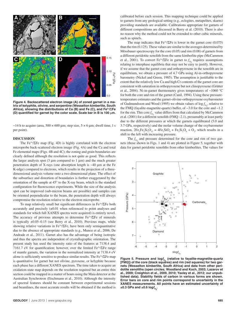
The distribution of Fe3+/ΣFe in a zoned garnet is shown in Figure 4D. The map corresponds to the ratio of the fl uorescence intensities at 7138.4 and 7161.7 eV, which can be converted to Fe3+/ΣFe using the calibration in Figure 3, and is thus quantitative. The color scale in Figure 4D indicates Fe3+/ΣFe for garnet but is not accurate for other minerals because the cali-bration is mineral specifi c. The average value of Fe3+/ΣFe obtained from the map is 0.075(15) for the core and 0.125(11) for the rim. The map took
A
D
B
C
0
1
2
7100 7200 7300
Nor
mal
ised
Inte
nsity
Energy (eV)
A
B
E2 E3 E4E1
0.00 0.05 0.10 0.15
Lesotho
Kimberley
Diavik
1.25
1.30
1.35
1.40
1.45
1.50
Fe3+/ΣFe
Inte
nsity
Rat
io
1GSA Data Repository item 2013188, Table DR1 (composition of phases in the sample of garnet peridotite from the Wesselton kimberlite used in this study), is available online at www.geosociety.org/pubs/ft2013.htm, or on request from [email protected] or Documents Secretary, GSA, P.O. Box 9140, Boulder, CO 80301, USA.
Figure 1. Elemental maps, acquired using an electron microprobe, showing distributions of Mg (A), Ca (B), Ti (C), and Fe (D) in a sample of garnet peridotite from the Wesselton kimberlite, South Africa. Im-ages (particularly C and D) show a zoned garnet, surrounded in places by kelyphite (pale blue in A and B, orange in C, and blue in D), in a matrix of olivine and orthopyroxene. Circular features in garnet are pits produced by laser ablation–inductively coupled plasma–mass spectrometry. Scale bar in C is 0.5 mm.
Figure 2. Fe K-edge XANES spectra of core (A) and rim (B) of the zoned garnet shown in Figure 4. Energies (E1–E4) at which images were recorded to produce Fe3+/ΣFe maps are indicated.
Figure 3. Correlation between the Fe K-edge XANES intensity ratio at 7138.4 and 7161.7 eV and Fe3+/ΣFe determined by Mössbauer spectros-copy of mantle garnets from various locations. Line is best fi t to data.
GEOLOGY | June 2013 | www.gsapubs.org 685
~14 h to acquire (area, 500 × 600 μm; step size, 5 × 6 μm; dwell time, 1 s per point).
DISCUSSIONThe Fe3+/ΣFe map (Fig. 4D) is highly correlated with the electron
microprobe back-scattered electron image (Fig. 4A) and the Ca and total Fe elemental maps (Figs. 4B and 4C); the zoning and grain boundaries are clearly defi ned although the resolution is not quite as good. This refl ects the larger analysis spot (3 μm compared to 1 μm) and the much greater penetration depth of X-rays (one absorption length is ~40 μm at the Fe K-edge) compared to electrons, which results in the projection of a three-dimensional analysis volume onto a two-dimensional plane. The effect of the subsurface and distortion of boundaries is further exaggerated by the orientation of the sample at 45° to the X-ray beam, which is the standard confi guration for fl uorescence experiments. While the size of the analysis spot can be improved (sub-micron beams are possible) and samples can be oriented perpendicular to the beam, the penetration depth will always compromise the resolution relative to the electron microprobe.
To map relatively small but signifi cant differences in Fe3+/ΣFe both accurately and precisely (±0.01 when referenced to point analyses and standards for which full XANES spectra were acquired) is entirely novel. The accuracy of previous attempts to determine Fe3+/ΣFe of minerals is typically ±0.05–0.15 (see Berry et al., 2010). Previous maps, while showing relative variations in Fe3+/ΣFe, have been only semiquantitative due to the absence of appropriate standards (e.g., Munoz et al., 2006; De Andrade et al., 2011). Garnet also has the advantage of being isotropic and thus the spectra are independent of crystallographic orientation. The present study has used the intensity ratio of the features at 7138.4 and 7161.7 eV for quantifi cation; however, over the limited Fe3+/ΣFe range of mantle garnets, the variation in the normalized intensity at 7138.4 eV alone is suffi ciently sensitive to produce similar results. The Fe3+/ΣFe map is quantitative for garnet but not olivine, pyroxene, or kelyphite because each phase has a different XANES spectrum. The time taken to acquire an oxidation-state map depends on the resolution required but an entire thin section could be mapped in a matter of hours using the Maia detector at the Australian Synchrotron (Etschmann et al., 2010). Although the intensity of spectral features should be constant between experimental sessions and beamlines, the most accurate results will be obtained if the method is
calibrated before each session. This mapping technique could be applied to garnets from any geological setting (e.g., eclogites, metapelites, skarns) providing standards are available. Calibrations appropriate for garnets of different compositions are discussed in Berry et al. (2010). There is also no reason why the method could not be extended to other cubic minerals, such as spinels.
The map indicates that Fe3+/ΣFe is lower in the garnet core (0.075) than the rim (0.125). These values are similar to the averages determined by Mössbauer spectroscopy for the core (0.05) and rim (0.08) of garnets from a different peridotite xenolith from the same kimberlite pipe (McCammon et al., 2001). To convert Fe3+/ΣFe in garnet to fO2
requires assumptions relating to interphase equilibria that may not be easy to justify. However, if we assume that the garnet core and orthopyroxene in the xenolith are in equilibrium, we obtain a pressure of 4.7 GPa using Al-in-orthopyroxene barometry (Nickel and Green, 1985). The assumption is justifi able to the extent that the relatively low Ca and high Cr contents of the garnet core are consistent with saturation in orthopyroxene but not clinopyroxene (Grütter et al., 2004). Ni-in-garnet thermometry gives temperatures of ~1060 °C for both the core and rim of the garnet (Canil, 1994). Using these pressure-temperature estimates and the garnet-olivine-orthopyroxene oxybarometer of Gudmundsson and Wood (1995) we obtain values of log fO2
, relative to the FMQ (fayalite-magnetite-quartz) buffer, of −3.0 for the core and −1.2 for the rim. This core fO2
value differs from that calculated by McCammon et al. (2001) for a different xenolith (FMQ –2.1), presumably at least partly due to the different pressures at which the garnets equilibrated (5.0 and 3.7 GPa, respectively) and the molar volume change of the oxybarometer reaction, 2Fe3Fe2Si3O12 = 4Fe2SiO4 + Fe2Si2O6 + O2, which results in a shift to the left with increasing pressure.
The fO2 and pressure determined for the core and rim of two gar-
nets (those shown in Figs. 1 and 4) are plotted in Figure 5, together with data for garnet peridotite xenoliths from other kimberlites. The values for
A B
C
0.05
0.10
0.15
0.0
D
Fe
3+/F
e
2
3
4
5
6
7
-6 -5 -4 -3 -2 -1 0 1 2
Pre
ssure
(G
Pa)
Carbonate
Diamond
Graphite
ΔlogfO2
Figure 4. Backscattered electron image (A) of zoned garnet in a ma-trix of kelyphite, olivine, and serpentine (Wesselton kimberlite, South Africa), showing the distributions of Ca (B) and Fe (C), and Fe3+/ΣFe (D) quantifi ed for garnet by the color scale. Scale bar in B is 100 μm.
Figure 5. Pressure and logfO2 (relative to fayalite-magnetite-quartz
[FMQ]) of the core (black squares) and rim (red squares) for two gar-nets (Wesselton kimberlite, South Africa) and data from other peri-dotite xenoliths (open circles; Woodland and Koch, 2003; Lazarov et al., 2009; Creighton et al., 2009, 2010; Yaxley et al., 2012; our unpub-lished data). Stability fi elds of carbon in various forms are shown. Error bars on core and rim points correspond to uncertainty in the XANES measurements. All points have an estimated uncertainty of ±0.3 GPa and ±0.6 logfO2
.

686 www.gsapubs.org | June 2013 | GEOLOGY
the cores are consistent with the trend defi ned by previous studies. The rims are more oxidized by several orders of magnitude in fO2
and repre-sent conditions that are suffi cient to destabilize diamond. Even though metasomatism of the lithospheric mantle is common (e.g., McGuire et al., 1991; Creighton et al., 2009; Lazarov et al., 2009; Creighton et al., 2010; Yaxley et al., 2012), metasomatic rims are rare because at mantle temperatures a rim is expected to rehomogenize rapidly with the core by diffusive re-equilibration, removing zoning and producing homogeneous grains. Preservation of these rims, and very narrow diffusion profi les at the core-rim interface, indicate that the metasomatism occurred very shortly (~103 yr) before the effects were quenched by entrapment and transport to the surface in a kimberlite (Griffi n et al., 1999). For a re-equilibrated garnet, the fO2
determined would be somewhere between that of the meta-somatic event and the original cratonic value (a mass-balanced average), masking the true fO2
of metasomatism. A metasomatic oxidizing trend has been reported for garnets from the Udachnaya East kimberlite (Russia) (Yaxley et al., 2012) that could easily be the result of such a process. Man-tle metasomatism may, therefore, be much more oxidizing than previously thought and much more likely to result in the breakdown of diamond.
ACKNOWLEDGMENTSWe thank Robert Rapp for assistance with the electron microprobe analyses.
Part of this research was undertaken at the XFM beamline of the Australian Syn-chrotron, Victoria, Australia. The work was partly funded by awards of an Austra-lian Research Council (ARC) Future Fellowship to Yaxley, an ARC Linkage Grant (with partner organization AMIRA International) to Yaxley and Berry, and an Aus-tralian National University Ph.D. studentship to Hanger.
REFERENCES CITEDBerry, A.J., O’Neill, H.St.C., Jayasuriya, K.D., Campbell, S.J., and Foran, G.J.,
2003, XANES calibrations for the oxidation state of iron in a silicate glass: The American Mineralogist, v. 88, p. 967–977.
Berry, A.J., O’Neill, H.St.C., Scott, D.R., Foran, G.J., and Shelley, J.M.G., 2006, The effect of composition on Cr2+/Cr3+ in silicate melts: The American Min-eralogist, v. 91, p. 1901–1908, doi:10.2138/am.2006.2097.
Berry, A.J., Danyushevsky, L.V., O’Neill, H.St.C., Newville, M., and Sutton, S.R., 2008, The oxidation state of iron in komatiitic melt inclusions indicates hot Archaean mantle: Nature, v. 455, p. 960–963, doi:10.1038/nature07377.
Berry, A.J., Yaxley, G.M., Woodland, A.B., and Foran, G.J., 2010, A XANES cali-bration for determining the oxidation state of iron in mantle garnet: Chemi-cal Geology, v. 278, p. 31–37, doi:10.1016/j.chemgeo.2010.08.019.
Borfecchia, E., Mino, L., Gianolio, D., Groppo, C., Malaspina, N., Martinez-Criado, G., Sans, J.A., Poli, S., Castelli, D., and Lamberti, C., 2012, Iron oxidation state in garnet from a subduction setting: A micro-XANES and electron microprobe (“fl ank method”) comparative study: Journal of Ana-lytical Atomic Spectrometry, v. 27, p. 1725–1733, doi:10.1039/c2ja30149k.
Canil, D., 1994, An experimental calibration of the nickel in garnet geothermom-eter with applications: Contributions to Mineralogy and Petrology, v. 117, p. 410–420, doi:10.1007/BF00307274.
Creighton, S., Stachel, T., Matveev, S., Höfer, H., McCammon, C., and Luth, R.W., 2009, Oxidation of the Kaapvaal lithospheric mantle driven by meta-somatism: Contributions to Mineralogy and Petrology, v. 157, p. 491–504, doi:10.1007/s00410-008-0348-3.
Creighton, S., Stachel, T., Eichenberg, D., and Luth, R., 2010, Oxidation state of the lithospheric mantle beneath Diavik diamond mine, central Slave craton, NWT, Canada: Contributions to Mineralogy and Petrology, v. 159, p. 645–657, doi:10.1007/s00410-009-0446-x.
De Andrade, V., Susini, J., Salome, M., Beraldin, O., Rigault, C., Heymes, T., Lewin, E., and Vidal, O., 2011, Submicrometer hyperspectral X-ray im-aging of heterogeneous rocks and geomaterials: Applications at the Fe K-edge: Analytical Chemistry, v. 83, p. 4220–4227, doi:10.1021/ac200559r.
Delaney, J.S., Dyar, M.D., Sutton, S.R., and Bajt, S., 1998, Redox ratios with rel-evant resolution: Solving an old problem by using the synchrotron micro-XANES probe: Geology, v. 26, p. 139–142, doi:10.1130/0091-7613(1998)026<0139:RRWRRS>2.3.CO;2.
Etschmann, B.E., and 14 others, 2010, Reduced As components in highly oxi-dized environments: Evidence from full spectral XANES imaging using
the Maia massively parallel detector: The American Mineralogist, v. 95, p. 884–887, doi:10.2138/am.2010.3469.
Griffi n, W.L., Shee, S.R., Ryan, C.G., Win, T.T., and Wyatt, B.A., 1999, Harz-burgite to lherzolite and back again: Metasomatic processes in ultramafi c xenoliths from the Wesselton kimberlite, Kimberley, South Africa: Con-tributions to Mineralogy and Petrology, v. 134, p. 232–250, doi:10.1007/s004100050481.
Grütter, H.S., Gurney, J.J., Menzies, A.H., and Winter, F., 2004, An updated clas-sifi cation scheme for mantle-derived garnet, for use by diamond explorers: Lithos, v. 77, p. 841–857, doi:10.1016/j.lithos.2004.04.012.
Gudmundsson, G., and Wood, B.J., 1995, Experimental tests of garnet perido-tite oxygen barometry: Contributions to Mineralogy and Petrology, v. 119, p. 56–67, doi:10.1007/BF00310717.
Lazarov, M., Woodland, A.B., and Brey, G.P., 2009, Thermal state and redox con-ditions of the Kaapvaal mantle: A study of xenoliths from the Finsch Mine, South Africa: Lithos, v. 112, p. 913–923, doi:10.1016/j.lithos.2009.03.035.
McCammon, C.A., Griffi n, W.L., Shee, S.R., and O’Neill, H.S.C., 2001, Oxidation during metasomatism in ultramafi c xenoliths from the Wesselton kimberlite, South Africa: Implications for the survival of diamond: Contributions to Min-eralogy and Petrology, v. 141, p. 287–296, doi:10.1007/s004100100244.
McGuire, A.V., Dyar, M.D., and Nielson, J.E., 1991, Metasomatic oxidation of upper mantle peridotite: Contributions to Mineralogy and Petrology, v. 109, p. 252–264.
Métrich, N., Berry, A.J., O’Neill, H.St.C., and Susini, J., 2009, The oxidation state of sulfur in synthetic and natural glasses determined by X-ray absorp-tion spectroscopy: Geochimica et Cosmochimica Acta, v. 73, p. 2382–2399, doi:10.1016/j.gca.2009.01.025.
Munoz, M., de Andrade, V., Vidal, O., Lewin, E., Pascarelli, S., and Susini, J., 2006, Redox and speciation micromapping using dispersive X-ray absorp-tion spectroscopy: Application to iron in chlorite mineral of a metamorphic rock thin section: Geochemistry Geophysics Geosystems, v. 7, Q11020, doi:10.1029/2006GC001381.
Nickel, K.G., and Green, D.H., 1985, Empirical geothermobarometry for garnet peridotites and implications for the nature of the lithosphere, kimberlites and diamonds: Earth and Planetary Science Letters, v. 73, p. 158–170, doi:10.1016/0012-821X(85)90043-3.
Paterson, D., de Jonge, M.D., Howard, D.L., Lewis, W., McKinlay, J., Starritt, A., Kusel, M., Ryan, C.G., Kirkham, R., Moorhead, G., and Siddons, D.P., 2011, The X-ray fl uorescence microscopy beamline at the Australian syn-chrotron: American Institute of Physics Conference Proceedings, v. 1365, p. 219–222.
Pickering, I.J., Prince, R.C., Salt, D.E., and George, G.N., 2000, Quantitative, chemically specifi c imaging of selenium transformation in plants: Proceed-ings of the National Academy of Sciences of the United States of America, v. 97, p. 10,717–10,722, doi:10.1073/pnas.200244597.
Sutton, S.R., Bajt, S., Delaney, J., Schulze, D., and Tokunaga, T., 1995, Synchro-tron X-ray fl uorescence microprobe: Quantifi cation and mapping of mixed valence state samples using micro-XANES: The Review of Scientifi c In-struments, v. 66, p. 1464–1467, doi:10.1063/1.1145942.
Wilke, M., Farges, F., Petit, P.-E., Brown, G.E., Jr., and Martin, F., 2001, Oxida-tion state and coordination of Fe in minerals: An Fe K-XANES spectro-scopic study: The American Mineralogist, v. 86, p. 714–730.
Woodland, A.B., and Koch, M., 2003, Variation in oxygen fugacity with depth in the upper mantle beneath the Kaapvaal craton, South Africa: Earth and Planetary Science Letters, v. 214, p. 295–310, doi:10.1016/S0012-821X(03)00379-0.
Yang, P., and Rivers, T., 2001, Chromium and manganese zoning in pelitic garnet and kyanite: Spiral, overprint, and oscillatory(?) zoning patterns and the role of growth rate: Journal of Metamorphic Petrology, v. 19, p. 455–474, doi:10.1046/j.0263-4929.2001.00323.x.
Yaxley, G.M., Berry, A.J., Kamenetsky, V., and Woodland, A.B., 2012, An oxygen fugacity profi le through the Siberian Craton: Fe K-edge XANES determinations of Fe3+/ΣFe in garnets in peridotite xenoliths from the Udachnaya East kimberlite: Lithos, v. 140–141, p. 142–151, doi:10.1016/j.lithos.2012.01.016.
Manuscript received 9 October 2012Revised manuscript received 8 January 2013Manuscript accepted 20 January 2013
Printed in USA


![Quantitative symplectic geometry - UniNEmembers.unine.ch/felix.schlenk/Maths/Papers/cap12.pdf · The following theorem from Gromov’s seminal paper [40], which initiated quantitative](https://static.fdocument.org/doc/165x107/5ea11b398cba9f44f01f50c4/quantitative-symplectic-geometry-the-following-theorem-from-gromovas-seminal.jpg)