Qianqian Fang, Stefan Carp, Mark Martino, Richard Moore, Daniel Kopans, David Boas Optics Division,...
25
Qianqian Fang, Stefan Carp, Mark Martino, Richard Moore, Daniel Kopans, David Boas Optics Division, Martinos Center for Biomedical Imaging, Massachusetts General Hospital, Harvard Medical School NIH R01-CA97305, R01-CA142575 3D OPTICAL IMAGING AND DIGITAL XR AY OF BREAST LESIONS HbT TOMO slice μM A F T A F T Fang et al Radiology 2011
-
Upload
silvia-stanley -
Category
Documents
-
view
222 -
download
2
Transcript of Qianqian Fang, Stefan Carp, Mark Martino, Richard Moore, Daniel Kopans, David Boas Optics Division,...
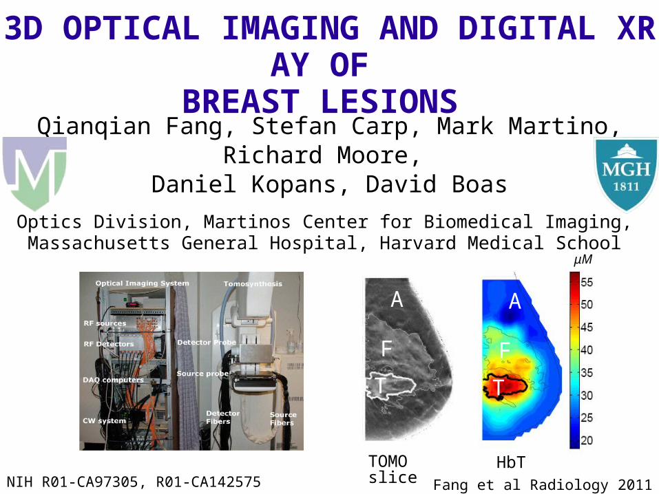
Qianqian Fang, Stefan Carp, Mark Martino,Richard Moore,
Daniel Kopans, David BoasOptics Division, Martinos Center for Biomedical Imaging, Massachusetts General Hospital, Harvard Medical School
NIH R01-CA97305, R01-CA142575
3D OPTICAL IMAGING AND DIGITAL XRAY OF BREAST LESIONS
HbTTOMO slice
μM
A
F
T
A
F
T
Fang et al Radiology 2011
Background Motivation and goal A multi-modality approach
Technology development Combined optical and tomosynthesis imaging system Multi-modality imaging data analysis
Clinical trials and findings Imaging healthy subjects Imaging malignant and benign lesions Statistical group analysis
Conclusions
Outline
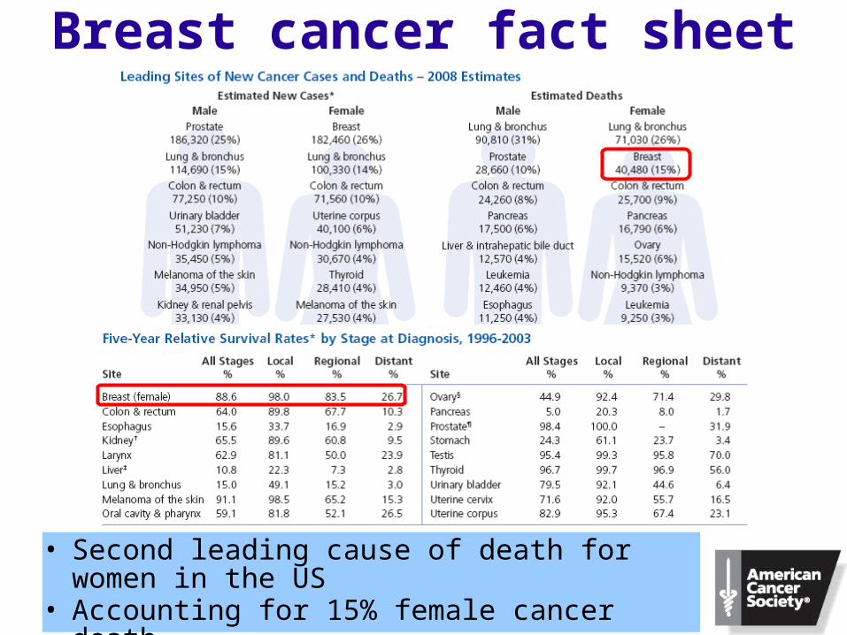
• Second leading cause of death for women in the US• Accounting for 15% female cancer death• Early detection is critical
Breast cancer fact sheet
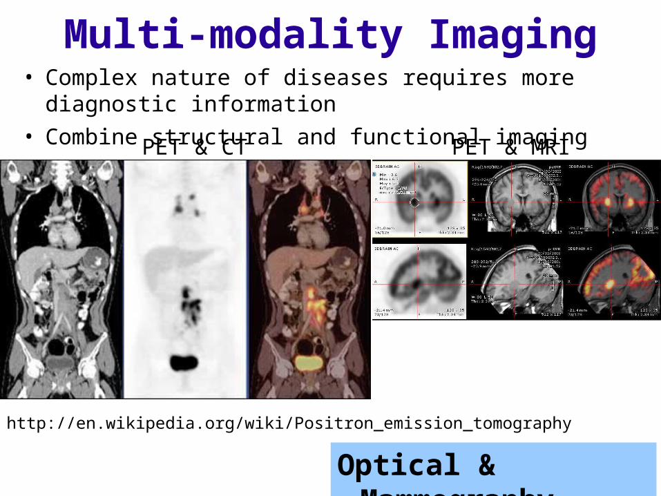
• Complex nature of diseases requires more diagnostic information• Combine structural and functional imaging
http://en.wikipedia.org/wiki/Positron_emission_tomography
Multi-modality Imaging
Optical & Mammography
PET & CT PET & MRI
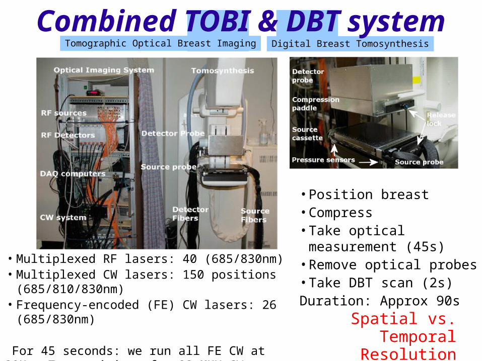
• Position breast• Compress• Take optical measurement (45s)• Remove optical probes• Take DBT scan (2s)Duration: Approx 90s
• Multiplexed RF lasers: 40 (685/830nm)• Multiplexed CW lasers: 150 positions (685/810/830nm)• Frequency-encoded (FE) CW lasers: 26 (685/830nm)
For 45 seconds: we run all FE CW at 20Hz, 7 repetitions for 22 MUX CW positions and 2 repetition for RF
Tomographic Optical Breast Imaging Digital Breast Tomosynthesis
Combined TOBI & DBT system
Spatial vs. Temporal Resolution
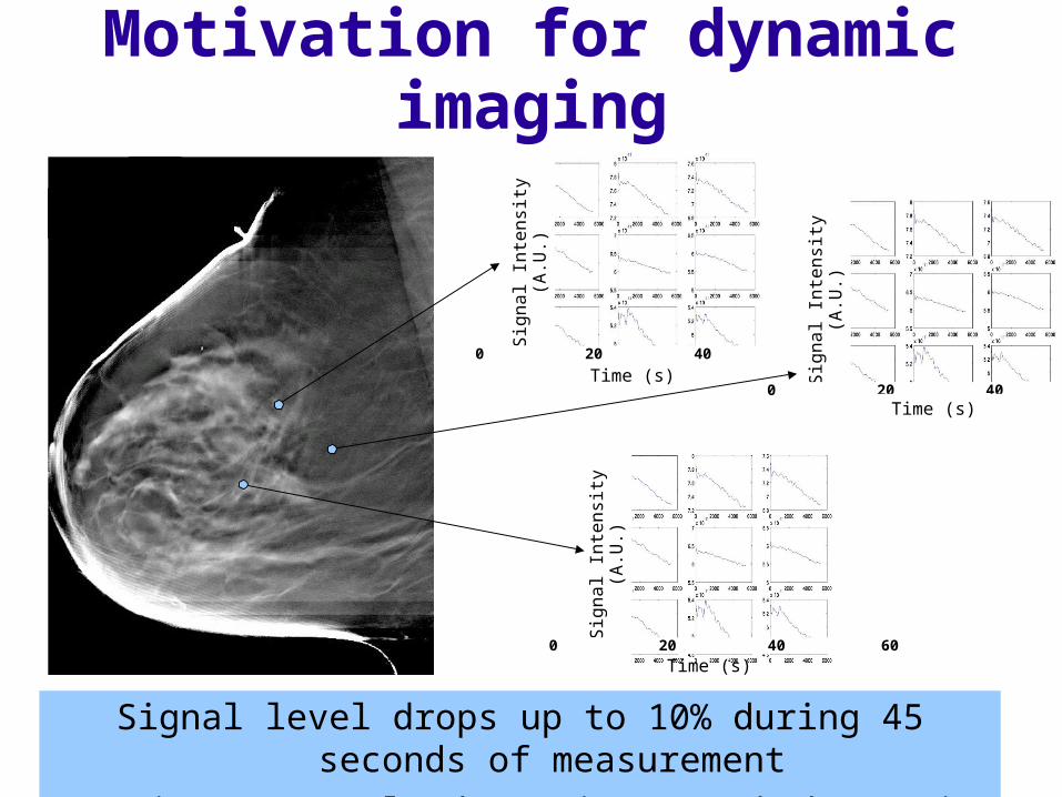
Motivation for dynamic imaging
Signal level drops up to 10% during 45 seconds of measurementDynamic process leads to increased absorption
Sign
al In
tens
ity (A
.U.)
0 20 40 60
Time (s) Sign
al In
tens
ity (A
.U.)
0 20 40 60 Time (s)
Sign
al In
tens
ity (A
.U.)
0 20 40 60
Time (s)
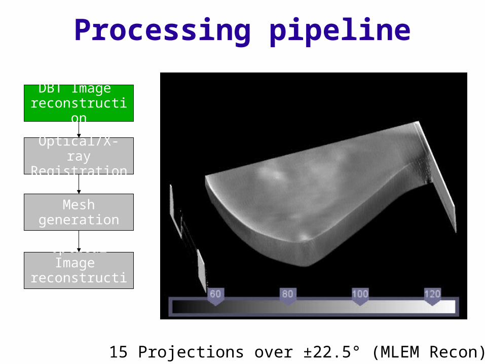
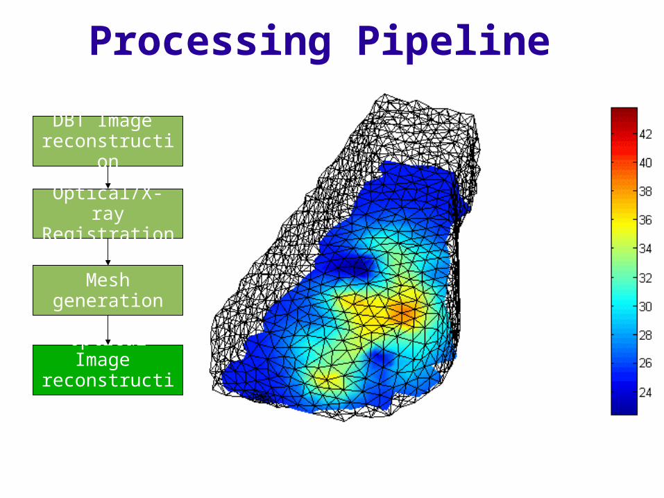
Processing pipeline
DBT Image reconstruction
Optical/X-rayRegistration
Mesh generation
Optical Image reconstruction
15 Projections over ±22.5° (MLEM Recon)
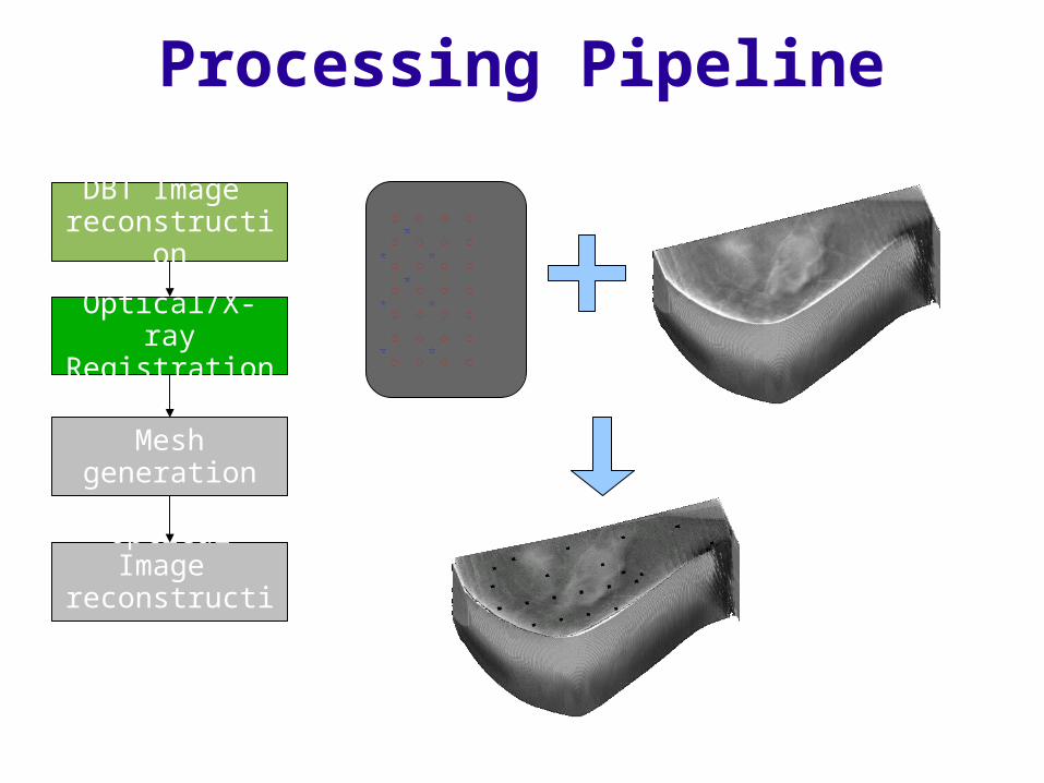
Processing Pipeline
DBT Image reconstruction
Optical/X-rayRegistration
Mesh generation
Optical Image reconstruction
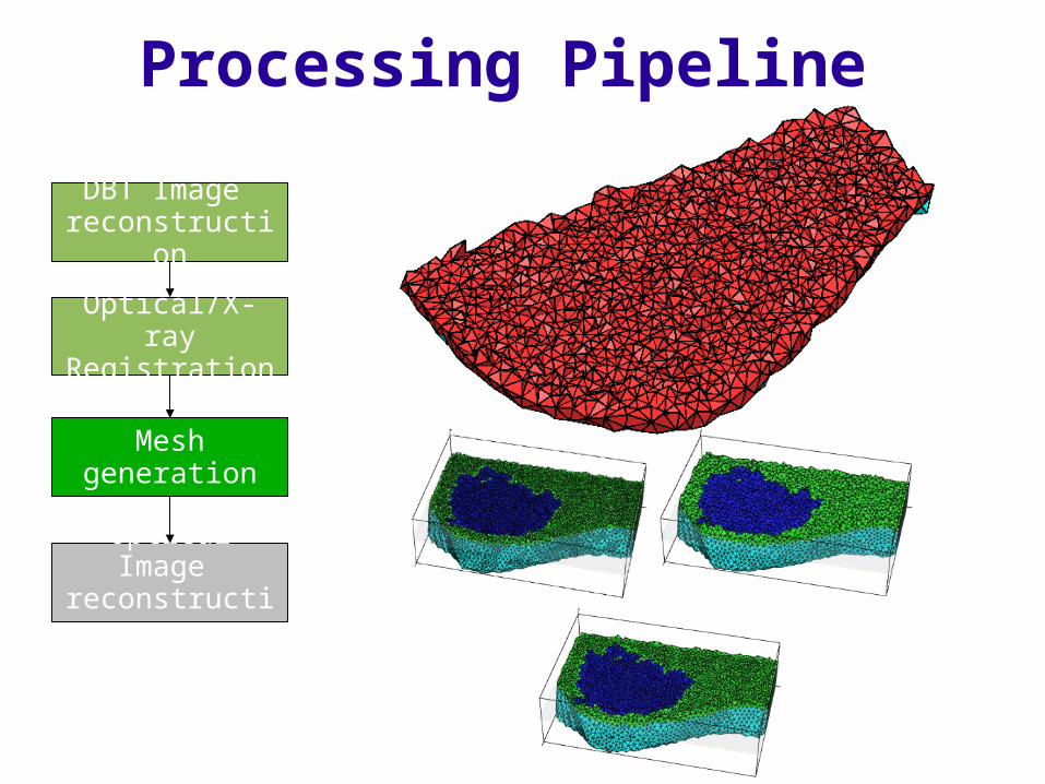
Processing Pipeline
DBT Image reconstruction
Optical/X-rayRegistration
Mesh generation
Optical Image reconstruction
Processing Pipeline
DBT Image reconstruction
Optical/X-rayRegistration
Mesh generation
Optical Image reconstruction
Background and significance Motivations and goals A multi-modality approach
Technology development Combined optical and tomosynthesis imaging system Multi-modality imaging data analysis
Clinical trials and findings Imaging healthy subjects Imaging malignant and benign lesions Statistical group analysis
Conclusions
Outline
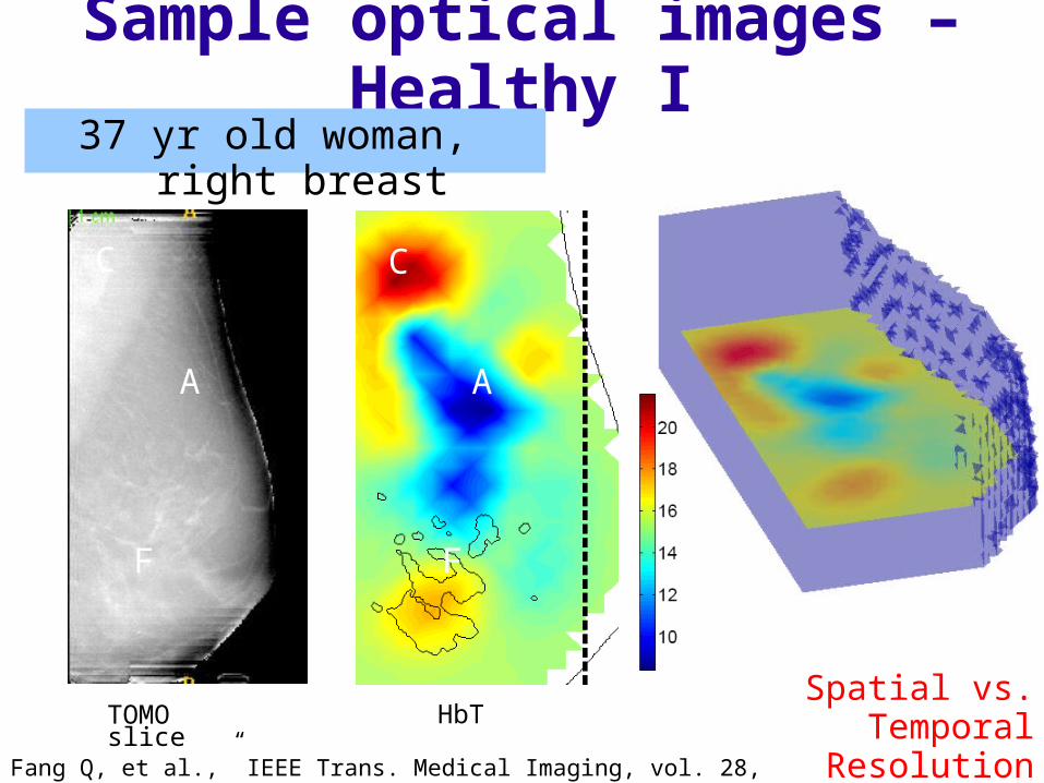
Sample optical images – Healthy I37 yr old woman, right breast
Fang Q, et al.,” IEEE Trans. Medical Imaging, vol. 28, issue 1, pp. 30 – 42, Jan. 2009.
TOMO slice HbT
C
F
A
C
F
A
Spatial vs. Temporal Resolution
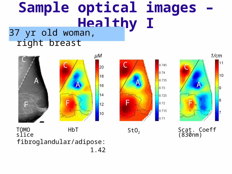
TOMO slice HbT StO2 Scat. Coeff (830nm)
fibroglandular/adipose: 1.42 1.02 1.28
μM 1/cmC
F
A
C
F
A
C
F
A
C
F
A
Sample optical images – Healthy I37 yr old woman, right breast
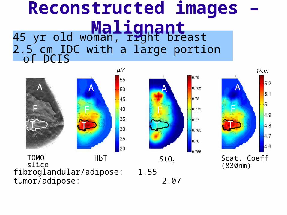
45 yr old woman, right breast2.5 cm IDC with a large portion of DCIS
StO2Scat. Coeff (830nm)HbTTOMO slice
μM 1/cm
fibroglandular/adipose: 1.55 1.00 1.04tumor/adipose: 2.07 0.99 1.09
A
F
T
A
F
T
A
F
T
A
F
T
Reconstructed images – Malignant
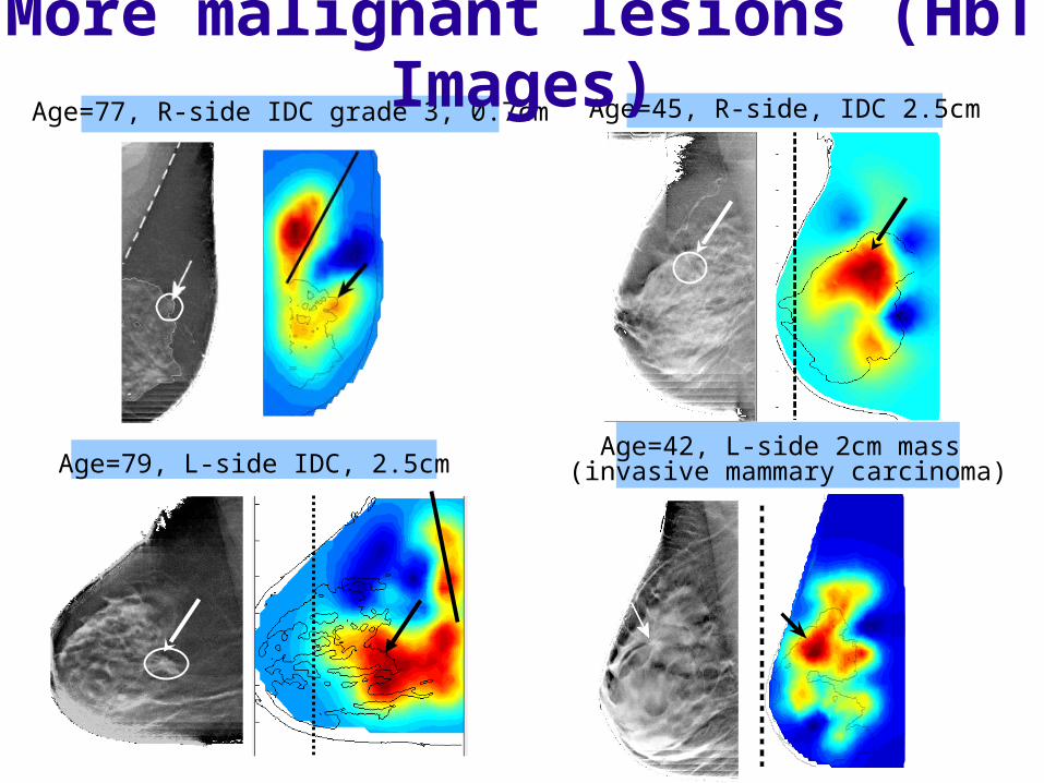
Age=79, L-side IDC, 2.5cm
Age=77, R-side IDC grade 3, 0.7cm
Age=42, L-side 2cm mass (invasive mammary carcinoma)
Age=45, R-side, IDC 2.5cm
More malignant lesions (HbT Images)
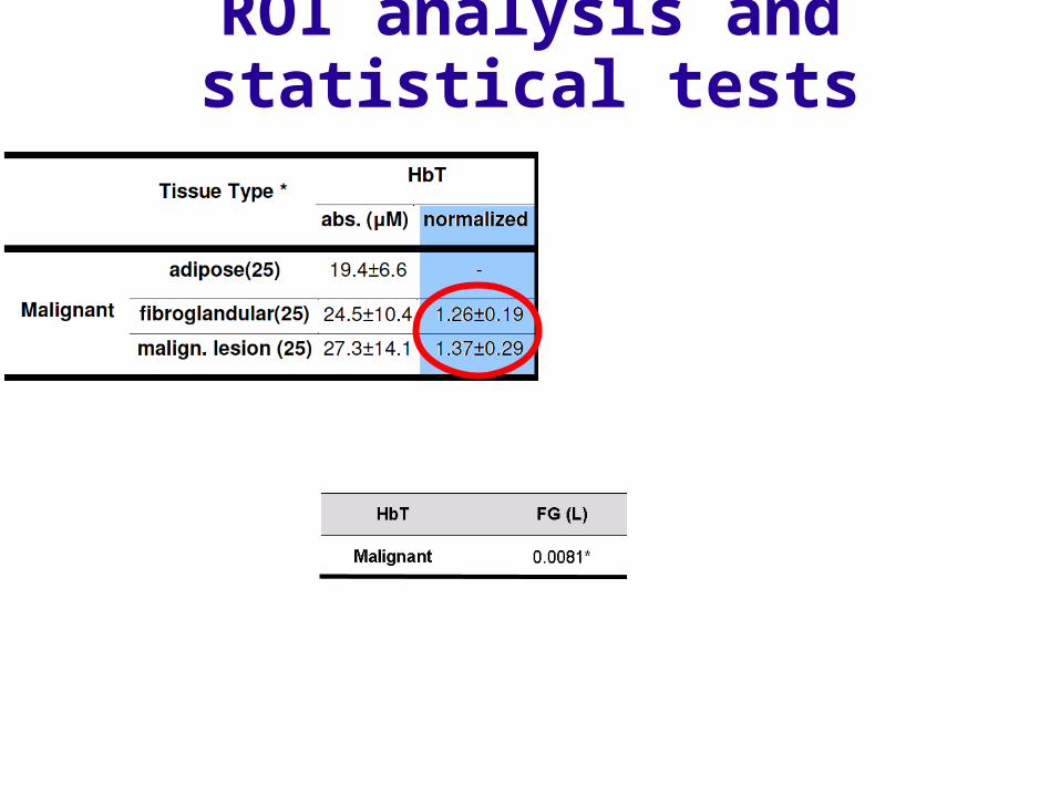
ROI analysis and statistical tests

42 years old woman: 8mm fibroadenoma
Solid Benign Lesions: Fibroadenomas
HbT
(μM
)
39 years old woman: 16mm fibroadenoma
HbT
(μM
)

Left breast of a 49-year-old womanTwo cysts 29x11x26mm and 12x9x5mm at the pointed locations
Benign Lesions: cysts
(μM) 1/cm
TOMO slice HbT StO2 Scat. Coeff (830nm)
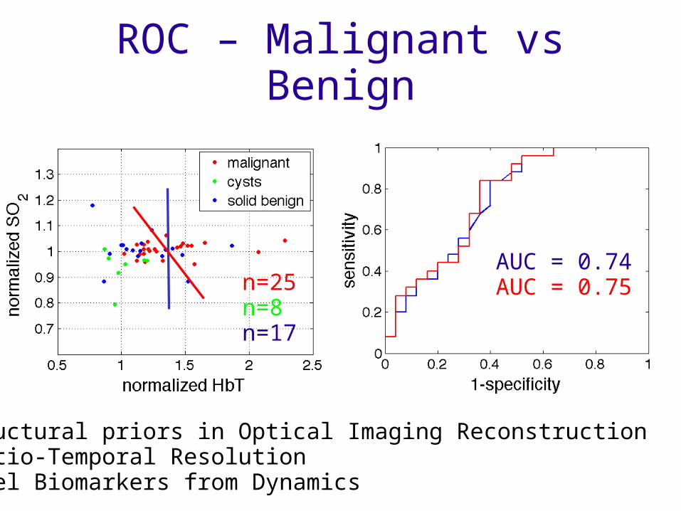
ROC – Malignant vs Benign
AUC = 0.74AUC = 0.75n=25
n=8n=17
• Structural priors in Optical Imaging Reconstruction• Spatio-Temporal Resolution• Novel Biomarkers from Dynamics
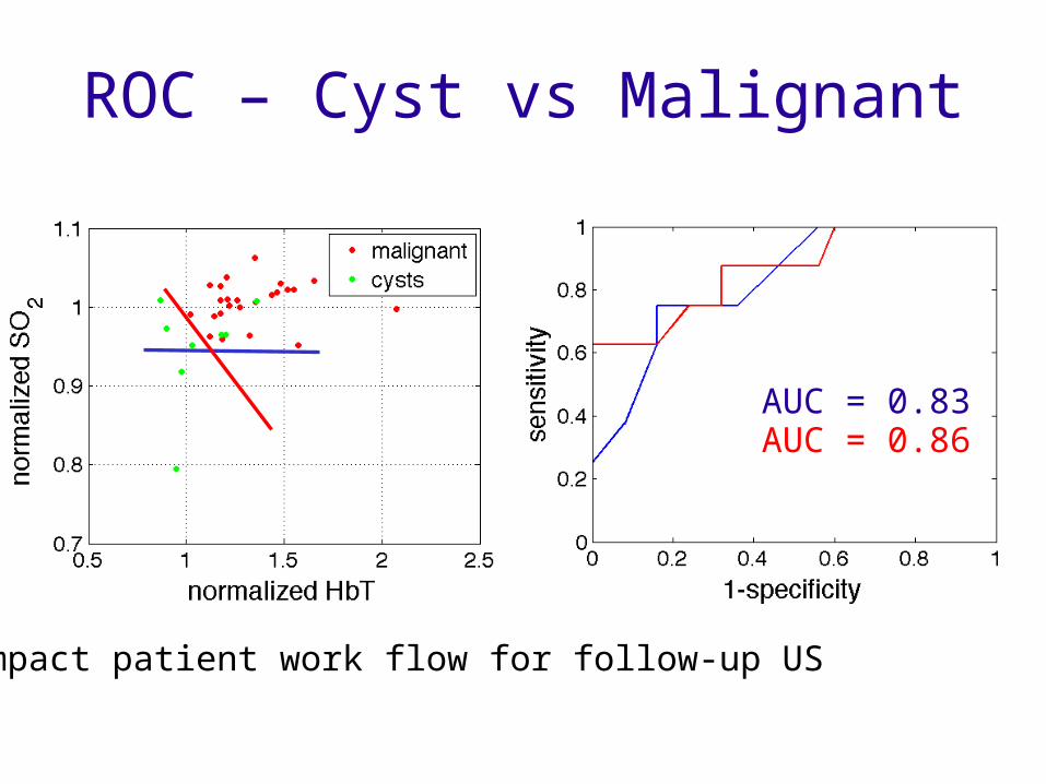
ROC – Cyst vs Malignant
AUC = 0.83AUC = 0.86
Impact patient work flow for follow-up US
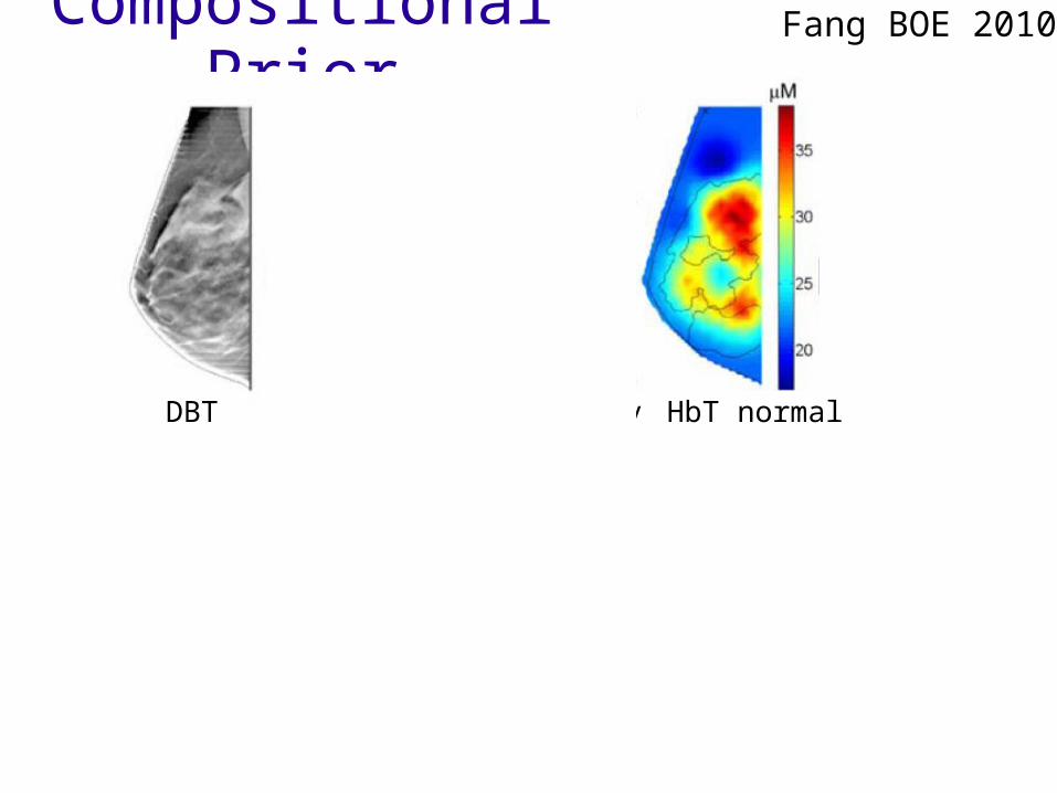
Compositional Prior Fang BOE 2010
DBT Cf HbT binary HbT normal
HbT Compositional Prior

Compositional Prior Fang BOE 2010
DBT HbT comp HbT normal DBT HbT comp HbT normal
Prior improves symmetry of optical properties between left and right breasts!
Residual comparable or reduced… degeneracy of ill-posed reconstruction
StO2Scat. Coeff (830nm)HbTTOMO slice
μM 1/cm
A
F
T
A
F
T
A
F
T
A
F
T
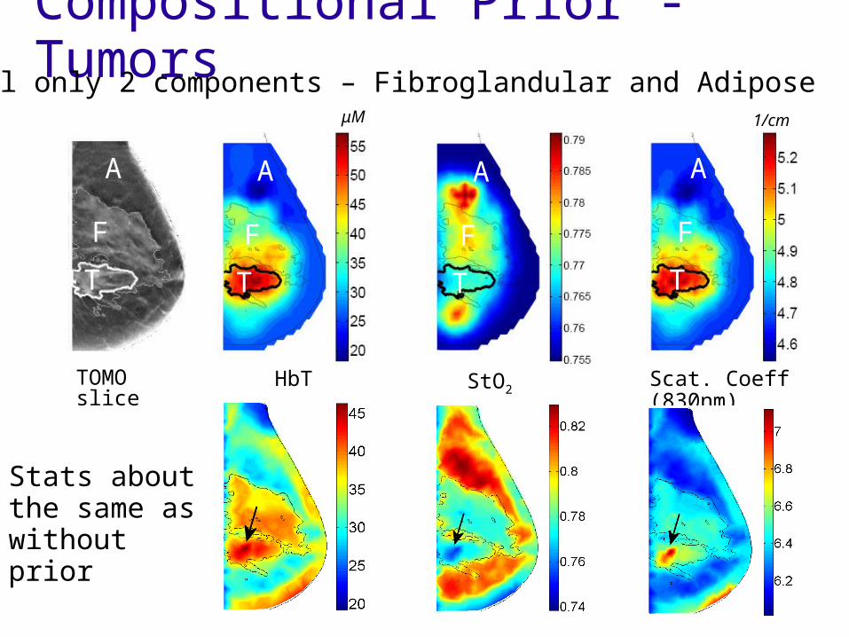
Compositional Prior - TumorsStill only 2 components – Fibroglandular and Adipose
Stats about the same as without prior
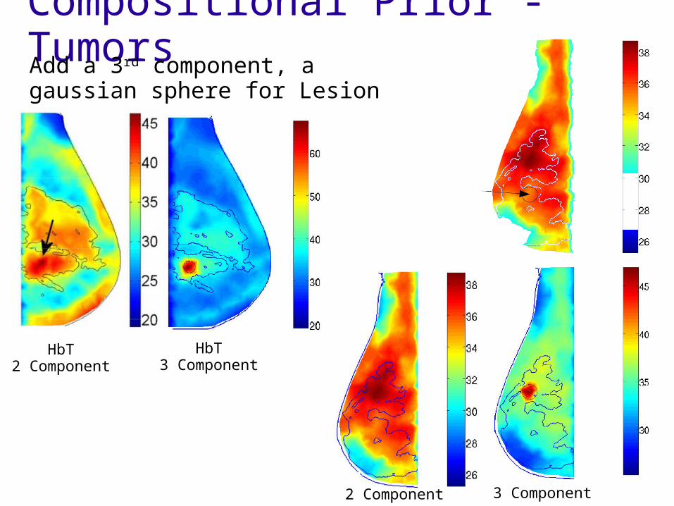
Compositional Prior - TumorsAdd a 3rd component, a gaussian sphere for Lesion
HbT2 Component
HbT3 Component
2 Component 3 Component
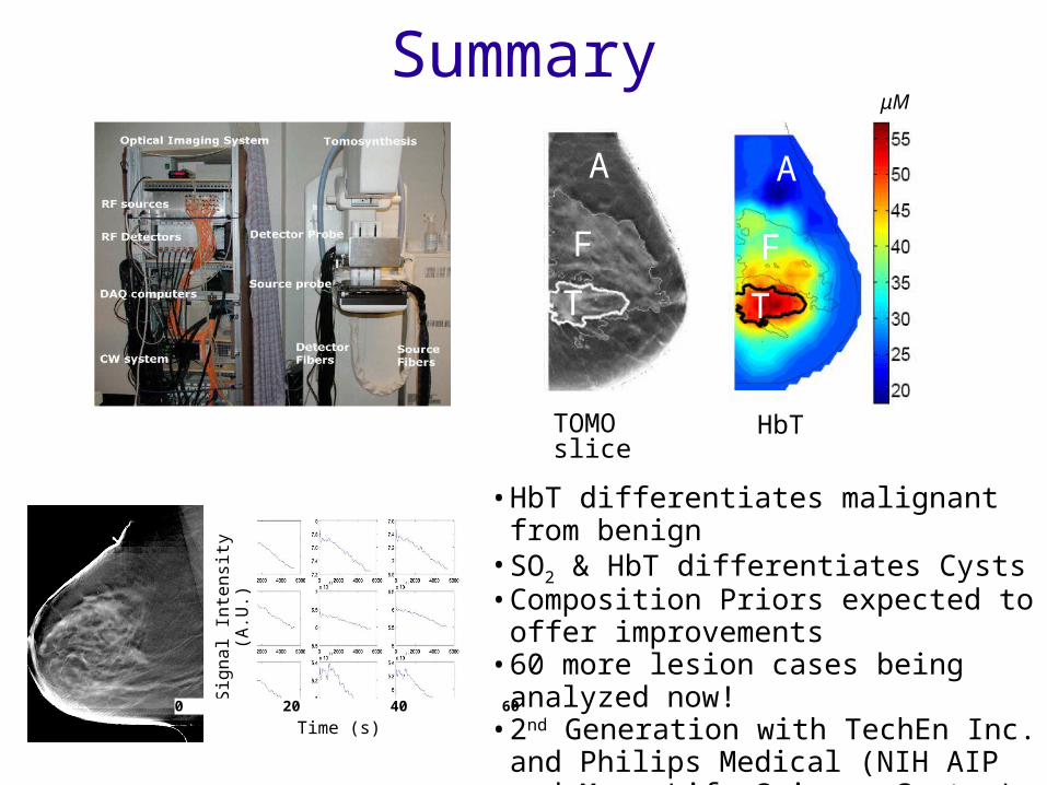
Summary
HbTTOMO slice
μM
A
F
T
A
F
T
Sign
al In
tens
ity (A
.U.)
0 20 40 60
Time (s)
• HbT differentiates malignant from benign• SO2 & HbT differentiates Cysts• Composition Priors expected to offer
improvements• 60 more lesion cases being analyzed
now!• 2nd Generation with TechEn Inc. and
Philips Medical (NIH AIP and Mass Life Science Center)

![arXiv:1511.07635v1 [math.AG] 24 Nov 2015francavi/lavori/fk.pdfthis problem with us. Theorem 1.1 is proven by applying Ratner’s theory (resp. Moore ergodicity theorem) to the linear](https://static.fdocument.org/doc/165x107/5f868b848ed46b5bd06526f7/arxiv151107635v1-mathag-24-nov-francavilavorifkpdf-this-problem-with-us.jpg)