PHOENIX WOUND MATRIX™ - RenovoDerm
7
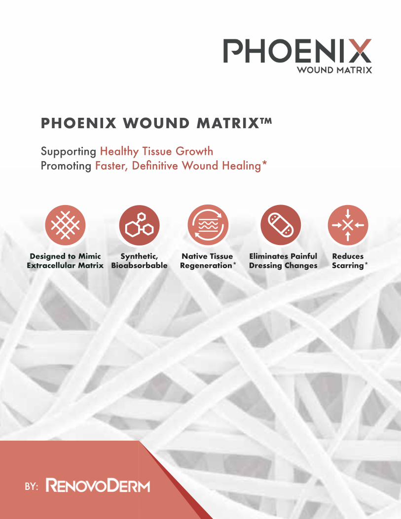
BY: PHOENIX WOUND MATRIX™ Supporting Healthy Tissue Growth Promoting Faster, Definitive Wound Healing* Designed to Mimic Extracellular Matrix Synthetic, Bioabsorbable Native Tissue Regeneration* Eliminates Painful Dressing Changes Reduces Scarring*
Transcript of PHOENIX WOUND MATRIX™ - RenovoDerm
BY:
PHOENIX WOUND MATRIX™
Supporting Healthy Tissue GrowthPromoting Faster, Definitive Wound Healing*
Designed to MimicExtracellular Matrix
Synthetic, Bioabsorbable
Native Tissue Regeneration*
Eliminates PainfulDressing Changes
Reduces Scarring*

Electrospun DevicesRenovoDerm was established to address the current performance gaps with yesterday’ssynthetics and biologics.
Biomimetic, synthetic, electrospun scaffolds provide the starting point for in situ tissue engineering (TE).
Performance Gap
What Are Electrospun Devices?• 1 μm = 0.000001 m
• Typical Fibroblast is 10 - 15 μm across
• Fibers comprising traditional dressings have 50+ μm diameters – far too large for a fibroblast to adhere to
• RenovoDerm’s Phoenix Wound Matrix fibers are only 5 μm
Designed To• Replicate the structure of the native
dermal ECM.
• Promote cell adherence and proliferation
SyntheticsBiointegration, Infection, Fibrosis...
BiologicsDegradation, Safety
Sourcing, Cost...
The Phoenix Wound Matrix
Dermal ECM The Phoenix Wound Matrix with Fibroblast
We have over a decade of experience crafting polymer scaffolds that allow individual cells, cell clusters, and cellular systems to express phenotypes favorable for regenerationof healthy tissue. Our extensive experience working with cancer cells and stem cells and our ongoing work with researchers addressing other adherent cell diseases gives us a solid understanding of the properties our scaffolds must possess tocomplete their tasks.
Who We AreNanofiber Solutions (NFS) – The Parent Company
NFS is redefining tissue engineering by addressing the problems associated with bothsynthetic and biologic devices while incorporating their respective advantages. Followingover a decade of research and development, including numerous (and still ongoing)human and animal studies, NFS has launched four medical device companies, eachtailoring NFS’s proprietary and patented technology to meet the specific requirements oftheir respective clinical area:
RenovoDermWound management
Atreon OrthopedicsJoint Repair
Tarian MedicalHernia repair
Vascular GenesisCardiovascular and peripheral vascular repair
• Comprised of synthetic polymers – more cost effective and more consistent than human/animal derivatives
1 2
Phoenix Wound Matrix® is a fully bioabsorbable advanced wound care device (CTP) designed to harness and support the wound healing process, allowing for the regeneration of functional, native tissue in the wound bed. *
Using proprietary technology, the Phoenix is designed to:
• Mimic native ECM morphology• Promote wound healing through all phases without
stalling in the inflammatory phase• Facilitate cellular adherence and infiltration• Support capillary growth, cellular proliferation,
and native mechanochemical behavior, including intercellular communication
• Regenerate fully-functional tissue
Other Advantages of the Phoenix Wound Matrix: Easy to handle and apply, no re-hydrationAnchor with physician’s/QHP’s preferred methodRe-apply q7 days prnNo contraindicationsReadily available off-the-shelfDegradation within 14 days via hydrolysisConvenient sizes 16 mm disc, 1.5 x 2 cm2, 2.5 x 2.5 cm2, 3 x 4 cm2, 5 x 5 cm2, 7 x 7 cm2, 10 x 10 cm2, 10 x 20 cm2
The Phoenix Wound Matrix has FDA clearance for use in the treatment of both acute and chronic, partial and full-thickness wounds, including: diabetic ulcers, venous stasis ulcers, arterial insufficiency ulcers, pressure ulcers, tunneled/undermined wounds, trauma wounds, second degree burns, and surgical wounds (e.g., post-Mohs, post-laser, wound dehiscence, donor sites/grafts)
ConclusionsThe Phoenix Wound Matrix gradually degrades in the wound bed after it is placed. Thisdegradation is designed to acidify the wound bed and to allow for gradual regrowth ofnative ECM structures to promote regeneration of healthy tissue.
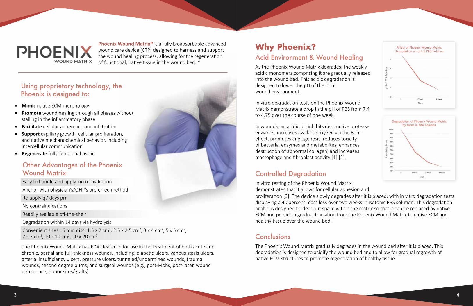
Acid Environment & Wound HealingAs the Phoenix Wound Matrix degrades, the weakly acidic monomers comprising it are gradually released into the wound bed. This acidic degradation is designed to lower the pH of the local wound environment.
In vitro degradation tests on the Phoenix Wound Matrix demonstrate a drop in the pH of PBS from 7.4 to 4.75 over the course of one week.
In wounds, an acidic pH inhibits destructive protease enzymes, increases available oxygen via the Bohr effect, promotes angiogenesis, reduces toxicity of bacterial enzymes and metabolites, enhances destruction of abnormal collagen, and increases macrophage and fibroblast activity [1] [2].
Controlled DegradationIn vitro testing of the Phoenix Wound Matrix demonstrates that it allows for cellular adhesion and
Why Phoenix?
proliferation [3]. The device slowly degrades after it is placed, with in vitro degradation tests displaying a 40 percent mass loss over two weeks in isotonic PBS solution. This degradation profile is designed to clear out space within the matrix so that it can be replaced by native ECM and provide a gradual transition from the Phoenix Wound Matrix to native ECM and healthy tissue over the wound bed.
3 4
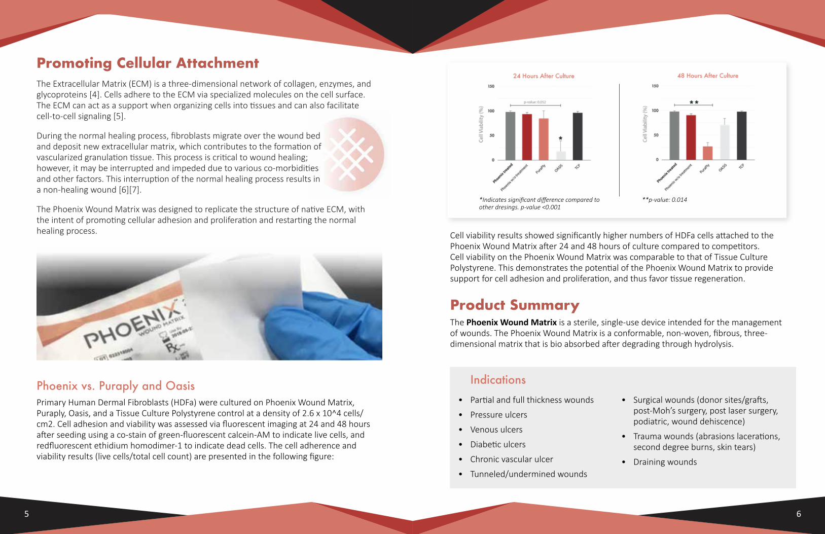
*Indicates significant difference compared to other dresings. p-value <0.001
**p-value: 0.014
Promoting Cellular AttachmentThe Extracellular Matrix (ECM) is a three-dimensional network of collagen, enzymes, andglycoproteins [4]. Cells adhere to the ECM via specialized molecules on the cell surface. The ECM can act as a support when organizing cells into tissues and can also facilitate cell-to-cell signaling [5].
During the normal healing process, fibroblasts migrate over the wound bed and deposit new extracellular matrix, which contributes to the formation of vascularized granulation tissue. This process is critical to wound healing; however, it may be interrupted and impeded due to various co-morbidities and other factors. This interruption of the normal healing process results in a non-healing wound [6][7].
The Phoenix Wound Matrix was designed to replicate the structure of native ECM, with the intent of promoting cellular adhesion and proliferation and restarting the normal healing process.
Phoenix vs. Puraply and OasisPrimary Human Dermal Fibroblasts (HDFa) were cultured on Phoenix Wound Matrix, Puraply, Oasis, and a Tissue Culture Polystyrene control at a density of 2.6 x 10^4 cells/cm2. Cell adhesion and viability was assessed via fluorescent imaging at 24 and 48 hours after seeding using a co-stain of green-fluorescent calcein-AM to indicate live cells, and redfluorescent ethidium homodimer-1 to indicate dead cells. The cell adherence and viability results (live cells/total cell count) are presented in the following figure:
Product SummaryThe Phoenix Wound Matrix is a sterile, single-use device intended for the management of wounds. The Phoenix Wound Matrix is a conformable, non-woven, fibrous, three-dimensional matrix that is bio absorbed after degrading through hydrolysis.
Indications• Partial and full thickness wounds
• Pressure ulcers
• Venous ulcers
• Diabetic ulcers
• Chronic vascular ulcer
• Tunneled/undermined wounds
• Surgical wounds (donor sites/grafts, post-Moh’s surgery, post laser surgery, podiatric, wound dehiscence)
• Trauma wounds (abrasions lacerations, second degree burns, skin tears)
• Draining wounds
Cell viability results showed significantly higher numbers of HDFa cells attached to the Phoenix Wound Matrix after 24 and 48 hours of culture compared to competitors. Cell viability on the Phoenix Wound Matrix was comparable to that of Tissue Culture Polystyrene. This demonstrates the potential of the Phoenix Wound Matrix to provide support for cell adhesion and proliferation, and thus favor tissue regeneration.
5 6
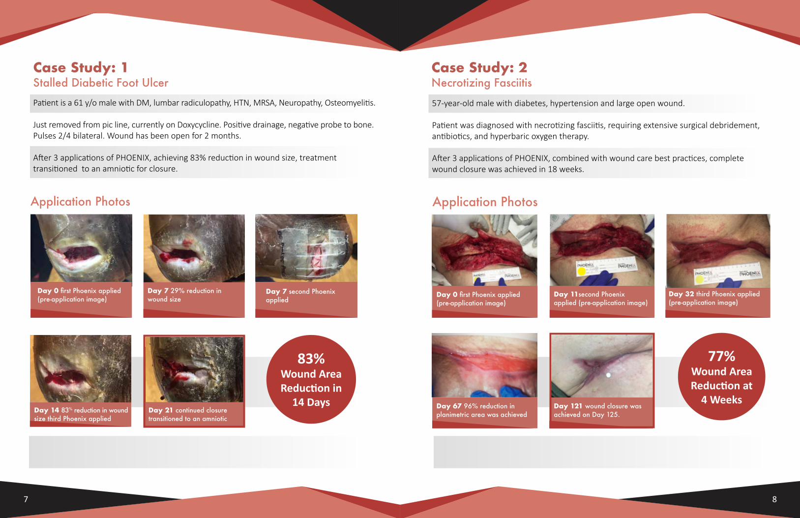
Day 0 first Phoenix applied (pre-application image)
Day 7 29% reduction in wound size
Day 7 second Phoenix applied
Day 14 83% reduction in wound size third Phoenix applied
3rd Phoenix Application
Day 21 continued closure transitioned to an amniotic
Application Photos
Case Study: 1Stalled Diabetic Foot Ulcer
Patient is a 61 y/o male with DM, lumbar radiculopathy, HTN, MRSA, Neuropathy, Osteomyelitis.
Just removed from pic line, currently on Doxycycline. Positive drainage, negative probe to bone. Pulses 2/4 bilateral. Wound has been open for 2 months.
After 3 applications of PHOENIX, achieving 83% reduction in wound size, treatment transitioned to an amniotic for closure.
Case Study: 2Necrotizing Fasciitis
Day 0 first Phoenix applied (pre-application image)
Day 11second Phoenix applied (pre-application image)
Day 32 third Phoenix applied (pre-application image)
Day 67 96% reduction in planimetric area was achieved
Day 121 wound closure was achieved on Day 125.
57-year-old male with diabetes, hypertension and large open wound.
Patient was diagnosed with necrotizing fasciitis, requiring extensive surgical debridement, antibiotics, and hyperbaric oxygen therapy.
After 3 applications of PHOENIX, combined with wound care best practices, complete wound closure was achieved in 18 weeks.
Application Photos
7 8
83% Wound Area Reduction in
14 Days
77% Wound Area Reduction at
4 Weeks
2. Anchor Matrix
Place the matrix in the wound and gently smooth it onto the wound bed to ensure direct contact. Anchor with physician’s/QHP’s preferred method of fixation: staples, sutures, surgical glue, or reinforced adhesive skin closures (e.g., Steri-Strip™). Gently rinse the matrix and wound bed with sterile saline.
Application Guide 1. Trim the Matrix (Optional)
Trim the Phoenix Wound Matrix to-size such that the edges match the edges of the wound bed.
3. Non-Adherent Dressing Apply an appropriate non-adherent dressing over the Phoenix Wound Matrix to bolster it firmly in contact with the wound bed.
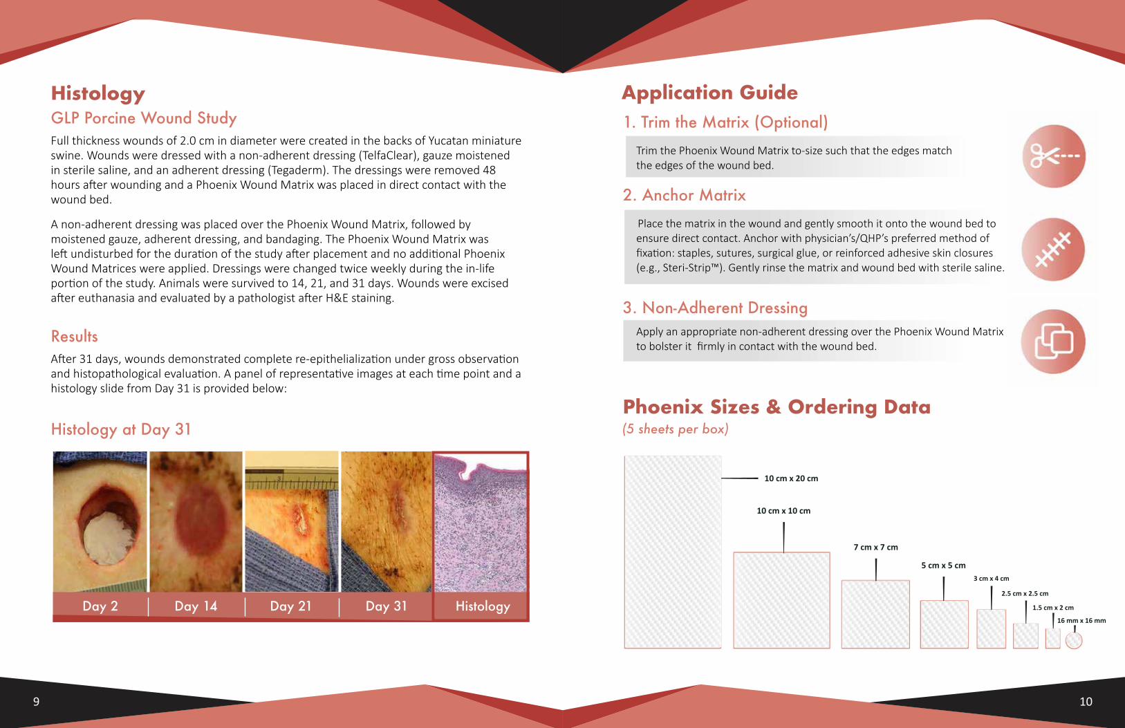
Histology at Day 31
Day 2 Day 14 Day 21 Day 31
HistologyGLP Porcine Wound StudyFull thickness wounds of 2.0 cm in diameter were created in the backs of Yucatan miniature swine. Wounds were dressed with a non-adherent dressing (TelfaClear), gauze moistened in sterile saline, and an adherent dressing (Tegaderm). The dressings were removed 48 hours after wounding and a Phoenix Wound Matrix was placed in direct contact with the wound bed.
A non-adherent dressing was placed over the Phoenix Wound Matrix, followed by moistened gauze, adherent dressing, and bandaging. The Phoenix Wound Matrix was left undisturbed for the duration of the study after placement and no additional Phoenix Wound Matrices were applied. Dressings were changed twice weekly during the in-life portion of the study. Animals were survived to 14, 21, and 31 days. Wounds were excised after euthanasia and evaluated by a pathologist after H&E staining.
ResultsAfter 31 days, wounds demonstrated complete re-epithelialization under gross observation and histopathological evaluation. A panel of representative images at each time point and a histology slide from Day 31 is provided below:
Histology
9 10
Phoenix Sizes & Ordering Data(5 sheets per box)
10 cm x 20 cm
10 cm x 10 cm
7 cm x 7 cm
5 cm x 5 cm3 cm x 4 cm
2.5 cm x 2.5 cm
1.5 cm x 2 cm
16 mm x 16 mm
*All claims are supported by clinical case studies, a GLP porcine animal study, and/or veterinary case studies.
Phoenix Wound Matrix is a registered trademark of RenovoDerm.
©2020 RenovoDerm Inc. All rights reserved. Printed in the USA.
References:1. Nagoba BS, Suryawanshi NM, Wadher B, Selkar S. Acidic Environment and Wound Healing: A Review. Wounds. 2015;27(1):5-11.2. Porporato PE, Payen VL, Saedeleer CJD, et al. Lactate stimulates angiogenesis and accelerates the healing of superficial and ischemic wounds in mice. Angiogenesis. 2012;15(4):581-592. doi:10.1007/s10456-012-9282-0.3. Manufacturer-Sponsored White Paper – “Phoenix Wound Matrix: Cell Adherence and Proliferation”.4. Theocharis, A. D.; Skandalis, S. S.; Gialeli, C.; Karamanos, N. K. (2016). “Extracellular matrix structure”. Advanced Drug Delivery Reviews. 97: 4–27.5. Lodish, Harvey; Berk, Arnold; Matsudaira, Paul; Kaiser, Chris A.; Krieger, Monty; Scott, Matthew P.; Zipursky, Lawrence; Darnell, James (2003). Molecular Cell Biology (5th ed.). W.H. Freeman.6. Takeo M, Lee W, Ito M. Wound healing and skin regeneration. Cold Spring Harb Perspect Med. 2015;5(1):a023267. Published . doi:10.1101/cshperspect.a0232677. Demidova-Rice TN, Hamblin MR, Herman IM. Acute and impaired wound healing: pathophysiology and current methods for drug delivery, part 1: normal and chronic wounds: biology, causes, and approaches to care. Adv Skin Wound Care. 2012;25(7):304–314. doi:10.1097/01.ASW.0000416006.55218.d08. Manufacturer-Sponsored White Paper – “Phoenix Wound Matrix: Cell Adherence and Proliferation”.9. Nagoba BS, Suryawanshi NM, Wadher B, Selkar S. Acidic Environment and Wound Healing: A Review. Wounds. 2015;27(1):5-11.10. Porporato PE, Payen VL, Saedeleer CJD, et al. Lactate stimulates angiogenesis and accelerates the healing of superficial and ischemic wounds in mice. Angiogenesis. 2012;15(4):581-592. doi:10.1007/s10456-012-9282-0.
RenovoDerm is dedicated to building an advanced line of wound care products thatenhance tissue regeneration and promote rapid and definitive wound healing.
For product information and technical, medical or reimbursement questions, please call (614) 602-1852 or visit www.phoenixmatrix.tech