PGC-1α Regulates Normal and Pathological Angiogenesis in the Retina
11
VASCULAR BIOLOGY, ATHEROSCLEROSIS, AND ENDOTHELIUM BIOLOGY PGC-1a Regulates Normal and Pathological Angiogenesis in the Retina Magali Saint-Geniez,* Aihua Jiang, y Stephanie Abend,* Laura Liu, y Harry Sweigard, z Kip M. Connor, z and Zoltan Arany y From the Schepens Eye Research Institute,* Department of Ophthalmology, z Massachusetts Eye and Ear Infirmary, Harvard Medical School, Boston; and The Cardiovascular Institute, y Beth Israel Deaconess Medical Center, Boston Accepted for publication September 4, 2012. Address correspondence to Zoltan Arany, M.D., Ph.D., ECLS906, Beth Israel Deaconess Medical Center, 330 Brookline Ave, Boston, MA 02215. E-mail: zarany@ bidmc.harvard.edu. Neovascular diseases of the eye are the most common causes of blindness worldwide. The mechanisms underlying pathological neovascularization in the retina remain incompletely understood. PGC-1a is a transcriptional coactivator that plays a central role in the regulation of cellular metabolism. In skeletal muscle, PGC-1a induces VEGFA expression and powerfully promotes angiogenesis, suggesting a similar role in other tissues. This study investigates the role of PGC-1a during normal and patho- logical vascularization in the retina. We show that PGC-1a induces the expression of VEGFA in numerous retinal cells, and that PGC-1a expression is strongly induced during postnatal retinal development, coincident with VEGFA expression and angiogenesis. PGC-1a / mice have a significant reduction of early retinal vascular outgrowth, and reduced density of capillaries and number of main arteries and veins as adults. In the oxygen-induced retinopathy model of retinopathy of prematurity, PGC-1a expression is dramatically induced in the inner nuclear layer of the retina, sug- gesting that PGC-1a drives pathological neovascularization. In support of this, PGC-1a / mice subjected to oxygen-induced retinopathy had decreased expression of VEGFA and were protected against pathological neovascularization. These results demonstrate that PGC-1a regulates VEGFA in the retina and is required for normal vessel development and for pathological neovascularization. The data highlight PGC-1a as a novel target in the treatment of neovascular diseases of the eye. (Am J Pathol 2013, 182: 255e265; http://dx.doi.org/10.1016/j.ajpath.2012.09.003) The mature retina is a highly metabolic neural tissue with the highest oxygen consumption per unit weight of any human tissue. 1 In most mammals, the adult retina is vascularized by two independent circulatory systems: the retinal vessels that supply the inner part of the retina, and the choroidal vessels that supply the deeper section of the retina. 2 Retinal blood vessels normally are quiescent with respect to growth. 3 However, in a variety of retinal pathologies, such as prolif- erative diabetic retinopathy, venous occlusion, age-related macular degeneration, and retinopathy of prematurity (ROP), abnormal angiogenic growth of vessels into the retinal tissue and/or the vitreous leads to serious vision loss. ROP, the leading cause of blindness in premature and very- low-birth-weight infants, 4 is caused by disorganized retinal vascular growth. 5 Vascularization of the retina in humans is normally complete and stabilized at term. By contrast, the immature vessels of preterm infants placed in hyperoxic incubators regress in the face of increased oxygen tension (vaso-obliterative phase). 6 Once returned to normoxia, the insufficiently perfused retina becomes ischemic, and dramatically induces vascular endothelial growth factor A (VEGFA) and abnormal vessel growth (vaso-proliferative phase). Current approaches to the treatment of ROP only reduce the incidence of blindness by 25%, and are destructive to the retina. 7 More recent efforts with anti-VEGF therapy have yielded promising results, underscoring the importance of VEGFA in this disease. 8 Understanding the pathophysi- ology of ROP, and identifying potentially targetable Supported by the March of Dimes (Z.A.), the Harvard Catalyst j The Harvard Clinical and Translational Science Center (Z.A. and M.S.-G.), the National Heart, Lung, and Blood Institute (Z.A.), the American Heart Association (A.J.), the American Diabetes Association (Z.A.), the Ellison Foundation (Z.A.), the National Eye Institute Training Grant (H.S.) and Core Grant for Vision Research (K.M.C.), Research to Prevent Blindness unrestricted grant to Harvard Department of Ophthalmology (K.M.C.), the NIH R01EY022084 (K.M.C.), and financial contributions from Harvard University and its affiliated academic health care centers. M.S.-G. and A.J. contributed equally to this work. Copyright ª 2013 American Society for Investigative Pathology. Published by Elsevier Inc. All rights reserved. http://dx.doi.org/10.1016/j.ajpath.2012.09.003 ajp.amjpathol.org The American Journal of Pathology, Vol. 182, No. 1, January 2013
Transcript of PGC-1α Regulates Normal and Pathological Angiogenesis in the Retina
The American Journal of Pathology, Vol. 182, No. 1, January 2013
ajp.amjpathol.org
VASCULAR BIOLOGY, ATHEROSCLEROSIS, AND ENDOTHELIUM BIOLOGY
PGC-1a Regulates Normal and Pathological Angiogenesisin the RetinaMagali Saint-Geniez,* Aihua Jiang,y Stephanie Abend,* Laura Liu,y Harry Sweigard,z Kip M. Connor,z and Zoltan Aranyy
From the Schepens Eye Research Institute,* Department of Ophthalmology,z Massachusetts Eye and Ear Infirmary, Harvard Medical School, Boston; andThe Cardiovascular Institute,y Beth Israel Deaconess Medical Center, Boston
Accepted for publication
C
P
h
September 4, 2012.
Address correspondence toZoltan Arany, M.D., Ph.D.,ECLS906, Beth IsraelDeaconess Medical Center, 330Brookline Ave, Boston,MA 02215. E-mail: [email protected].
opyright ª 2013 American Society for Inve
ublished by Elsevier Inc. All rights reserved
ttp://dx.doi.org/10.1016/j.ajpath.2012.09.003
Neovascular diseases of the eye are the most common causes of blindness worldwide. The mechanismsunderlying pathological neovascularization in the retina remain incompletely understood. PGC-1a isa transcriptional coactivator that plays a central role in the regulation of cellular metabolism. Inskeletal muscle, PGC-1a induces VEGFA expression and powerfully promotes angiogenesis, suggestinga similar role in other tissues. This study investigates the role of PGC-1a during normal and patho-logical vascularization in the retina. We show that PGC-1a induces the expression of VEGFA innumerous retinal cells, and that PGC-1a expression is strongly induced during postnatal retinaldevelopment, coincident with VEGFA expression and angiogenesis. PGC-1a�/� mice have a significantreduction of early retinal vascular outgrowth, and reduced density of capillaries and number ofmain arteries and veins as adults. In the oxygen-induced retinopathy model of retinopathy ofprematurity, PGC-1a expression is dramatically induced in the inner nuclear layer of the retina, sug-gesting that PGC-1a drives pathological neovascularization. In support of this, PGC-1a�/� micesubjected to oxygen-induced retinopathy had decreased expression of VEGFA and were protectedagainst pathological neovascularization. These results demonstrate that PGC-1a regulates VEGFA in theretina and is required for normal vessel development and for pathological neovascularization. The datahighlight PGC-1a as a novel target in the treatment of neovascular diseases of the eye. (Am J Pathol2013, 182: 255e265; http://dx.doi.org/10.1016/j.ajpath.2012.09.003)
Supported by the March of Dimes (Z.A.), the Harvard Catalyst j TheHarvard Clinical and Translational Science Center (Z.A. and M.S.-G.), theNational Heart, Lung, and Blood Institute (Z.A.), the American HeartAssociation (A.J.), the American Diabetes Association (Z.A.), the EllisonFoundation (Z.A.), the National Eye Institute Training Grant (H.S.) andCore Grant for Vision Research (K.M.C.), Research to Prevent Blindnessunrestricted grant to Harvard Department of Ophthalmology (K.M.C.), theNIH R01EY022084 (K.M.C.), and financial contributions from HarvardUniversity and its affiliated academic health care centers.
M.S.-G. and A.J. contributed equally to this work.
The mature retina is a highly metabolic neural tissue with thehighest oxygen consumption per unit weight of any humantissue.1 In most mammals, the adult retina is vascularized bytwo independent circulatory systems: the retinal vessels thatsupply the inner part of the retina, and the choroidal vesselsthat supply the deeper section of the retina.2 Retinal bloodvessels normally are quiescent with respect to growth.3
However, in a variety of retinal pathologies, such as prolif-erative diabetic retinopathy, venous occlusion, age-relatedmacular degeneration, and retinopathy of prematurity(ROP), abnormal angiogenic growth of vessels into theretinal tissue and/or the vitreous leads to serious vision loss.
ROP, the leading cause of blindness in premature and very-low-birth-weight infants,4 is caused by disorganized retinalvascular growth.5 Vascularization of the retina in humans isnormally complete and stabilized at term. By contrast, theimmature vessels of preterm infants placed in hyperoxicincubators regress in the face of increased oxygen tension(vaso-obliterative phase).6 Once returned to normoxia, the
stigative Pathology.
.
insufficiently perfused retina becomes ischemic, anddramatically induces vascular endothelial growth factor A(VEGFA) and abnormal vessel growth (vaso-proliferativephase). Current approaches to the treatment of ROP onlyreduce the incidence of blindness by 25%, and are destructiveto the retina.7 More recent efforts with anti-VEGF therapyhave yielded promising results, underscoring the importanceof VEGFA in this disease.8 Understanding the pathophysi-ology of ROP, and identifying potentially targetable
Saint-Geniez et al
pathways that regulate retinal VEGFA and angiogenesis, istherefore of great interest.
The transcription factor hypoxia inducible factor-1a (HIF-1a) has long been proposed as the main force driving retinalvascularization in both normal and pathological conditions.9
However, this concept has recently been challenged. Forexample, using genetically modified mice in which theVEGFA promoter lacks the HIF-responsive element andtherefore is unable to produce VEGFA in response to HIF-1a,Vinores et al10 showed that HIF-1aedependent VEGFAregulation was surprisingly not required for normal retinalvascularization. We recently described in skeletal musclea novel and HIF-independent pathway that powerfully regu-lates angiogenesis, involving the transcriptional coactivatorPPARg-coactivator-1a (PPARGC1A; alias PGC-1a).11
PGC-1a is a powerful regulator of mitochondria and oxida-tive metabolism in numerous tissues (reviewed in Kelly andScarpulla,12 Lin et al,13 Handschin and Spiegelman,14 andArany15). In addition, we recently showed that PGC-1a alsoregulates an angiogenic program, including VEGFA andother angiogenic factors, in cultured muscle cells and skeletalmuscle in vivo.11 Transgenic expression of PGC-1a in skel-etalmuscle dramatically increasedmicrovascular density, andwas protective in a model of skeletal muscle ischemia. PGC-1a, a major regulator of mitochondrial function, thus alsocontrols a novel and powerful angiogenic pathway in skeletalmuscle, suggesting that it may also do so in other tissues.
Despite the fact that the retina is one of the most metabol-ically active and vascularized tissues in the body, little isknown about the role of PGC-1a in the eye. Only one reportrecently implicated PGC-1a in the survival of photoreceptorcells to light-induced damage.16 The identification of PGC-1aas a novel regulator of angiogenesis, and its important role inmetabolism, led us to suspect that it may play a role in retinalvascular development and in ROP. We show here that PGC-1a is expressed in the retina, that its expression risesdramatically within the developing retina, and that PGC-1acan induce VEGFA in retinal cells. Moreover, PGC-1a�/�
mice have delayed retinal vasculature outgrowth and areprotected from pathological neovascularization in a model ofproliferative retinopathy similar to ROP.
Materials and Methods
Animals
All animal experiments were approved by the Schepens EyeResearch Institutional Animal Care and Use Committee.PGC-1aedeficient mice have been previously described.17
Oxygen-induced retinopathy (OIR) was induced by placingpostnatal day (P) 7 mice in 75� 2% oxygen for 5 consecutivedays, as previously described.18
Cells and Reagents
The human Müller cell line, MIO-M1 (a gift from G. AstridLimb, Institute of Ophthalmology and Moorfields Eye
256
Hospital, London, UK), and the murine cone cell line,661W (a gift from Dr. Al-Ubaidi, Department of CellBiology, University of Oklahoma Health Sciences Center)were maintained in Dulbecco’s modified Eagle’s medium(DMEM) (Invitrogen/Gibco, Carlsbad, CA) supplementedwith 100 IU/mL penicillin, 100 mg/mL streptomycin (allfrom Gibco), and 10% heat-inactivated fetal bovine serum(Atlanta Biologicals, Lawrenceville, GA). The rat ganglioncell line, RGC-5, was maintained in DMEM with 1.0 g/Lglucose (Invitrogen/Gibco) with 100 IU/mL penicillin,100 mg/mL streptomycin (all from Gibco), and 10% heat-inactivated fetal bovine serum (Atlanta Biologicals). ARPE-19 cells (American Type Culture Collection, Manassas, VA)were maintained in DMEM/F12 medium (Lonza, Basel,Switzerland), 100 IU/mL penicillin, 100 mg/mL strepto-mycin (all from Gibco), and 10% heat-inactivated fetalbovine serum (Atlanta Biologicals). RGC5 differentiationwas induced by 1 mmol/L staurosporine in DMEM (withoutserum or antibiotics) for 1 hour at 37�C. All cultures weremaintained in a humidified atmosphere of 95% air and 5%CO2 at 37�C. Retrovirus and adenoviruses overexpressingPGC-1a have been described.19,20 For PGC-1a knock-down, MIO-M1s were infected with lentivirus carryinghuman PGC-1a short hairpin (shPGC-1a) plasmid or greenfluorescent protein (GFP) short hairpin (shGFP) plasmid(obtained from the RNAi Consortium at the Broad Instituteof Harvard and MIT) and selected for puromycin resistance.PGC-1a expression was confirmed by quantitative PCR(qPCR) of the PGC-1a gene as well as its downstreamtarget genes. PGC-1a expression at protein level wasmeasured by Western blotting. MIO-M1 cells were starvedin DMEM with 0.5% FBS overnight and then kept innormoxia or hypoxia (complete deprivation of oxygen) for24 hours before harvesting for qPCR or enzyme-linkedimmunosorbent assay for VEGFA expression levels.VEGFA enzyme-linked immunosorbent assay was per-formed on conditioned media using the Human VEGFQuantikine kit (R&D Systems, Minneapolis, MN) accordingto the manufacturer’s instructions.
qPCR
Total RNA was isolated from tissue and cells using RNA-Bee solution (Iso-Tex Diagnostics, Friendswood, TX) or theTurboCapture 384 mRNA Kit (Qiagen, Valencia, CA).RNA was reverse-transcribed using iScript (Bio-RadLaboratories, Hercules, CA) or the High Capacity cDNAReverse Transcription Kit (Applied Biosystems, Foster City,CA). qPCR reactions were performed using the SYBRGreen Master Mix and the ABI Prism 9700 SequenceDetection System (Applied Biosystems) or the CFX384Real-Time System (Bio-Rad) according to the manufac-turers’ instructions. To ensure accurate gene quantification,the PCR data were normalized to the mean of multipleinternal control genes (HRPT, B2M, ACTB, and TBP),21 andgene expression was calculated according to the DDCT
ajp.amjpathol.org - The American Journal of Pathology
PGC-1a and Retinal Angiogenesis
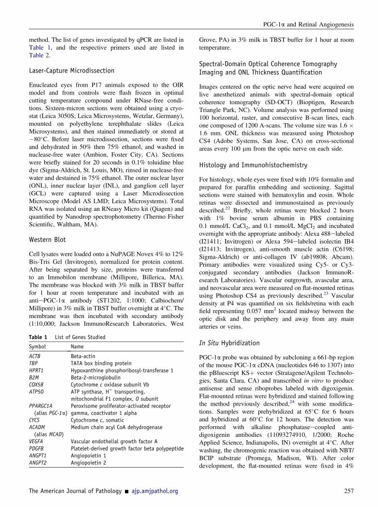
method. The list of genes investigated by qPCR are listed inTable 1, and the respective primers used are listed inTable 2.
Laser-Capture Microdissection
Enucleated eyes from P17 animals exposed to the OIRmodel and from controls were flash frozen in optimalcutting temperature compound under RNase-free condi-tions. Sixteen-micron sections were obtained using a cryo-stat (Leica 3050S; Leica Microsystems, Wetzlar, Germany),mounted on polyethylene terephthalate slides (LeicaMicrosystems), and then stained immediately or stored at�80�C. Before laser microdissection, sections were fixedand dehydrated in 50% then 75% ethanol, and washed innuclease-free water (Ambion, Foster City, CA). Sectionswere briefly stained for 20 seconds in 0.1% toluidine bluedye (Sigma-Aldrich, St. Louis, MO), rinsed in nuclease-freewater and destained in 75% ethanol. The outer nuclear layer(ONL), inner nuclear layer (INL), and ganglion cell layer(GCL) were captured using a Laser MicrodissectionMicroscope (Model AS LMD; Leica Microsystems). TotalRNA was isolated using an RNeasy Micro kit (Qiagen) andquantified by Nanodrop spectrophotometry (Thermo FisherScientific, Waltham, MA).
Western Blot
Cell lysates were loaded onto a NuPAGE Novex 4% to 12%Bis-Tris Gel (Invitrogen), normalized for protein content.After being separated by size, proteins were transferredto an Immobilon membrane (Millipore, Billerica, MA).The membrane was blocked with 3% milk in TBST bufferfor 1 hour at room temperature and incubated with anantiePGC-1a antibody (ST1202, 1:1000; Calbiochem/Millipore) in 3% milk in TBST buffer overnight at 4�C. Themembrane was then incubated with secondary antibody(1:10,000; Jackson ImmunoResearch Laboratories, West
Table 1 List of Genes Studied
Symbol Name
ACTB Beta-actinTBP TATA box binding proteinHPRT1 Hypoxanthine phosphoribosyl-transferase 1B2M Beta-2-microglobulinCOX5B Cytochrome c oxidase subunit VbATP5O ATP synthase, Hþ transporting,
mitochondrial F1 complex, O subunitPPARGC1A(alias PGC-1a)
Peroxisome proliferator-activated receptorgamma, coactivator 1 alpha
CYCS Cytochrome c, somaticACADM(alias MCAD)
Medium chain acyl CoA dehydrogenase
VEGFA Vascular endothelial growth factor APDGFB Platelet-derived growth factor beta polypeptideANGPT1 Angiopoietin 1ANGPT2 Angiopoietin 2
The American Journal of Pathology - ajp.amjpathol.org
Grove, PA) in 3% milk in TBST buffer for 1 hour at roomtemperature.
Spectral-Domain Optical Coherence TomographyImaging and ONL Thickness Quantification
Images centered on the optic nerve head were acquired onlive anesthetized animals with spectral-domain opticalcoherence tomography (SD-OCT) (Bioptigen, ResearchTriangle Park, NC). Volume analysis was performed using100 horizontal, raster, and consecutive B-scan lines, eachone composed of 1200 A-scans. The volume size was 1.6 �1.6 mm. ONL thickness was measured using PhotoshopCS4 (Adobe Systems, San Jose, CA) on cross-sectionalareas every 100 mm from the optic nerve on each side.
Histology and Immunohistochemistry
For histology, whole eyes were fixed with 10% formalin andprepared for paraffin embedding and sectioning. Sagittalsections were stained with hematoxylin and eosin. Wholeretinas were dissected and immunostained as previouslydescribed.22 Briefly, whole retinas were blocked 2 hourswith 1% bovine serum albumin in PBS containing0.1 mmol/L CaCl2, and 0.1 mmol/L MgCl2 and incubatedovernight with the appropriate antibody: Alexa 488elabeled(I21411; Invitrogen) or Alexa 594elabeled isolectin IB4(I21413; Invitrogen), anti-smooth muscle actin (C6198;Sigma-Aldrich) or anti-collagen IV (ab19808; Abcam).Primary antibodies were visualized using Cy5- or Cy3-conjugated secondary antibodies (Jackson ImmunoR-esearch Laboratories). Vascular outgrowth, avascular area,and neovascular area were measured on flat-mounted retinasusing Photoshop CS4 as previously described.23 Vasculardensity at P4 was quantified on six fields/retina with eachfield representing 0.057 mm2 located midway between theoptic disk and the periphery and away from any mainarteries or veins.
In Situ Hybridization
PGC-1a probe was obtained by subcloning a 661-bp regionof the mouse PGC-1a cDNA (nucleotides 646 to 1307) intothe pBluescript KSþ vector (Stratagene/Agilent Technolo-gies, Santa Clara, CA) and transcribed in vitro to produceantisense and sense riboprobes labeled with digoxigenin.Flat-mounted retinas were hybridized and stained followingthe method previously described,24 with some modifica-tions. Samples were prehybridized at 65�C for 6 hoursand hybridized at 60�C for 12 hours. The detection wasperformed with alkaline phosphataseecoupled anti-digoxigenin antibodies (11093274910, 1/2000; RocheApplied Science, Indianapolis, IN) overnight at 4�C. Afterwashing, the chromogenic reaction was obtained with NBT/BCIP substrate (Promega, Madison, WI). After colordevelopment, the flat-mounted retinas were fixed in 4%
257
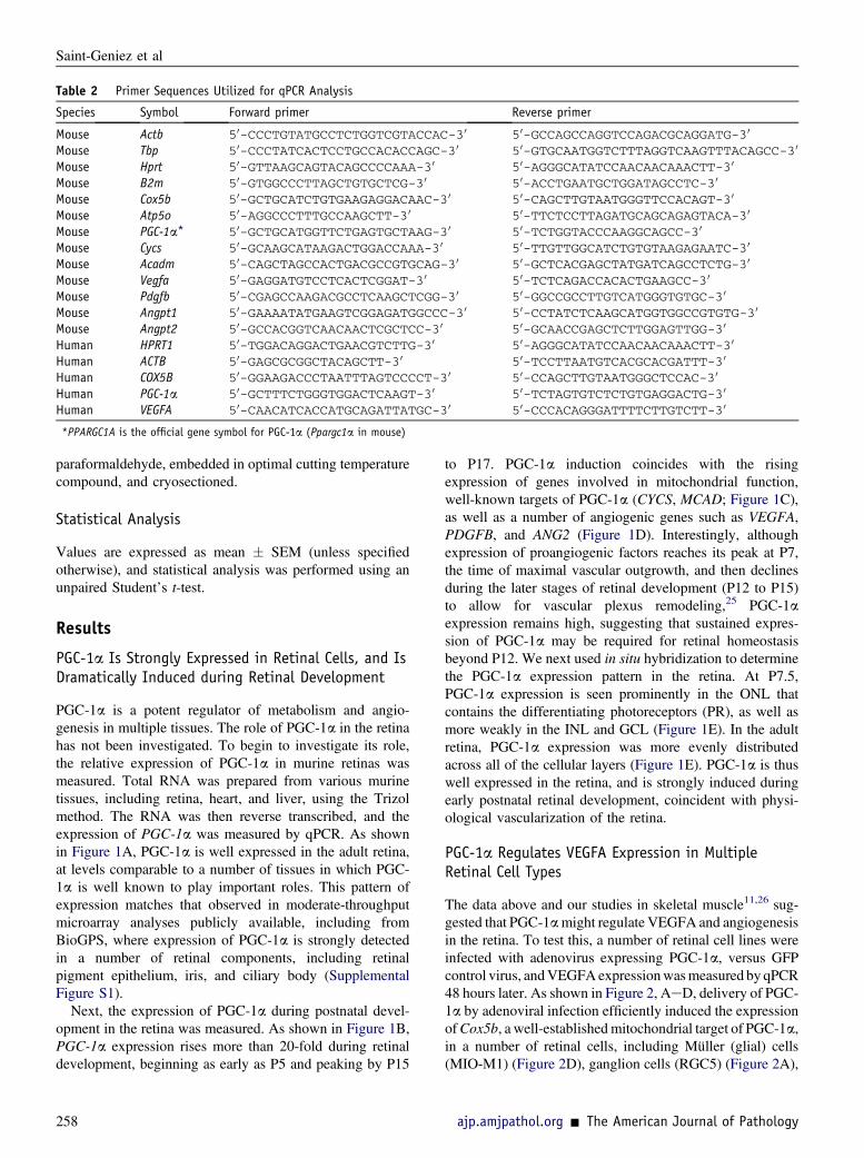
Table 2 Primer Sequences Utilized for qPCR Analysis
Species Symbol Forward primer Reverse primer
Mouse Actb 50-CCCTGTATGCCTCTGGTCGTACCAC-30 50-GCCAGCCAGGTCCAGACGCAGGATG-30
Mouse Tbp 50-CCCTATCACTCCTGCCACACCAGC-30 50-GTGCAATGGTCTTTAGGTCAAGTTTACAGCC-30
Mouse Hprt 50-GTTAAGCAGTACAGCCCCAAA-30 50-AGGGCATATCCAACAACAAACTT-30
Mouse B2m 50-GTGGCCCTTAGCTGTGCTCG-30 50-ACCTGAATGCTGGATAGCCTC-30
Mouse Cox5b 50-GCTGCATCTGTGAAGAGGACAAC-30 50-CAGCTTGTAATGGGTTCCACAGT-30
Mouse Atp5o 50-AGGCCCTTTGCCAAGCTT-30 50-TTCTCCTTAGATGCAGCAGAGTACA-30
Mouse PGC-1a* 50-GCTGCATGGTTCTGAGTGCTAAG-30 50-TCTGGTACCCAAGGCAGCC-30
Mouse Cycs 50-GCAAGCATAAGACTGGACCAAA-30 50-TTGTTGGCATCTGTGTAAGAGAATC-30
Mouse Acadm 50-CAGCTAGCCACTGACGCCGTGCAG-30 50-GCTCACGAGCTATGATCAGCCTCTG-30
Mouse Vegfa 50-GAGGATGTCCTCACTCGGAT-30 50-TCTCAGACCACACTGAAGCC-30
Mouse Pdgfb 50-CGAGCCAAGACGCCTCAAGCTCGG-30 50-GGCCGCCTTGTCATGGGTGTGC-30
Mouse Angpt1 50-GAAAATATGAAGTCGGAGATGGCCC-30 50-CCTATCTCAAGCATGGTGGCCGTGTG-30
Mouse Angpt2 50-GCCACGGTCAACAACTCGCTCC-30 50-GCAACCGAGCTCTTGGAGTTGG-30
Human HPRT1 50-TGGACAGGACTGAACGTCTTG-30 50-AGGGCATATCCAACAACAAACTT-30
Human ACTB 50-GAGCGCGGCTACAGCTT-30 50-TCCTTAATGTCACGCACGATTT-30
Human COX5B 50-GGAAGACCCTAATTTAGTCCCCT-30 50-CCAGCTTGTAATGGGCTCCAC-30
Human PGC-1a 50-GCTTTCTGGGTGGACTCAAGT-30 50-TCTAGTGTCTCTGTGAGGACTG-30
Human VEGFA 50-CAACATCACCATGCAGATTATGC-30 50-CCCACAGGGATTTTCTTGTCTT-30
*PPARGC1A is the official gene symbol for PGC-1a (Ppargc1a in mouse)
Saint-Geniez et al
paraformaldehyde, embedded in optimal cutting temperaturecompound, and cryosectioned.
Statistical Analysis
Values are expressed as mean � SEM (unless specifiedotherwise), and statistical analysis was performed using anunpaired Student’s t-test.
Results
PGC-1a Is Strongly Expressed in Retinal Cells, and IsDramatically Induced during Retinal Development
PGC-1a is a potent regulator of metabolism and angio-genesis in multiple tissues. The role of PGC-1a in the retinahas not been investigated. To begin to investigate its role,the relative expression of PGC-1a in murine retinas wasmeasured. Total RNA was prepared from various murinetissues, including retina, heart, and liver, using the Trizolmethod. The RNA was then reverse transcribed, and theexpression of PGC-1a was measured by qPCR. As shownin Figure 1A, PGC-1a is well expressed in the adult retina,at levels comparable to a number of tissues in which PGC-1a is well known to play important roles. This pattern ofexpression matches that observed in moderate-throughputmicroarray analyses publicly available, including fromBioGPS, where expression of PGC-1a is strongly detectedin a number of retinal components, including retinalpigment epithelium, iris, and ciliary body (SupplementalFigure S1).
Next, the expression of PGC-1a during postnatal devel-opment in the retina was measured. As shown in Figure 1B,PGC-1a expression rises more than 20-fold during retinaldevelopment, beginning as early as P5 and peaking by P15
258
to P17. PGC-1a induction coincides with the risingexpression of genes involved in mitochondrial function,well-known targets of PGC-1a (CYCS, MCAD; Figure 1C),as well as a number of angiogenic genes such as VEGFA,PDGFB, and ANG2 (Figure 1D). Interestingly, althoughexpression of proangiogenic factors reaches its peak at P7,the time of maximal vascular outgrowth, and then declinesduring the later stages of retinal development (P12 to P15)to allow for vascular plexus remodeling,25 PGC-1aexpression remains high, suggesting that sustained expres-sion of PGC-1a may be required for retinal homeostasisbeyond P12. We next used in situ hybridization to determinethe PGC-1a expression pattern in the retina. At P7.5,PGC-1a expression is seen prominently in the ONL thatcontains the differentiating photoreceptors (PR), as well asmore weakly in the INL and GCL (Figure 1E). In the adultretina, PGC-1a expression was more evenly distributedacross all of the cellular layers (Figure 1E). PGC-1a is thuswell expressed in the retina, and is strongly induced duringearly postnatal retinal development, coincident with physi-ological vascularization of the retina.
PGC-1a Regulates VEGFA Expression in MultipleRetinal Cell Types
The data above and our studies in skeletal muscle11,26 sug-gested that PGC-1amight regulate VEGFA and angiogenesisin the retina. To test this, a number of retinal cell lines wereinfected with adenovirus expressing PGC-1a, versus GFPcontrol virus, andVEGFAexpressionwasmeasured by qPCR48 hours later. As shown in Figure 2, AeD, delivery of PGC-1a by adenoviral infection efficiently induced the expressionofCox5b, a well-establishedmitochondrial target of PGC-1a,in a number of retinal cells, including Müller (glial) cells(MIO-M1) (Figure 2D), ganglion cells (RGC5) (Figure 2A),
ajp.amjpathol.org - The American Journal of Pathology
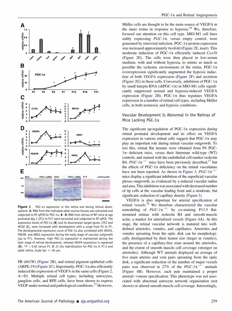
Figure 1 PGC-1a expression in the retina and during retinal devel-
opment. A: RNA from the indicated adult murine tissues was extracted and
subjected to RT-qPCR for PGC-1a. BeD: RNA from retinas of WT mice at age
postnatal day 1 (P1) to P17 were harvested and subjected to RT-qPCR. The
expression levels of PGC-1a (B) and its downstream target genes, CYCS and
MCAD (C), were increased with development with a surge from P3 to P7.
The developmental expression curve of PGC-1a also correlated with VEGFA,
PDGFB, and ANG2 expression during the early stage of vascular outgrowth
(up to P7). However, high PGC-1a expression is maintained during the
later stage of retinal development, whereas VEGFA expression is repressed
(D). *P < 0.05 versus P1. E: In situ hybridization for PGC-1a in P7.5 and
adult retina. Scale bar Z 50 mm.
PGC-1a and Retinal Angiogenesis
PR (661W) (Figure 2B), and retinal pigment epithelial cells(ARPE-19) (Figure 2C). Importantly, PGC-1a also efficientlyinduced the expression of VEGFA in the same cells (Figure 2,AeD). Multiple retinal cell types, including astrocytes,ganglion cells, and RPE cells, have been shown to expressVEGFunder normal and pathological conditions.27However,
The American Journal of Pathology - ajp.amjpathol.org
Müller cells are thought to be the main source of VEGFA inthe inner retina in response to hypoxia.28 We, therefore,focused our attention on this cell type. MIO-M1 cell linesstably expressing PGC-1a, versus empty control, weregenerated by retroviral infection. PGC-1a protein expressionwas increased approximately twofold (Figure 2E, inset). Thismoderate induction of PGC-1a efficiently induced Cox5b(Figure 2E). The cells were then placed in low-serummedium, with and without hypoxia, to mimic as much aspossible the ischemic environment of the retina. PGC-1aoverexpression significantly augmented the hypoxic induc-tion of both VEGFA expression (Figure 2F) and secretion(Figure 2G) in these cells. Conversely, inhibition of PGC-1aby small hairpin RNA (shPGC-1a) in MIO-M1 cells signifi-cantly suppressed normal and hypoxia-induced VEGFAexpression (Figure 2H). PGC-1a thus regulates VEGFAexpression in a number of retinal cell types, including Müllercells, in both normoxic and hypoxic conditions.
Vascular Development Is Abnormal in the Retinas ofMice Lacking PGC-1a
The significant up-regulation of PGC-1a expression duringretinal postnatal development and its effect on VEGFAexpression in various retinal cells suggest that PGC-1a mayplay an important role during retinal vascular outgrowth. Totest this, retinal flat mounts were obtained from P4 PGC-1aedeficient mice, versus their littermate wild-type (WT)controls, and stained with the endothelial cell marker isolectinB4. PGC-1a�/� mice have been previously described,17 butthe effects of PGC-1a deficiency on the retinal vasculaturehave not been reported. As shown in Figure 3, PGC-1a�/�
mice display a significant inhibition of the superficial vascularplexus outgrowth, as evidenced by a reduced vascular radiusand area. This inhibitionwas associatedwith decreased numberof tip cells at the vascular leading front and a moderate, butsignificant, reduction of capillary density (Figure 3).
VEGFA is also important for arterial specification ofretinal vessels.29 We therefore characterized the vascularremodeling of PGC-1a�/� by co-staining P13.5 flat-mounted retinas with isolectin B4 and smooth-muscleactin, a marker for arterialized vessels (Figure 4A). At thisstage, the retinal vascular network is matured into well-defined arterioles, venules, and capillaries. Arterioles andvenules sprouting from the optic disk can be morphologi-cally distinguished by their lumen size (larger in venules),the presence of a capillary-free zone around the arterioles,and the extent of smooth muscle cell coverage (stronger onarterioles). Although WT animals displayed an average offive main arteries and vein pairs sprouting from the opticdisk, a significant reduction of the number of major vesselspairs was observed in 27% of the PGC-1a�/� animals(Figure 4B). However, each pair maintained a properarterialevenous specification. This phenotype was not asso-ciated with abnormal astrocyte network organization (notshown) or altered smooth muscle cell coverage. Interestingly,
259
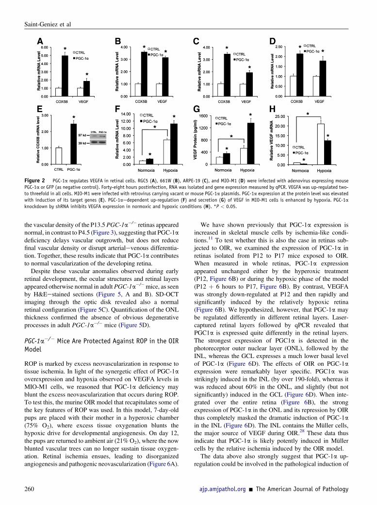
Figure 2 PGC-1a regulates VEGFA in retinal cells. RGC5 (A), 661W (B), ARPE-19 (C), and MIO-M1 (D) were infected with adenovirus expressing mouse
PGC-1a or GFP (as negative control). Forty-eight hours postinfection, RNA was isolated and gene expression measured by qPCR. VEGFA was up-regulated two-
to threefold in all cells. MIO-M1 were infected with retrovirus carrying vacant or mouse PGC-1a plasmids. PGC-1a expression at the protein level was elevated
with induction of its target genes (E). PGC-1aedependent up-regulation (F) and secretion (G) of VEGF in MIO-M1 cells is enhanced by hypoxia. PGC-1a
knockdown by shRNA inhibits VEGFA expression in normoxic and hypoxic conditions (H). *P < 0.05.
Saint-Geniez et al
the vascular density of the P13.5 PGC-1a�/� retinas appearednormal, in contrast to P4.5 (Figure 3), suggesting that PGC-1adeficiency delays vascular outgrowth, but does not reducefinal vascular density or disrupt arterialevenous differentia-tion. Together, these results indicate that PGC-1a contributesto normal vascularization of the developing retina.
Despite these vascular anomalies observed during earlyretinal development, the ocular structures and retinal layersappeared otherwise normal in adult PGC-1a�/�mice, as seenby H&Eestained sections (Figure 5, A and B). SD-OCTimaging through the optic disk revealed also a normalretinal configuration (Figure 5C). Quantification of the ONLthickness confirmed the absence of obvious degenerativeprocesses in adult PGC-1a�/� mice (Figure 5D).
PGC-1a�/� Mice Are Protected Against ROP in the OIRModel
ROP is marked by excess neovascularization in response totissue ischemia. In light of the synergetic effect of PGC-1aoverexpression and hypoxia observed on VEGFA levels inMIO-M1 cells, we reasoned that PGC-1a deficiency mayblunt the excess neovascularization that occurs during ROP.To test this, the murine OIR model that recapitulates some ofthe key features of ROP was used. In this model, 7-day-oldpups are placed with their mother in a hyperoxic chamber(75% O2), where excess tissue oxygenation blunts thehypoxic drive for developmental angiogenesis. On day 12,the pups are returned to ambient air (21% O2), where the nowblunted vascular trees can no longer sustain tissue oxygen-ation. Retinal ischemia ensues, leading to disorganizedangiogenesis and pathogenic neovascularization (Figure 6A).
260
We have shown previously that PGC-1a expression isincreased in skeletal muscle cells by ischemia-like condi-tions.11 To test whether this is also the case in retinas sub-jected to OIR, we examined the expression of PGC-1a inretinas isolated from P12 to P17 mice exposed to OIR.When measured in whole retinas, PGC-1a expressionappeared unchanged either by the hyperoxic treatment(P12, Figure 6B) or during the hypoxic phase of the model(P12 þ 6 hours to P17, Figure 6B). By contrast, VEGFAwas strongly down-regulated at P12 and then rapidly andsignificantly induced by the relatively hypoxic retina(Figure 6B). We hypothesized, however, that PGC-1a maybe regulated differently in different retinal layers. Laser-captured retinal layers followed by qPCR revealed thatPGC1a is expressed quite differently in the retinal layers.The strongest expression of PGC1a is detected in thephotoreceptor outer nuclear layer (ONL), followed by theINL, whereas the GCL expresses a much lower basal levelof PGC-1a (Figure 6D). The effects of OIR on PGC-1aexpression were remarkably layer specific. PGC1a wasstrikingly induced in the INL (by over 190-fold), whereas itwas reduced about 60% in the ONL, and slightly (but notsignificantly) induced in the GCL (Figure 6D). When inte-grated over the entire retina (Figure 6B), the strongexpression of PGC-1a in the ONL and its repression by OIRthus completely masked the dramatic induction of PGC-1ain the INL (Figure 6D). The INL contains the Müller cells,the major source of VEGF during OIR.28 These data thusindicate that PGC-1a is likely potently induced in Müllercells by the relative ischemia induced by the OIR model.The data above also strongly suggest that PGC-1a up-
regulation could be involved in the pathological induction of
ajp.amjpathol.org - The American Journal of Pathology
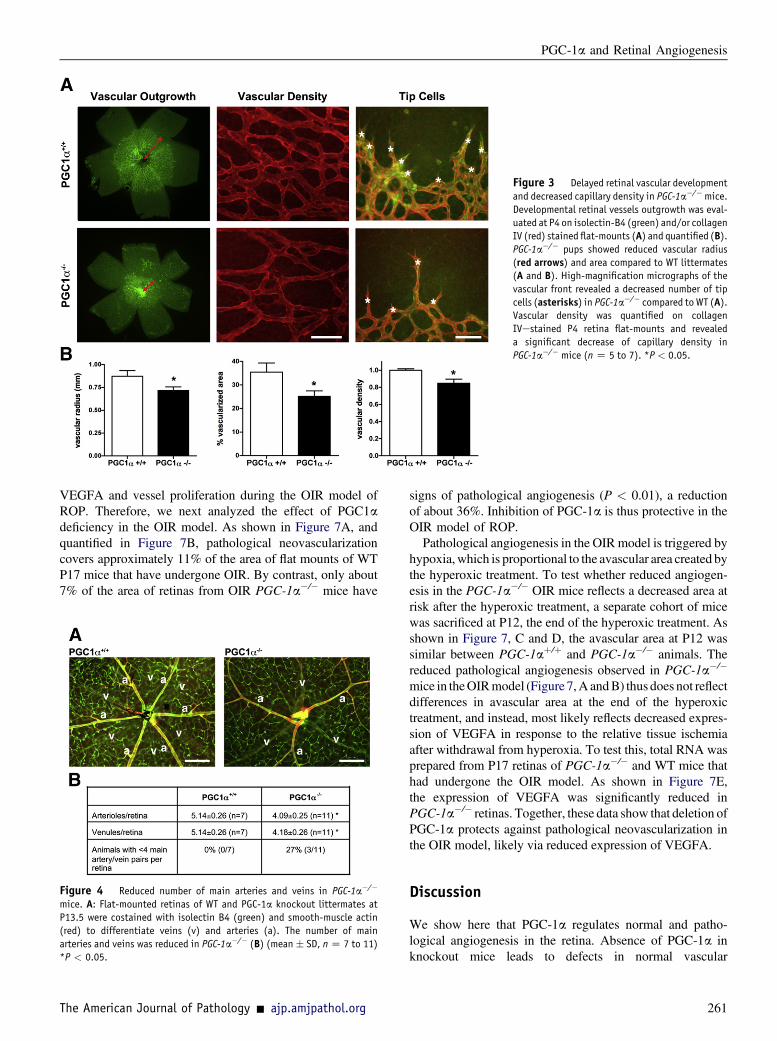
Figure 3 Delayed retinal vascular development
and decreased capillary density in PGC-1a�/�mice.
Developmental retinal vessels outgrowth was eval-
uated at P4 on isolectin-B4 (green) and/or collagen
IV (red) stained flat-mounts (A) and quantified (B).PGC-1a�/� pups showed reduced vascular radius
(red arrows) and area compared to WT littermates
(A and B). High-magnification micrographs of the
vascular front revealed a decreased number of tip
cells (asterisks) in PGC-1a�/� compared to WT (A).Vascular density was quantified on collagen
IVestained P4 retina flat-mounts and revealed
a significant decrease of capillary density in
PGC-1a�/� mice (n Z 5 to 7). *P < 0.05.
PGC-1a and Retinal Angiogenesis
VEGFA and vessel proliferation during the OIR model ofROP. Therefore, we next analyzed the effect of PGC1adeficiency in the OIR model. As shown in Figure 7A, andquantified in Figure 7B, pathological neovascularizationcovers approximately 11% of the area of flat mounts of WTP17 mice that have undergone OIR. By contrast, only about7% of the area of retinas from OIR PGC-1a�/� mice have
Figure 4 Reduced number of main arteries and veins in PGC-1a�/�
mice. A: Flat-mounted retinas of WT and PGC-1a knockout littermates at
P13.5 were costained with isolectin B4 (green) and smooth-muscle actin
(red) to differentiate veins (v) and arteries (a). The number of main
arteries and veins was reduced in PGC-1a�/� (B) (mean � SD, nZ 7 to 11)
*P < 0.05.
The American Journal of Pathology - ajp.amjpathol.org
signs of pathological angiogenesis (P < 0.01), a reductionof about 36%. Inhibition of PGC-1a is thus protective in theOIR model of ROP.
Pathological angiogenesis in the OIR model is triggered byhypoxia,which is proportional to the avascular area created bythe hyperoxic treatment. To test whether reduced angiogen-esis in the PGC-1a�/� OIR mice reflects a decreased area atrisk after the hyperoxic treatment, a separate cohort of micewas sacrificed at P12, the end of the hyperoxic treatment. Asshown in Figure 7, C and D, the avascular area at P12 wassimilar between PGC-1aþ/þ and PGC-1a�/� animals. Thereduced pathological angiogenesis observed in PGC-1a�/�
mice in theOIRmodel (Figure 7,A andB) thus does not reflectdifferences in avascular area at the end of the hyperoxictreatment, and instead, most likely reflects decreased expres-sion of VEGFA in response to the relative tissue ischemiaafter withdrawal from hyperoxia. To test this, total RNA wasprepared from P17 retinas of PGC-1a�/� and WT mice thathad undergone the OIR model. As shown in Figure 7E,the expression of VEGFA was significantly reduced inPGC-1a�/� retinas. Together, these data show that deletion ofPGC-1a protects against pathological neovascularization inthe OIR model, likely via reduced expression of VEGFA.
Discussion
We show here that PGC-1a regulates normal and patho-logical angiogenesis in the retina. Absence of PGC-1a inknockout mice leads to defects in normal vascular
261
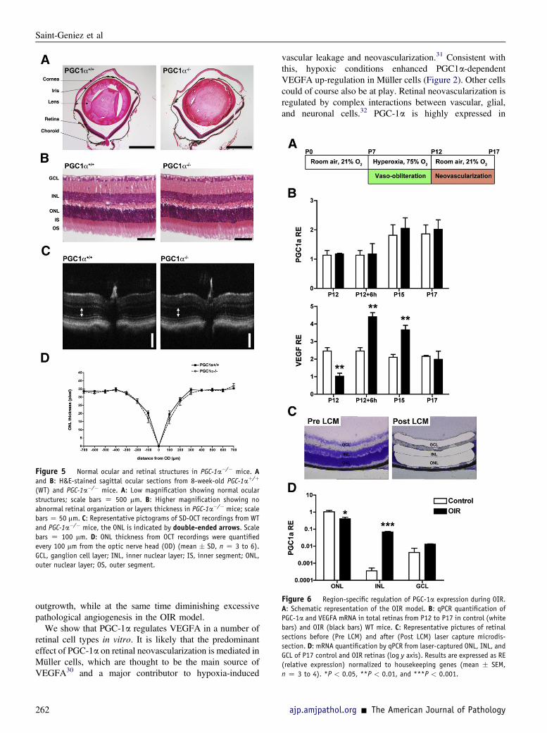
Figure 6 Region-specific regulation of PGC-1a expression during OIR.
A: Schematic representation of the OIR model. B: qPCR quantification of
PGC-1a and VEGFA mRNA in total retinas from P12 to P17 in control (white
bars) and OIR (black bars) WT mice. C: Representative pictures of retinal
sections before (Pre LCM) and after (Post LCM) laser capture microdis-
section. D: mRNA quantification by qPCR from laser-captured ONL, INL, and
GCL of P17 control and OIR retinas (log y axis). Results are expressed as RE
(relative expression) normalized to housekeeping genes (mean � SEM,
n Z 3 to 4). *P < 0.05, **P < 0.01, and ***P < 0.001.
Figure 5 Normal ocular and retinal structures in PGC-1a�/� mice. Aand B: H&E-stained sagittal ocular sections from 8-week-old PGC-1aþ/þ
(WT) and PGC-1a�/� mice. A: Low magnification showing normal ocular
structures; scale bars Z 500 mm. B: Higher magnification showing no
abnormal retinal organization or layers thickness in PGC-1a�/� mice; scale
bars Z 50 mm. C: Representative pictograms of SD-OCT recordings from WT
and PGC-1a�/� mice, the ONL is indicated by double-ended arrows. Scalebars Z 100 mm. D: ONL thickness from OCT recordings were quantified
every 100 mm from the optic nerve head (OD) (mean � SD, n Z 3 to 6).
GCL, ganglion cell layer; INL, inner nuclear layer; IS, inner segment; ONL,
outer nuclear layer; OS, outer segment.
Saint-Geniez et al
outgrowth, while at the same time diminishing excessivepathological angiogenesis in the OIR model.
We show that PGC-1a regulates VEGFA in a number ofretinal cell types in vitro. It is likely that the predominanteffect of PGC-1a on retinal neovascularization is mediated inMüller cells, which are thought to be the main source ofVEGFA30 and a major contributor to hypoxia-induced
262
vascular leakage and neovascularization.31 Consistent withthis, hypoxic conditions enhanced PGC1a-dependentVEGFA up-regulation in Müller cells (Figure 2). Other cellscould of course also be at play. Retinal neovascularization isregulated by complex interactions between vascular, glial,and neuronal cells.32 PGC-1a is highly expressed in
ajp.amjpathol.org - The American Journal of Pathology

Figure 7 PGC-1a�/� mice are protected
against OIR. Oxygen-induced retinopathy (OIR)
was induced by exposing PGC-1aþ/þ (WT) and
PGC-1a�/� mouse pups to 75% oxygen from P7 to
P12. A and B: At P17, retinas were harvested and
stained with isolectin-B4. The vaso-obliterated
areas (yellow outline) and neovascular areas
(marked in white) were quantified using Photo-
shop CS4 software. Loss of PGC-1a did not affect
the avascular zone but significantly reduced the
neovascular area (n Z 7 to 9). **P < 0.01. C and
D: Representative pictograms (C) and quantifica-
tion (D) of the central vaso-obliteration induced
by hyperoxia at P12 (n Z 7 to 12). ns, no
statistical difference. E: qPCR analysis of total
retina mRNA isolated from P17 animals exposed to
OIR model revealed a significant reduction in
Cox5b and VEGFA expression in PGC-1a�/� mice
compared to PGC-1aþ/þ (n Z 4). *P < 0.05,
***P < 0.001.
PGC-1a and Retinal Angiogenesis
developing PR (Figure 1) and was recently shown to beinvolved in PR susceptibility to light damage.16 Even thoughthe PR do not appear to be a major source of proangiogenicmolecules even under severe hypoxia, the hypoxic state ofthe PR can affect VEGFA expression and the subsequentangiogenic response.33 However, since PGC-1a deficiencydoes not cause retinal degeneration (Figure 5 and Eggeret al16), it seems unlikely that PGC-1aedependent modula-tion of PR metabolism drives the reduced oxygen-dependentneovascularization that we observed. PGC-1a could also beinvolved in the homeostasis of endothelial cells.34 However,PGC-1a expression in endothelial cells is extremely lowwhen compared to other cell types and, in particular, toMüller cells (Supplemental Figure S2), suggesting that therole of PGC-1a is likely much greater inMüller cells. Finally,it is formally possible that some aspect of the phenotypeobserved here reflects alterations in systemic metabolism inthe PGC-1a�/� mice. The simplest interpretation of ourfindings, however, is that the predominant effect of PGC-1ais mediated in Müller cells, which are thought to be thestrongest secretors of VEGFA.30 Precise characterization offunction of PGC-1a in each cell type will require cell-specificdeletion with available floxed alleles of PGC-1a.
Angiogenesis in the retina, in particular in response tophysiological and pathological hypoxia has been thought tobe mediated primarily by the HIF-1 pathway, even thoughevidence suggests that other pathways must exist.10 Wehave shown previously that the induction of VEGFA andangiogenic factors by PGC-1a in skeletal muscle is
The American Journal of Pathology - ajp.amjpathol.org
independent of HIF-1a, occurring instead by coactivation ofthe orphan nuclear receptor estrogen-related receptora (ERRa).11 The data presented here demonstrate that thispathway is also likely at play in the retina. Interestingly, wedid not observe in PGC-1aedeficient mice any changes inthe extent of oxygen-induced vaso-obliteration in the OIRmodel, suggesting that, contrary to what occurs with HIF-1proteins,35 PGC-1a is not involved in the maintenance ofthe trophic factors that prevent microvascular obliteration. Itwill be of interest to study retinal development and responseto OIR in ERRa�/� mice. It will also ultimately be ofinterest to attempt to intervene pharmacologically in thisnovel pathway. Screens to identify small molecules thatmodulate PGC-1a have yielded some results, for example,but remain in the early stages.36
Some aspects of the phenotypes we observed in unchal-lenged PGC-1a�/� mice were incompletely penetrant (eg,Figure 4). Thismay reflect effects of variable genetic modifiersand/or variable compensation by other pathways. PGC-1b, forexample, regulates many of the same pathways as PGC-1a,including angiogenesis in skeletal muscle.37 We did notobserve compensatory up-regulation of PGC-1b mRNA inPGC-1a�/� mice, but compensation may have occurred at theposttranscriptional level. It will be of interest, therefore, toevaluate deletion of both PGC-1a and b simultaneously. Thiswill require conditional deletion in the appropriate cell type,since PGC-1a/b doubly deleted mice die at birth.38
The PGC-1aeregulated pathway in the eye likelyprovides an important node for regulation by multiple
263
Saint-Geniez et al
physiological cues. Hypoxia is not the only challenge thatischemic tissues face. Insufficient blood flow also leads todeprivation of nutrients, pH changes, dramatic changes inintermediate metabolism and cellular redox state, andaccumulation of numerous extracellular molecules. The rolethat these metabolic events play in regulating angiogenesisis not well understood. For example, Sapieha et al39 recentlyshowed that the hypoxic retina accumulates extracellularsuccinate, which directly stimulates ganglion cells to secreteVEGFA and other angiogenic factors independently fromthe HIF pathway. Thus, the complex metabolic events thatoccur during ischemia are clearly linked to the angiogenicresponse, but the mechanisms involved remain incompletelyresolved. PGC-1a is extensively regulated, both at thetranscriptional and posttranslational levels, by upstreamsignaling cascades including cAMP, mitogen-activatedprotein kinase, and powerful metabolic sensors such asAMP-activated protein kinase and sirtuin.14 PGC-1a thusintegrates multiple metabolic signals in addition to hypoxia,and is in an ideal position to link those signals to angio-genesis in the eye.
In summary, we show here that PGC-1a regulatesVEGFA and angiogenesis in the retina under both physio-logical and pathophysiological conditions. Further study ofthis novel pathway may lead to new insight and approachesto treating a variety of retinal pathologies marked by inap-propriate and deranged neovascularization, including ROP,but also “wet” aged-related macular degeneration and dia-betic retinopathy.
Acknowledgments
We thank Drs. Andrea Giani and Aristomenis Thanos(Massachusetts Eye and Ear Infirmary, Boston) for theirassistance in SD-OCT image acquisition.
Supplemental Data
Supplemental material for this article can be found at http://dx.doi.org/10.1016/j.ajpath.2012.09.003.
References
1. Yu DY, Cringle SJ: Oxygen distribution and consumption within theretina in vascularised and avascular retinas and in animal models ofretinal disease. Prog Retin Eye Res 2001, 20:175e208
2. Saint-Geniez M, D’Amore PA: Development and pathology of thehyaloid, choroidal and retinal vasculature. Int J Dev Biol 2004, 48:1045e1058
3. Engerman RL, Pfaffenbach D, Davis MD: Cell turnover of capillaries.Lab Invest 1967, 17:738e743
4. Phelps DL: Retinopathy of prematurity: an estimate of vision loss inthe United Statese1979. Pediatrics 1981, 67:924e925
5. Sapieha P, Joyal JS, Rivera JC, Kermorvant-Duchemin E, Sennlaub F,Hardy P, Lachapelle P, Chemtob S: Retinopathy of prematurity:understanding ischemic retinal vasculopathies at an extreme of life.J Clin Invest 2012, 120:3022e3032
264
6. Alon T, Hemo I, Itin A, Pe’er J, Stone J, Keshet S: Vascular endo-thelial growth factor acts as a survival factor for newly formed retinalvessels and has implications for retinopathy of prematurity. Nat Med1995, 1:1024e1028
7. Chen J, Stahl A, Hellstrom A, Smith LE: Current update on retinop-athy of prematurity: screening and treatment. Curr Opin Pediatr 2011,23:173e178
8. Mintz-Hittner HA, Kennedy KA, Chuang AZ, BEAT-ROP Coopera-tive Group: Efficacy of intravitreal bevacizumab for stage 3þ reti-nopathy of prematurity. N Engl J Med 2011, 364:603e615
9. Arjamaa O, Nikinmaa M: Oxygen-dependent diseases in the retina:role of hypoxia-inducible factors. Exp Eye Res 2006, 83:473e483
10. Vinores SA, Xiao WH, Aslam S, Shen J, Oshima Y, Nambu H, Liu H,Carmeliet P, Campochiaro PA: Implication of the hypoxia responseelement of the Vegf promoter in mouse models of retinal and choroidalneovascularization, but not retinal vascular development. J CellPhysiol 2006, 206:749e758
11. Arany Z, Foo SY, Ma Y, Ruas JL, Bommi-Reddy A, Girnun G,Cooper M, Laznik D, Chinsomboon J, Rangwala SM, Baek KH,Rosenzweig A, Spiegelman BM: HIF-independent regulation of VEGFand angiogenesis by the transcriptional coactivator PGC-1alpha.Nature 2008, 451:1008e1012
12. Kelly DP, Scarpulla RC: Transcriptional regulatory circuits control-ling mitochondrial biogenesis and function. Genes Dev 2004, 18:357e368
13. Lin J, Handschin C, Spiegelman BM: Metabolic control through thePGC-1 family of transcription coactivators. Cell Metab 2005, 1:361e370
14. Handschin C, Spiegelman BM: Peroxisome proliferator-activatedreceptor gamma coactivator 1 coactivators, energy homeostasis, andmetabolism. Endocr Rev 2006, 27:728e735
15. Arany Z: PGC-1 coactivators and skeletal muscle adaptations in healthand disease. Curr Opin Genet Dev 2008, 18:426e434
16. Egger A, Samardzija M, Sothilingam V, Tanimoto N, Lange C,Salatino S, Fang L, Garcia-Garrido M, Beck S, Okoniewski MJ,Neutzner A, Seeliger MW, Grimm C, Handschin C: PGC-1alphadetermines light damage susceptibility of the murine retina. PLoS One2012, 7:e31272
17. Lin J, Wu PH, Tarr PT, Lindenberg KS, St-Pierre J, Zhang CY,Mootha VK, Jager S, Vianna CR, Reznick RM, Cui L, Manieri M,Donovan MX, Wu Z, Cooper MP, Fan MC, Rohas LM, Zavacki AM,Cinti S, Shulman GI, Lowell BB, Krainc D, Spiegelman BM: Defectsin adaptive energy metabolism with CNS-linked hyperactivity inPGC-1alpha null mice. Cell 2004, 119:121e135
18. Smith LE, Wesolowski E, McLellan A, Kostyk SK, D’Amato R,Sullivan R, D’Amore PA: Oxygen-induced retinopathy in the mouse.Invest Ophthalmol Vis Sci 1994, 35:101e111
19. Vega RB, Huss JM, Kelly DP: The coactivator PGC-1 cooperates withperoxisome proliferator-activated receptor alpha in transcriptionalcontrol of nuclear genes encoding mitochondrial fatty acid oxidationenzymes. Mol Cell Biol 2000, 20:1868e1876
20. Puigserver P, Wu Z, Park CW, Graves R, Wright M, Spiegelman BM:A cold-inducible coactivator of nuclear receptors linked to adaptivethermogenesis. Cell 1998, 92:829e839
21. Vandesompele J, De Preter K, Pattyn F, Poppe B, Van Roy N,De Paepe A, Speleman F: Accurate normalization of real-time quan-titative RT-PCR data by geometric averaging of multiple internalcontrol genes. Genome Biol 2002, 3:RESEARCH0034
22. Saint-Geniez M, Maharaj AS, Walshe TE, Tucker BA, Sekiyama E,Kurihara T, Darland DC, Young MJ, D’Amore PA: EndogenousVEGF is required for visual function: evidence for a survival role onMüller cells and photoreceptors. PLoS ONE 2008, 3:e3554
23. Connor KM, Krah NM, Dennison RJ, Aderman CM, Chen J,Guerin KI, Sapieha P, Stahl A, Willett KL, Smith LE: Quantification ofoxygen-induced retinopathy in the mouse: a model of vessel loss,vessel regrowth and pathological angiogenesis. Nat Protoc 2009, 4:1565e1573
ajp.amjpathol.org - The American Journal of Pathology
PGC-1a and Retinal Angiogenesis
24. Saint-Geniez M, Argence CB, Knibiehler B, Audigier Y: The msr/apjgene encoding the apelin receptor is an early and specific marker ofthe venous phenotype in the retinal vasculature. Gene Expr Patterns2003, 3:467e472
25. Fruttiger M: Development of the retinal vasculature. Angiogenesis2007, 10:77e88
26. Chinsomboon J, Ruas J, Gupta RK, Thom R, Shoag J, Rowe GC,Sawada N, Raghuram S, Arany Z: The transcriptional coactivatorPGC-1a mediates exercise-induced angiogenesis in skeletal muscle.Proc Natl Acad Sci U S A 2009, 106:21401e21406
27. Gariano RF, Gardner TW: Retinal angiogenesis in development anddisease. Nature 2005, 438:960e966
28. Pierce EA, Avery RL, Foley ED, Aiello LP, Smith LE: Vascularendothelial growth factor/vascular permeability factor expression ina mouse model of retinal neovascularization. Proc Natl Acad Sci U S A1995, 92:905e909
29. Stalmans I, Ng YS, Rohan R, Fruttiger M, Bouche A, Yuce A,Fujisawa H, Hermans B, Shani M, Jansen S, Hicklin D, Anderson DJ,Gardiner T, Hammes HP, Moons L, Dewerchin M, Collen D,Carmeliet P, D’Amore PA: Arteriolar and venular patterning in retinasof mice selectively expressing VEGF isoforms. J Clin Invest 2002,109:327e336
30. Stone J, Itin A, Alon T, Pe’er J, Gnessin H, Chan-Ling T, Keshet E:Development of retinal vasculature is mediated by hypoxia-inducedvascular endothelial growth factor (VEGF) expression by neuroglia.J Neurosci 1995, 15:4738e4747
31. Bai Y, Ma JX, Guo J, Wang J, Zhu M, Chen Y, Le YZ: Muller cell-derived VEGF is a significant contributor to retinal neovascularization.J Pathol 2009, 219:446e454
The American Journal of Pathology - ajp.amjpathol.org
32. Vessey KA, Wilkinson-Berka JL, Fletcher EL: Characterization ofretinal function and glial cell response in a mouse model of oxygen-induced retinopathy. J Comp Neurol 2011, 519:506e527
33. Lahdenranta J, Pasqualini R, Schlingemann RO, Hagedorn M,Stallcup WB, Bucana CD, Sidman RL, Arap W: An anti-angiogenicstate in mice and humans with retinal photoreceptor cell degenera-tion. Proc Natl Acad Sci U S A 2001, 98:10368e10373
34. Valle I, Alvarez-Barrientos A, Arza E, Lamas S, Monsalve M: PGC-1alpha regulates the mitochondrial antioxidant defense system invascular endothelial cells. Cardiovasc Res 2005, 66:562e573
35. Duan LJ, Takeda K, Fong GH: Prolyl hydroxylase domain protein 2(PHD2) mediates oxygen-induced retinopathy in neonatal mice. Am JPathol 2011, 178:1881e1890
36. Arany Z, Wagner BK, Ma Y, Chinsomboon J, Laznik D,Spiegelman BM: Gene expression-based screening identifies micro-tubule inhibitors as inducers of PGC-1alpha and oxidative phosphor-ylation. Proc Natl Acad Sci U S A 2008, 105:4721e4726
37. RoweGC, JangC, Patten IS, Arany Z: PGC-1b regulates angiogenesis inskeletal muscle. Am J Physiol Endocrinol Metab 2011, 301:E155eE163
38. Lai L, Leone TC, Zechner C, Schaeffer PJ, Kelly SM, Flanagan DP,Medeiros DM, Kovacs A, Kelly DP: Transcriptional coactivatorsPGC-1alpha and PGC-lbeta control overlapping programs requiredfor perinatal maturation of the heart. Genes Dev 2008, 22:1948e1961
39. Sapieha P, Sirinyan M, Hamel D, Zaniolo K, Joyal JS, Cho JH,Honore JC, Kermorvant-Duchemin E, Varma DR, Tremblay S,Leduc M, Rihakova L, Hardy P, Klein WH, Mu X, Mamer O,Lachapelle P, Di Polo A, Beausejour C, Andelfinger G, Mitchell G,Sennlaub F, Chemtob S: The succinate receptor GPR91 in neurons hasa major role in retinal angiogenesis. Nat Med 2008, 14:1067e1076
265