Clinical aspects of vestibular and oculomotor...
57
Clinical aspects of vestibular and ocular motor physiology: bringing physiology and anatomy to the bedside Skews Nystagmus Tilts [email protected]
Transcript of Clinical aspects of vestibular and oculomotor...
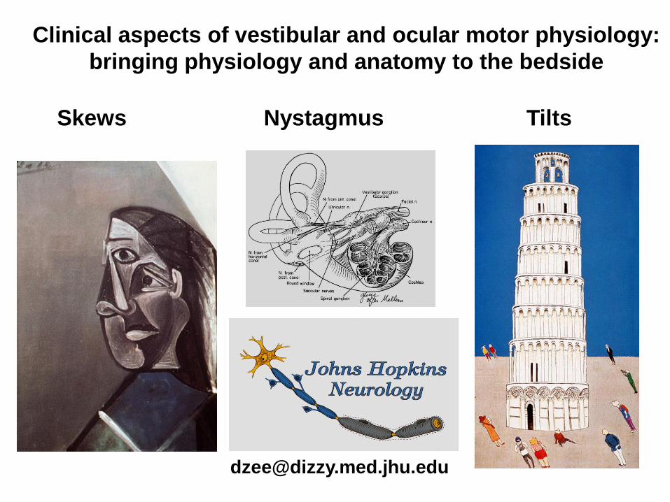
Clinical aspects of vestibular and ocular motor physiology:
bringing physiology and anatomy to the bedside
Skews Nystagmus Tilts
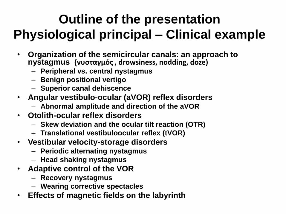
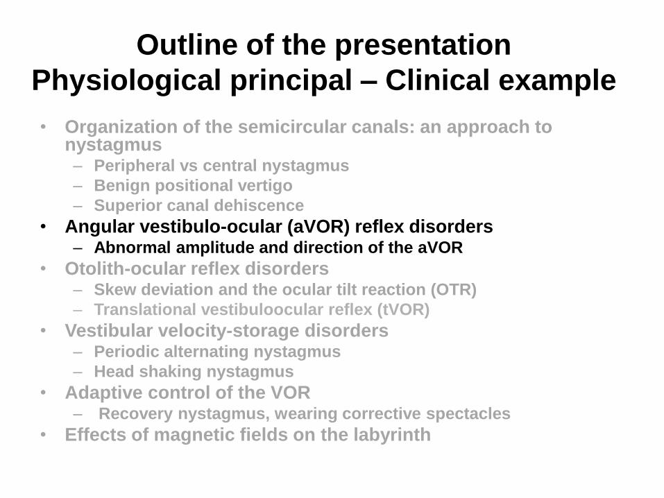
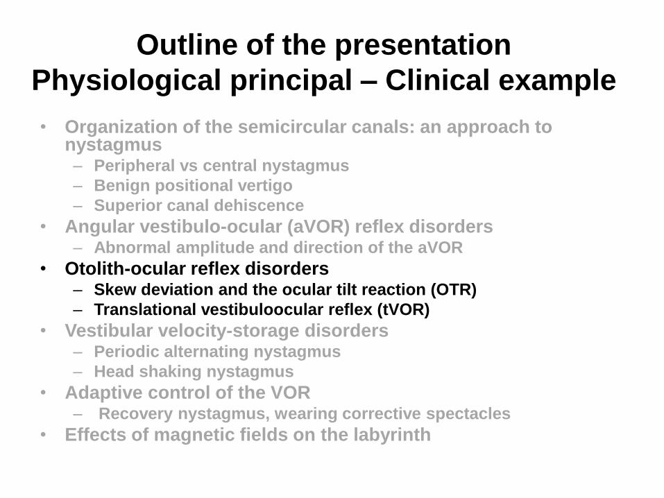
Outline of the presentation
Physiological principal – Clinical example
• Organization of the semicircular canals: an approach to nystagmus (νυσταγμός , drowsiness, nodding, doze) – Peripheral vs. central nystagmus
– Benign positional vertigo
– Superior canal dehiscence
• Angular vestibulo-ocular (aVOR) reflex disorders – Abnormal amplitude and direction of the aVOR
• Otolith-ocular reflex disorders – Skew deviation and the ocular tilt reaction (OTR)
– Translational vestibuloocular reflex (tVOR)
• Vestibular velocity-storage disorders – Periodic alternating nystagmus
– Head shaking nystagmus
• Adaptive control of the VOR – Recovery nystagmus
– Wearing corrective spectacles
• Effects of magnetic fields on the labyrinth
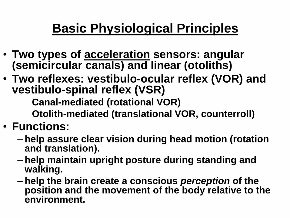
Basic Physiological Principles
• Two types of acceleration sensors: angular (semicircular canals) and linear (otoliths)
• Two reflexes: vestibulo-ocular reflex (VOR) and vestibulo-spinal reflex (VSR) Canal-mediated (rotational VOR)
Otolith-mediated (translational VOR, counterroll)
• Functions: – help assure clear vision during head motion (rotation
and translation).
– help maintain upright posture during standing and walking.
– help the brain create a conscious perception of the position and the movement of the body relative to the environment.
Outline of the presentation
Physiological principal – Clinical example
• Organization of the semicircular canals: an approach to nystagmus (νυσταγμός , drowsiness, nodding, doze) – Peripheral vs. central nystagmus
– Benign positional vertigo
– Superior canal dehiscence
• Angular vestibulo-ocular (aVOR) reflex disorders – Abnormal amplitude and direction of the aVOR
• Otolith-ocular reflex disorders – Skew deviation and the ocular tilt reaction (OTR)
– Translational vestibuloocular reflex (tVOR)
• Vestibular velocity-storage disorders – Periodic alternating nystagmus
– Head shaking nystagmus
• Adaptive control of the VOR – Recovery nystagmus, wearing corrective spectacles
• Effects of magnetic fields on the labyrinth
Peripheral
Central
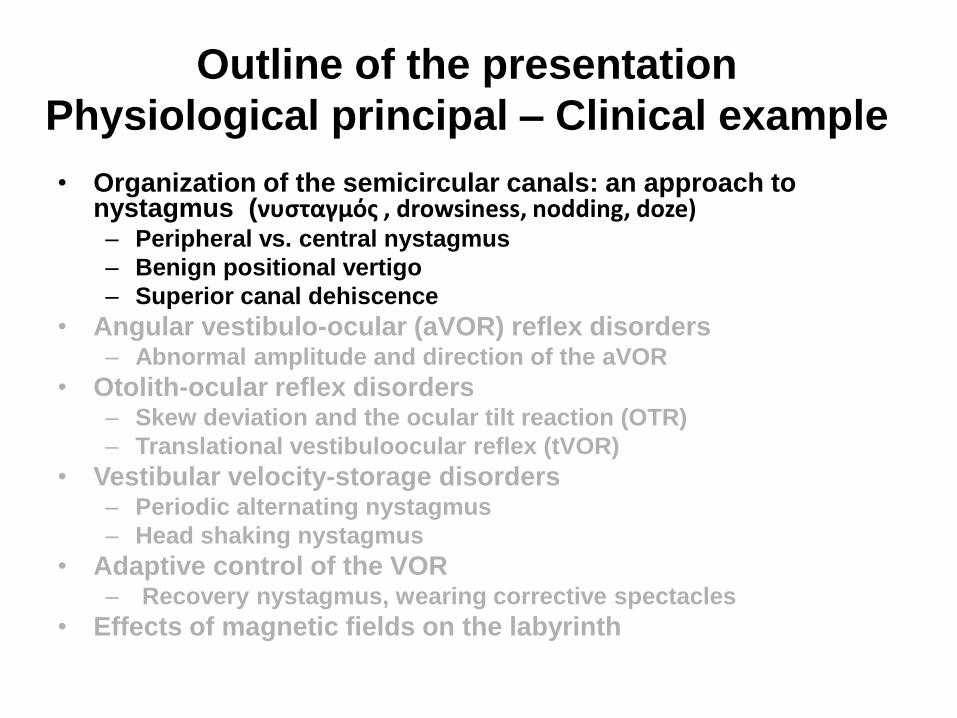
Ewald’s First Law: Eyes (head) rotates in a plane parallel to that of rotation of the head
(detected by the SCC in that plane) and so stabilizes gaze (eye in space) around all three
axes of head rotation
SCC Organization: a guide to nystagmus
Work of Ewald and of Flourens
Arrows indicate
direction of slow
phase with
stimulation
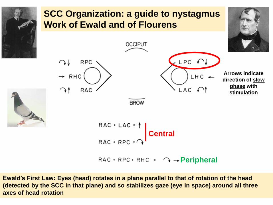
Ophthalmoscope and spontaneous
nystagmus
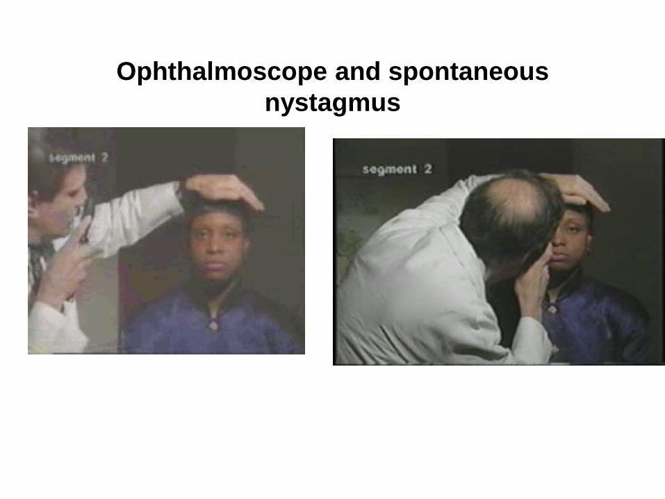
Frenzel Lenses (best used with room lights off) to remove
fixation
Look for nystagmus
Strupp, Neurology, 2014
•Peripheral vestibular nystagmus is increased or brought out by removal of fixation
•Alexander’s Law: Peripheral vestibular nystagmus increases in intensity when
looking in the direction of quick phase
Young man presents with acute onset of sustained vertigo, nausea,
vomiting, imbalance, without hearing symptoms.
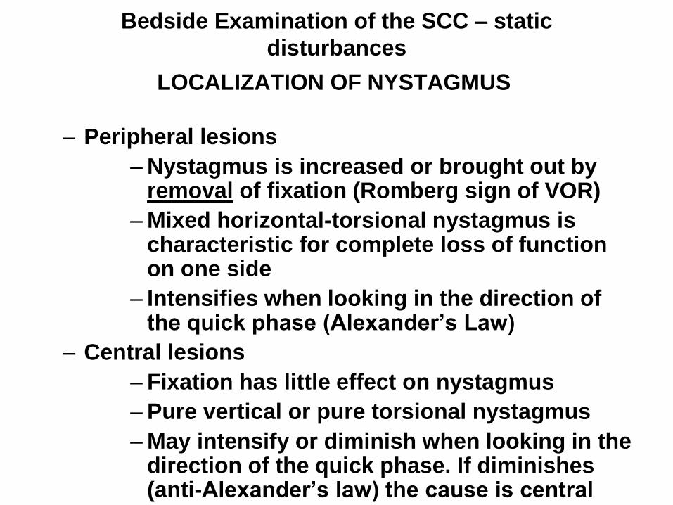
Bedside Examination of the SCC – static
disturbances
LOCALIZATION OF NYSTAGMUS
– Peripheral lesions
– Nystagmus is increased or brought out by removal of fixation (Romberg sign of VOR)
– Mixed horizontal-torsional nystagmus is characteristic for complete loss of function on one side
– Intensifies when looking in the direction of the quick phase (Alexander’s Law)
– Central lesions
– Fixation has little effect on nystagmus
– Pure vertical or pure torsional nystagmus
– May intensify or diminish when looking in the direction of the quick phase. If diminishes (anti-Alexander’s law) the cause is central
Benign Paroxysmal Positional Vertigo (BPPV)
• Easily diagnosed by history and exam
• Pathophysiology well understood
• Easily treated
• Patients gratified
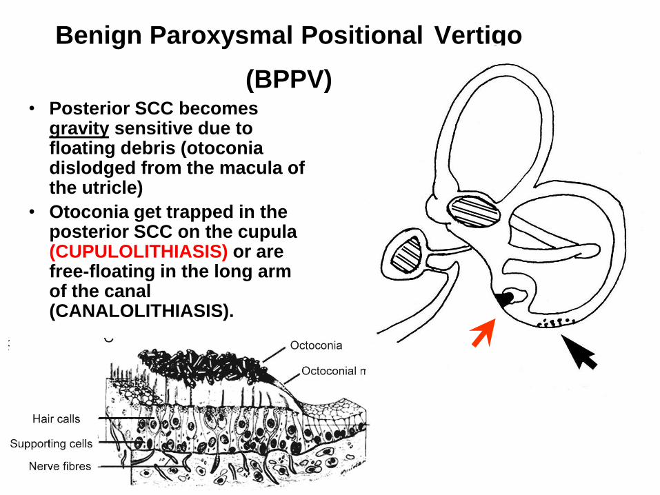
Benign Paroxysmal Positional Vertigo
(BPPV) • Posterior SCC becomes
gravity sensitive due to floating debris (otoconia dislodged from the macula of the utricle)
• Otoconia get trapped in the posterior SCC on the cupula (CUPULOLITHIASIS) or are free-floating in the long arm of the canal (CANALOLITHIASIS).
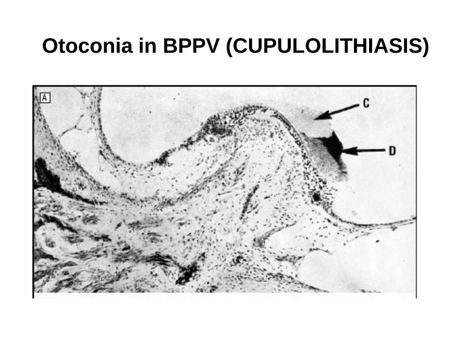
Otoconia in BPPV (CUPULOLITHIASIS)
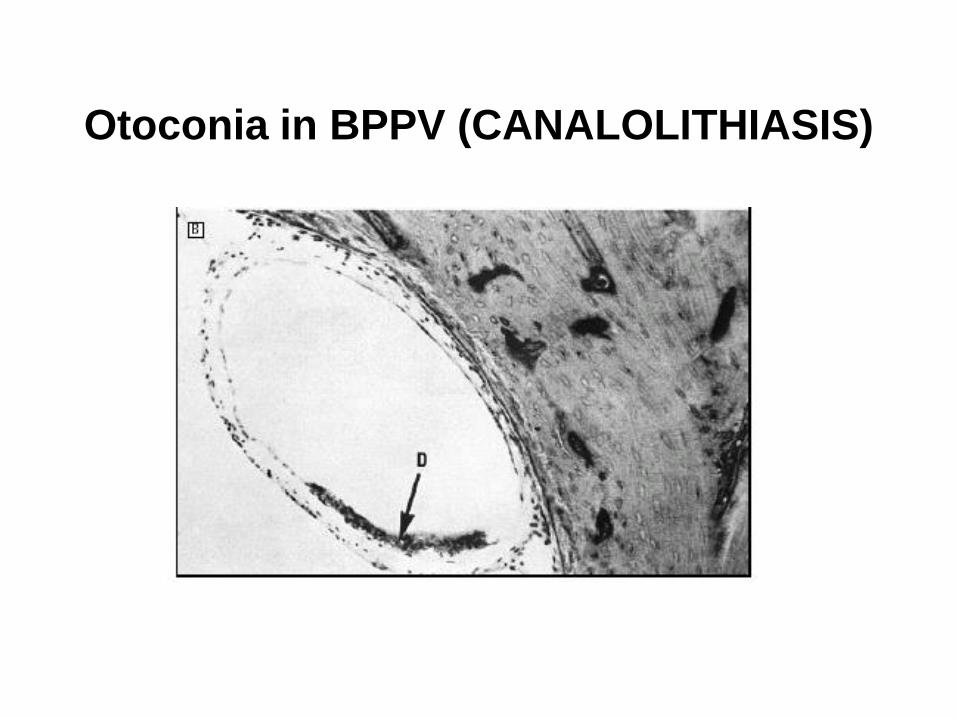
Otoconia in BPPV (CANALOLITHIASIS)
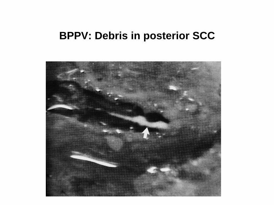
BPPV: Debris in posterior SCC
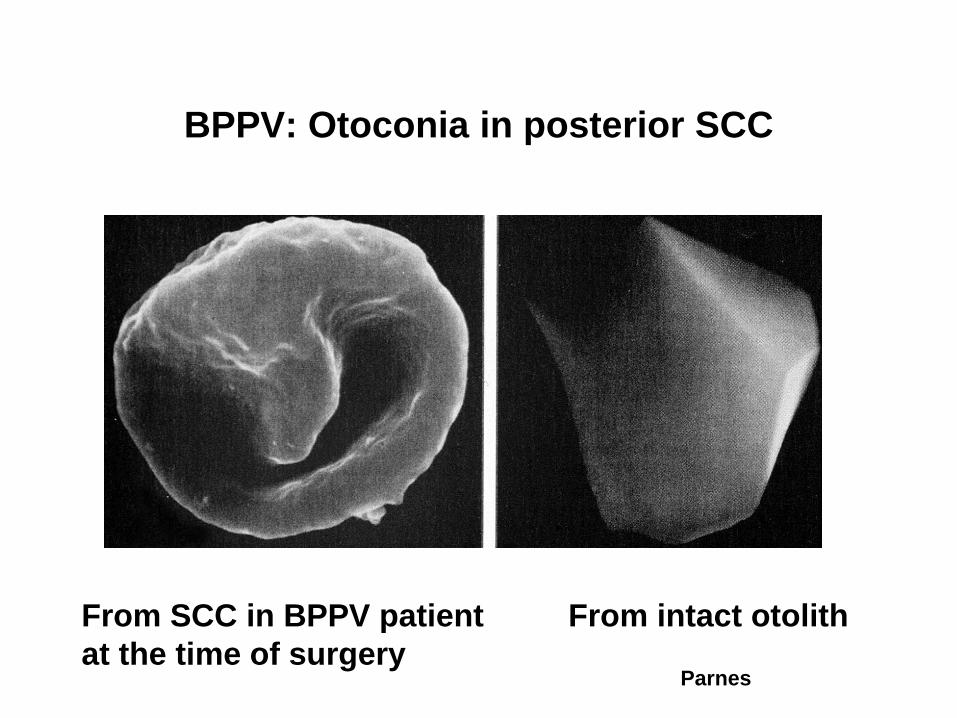
BPPV: Otoconia in posterior SCC
From intact otolith From SCC in BPPV patient
at the time of surgery Parnes
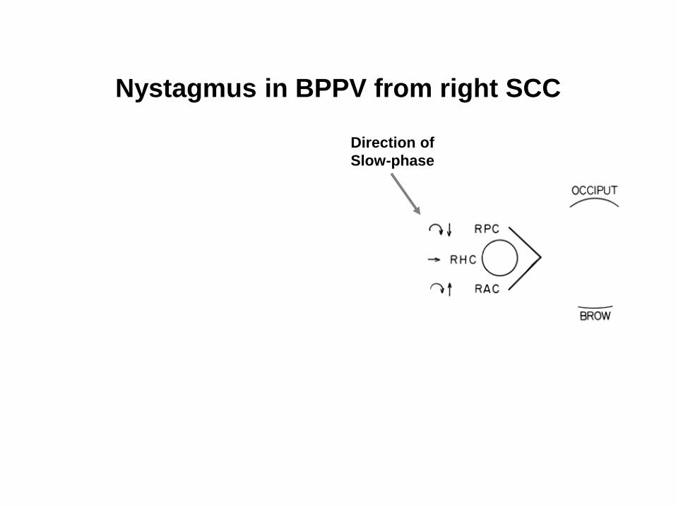
Nystagmus in BPPV from right SCC
Direction of
Slow-phase
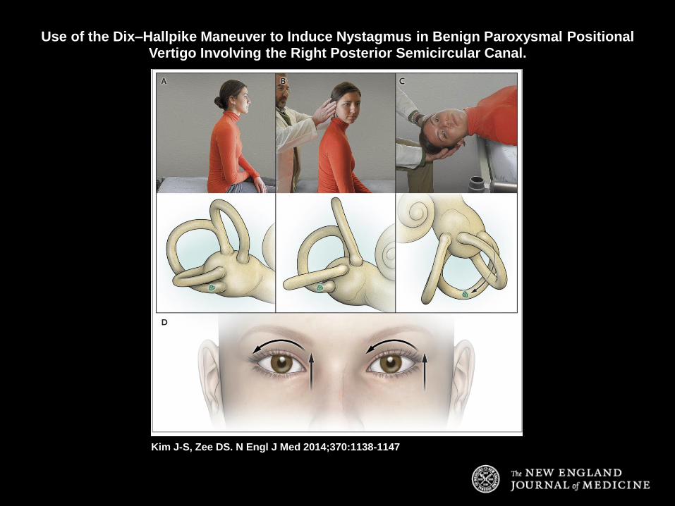
Use of the Dix–Hallpike Maneuver to Induce Nystagmus in Benign Paroxysmal Positional Vertigo Involving the Right Posterior Semicircular Canal.
Kim J-S, Zee DS. N Engl J Med 2014;370:1138-1147
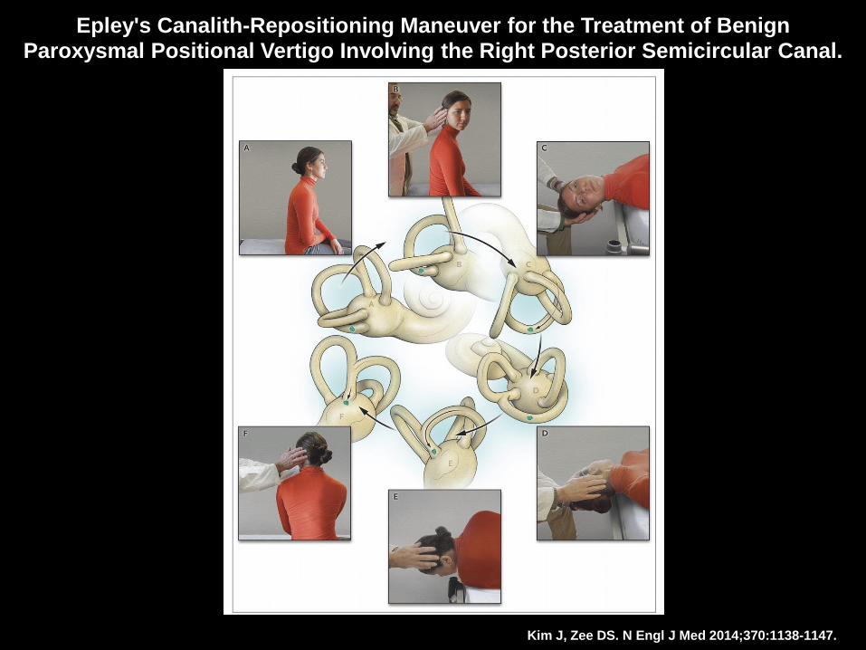
Epley's Canalith-Repositioning Maneuver for the Treatment of Benign Paroxysmal Positional Vertigo Involving the Right Posterior Semicircular Canal.
Kim J, Zee DS. N Engl J Med 2014;370:1138-1147.
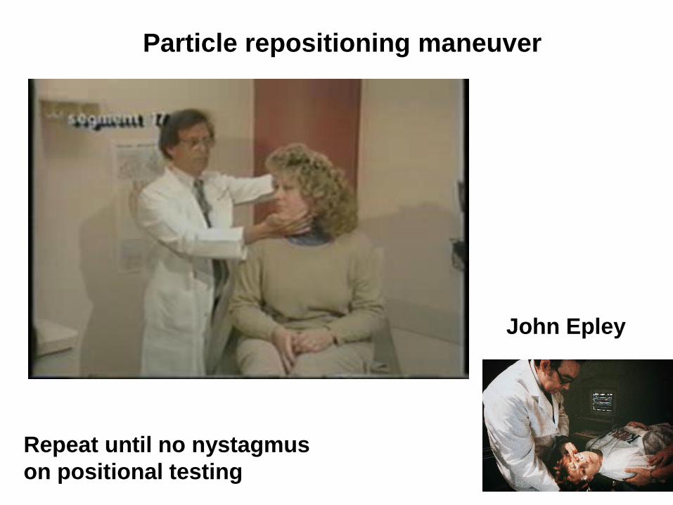
Particle repositioning maneuver
John Epley
Repeat until no nystagmus
on positional testing
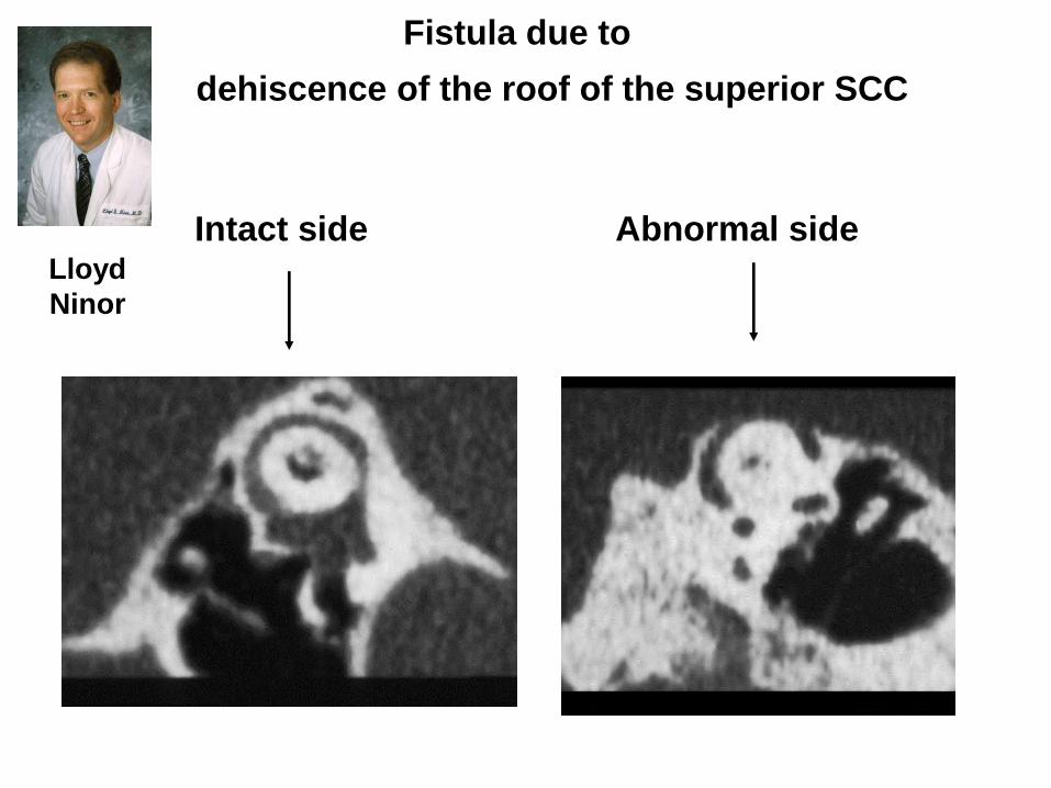
Fistula due to
Intact side Abnormal side
dehiscence of the roof of the superior SCC
Lloyd
Ninor
Outline of the presentation
Physiological principal – Clinical example
• Organization of the semicircular canals: an approach to nystagmus – Peripheral vs central nystagmus
– Benign positional vertigo
– Superior canal dehiscence
• Angular vestibulo-ocular (aVOR) reflex disorders – Abnormal amplitude and direction of the aVOR
• Otolith-ocular reflex disorders – Skew deviation and the ocular tilt reaction (OTR)
– Translational vestibuloocular reflex (tVOR)
• Vestibular velocity-storage disorders – Periodic alternating nystagmus
– Head shaking nystagmus
• Adaptive control of the VOR – Recovery nystagmus, wearing corrective spectacles
• Effects of magnetic fields on the labyrinth
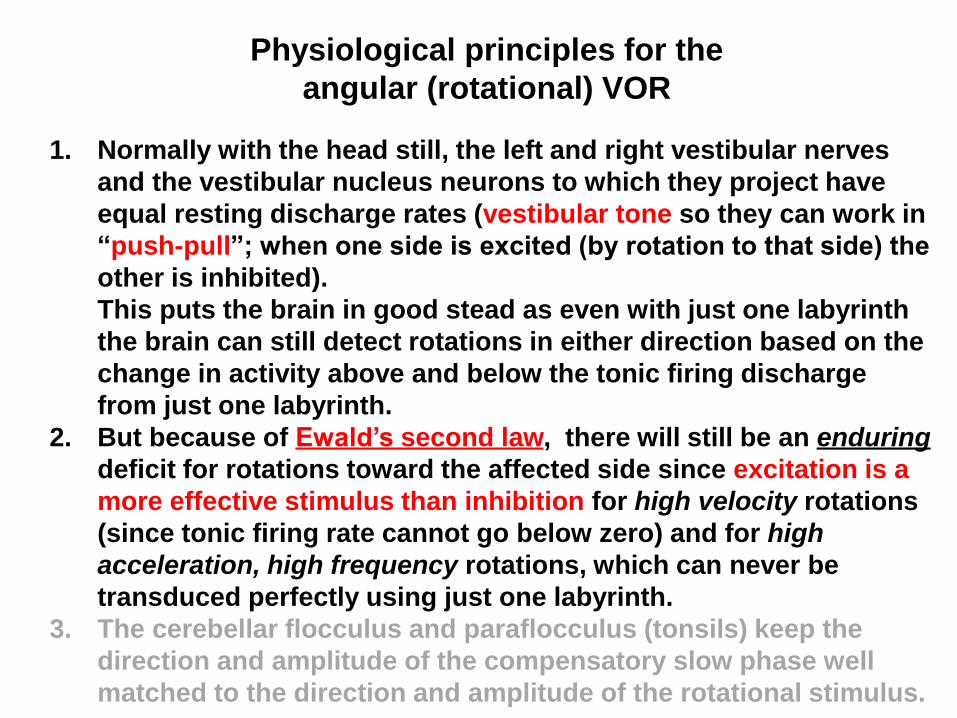
Physiological principles for the
angular (rotational) VOR
1. Normally with the head still, the left and right vestibular nerves
and the vestibular nucleus neurons to which they project have
equal resting discharge rates (vestibular tone so they can work in
“push-pull”; when one side is excited (by rotation to that side) the
other is inhibited).
This puts the brain in good stead as even with just one labyrinth
the brain can still detect rotations in either direction based on the
change in activity above and below the tonic firing discharge
from just one labyrinth.
2. But because of Ewald’s second law, there will still be an enduring
deficit for rotations toward the affected side since excitation is a
more effective stimulus than inhibition for high velocity rotations
(since tonic firing rate cannot go below zero) and for high
acceleration, high frequency rotations, which can never be
transduced perfectly using just one labyrinth.
3. The cerebellar flocculus and paraflocculus (tonsils) keep the
direction and amplitude of the compensatory slow phase well
matched to the direction and amplitude of the rotational stimulus.
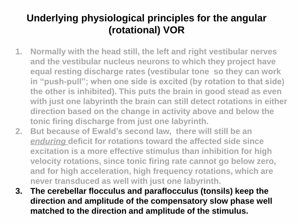
Underlying physiological principles for the angular
(rotational) VOR
1. Normally with the head still, the left and right vestibular nerves
and the vestibular nucleus neurons to which they project have
equal resting discharge rates (vestibular tone so they can work
in “push-pull”; when one side is excited (by rotation to that side)
the other is inhibited). This puts the brain in good stead as even
with just one labyrinth the brain can still detect rotations in either
direction based on the change in activity above and below the
tonic firing discharge from just one labyrinth.
2. But because of Ewald’s second law, there will still be an
enduring deficit for rotations toward the affected side since
excitation is a more effective stimulus than inhibition for high
velocity rotations, since tonic firing rate cannot go below zero,
and for high acceleration, high frequency rotations, which are
never transduced as well with just one labyrinth.
3. The cerebellar flocculus and paraflocculus (tonsils) keep the
direction and amplitude of the compensatory slow phase well
matched to the direction and amplitude of the stimulus.
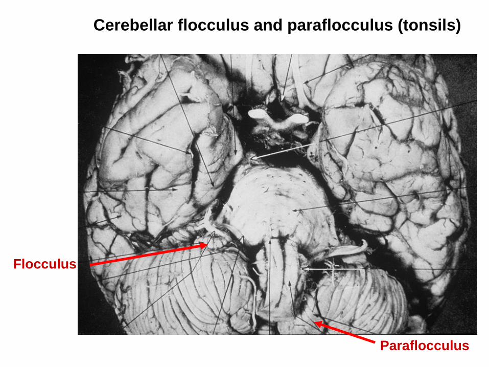
Cerebellar flocculus and paraflocculus (tonsils)
Flocculus
Paraflocculus
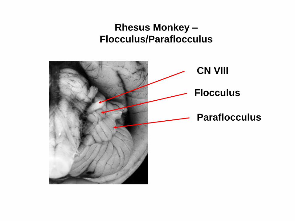
Rhesus Monkey –
Flocculus/Paraflocculus
Flocculus
Paraflocculus
CN VIII
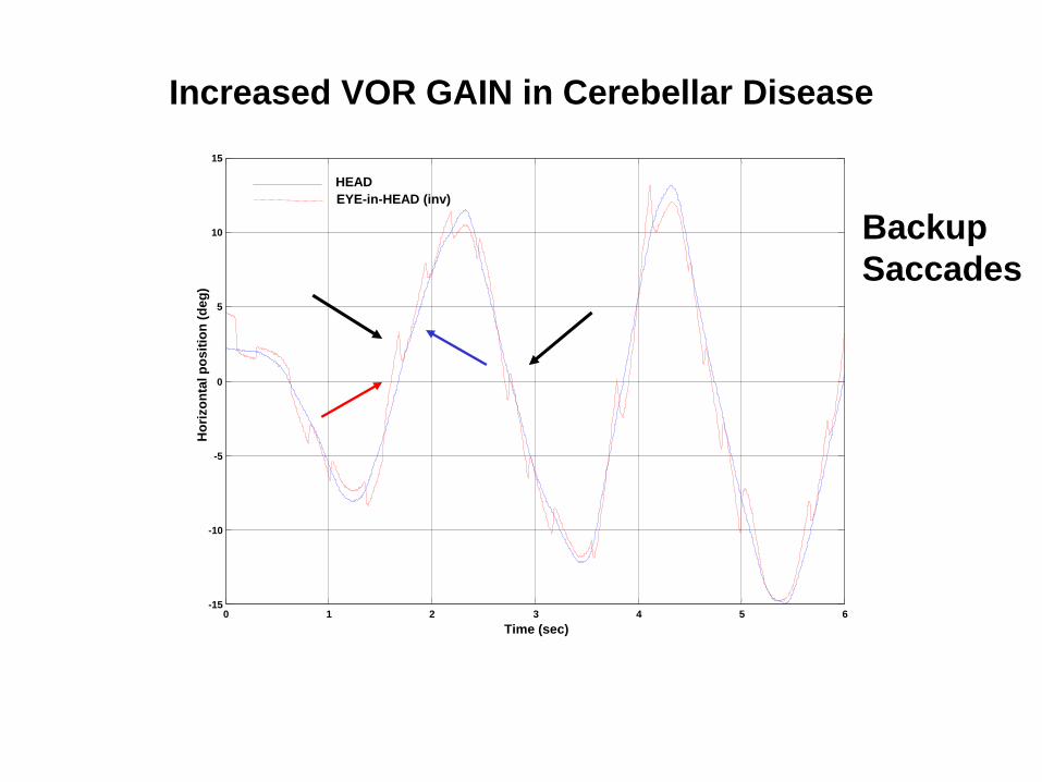
Increased VOR GAIN in Cerebellar Disease
0 1 2 3 4 5 6 -15
-10
-5
0
5
10
15
Time (sec)
Ho
rizo
nta
l p
os
itio
n (
de
g)
HEAD
EYE-in-HEAD (inv)
Backup
Saccades
-400
-300
-200
-100
0
100
200
300
400
-400 -200 0 200 400
Ve
rtic
al V
elo
cit
y (d
eg
/se
c)
Horizontal Velocity (deg/sec)
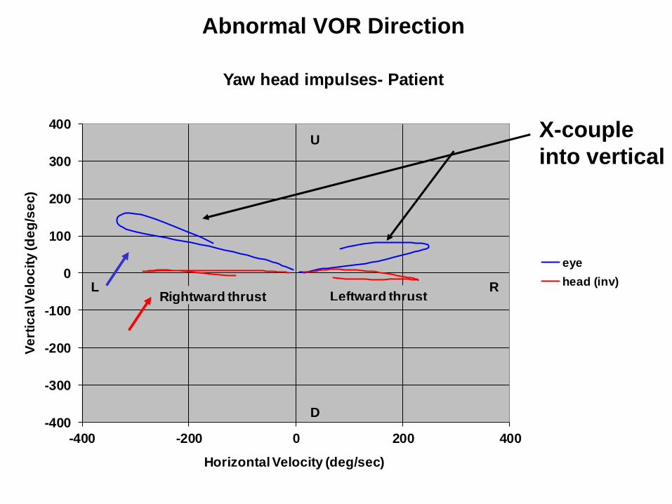
Yaw head impulses- Patient
eye
head (inv)
Rightward thrust Leftward thrust
U
D
L R
Abnormal VOR Direction
X-couple
into vertical
Outline of the presentation
Physiological principal – Clinical example
• Organization of the semicircular canals: an approach to nystagmus – Peripheral vs central nystagmus
– Benign positional vertigo
– Superior canal dehiscence
• Angular vestibulo-ocular (aVOR) reflex disorders – Abnormal amplitude and direction of the aVOR
• Otolith-ocular reflex disorders – Skew deviation and the ocular tilt reaction (OTR)
– Translational vestibuloocular reflex (tVOR)
• Vestibular velocity-storage disorders – Periodic alternating nystagmus
– Head shaking nystagmus
• Adaptive control of the VOR – Recovery nystagmus, wearing corrective spectacles
• Effects of magnetic fields on the labyrinth
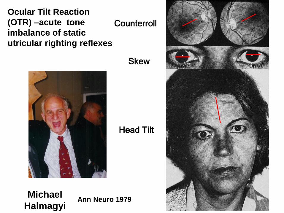
Ann Neuro 1979 Michael
Halmagyi
Head Tilt
Skew
Counterroll
Ocular Tilt Reaction
(OTR) –acute tone
imbalance of static
utricular righting reflexes
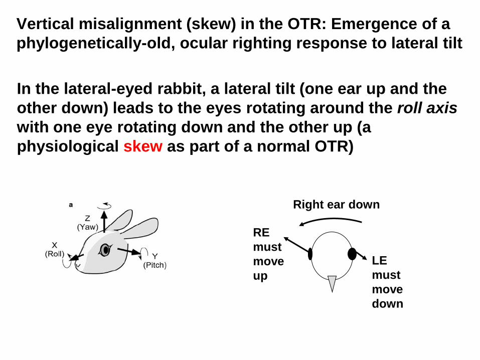
In the lateral-eyed rabbit, a lateral tilt (one ear up and the
other down) leads to the eyes rotating around the roll axis
with one eye rotating down and the other up (a
physiological skew as part of a normal OTR)
Right ear down
LE
must
move
down
RE
must
move
up
Vertical misalignment (skew) in the OTR: Emergence of a
phylogenetically-old, ocular righting response to lateral tilt
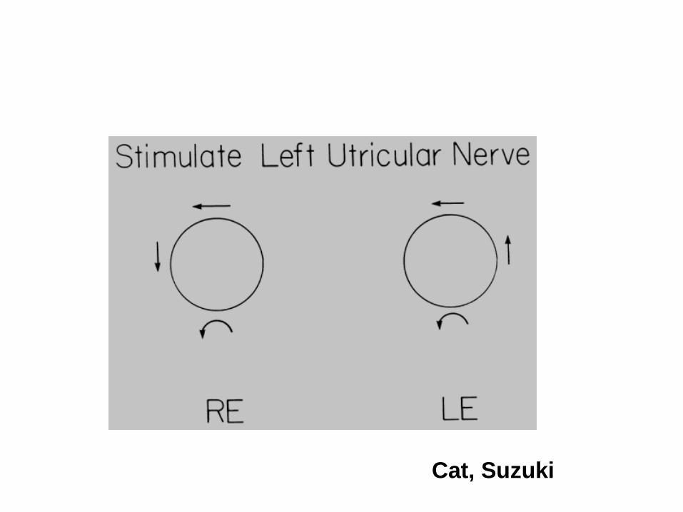
Cat, Suzuki
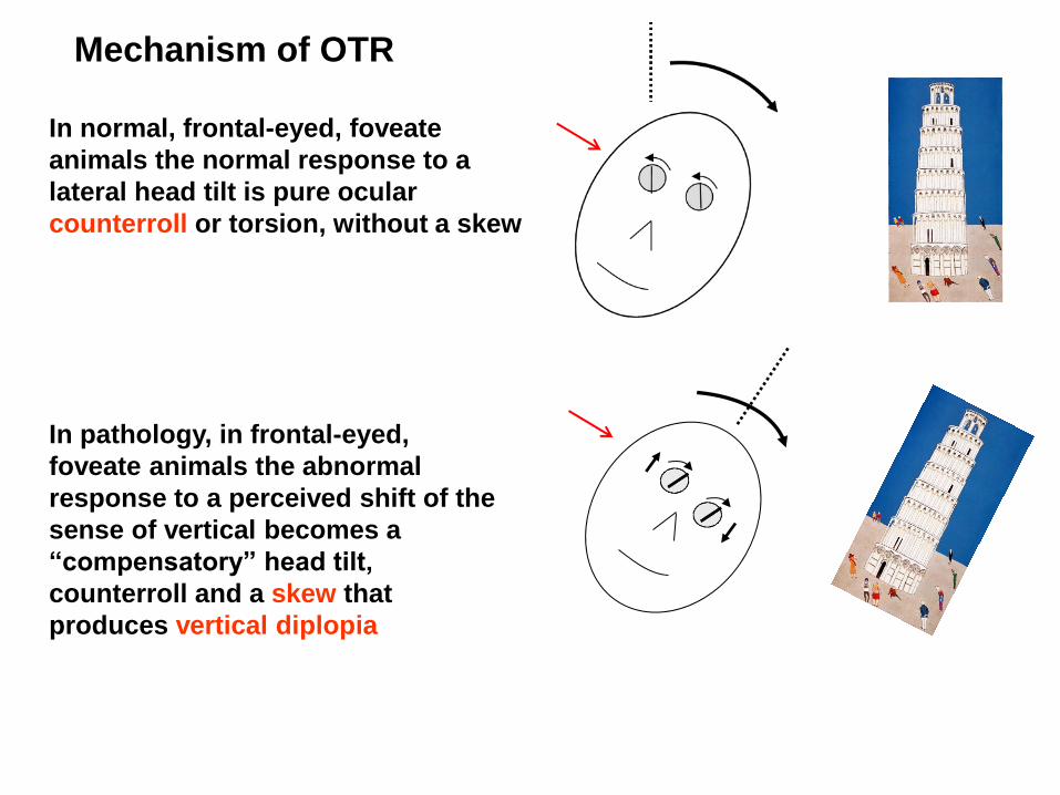
In normal, frontal-eyed, foveate
animals the normal response to a
lateral head tilt is pure ocular
counterroll or torsion, without a skew
In pathology, in frontal-eyed,
foveate animals the abnormal
response to a perceived shift of the
sense of vertical becomes a
“compensatory” head tilt,
counterroll and a skew that
produces vertical diplopia
Mechanism of OTR
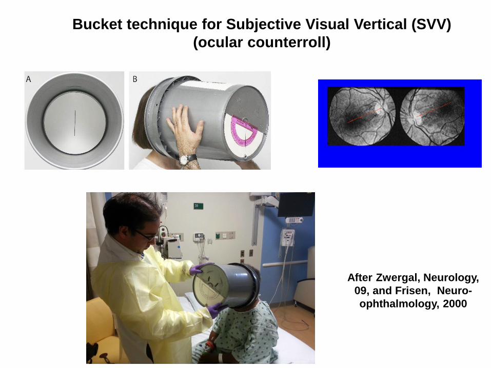
Bucket technique for Subjective Visual Vertical (SVV)
(ocular counterroll)
After Zwergal, Neurology,
09, and Frisen, Neuro-
ophthalmology, 2000
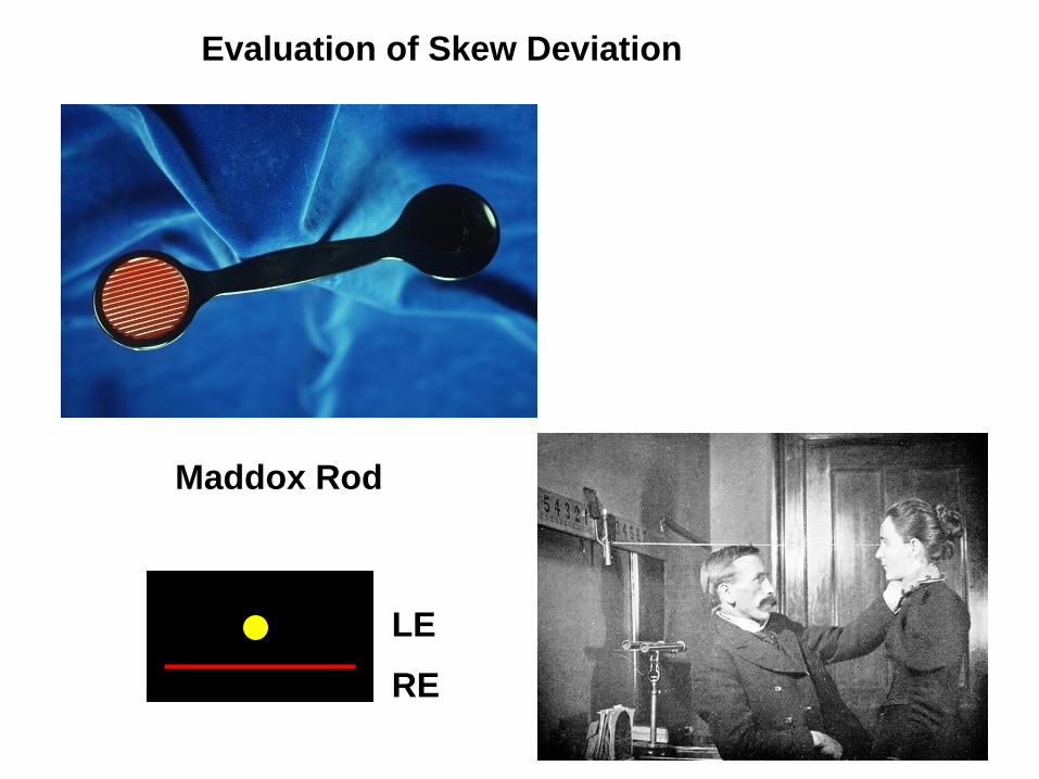
Evaluation of Skew Deviation
Maddox Rod
RE
LE
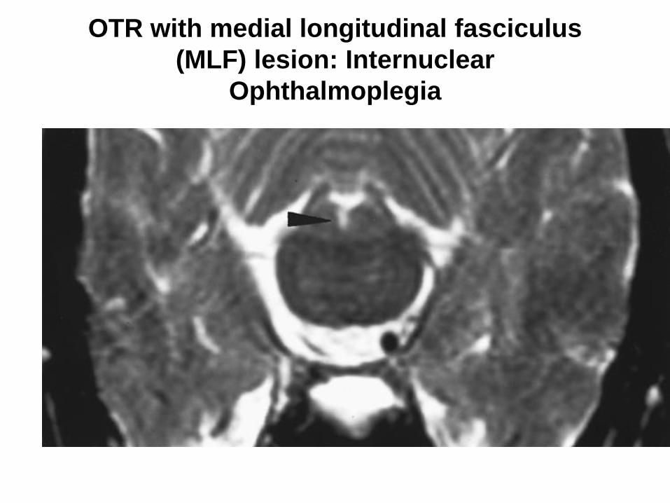
OTR with medial longitudinal fasciculus
(MLF) lesion: Internuclear
Ophthalmoplegia
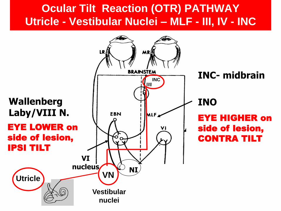
Wallenberg Laby/VIII N.
NI
VI nucleus
Vestibular
nuclei
VN
INC
EYE LOWER on
side of lesion,
IPSI TILT
EYE HIGHER on
side of lesion,
CONTRA TILT
Utricle
INC- midbrain
Ocular Tilt Reaction (OTR) PATHWAY
Utricle - Vestibular Nuclei – MLF - III, IV - INC
INO
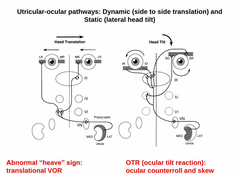
Utricular-ocular pathways: Dynamic (side to side translation) and
Static (lateral head tilt)
Abnormal “heave” sign:
translational VOR
OTR (ocular tilt reaction):
ocular counterroll and skew
Outline of the presentation
Physiological principal – Clinical example
• Organization of the semicircular canals: an approach to nystagmus – Peripheral vs central nystagmus
– Benign positional vertigo
– Superior canal dehiscence
• Angular vestibulo-ocular (aVOR) reflex disorders – Abnormal amplitude and direction of the aVOR
• Otolith-ocular reflex disorders – Skew deviation and the ocular tilt reaction (OTR)
– Translational vestibuloocular reflex (tVOR)
• Vestibular velocity-storage disorders – Periodic alternating nystagmus
• Adaptive control of the VOR – Recovery nystagmus, wearing corrective spectacles
• Effects of magnetic fields on the labyrinth
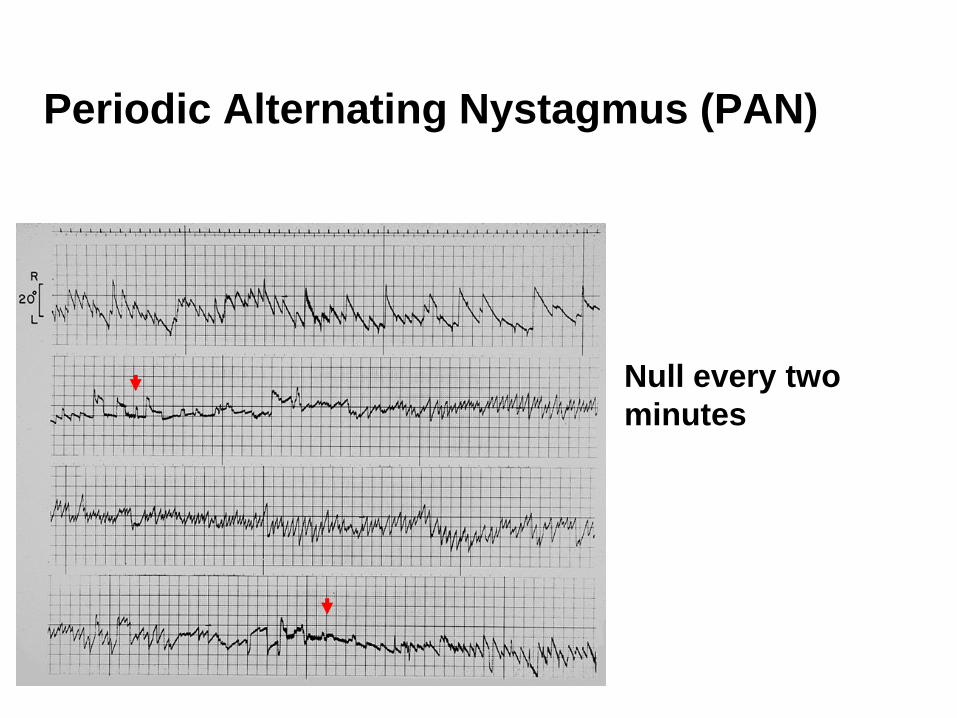
Periodic Alternating Nystagmus (PAN)
Null every two
minutes
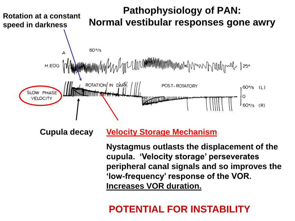
Pathophysiology of PAN:
Normal vestibular responses gone awry
Cupula decay Velocity Storage Mechanism
Nystagmus outlasts the displacement of the
cupula. ‘Velocity storage’ perseverates
peripheral canal signals and so improves the
‘low-frequency’ response of the VOR.
Increases VOR duration.
Rotation at a constant
speed in darkness
POTENTIAL FOR INSTABILITY
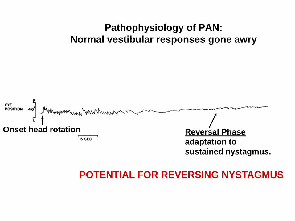
Pathophysiology of PAN:
Normal vestibular responses gone awry
Onset head rotation Reversal Phase
adaptation to
sustained nystagmus.
POTENTIAL FOR REVERSING NYSTAGMUS
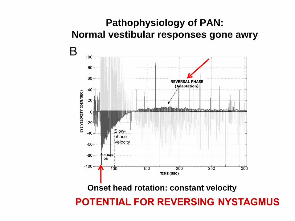
Pathophysiology of PAN:
Normal vestibular responses gone awry
Onset head rotation: constant velocity
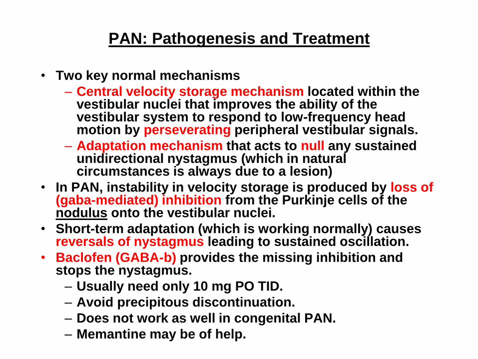
PAN: Pathogenesis and Treatment
• Two key normal mechanisms
– Central velocity storage mechanism located within the vestibular nuclei that improves the ability of the vestibular system to respond to low-frequency head motion by perseverating peripheral vestibular signals.
– Adaptation mechanism that acts to null any sustained unidirectional nystagmus (which in natural circumstances is always due to a lesion)
• In PAN, instability in velocity storage is produced by loss of (gaba-mediated) inhibition from the Purkinje cells of the nodulus onto the vestibular nuclei.
• Short-term adaptation (which is working normally) causes reversals of nystagmus leading to sustained oscillation.
• Baclofen (GABA-b) provides the missing inhibition and stops the nystagmus.
– Usually need only 10 mg PO TID.
– Avoid precipitous discontinuation.
– Does not work as well in congenital PAN.
– Memantine may be of help.
Outline of the presentation
Physiological principal – Clinical example
• Organization of the semicircular canals: an approach to nystagmus – Peripheral vs central nystagmus
– Benign positional vertigo
– Superior canal dehiscence
• Angular vestibulo-ocular (aVOR) reflex disorders – Abnormal amplitude and direction of the aVOR
• Otolith-ocular reflex disorders – Skew deviation and the ocular tilt reaction (OTR)
– Translational vestibuloocular reflex (tVOR)
• Vestibular velocity-storage disorders – Periodic alternating nystagmus
– Head shaking nystagmus
• Adaptive control of the VOR – Wearing corrective spectacles
• Effects of magnetic fields on the labyrinth
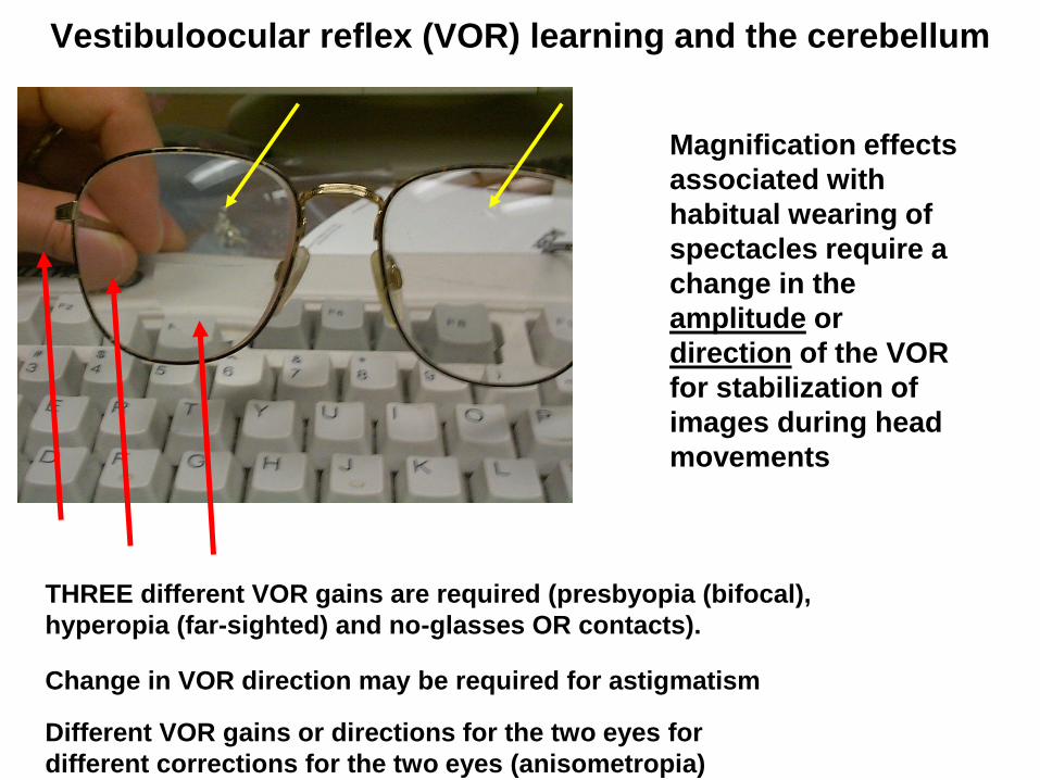
Vestibuloocular reflex (VOR) learning and the cerebellum
Magnification effects
associated with
habitual wearing of
spectacles require a
change in the
amplitude or
direction of the VOR
for stabilization of
images during head
movements
Change in VOR direction may be required for astigmatism
Different VOR gains or directions for the two eyes for
different corrections for the two eyes (anisometropia)
THREE different VOR gains are required (presbyopia (bifocal),
hyperopia (far-sighted) and no-glasses OR contacts).

Rhesus Monkey –
Flocculus/Paraflocculus
Flocculus
Paraflocculus
CN VIII
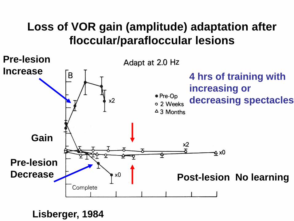
Loss of VOR gain (amplitude) adaptation after
floccular/parafloccular lesions
Pre-lesion
Increase
Pre-lesion
Decrease Post-lesion No learning
4 hrs of training with
increasing or
decreasing spectacles
Lisberger, 1984
Gain
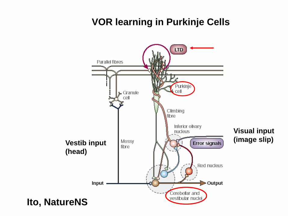
VOR learning in Purkinje Cells
Vestib input
(head)
Visual input
(image slip)
Ito, NatureNS
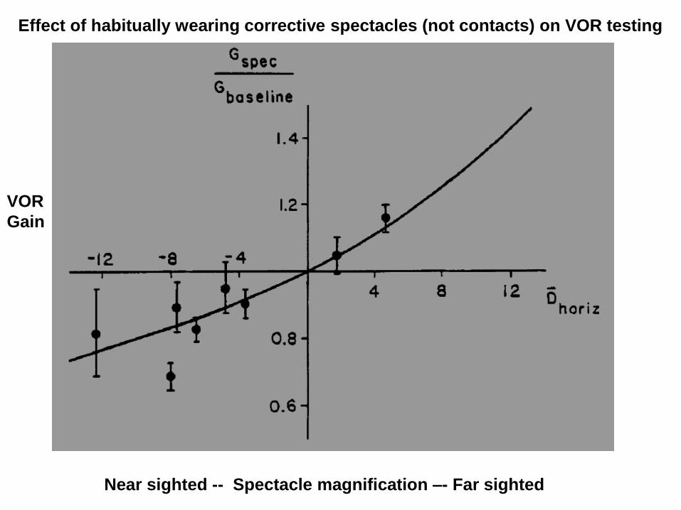
Near sighted -- Spectacle magnification –- Far sighted
VOR
Gain
Effect of habitually wearing corrective spectacles (not contacts) on VOR testing
Outline of the presentation
Physiological principal – Clinical example
• Organization of the semicircular canals: an approach to nystagmus – Peripheral vs central nystagmus
– Benign positional vertigo
– Superior canal dehiscence
• Angular vestibulo-ocular (aVOR) reflex disorders – Abnormal amplitude and direction of the aVOR
• Otolith-ocular reflex disorders – Skew deviation and the ocular tilt reaction (OTR)
– Translational vestibuloocular reflex (tVOR)
• Vestibular velocity-storage disorders – Periodic alternating nystagmus
– Head shaking nystagmus
• Adaptive control of the VOR – Wearing corrective spectacles
• Effects of magnetic fields on the labyrinth
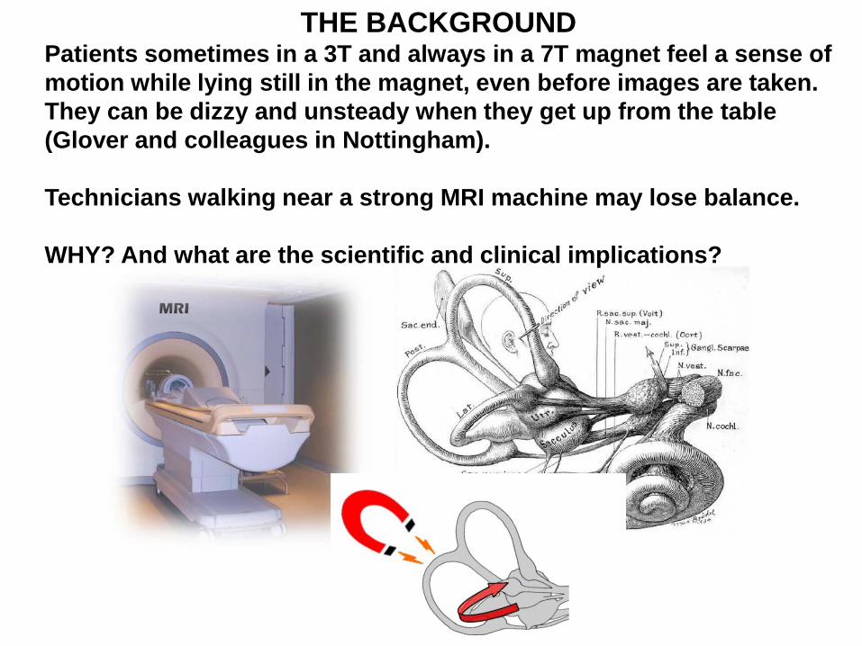
THE BACKGROUND
Patients sometimes in a 3T and always in a 7T magnet feel a sense of
motion while lying still in the magnet, even before images are taken.
They can be dizzy and unsteady when they get up from the table
(Glover and colleagues in Nottingham).
Technicians walking near a strong MRI machine may lose balance.
WHY? And what are the scientific and clinical implications?
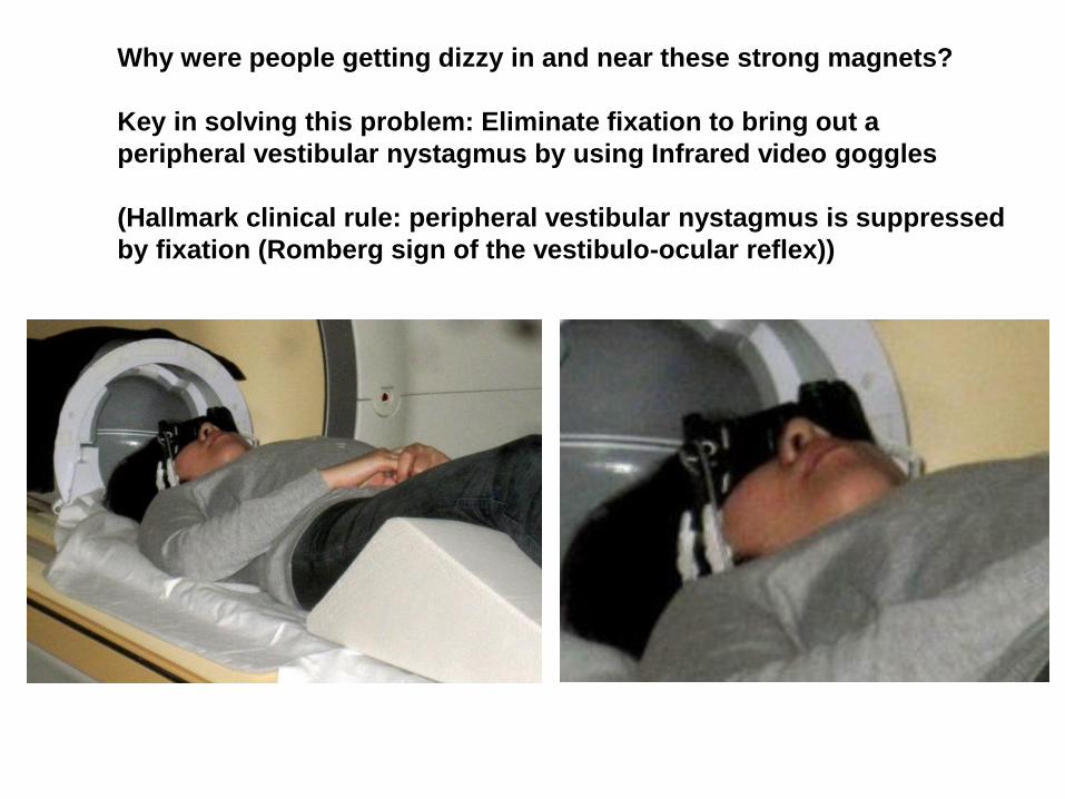
Why were people getting dizzy in and near these strong magnets?
Key in solving this problem: Eliminate fixation to bring out a
peripheral vestibular nystagmus by using Infrared video goggles
(Hallmark clinical rule: peripheral vestibular nystagmus is suppressed
by fixation (Romberg sign of the vestibulo-ocular reflex))
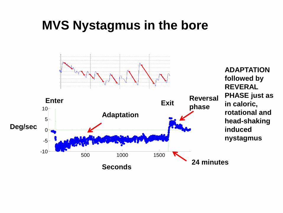
MVS Nystagmus in the bore
500 1000 1500-10
-5
0
5
10
Slo
w P
hase E
ye V
elo
cit
y(d
eg
rees/s
eco
nd
)
Time (seconds)
Seconds
Deg/sec
24 minutes
Reversal
phase
ADAPTATION
followed by
REVERAL
PHASE just as
in caloric,
rotational and
head-shaking
induced
nystagmus
Enter
Adaptation
Exit
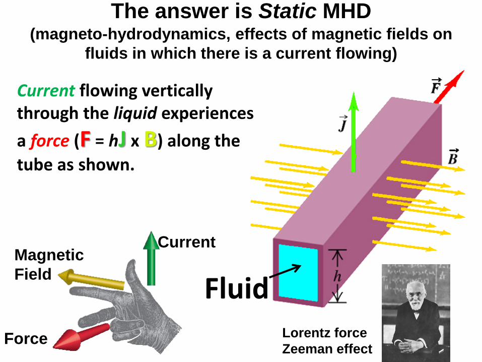
Current flowing vertically through the liquid experiences
a force (F = hJ x B) along the
tube as shown.
The answer is Static MHD (magneto-hydrodynamics, effects of magnetic fields on
fluids in which there is a current flowing)
Fluid
Current Magnetic
Field
Force Lorentz force
Zeeman effect
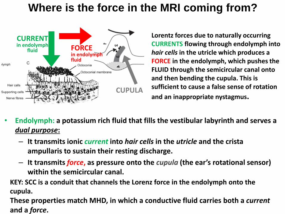
Where is the force in the MRI coming from?
Lorentz forces due to naturally occurring CURRENTS flowing through endolymph into hair cells in the utricle which produces a FORCE in the endolymph, which pushes the FLUID through the semicircular canal onto and then bending the cupula. This is sufficient to cause a false sense of rotation
and an inappropriate nystagmus.
CURRENT in endolymph
fluid FORCE in endolymph fluid
• Endolymph: a potassium rich fluid that fills the vestibular labyrinth and serves a dual purpose:
– It transmits ionic current into hair cells in the utricle and the crista ampullaris to sustain their resting discharge.
– It transmits force, as pressure onto the cupula (the ear’s rotational sensor) within the semicircular canal.
KEY: SCC is a conduit that channels the Lorenz force in the endolymph onto the cupula.
These properties match MHD, in which a conductive fluid carries both a current and a force.
CUPULA
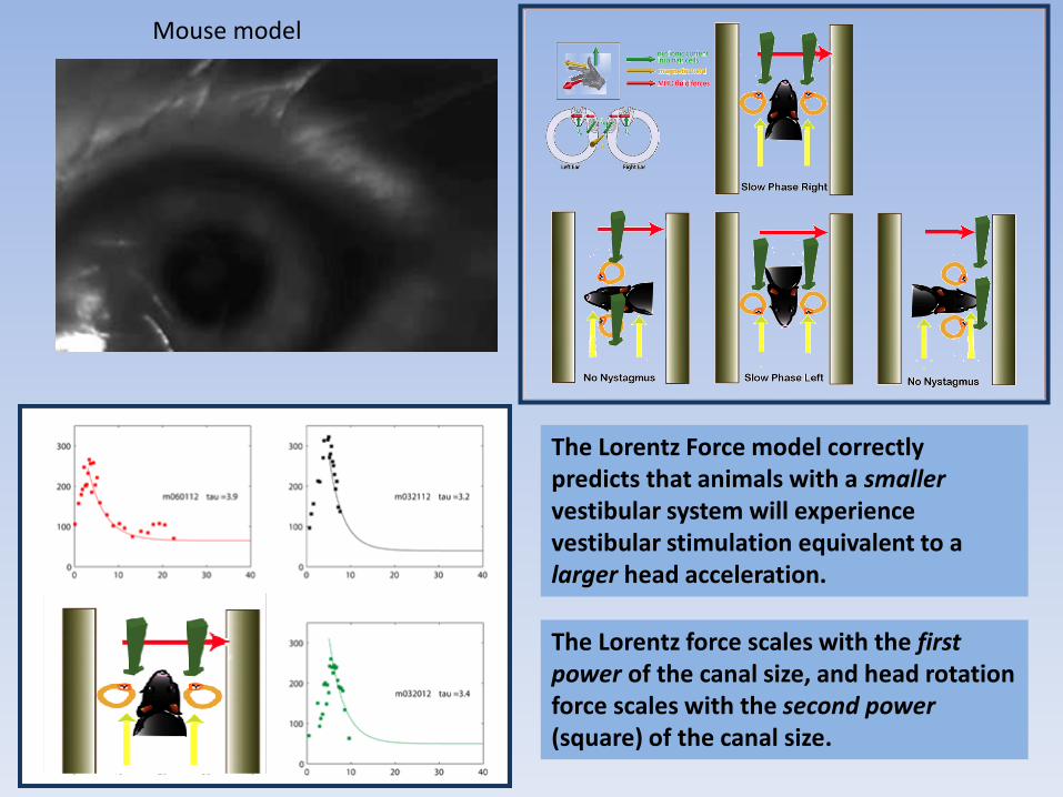
Mouse model
The Lorentz Force model correctly predicts that animals with a smaller vestibular system will experience vestibular stimulation equivalent to a larger head acceleration.
The Lorentz force scales with the first power of the canal size, and head rotation force scales with the second power (square) of the canal size.
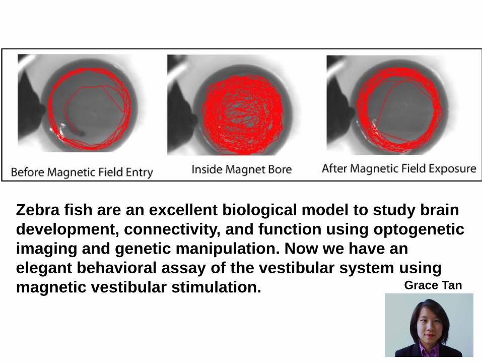
Zebra fish are an excellent biological model to study brain
development, connectivity, and function using optogenetic
imaging and genetic manipulation. Now we have an
elegant behavioral assay of the vestibular system using
magnetic vestibular stimulation. Grace Tan