Page 1 of 38 Diabetesdiabetes.diabetesjournals.org/content/diabetes/early/2016/08/19/db... ·...
38
1 Salsalate (salicylate) uncouples mitochondria, improves glucose homeostasis, and reduces liver lipids independent of AMPK β1 Brennan K. Smith 1 , Rebecca J. Ford 1 , Eric M. Desjardins 1 , Alex E. Green 1 , Meghan C. Hughes 3 , Vanessa P. Houde 1 , Emily A. Day 1 , Katarina Marcinko 1 , Justin D. Crane 1 , Emilio P. Mottillo 1 , Christopher G.R. Perry 3 , Bruce E. Kemp 4,5 , Mark A. Tarnopolsky 6 and Gregory R. Steinberg 1,2,* 1 Division of Endocrinology and Metabolism, Department of Medicine, McMaster University, Hamilton, Canada 2 Department of Biochemistry and Biomedical Sciences, McMaster University, Hamilton, Canada 3 Muscle Health Research Centre, School of Kinesiology and Health Science, York University, Toronto, Canada 4 Protein Chemistry and Metabolism, St Vincent's Institute and Department of Medicine, University of Melbourne 5 Mary MacKillop Institute for Health Research Australian Catholic University, Victoria Parade, Fitzroy, Victoria, Australia 6 Department of Pediatrics, McMaster University, Hamilton, Canada *Correspondence to: Gregory R. Steinberg Division of Endocrinology and Metabolism, Department of Medicine, McMaster University 1280 Main St. West, Hamilton, Ontario, L8N 3Z5, Canada Tel: 905.525.9140 ext. 21691 Fax: +1 905 777 7856 E-mail: [email protected] Word count: 3511 Number of figures: 5 Running title: AMPK-independent effects of salicylate on metabolic homeostasis Page 1 of 38 Diabetes Diabetes Publish Ahead of Print, published online August 23, 2016 Diabetes Publish Ahead of Print, published online August 23, 2016 Diabetes Publish Ahead of Print, published online August 23, 2016 Diabetes Publish Ahead of Print, published online August 23, 2016
Transcript of Page 1 of 38 Diabetesdiabetes.diabetesjournals.org/content/diabetes/early/2016/08/19/db... ·...
1
Salsalate (salicylate) uncouples mitochondria, improves glucose homeostasis, and reduces liver
lipids independent of AMPK β1
Brennan K. Smith1, Rebecca J. Ford
1, Eric M. Desjardins
1, Alex E. Green
1, Meghan C. Hughes
3,
Vanessa P. Houde1, Emily A. Day
1, Katarina Marcinko
1, Justin D. Crane
1, Emilio P. Mottillo
1,
Christopher G.R. Perry3, Bruce E. Kemp
4,5, Mark A. Tarnopolsky
6 and Gregory R. Steinberg
1,2,*
1Division of Endocrinology and Metabolism, Department of Medicine, McMaster University,
Hamilton, Canada 2Department of Biochemistry and Biomedical Sciences, McMaster University,
Hamilton, Canada 3Muscle Health Research Centre, School of Kinesiology and Health Science, York University,
Toronto, Canada 4Protein Chemistry and Metabolism, St Vincent's Institute and Department of Medicine,
University of Melbourne 5Mary MacKillop Institute for Health Research Australian Catholic University, Victoria Parade,
Fitzroy, Victoria, Australia 6Department of Pediatrics, McMaster University,
Hamilton, Canada
*Correspondence to:
Gregory R. Steinberg
Division of Endocrinology and Metabolism, Department of Medicine, McMaster University
1280 Main St. West, Hamilton, Ontario, L8N 3Z5, Canada
Tel: 905.525.9140 ext. 21691
Fax: +1 905 777 7856
E-mail: [email protected]
Word count: 3511
Number of figures: 5
Running title: AMPK-independent effects of salicylate on metabolic homeostasis
Page 1 of 38 Diabetes
Diabetes Publish Ahead of Print, published online August 23, 2016 Diabetes Publish Ahead of Print, published online August 23, 2016 Diabetes Publish Ahead of Print, published online August 23, 2016 Diabetes Publish Ahead of Print, published online August 23, 2016
2
Abstract
Salsalate is a prodrug of salicylate that lowers blood glucose in patients with type 2 diabetes
(T2D) and reduces non-alcoholic fatty liver disease (NAFLD) in animal models; however, the
mechanism mediating these effects is unclear. Salicylate directly activates AMP-activated
protein kinase (AMPK) via the β1 subunit but whether salsalate requires AMPK β1 to improve
T2D and NAFLD has not been examined. Therefore, wild-type (WT) and AMPK β1 knockout
mice (AMPK β1KO) were treated with a salsalate dose resulting in clinically relevant serum
salicylate concentrations (~1 mM). Salsalate treatment increased oxygen consumption, lowered
fasting glucose, improved glucose tolerance and led to an ~55% reduction in liver lipid content;
effects observed in both WT and AMPK β1KO mice. To explain these AMPK-independent
effects, it was found that salicylate increases oligomycin-insensitive respiration (state 4o) and
directly increases mitochondrial proton conductance at clinical concentrations. This uncoupling
effect is tightly correlated with the suppression of de novo lipogenesis. Salicylate is also able to
stimulate brown adipose tissue respiration independent of UCP1. These data indicate that the
primary mechanism by which salsalate improves glucose homeostasis and NAFLD is via
salicylate-driven mitochondrial uncoupling.
Page 2 of 38Diabetes
3
Non-alcoholic fatty liver disease (NAFLD) is considered an important contributing factor to the
development of insulin resistance and type 2 diabetes (T2D) [1]. Despite the rising prevalence of
NAFLD and importance for the development of T2D, there are currently no pharmacological
approaches for the treatment of this disease [2].
Salsalate is a prodrug of salicylate and is hydrolyzed in the small intestine to produce two
molecules of salicylate [3, 4]. The circulating concentration of salicylate in humans administered
salsalate in T2D clinical trials is ~1 mM [5-8]. Salsalate has also been shown to improve
symptoms of NAFLD [9] and nonalcoholic steatohepatitis (NASH) in mice [10]. The mechanism
by which salsalate improves T2D and NAFLD is currently unclear, although multiple
mechanisms have been proposed [10-16]. The mechanism of action most commonly associated
with salsalate is the direct repressing effect of salicylate on IKKβ to reduce inflammation [11-
13]. However, the concentration of salicylate used in these studies non-specifically inhibits many
protein kinases through direct competition with their ATP binding sites [16-18]. In contrast to
kinase inhibition, salicylate has also been shown to directly activate AMP-activated protein
kinase (AMPK); a metabolic sensing enzyme important for regulating inflammation [19], liver
lipid metabolism [20], and brown fat thermogenesis [21, 22]. The effect of salicylate on AMPK
occurs via a direct interaction with the Ser108 residue of the β1 subunit [16, 23]. The most recent
proposal to explain the mechanism of salicylate veers away from AMPK and suggests that
salsalate can activate brown adipose tissue through activation of cAMP-dependent protein kinase
(PKA) [15].
Page 3 of 38 Diabetes
4
While salicylate directly activates AMPK via the β1 subunit, daily intraperitoneal (ip) injections
of salicylate (250 mg/kg) improved a marker of insulin resistance (HOMA-IR) in both WT and
AMPK β1KO mice fed a high fat diet [16]. As the dose of salicylate used in this study results in
serum concentrations of salicylate more than double the clinical levels following the oral intake
of salsalate (~2.4 mM compared to ~1.0 mM, respectively) we hypothesized that the AMPK β1-
independent effects may have been due to off-target kinase inhibition [16-18].
The purpose of this study was to investigate whether oral delivery of clinically relevant
concentrations of salsalate improves glucose homeostasis and reduces NAFLD through an
AMPK β1-dependent pathway. It was observed that salsalate improves whole-body glucose
homeostasis, reduces liver lipid content and improves adipose tissue inflammation independently
of AMPK β1. These diverse metabolic effects of salsalate are associated with the protonophoric
effects of salicylate and subsequent mitochondrial uncoupling and increased energy expenditure.
These data suggest that salicylate-driven mitochondrial uncoupling is the primary mechanism
mediating the beneficial effects of salsalate therapy on NAFLD and T2D.
RESEARCH DESIGN AND METHODS
Study approval. All animal procedures were approved by the McMaster University Animal
Ethics Research Board (Hamilton, Canada, AUP #: 12-12-44) and conform to the guide for the
care and use of laboratory animals published by the US National Institutes of Health.
Page 4 of 38Diabetes
5
Animals. WT and AMPK β1KO mice were started on a high fat diet (60% calories from fat) at 8
weeks of age. 4 weeks after the initiation of the HFD, half of the mice continued on the HFD
while the other half were switched onto a HFD supplemented with 2.5 g/kg salsalate. These diets
were maintained for 8 weeks until sacrifice (Supplementary Fig. 1A). The glucose tolerance test
was performed in 6 hour fasted mice after an intraperitoneal (i.p.) injection of glucose (0.8 g/kg).
The alanine tolerance test was performed in 16 hour fasted mice after i.p. injection of 2 g/kg of
alanine [24]. Blood glucose levels were determined from a small tail vein nick using a One
Touch Ultra Glucometer (LifeScan, Canada). Metabolic monitoring was performed in a
Comprehensive Lab Animal Monitoring System (Columbus Instruments, Columbus, OH) at
week 10. Non-moving VO2 measurements were taken under light anesthetic to remove activity
level confounding [25, 26]. To assess UCP1-mediated thermogenesis, CL-316,243 (CL; 0.033
nmol/g body weight) administration was performed as previously described [26]. In a subset of
animals, insulin (1 U/kg) was administered prior to sacrifice to examine insulin signalling in the
liver and 2-Deoxy-D-glucose (2DG) uptake into skeletal muscle and adipose tissue [27].
Analytical measurements. Serum salicylate concentrations were determined following
manufacturers’ instructions from a commercially available kit (Neogen Corporation). Liver
sections were stained for hematoxylin and eosin stain (H&E). Liver and tibialis anterior (TA)
samples were extracted by the Folch method to measure tissue triglyceride levels [28]. Primary
hepatocytes were freshly isolated by collagenase perfusion for the lipogenesis and respiration
measurements. Mitochondrial membrane potential (∆ψm) was measured using the TMRM stain
(20 nM, non-quenching) [29]. Primary hepatocyte de novo lipogenesis was measured similar to
previous reports using 3H acetate (Perkin Elmer) [20]. RT-qPCR was carried out as previously
Page 5 of 38 Diabetes
6
described to determine mRNA expression levels [19]. Briefly, epididymal adipose tissue was
lysed in TRIzol reagent (Invitrogen, CA, USA) to remove lipid, and the aqueous phase was
applied to an RNeasy kit (Qiagen, CA, USA) column for subsequent purification. Relative gene
expression was calculated using the comparative Ct (2-ΔΔCt
) method where values were
normalized to housekeeping gene (Ppia). Taqman®
primers F4/80 (Emr1, Mm00802529_m1),
Cluster of Differentiation 68 (Cd68, Mm00839636_g1), tumor necrosis factor alpha (Tnf-α,
Mm00443258_m1), Chemokine (C-C Motif) Ligand 2 (CCL2, Mm00441242_m1), interleukin-
1β (Il-1β, Mm00434228_m1), were purchased from Invitrogen. Western blotting was performed
similar to [9] and all antibodies were purchased from Cell Signalling. ATP concentration was
determined in freeze-clamped liver tissue according to manufacturer’s instruction (ab113849,
Abcam) [30].
Respiration methods. Mitochondrial respiration was measured by high-resolution respirometry
(Oroboros Oxygraph-2 k, Innsbruck, Austria) at 37 °C and room air saturated oxygen tension.
Permeabilized primary hepatocyte respiration was performed in MIRO5 buffer containing EGTA
(0.5 mM), MgCl2*6H2O (3 mM), K-lactobionate (60 mM), KH2PO4 (10 mM), HEPES (20 mM),
Sucrose (110 mM) and fatty acid free BSA (1 g/L). Primary hepatocytes were scraped into 2 ml
of respiration buffer and 800 µl of the suspension was quickly added to respiration chambers.
Digitonin (8.1 µM) was added to chambers to permeabilize the cells, and following a 5-minute
incubation period the assay was initiated. Permeabilized skeletal muscle fibres and epididymal
adipose tissue were prepared as previously described [31, 32]. BAT mitochondria were isolated
and respiration was performed similar to previous reports [33, 34].
Page 6 of 38Diabetes
7
Mitochondrial proton conductance. Isolated liver mitochondrial oxygen consumption rates and
∆ψm were measured simultaneously in the Oroboros system at 37 °C [35, 36]. Mitochondria
were isolated similar to [37] and experiments were run in Buffer Z containing K-MES (110
mM), KCl (35 mM), EGTA (1 mM), K2HPO4 (5 mM), MgCl2*6H2O (3 mM), BSA (0.5 mg/ml),
pH 7.1, 295 mOsm. Buffer Z was supplemented with carboxyatractyloside (1.5 µM), oligomycin
(1.25 µg/ml), GDP (0.5 mM), nigericin (0.1 µM), rotenone (5 µM) and succinate (6 mM). ∆ψ
was measured using electrodes sensitive to tetraphenylphosponium (TPP+) and the TPP+
electrode was calibrated by a 5-point titration (0.9-1.7 µM, every 2 µM) at the beginning of each
experiment. ∆ψm was lowered by titration of the complex II inhibitor malonate (0.1 to 5 mM) in
the absence or presence of 1 mM salicylate. Salicylate was also titrated directly into respiration
chambers.
Acute effects of salsalate in vivo. To assess the acute effects of salsalate on in vivo lipogenesis,
energy expenditure, VO2 and VCO2, mice were i.p. injected with either salsalate (Cayman
Chemicals) or vehicle. Salsalate was initially dissolved in 100% DMSO and then further
suspended in 20% 2-Hydroxypropyl-β-cyclodextrin (Sigma Aldrich) in saline down to a final
concentration of 5% DMSO. The vehicle control for these experiments was the same stock of
20% 2-Hydroxypropyl-β-cyclodextrin (in saline) with 5% DMSO. In vivo lipogenesis was
performed similar to previous reports [20, 38]. Mice were fasted for 15 hours and then re-fed for
2 hours. 20 µCi of 3H acetate (Perkin Elmer) was then i.p injected into the mouse. 15 minutes
later, the mice were i.p injected with salsalate or vehicle made up as above. 1 hour later the mice
were sacrificed (Supplementary Fig. 1B). The lipids were extracted by the Folch method and the
entire chloroform layer was counted for radioactivity. The liver was also examined for
Page 7 of 38 Diabetes
8
measurements of AMPK activation and ATP concentration. For acute changes in energy
expenditure, 1 hour after the administration of salsalate or vehicle, mice were lightly
anesthetized with an i.p. injection of 0.5 mg/g body weight Avertin (2,2,2-Tribromoethanol
dissolved in 2-methyl-2-butanol, Sigma Aldrich) to get non-moving measurements. This protocol
was undertaken to ensure that energy expenditure associated with activity (skeletal muscle
contraction) would not confound our energy expenditure data. The mice were then placed dorsal
side up onto an enclosed stationary treadmill and metabolic measurements were monitored over
a 12-minute period using Comprehensive Lab Animal Monitoring System (CLAMS, Columbus
Instruments, an indirect calorimetry system) (Supplementary Fig. 1C).
Statistical analyses. Values are reported as mean ± SEM. Data were analyzed using 2-way or 1-
way ANOVA with Bonferroni post hoc test or Students t test where indicated. Differences were
considered significant when P < 0.05.
RESULTS
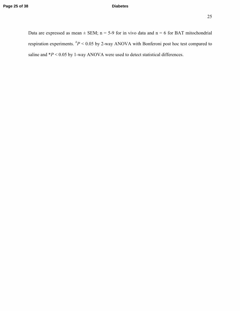
Salsalate treatment improves glucose homeostasis and reduces liver lipids independent of
AMPK β1. It was observed that 2.5 g/kg salsalate supplemented in a 60% high fat diet (HFD)
gave serum salicylate values of ~800-900 µM (Fig. 1A) matching clinical levels [5]. WT and
AMPK β1KO mice were fed a high fat diet for 4 weeks, followed by 8 weeks of HFD or HFD
with salsalate supplementation (HFD+SAL). Significant differences in body mass were not
observed (Fig. 1B), but salsalate supplementation significantly reduced percent adiposity and
increased percent lean mass (Fig. 1C and D). An increase in non-moving VO2 was observed
Page 8 of 38Diabetes
9
(Figure 1E) although examination of the mice in the free living state indicated no differences in
VO2, energy expenditure, VCO2, activity levels or food intake (Supplementary Figure 2).
Fasting glucose levels and glucose tolerance were improved by salsalate in both WT and AMPK
β1KO mice (Fig. 1F-H). Salsalate supplementation improved alanine tolerance (Fig. 1I and J)
suggesting reductions in hepatic gluconeogenesis. Reductions in liver lipid content in both WT
and AMPK β1KO mice were also observed (Fig. 1K and L). Lipid levels in the tibialis anterior
muscle were not significantly reduced by salsalate (Supplementary Figure 3). Consistent with
reductions in adiposity in both WT and AMPK β1KO mice (Fig. 1C), markers of adipose tissue
inflammation were generally reduced in both genotypes following treatment with salsalate
(Supplementary Figure 4). Insulin-stimulated 2DG uptake into skeletal muscle (Supplementary
Figure 5A) and inguinal white adipose tissue (Supplementary Figure 5B) was not different
between genotypes or following salsalate treatment. Liver AKT phosphorylation at Thr308, and
IRS1 phosphorylation at Tyr1222 were also unchanged (Supplementary Figure 6). Therefore, a
clinically relevant dose of salsalate increases non-moving VO2, improves glucose homeostasis,
lowers markers of adipose tissue inflammation, and reduces liver lipids independent of AMPK
β1.
Salicylate uncouples permeabilized primary hepatocytes. To explore AMPK β1-independent
mechanisms by which salsalate may increase VO2, reduce liver lipid content and improve
glucose homeostasis, the ability of salicylate to uncouple mitochondria at clinical concentrations
in permeabilized primary hepatocytes was examined. To this end, permeabilized primary
hepatocyte respiration was stimulated with glutamate, malate and ADP to induce state 3
Page 9 of 38 Diabetes
10
respiration, and in a stepwise fashion the following were added: 1) cytochrome c to check for
outer mitochondrial membrane integrity [39], 2) GDP to inhibit uncoupling proteins [40, 41], 3)
oligomycin to inhibit ATP synthase [42], and 4) salicylate at increasing concentrations.
Salicylate increases respiration independent of uncoupling proteins and ATP synthase in a dose
dependent manner at concentrations starting as low as 0.1 mM in both WT (Fig. 2A and B) and
AMPK β1KO hepatocytes (Supplementary Figure 7A). Similar observations were also observed
in permeabilized skeletal muscle fibres (Supplementary Figure 7B) and epididymal white
adipose tissue (Supplementary Figure 7C). To further examine the concept of mitochondrial
uncoupling, ∆ψm was measured using TMRM staining and salicylate was observed to dose-
dependently decrease ∆ψm (Fig. 2C and D). Mitochondrial proton conductance assays were then
performed and in the presence of 1.0 mM salicylate proton current was elevated at a given
membrane potential (Figure 2E). Salicylate also directly increased mitochondrial proton
conductance (Figure 2F). Thus, the protonophoric effect of salicylate can explain the salicylate-
induced mitochondrial uncoupling, the increase in oxygen consumption in vitro and in vivo, and
the AMPK β1-independent effects of salsalate.
Subclinical salicylate treatment suppresses primary hepatocyte de novo lipogenesis.
Traditionally, the mechanism by which mitochondrial uncoupling is viewed to improve
metabolic health is associated with increases in substrate oxidation; however, the suppression of
anabolism may also play a role. Indeed, human subjects with NAFLD display a 5-fold increase
in rates of fatty acid synthesis (de novo lipogenesis (DNL)) [43, 44]. Therefore, the effects of
clinical salicylate concentrations on DNL were examined and correlated with mitochondrial
uncoupling.
Page 10 of 38Diabetes
11
Salicylate dose-dependently suppressed primary hepatocyte DNL at subclinical concentrations
(Fig. 3A), and this suppression in DNL correlated with the salicylate-driven increases in
uncoupled respiration (Fig. 3B and C). In addition, when hepatocytes were treated with two
classic mitochondrial uncouplers, 2, 4 dinitrophenol (DNP) and carbonyl cyanide-4-
(trifluoromethoxy) phenylhydrazone (FCCP), DNL rates were significantly reduced (Fig. 3D).
These data highlight the importance of ∆ψm in the maintenance of DNL.
To investigate a role for AMPK in mediating the DNL lowering effects of salicylate, DNL rates
were examined in AMPK β1KO primary hepatocytes. Additionally, AMPK is known to inhibit
DNL via phosphorylation of Ser79/212 on acetyl-CoA carboxylase (ACC) [45] and so the effects
of salicylate in a double knock-in model (ACC-DKI) harbouring serine to alanine mutations on
these two residues were analyzed [20]. DNL rates were inhibited by salicylate in primary
hepatocytes derived from AMPK β1KO and ACC-DKI mice indicating that salicylate inhibits
DNL independent of AMPK β1 and AMPK-ACC signalling (Fig. 3E).
Salsalate suppresses liver de novo lipogenesis in vivo. To investigate if these in vitro effects
regarding DNL occurred in vivo, a salsalate dose that gives rise to circulating salicylate
concentrations similar to human clinical data and the in vivo feeding study was first achieved.
An acute i.p. injection of 62.5 mg/kg salsalate in mice resulted in clinically relevant serum
concentrations of salicylate (Fig. 4A). This dose is equivalent to a 70 kg human ingesting 4.2 g of
salsalate, which is aligned with clinical dosing (3.5-4.5 g) [5, 7]. Following an acute treatment
with salsalate (62.5 mg/kg), non-moving VO2, VCO2 and energy expenditure were increased,
Page 11 of 38 Diabetes
12
effects that are characteristic of mitochondrial uncoupling (Fig. 4B-D, method outlined in
Supplementary Figure 1C). In addition, salsalate reduced in vivo DNL rates by ~25% (Fig. 4E,
method outlined in Supplementary Figure 1B) and these effects were independent of changes in
AMPK activation or ATP levels (Supplementary Figure 8). Collectively, these data suggest that
salsalate, via salicylate-driven mitochondrial uncoupling, increases energy expenditure and
suppresses DNL in vivo.
Salicylate increases respiration in brown adipose tissue mitochondria independent of
UCP1.
Brown adipose tissue (BAT) activation increases energy expenditure and strategies to increase
BAT activation have shown therapeutic promise for treating NAFLD and T2D [46, 47]; effects
which are largely dependent on the activation of uncoupling protein 1 (UCP1) [48].
Recently, it has been shown that salsalate directly activates BAT to increase energy expenditure
and improve HFD-induced metabolic dysfunction [15]. To further examine this proposed
mechanism of action, salsalate-treated mice were analyzed using an in vivo technique that
specifically detects UCP1-mediated thermogenesis [26]. Upon analysis, salsalate treatment did
not alter UCP1-mediated thermogenesis in response to β3 adrenergic stimulation in vivo (Figure
5A-D), suggesting salsalate-induced increases in whole body VO2 is not dependent on increases
in UCP1 activation. Therefore, UCP1-independent BAT mitochondrial activity was investigated
in response to salicylate. To this end, BAT mitochondria were isolated and respiration was
stimulated with palmitoyl-CoA (P-CoA, 30 µM), and then inhibited with the UCP1 inhibitor
GDP (2 mM) [34]. Salicylate dose-dependently increased oxygen consumption even when UCP1
Page 12 of 38Diabetes
13
was inhibited by GDP (Fig. 5E and F). These data indicate that salicylate increases BAT
mitochondrial oxygen consumption independent of UCP1.
DISCUSSION
Various mechanisms have been proposed to explain the beneficial health effects associated with
salsalate/salicylate. This is the first manuscript to suggest that salicylate-driven mitochondrial
uncoupling is the primary mechanism of action to explain the host of beneficial effects
associated with salicylate [11, 49] and salsalate [6-8, 14, 50, 51]. The present data suggest that
mitochondrial uncoupling due to the protonophoric properties of salicylate explains the
consistently observed increases in energy expenditure in murine models and human subjects
treated with salicylate-based compounds (present data and [6, 10, 11, 14, 15, 51-57]).
Classical studies from the 1950s observed that salicylate stimulates mitochondrial uncoupling
[52, 58] and previous work established that mitochondrial membrane potential (∆ψm) is reduced
by 1.0 mM salicylate [53]. Moreover, proton conductance is increased with 1.0 mM salicylate
treatment (present data and [53]) and these bioenergetic effects are likely due to the ability of
salicylate to act as a proton carrier [59, 60]. The current manuscript extends this bioenergetic
mechanism of salicylate into the regulation of whole body physiology.
From a therapeutic perspective, the beneficial effects of mitochondrial protonophores
(uncouplers) on T2D and NAFLD have been consistently observed [61-65]. The compounds
recently studied include a modified version of the classic protonophore, 2,4 dinitrophenol [61],
Page 13 of 38 Diabetes
14
which is structurally similar to salicylate [52, 66]. These mitochondrial uncouplers result in an
increase in energy expenditure similar to that obtained when treating with salsalate/salicylate
(present work [6, 10, 11, 14, 15, 51-57]) and also improve markers of NAFLD and T2D [61-65].
Therefore, considering mitochondrial uncouplers improve T2D and NAFLD, and increasing
energy expenditure is a potent mechanism to improve T2D and NAFLD [61, 62, 67-69], the
present work in combination with previous reports in both mice and humans is highly suggestive
that salicylate-driven mitochondrial uncoupling is the primary mechanism of action explaining
the improvement in T2D and NAFLD associated with salsalate treatment.
As a potential mechanism to explain why mitochondrial uncouplers improve T2D and NAFLD,
it was found that salsalate suppresses hepatic DNL in vivo. NAFLD is associated with increased
rates of hepatic DNL [43, 44] and this manuscript is the first to show that DNL can be
suppressed in vivo with an acute, therapeutically relevant dose of salsalate. DNL requires three
basic constituents; substrate (acetyl-CoA), reductive power (NADPH) and energy supply (ATP).
By reducing ∆ψm, salicylate and other uncoupling agents compromise the availability of all
three factors as the cell shifts to a catabolic state and downregulates anabolic activity [66, 70].
Considering the potential importance of DNL in human NAFLD, future work investigating
whether salsalate can suppress DNL in humans is warranted.
From a general mechanism standpoint, previous work in humans supplemented with high dose
aspirin [56] or salsalate [14] observed increases in both carbohydrate and fat oxidation. In vivo,
the only way carbohydrate and fat oxidation can simultaneously increase is if there is a change in
the overall demand of the system (i.e. uncoupling or increased ATP turnover). In the case of
Page 14 of 38Diabetes
15
salsalate, by providing a protonophore (salicylate) ∆ψm is reduced, an effect which allows fat
and carbohydrate catabolism to increase. In environments typified by over-nutrition, such as
those seen in much of the Western world, this uncoupling mechanism works to blunt the toxic
excess of substrate by siphoning fat and/or carbohydrate away from anabolic pathways (DNL) in
favour of oxidation. In other words, mitochondrial uncouplers suppress the toxic effects of
substrate oversupply by removing the substrate. The current results, in combination with our
previous studies indicating AMPK-independent effects of salicylate on HOMA-IR [16], suggest
that salicylate/salsalate-induced improvements in glucose homeostasis of HFD-fed mice are
likely mediated through mitochondrial uncoupling.
A limitation of this work is that definitive genetic evidence is unavailable given it is not possible
to remove mitochondria and therefore the uncoupling effects of salicylate. A further limitation is
the fact that it is difficult to discern which cell type or organ system is playing the most
prominent role in mediating the beneficial effects of salsalate. Inhibition of hepatic DNL has
showed therapeutic promise for NAFLD [71], as has promoting hepatic substrate consumption
[61], indicating that the liver may be important. However, increasing BAT mitochondrial activity
has been shown to spare the liver from substrate oversupply [46], and skeletal muscle is
responsible for the majority of glucose clearance [72]. Thus, further work is required to
investigate which organ system is playing the most prominent role.
In addition to the positive metabolic effects associated with mitochondrial uncouplers in the
presence of nutrient excess, significant negative side effects have been associated with
uncoupling agents, including 2,4 dinitrophenol [73, 74]. The present manuscript is an important
Page 15 of 38 Diabetes
16
message from not only a therapeutic perspective, but also from a potential side effects
perspective, and future work examining salicylate-based compounds should consider the fact that
salicylate uncouples mitochondria.
In summary, a therapeutically relevant dose of salsalate improves glucose homeostasis, lowers
adipose tissue inflammation and reduces liver lipid content, independent of AMPK β1. Instead of
AMPK it seems likely that the protonophoric effect of salicylate is the primary mechanism
underlying the metabolic improvements associated with salicylate [11, 49] and salsalate [6-8, 14,
50, 51] under conditions of nutrient excess. Future studies should consider this mechanism of
action when examining effects of salsalate on T2D and NAFLD.
AUTHOR CONTRIBUTIONS
BKS and GRS designed research studies, analyzed data and wrote manuscript. BKS, RJF, EAD,
EMD, KM, AEG, VPH, CGRP, MCH, JDC and EPM conducted experiments and analyzed data.
BKS, GRS, BEK, MAT, RJF, EAD, AEG, EMD, VPH, KM, JDC and EPM edited the
manuscript.
ACKNOWLEDGEMENTS
BKS is a recipient of a Canadian Institutes of Health Research Postdoctoral Fellowship and
Michael G. Degroote Postdoctoral Fellowship. These studies were supported by grants from the
Canadian Diabetes Association (GRS), the Canadian Institutes of Health Research (GRS) and the
Natural Sciences and Engineering Research Council of Canada (GRS). EPM is a Canadian
Diabetes Association postdoctoral fellow. AEG is a recipient of a Canadian Graduate
Page 16 of 38Diabetes
17
Scholarship from CIHR/MitoCanada. EMD is a recipient of an Ontario Graduate Scholarship
and Queen Elizabeth II Graduate Scholarship in Science and Technology. BEK is supported by
grants and a fellowships from the Australian Research Council (ARC) and the National Health
and Medical Research Council (NHMRC), supported in part by the Victorian Government’s
Operational Infrastructure. GRS is a Canada Research Chair in Metabolism and Obesity and the
J. Bruce Duncan Chair in Metabolic Diseases.
There are no conflicts of interest
Guarantor: Dr. Gregory R. Steinberg is the guarantor of this work and, as such, had full access to
all the data in the study and takes responsibility for the integrity of the data and the accuracy of
the data analysis.
Page 17 of 38 Diabetes
18
REFERENCE LIST
[1] Rinella ME. Nonalcoholic fatty liver disease: a systematic review. JAMA. 2015;313:2263-
73.
[2] Perry RJ, Samuel VT, Petersen KF, Shulman GI. The role of hepatic lipids in hepatic insulin
resistance and type 2 diabetes. Nature. 2014;510:84-91.
[3] Dromgoole SH, Cassell S, Furst DE, Paulus HE. Availability of salicylate from salsalate and
aspirin. Clin Pharmacol Ther. 1983;34:539-45.
[4] Dromgoole SH, Furst DE, Paulus HE. Metabolism of salsalate in normal subjects. J Pharm
Sci. 1984;73:1657-9.
[5] Fleischman A, Shoelson SE, Bernier R, Goldfine AB. Salsalate improves glycemia and
inflammatory parameters in obese young adults. Diabetes Care. 2008;31:289-94.
[6] Goldfine AB, Silver R, Aldhahi W, Cai D, Tatro E, Lee J, et al. Use of salsalate to target
inflammation in the treatment of insulin resistance and type 2 diabetes. Clin Transl Sci.
2008;1:36-43.
[7] Goldfine AB, Fonseca V, Jablonski KA, Pyle L, Staten MA, Shoelson SE. The effects of
salsalate on glycemic control in patients with type 2 diabetes: a randomized trial. Ann Intern
Med. 2010;152:346-57.
[8] Barzilay JI, Jablonski KA, Fonseca V, Shoelson SE, Goldfine AB, Strauch C, et al. The
impact of salsalate treatment on serum levels of advanced glycation end products in type 2
diabetes. Diabetes Care. 2014;37:1083-91.
[9] Ford RJ, Fullerton MD, Pinkosky SL, Day EA, Scott JW, Oakhill JS, et al. Metformin and
salicylate synergistically activate liver AMPK, inhibit lipogenesis and improve insulin
sensitivity. Biochem J. 2015;468:125-32.
[10] Liang W, Verschuren L, Mulder P, van der Hoorn JW, Verheij J, van Dam AD, et al.
Salsalate attenuates diet induced non-alcoholic steatohepatitis in mice by decreasing lipogenic
and inflammatory processes. Br J Pharmacol. 2015.
[11] Kim JK, Kim YJ, Fillmore JJ, Chen Y, Moore I, Lee J, et al. Prevention of fat-induced
insulin resistance by salicylate. J Clin Invest. 2001;108:437-46.
[12] Yuan M, Konstantopoulos N, Lee J, Hansen L, Li ZW, Karin M, et al. Reversal of obesity-
and diet-induced insulin resistance with salicylates or targeted disruption of Ikkbeta. Science.
2001;293:1673-7.
[13] Yin MJ, Yamamoto Y, Gaynor RB. The anti-inflammatory agents aspirin and salicylate
inhibit the activity of I(kappa)B kinase-beta. Nature. 1998;396:77-80.
[14] Meex RC, Phielix E, Moonen-Kornips E, Schrauwen P, Hesselink MK. Stimulation of
human whole-body energy expenditure by salsalate is fueled by higher lipid oxidation under
Page 18 of 38Diabetes
19
fasting conditions and by higher oxidative glucose disposal under insulin-stimulated conditions. J
Clin Endocrinol Metab. 2011;96:1415-23.
[15] van Dam AD, Nahon KJ, Kooijman S, van den Berg SM, Kanhai AA, Kikuchi T, et al.
Salsalate activates brown adipose tissue in mice. Diabetes. 2015;64:1544-54.
[16] Hawley SA, Fullerton MD, Ross FA, Schertzer JD, Chevtzoff C, Walker KJ, et al. The
ancient drug salicylate directly activates AMP-activated protein kinase. Science. 2012;336:918-
22.
[17] Alpert D, Vilcek J. Inhibition of IkappaB kinase activity by sodium salicylate in vitro does
not reflect its inhibitory mechanism in intact cells. J Biol Chem. 2000;275:10925-9.
[18] Steinberg GR, Dandapani M, Hardie DG. AMPK: mediating the metabolic effects of
salicylate-based drugs? Trends EndocrinolMetab. 2013;24:481-7.
[19] Galic S, Fullerton MD, Schertzer JD, Sikkema S, Marcinko K, Walkley CR, et al.
Hematopoietic AMPK beta1 reduces mouse adipose tissue macrophage inflammation and insulin
resistance in obesity. JClinInvest. 2011;121:4903-15.
[20] Fullerton MD, Galic S, Marcinko K, Sikkema S, Pulinilkunnil T, Chen ZP, et al. Single
phosphorylation sites in Acc1 and Acc2 regulate lipid homeostasis and the insulin-sensitizing
effects of metformin. Nat Med. 2013;19:1649-54.
[21] Mottillo EP, Desjardins EM, Crane JD, Smith BK, Green AE, Ducommun S, et al. Lack of
Adipocyte AMPK Exacerbates Insulin Resistance and Hepatic Steatosis through Brown and
Beige Adipose Tissue Function. Cell Metab. 2016;24:118-29.
[22] Zhang H, Guan M, Townsend KL, Huang TL, An D, Yan X, et al. MicroRNA-455 regulates
brown adipogenesis via a novel HIF1an-AMPK-PGC1alpha signaling network. EMBO Rep.
2015;16:1378-93.
[23] Calabrese MF, Rajamohan F, Harris MS, Caspers NL, Magyar R, Withka JM, et al.
Structural basis for AMPK activation: natural and synthetic ligands regulate kinase activity from
opposite poles by different molecular mechanisms. Structure. 2014;22:1161-72.
[24] Bujak AL, Crane JD, Lally JS, Ford RJ, Kang SJ, Rebalka IA, et al. AMPK activation of
muscle autophagy prevents fasting-induced hypoglycemia and myopathy during aging. Cell
Metab. 2015;21:883-90.
[25] O'Neill HM, Maarbjerg SJ, Crane JD, Jeppesen J, Jorgensen SB, Schertzer JD, et al. AMP-
activated protein kinase (AMPK) beta1beta2 muscle null mice reveal an essential role for AMPK
in maintaining mitochondrial content and glucose uptake during exercise. ProcNatlAcadSciUSA.
2011;108:16092-7.
[26] Crane JD, Mottillo EP, Farncombe TH, Morrison KM, Steinberg GR. A standardized
infrared imaging technique that specifically detects UCP1-mediated thermogenesis in vivo. Mol
Metab. 2014;3:490-4.
[27] Howlett KF, Andrikopoulos S, Proietto J, Hargreaves M. Exercise-induced muscle glucose
uptake in mice with graded, muscle-specific GLUT-4 deletion. Physiol Rep. 2013;1:e00065.
[28] Folch J, Lees M, Sloane Stanley GH. A simple method for the isolation and purification of
total lipides from animal tissues. J Biol Chem. 1957;226:497-509.
[29] Scaduto RC, Jr., Grotyohann LW. Measurement of mitochondrial membrane potential using
fluorescent rhodamine derivatives. Biophys J. 1999;76:469-77.
[30] Borowski LS, Dziembowski A, Hejnowicz MS, Stepien PP, Szczesny RJ. Human
mitochondrial RNA decay mediated by PNPase-hSuv3 complex takes place in distinct foci.
Nucleic Acids Res. 2013;41:1223-40.
Page 19 of 38 Diabetes
20
[31] Smith BK, Perry CG, Herbst EA, Ritchie IR, Beaudoin MS, Smith JC, et al. Submaximal
ADP-stimulated respiration is impaired in ZDF rats and recovered by resveratrol. JPhysiol. 2013.
[32] Paglialunga S, Ludzki A, Root-McCaig J, Holloway GP. In adipose tissue, increased
mitochondrial emission of reactive oxygen species is important for short-term high-fat diet-
induced insulin resistance in mice. Diabetologia. 2015;58:1071-80.
[33] Cannon B, Nedergaard J. Studies of thermogenesis and mitochondrial function in adipose
tissues. Methods Mol Biol. 2008;456:109-21.
[34] Shabalina IG, Petrovic N, de Jong JM, Kalinovich AV, Cannon B, Nedergaard J. UCP1 in
brite/beige adipose tissue mitochondria is functionally thermogenic. Cell Rep. 2013;5:1196-203.
[35] Fisher-Wellman KH, Lin CT, Ryan TE, Reese LR, Gilliam LA, Cathey BL, et al. Pyruvate
dehydrogenase complex and nicotinamide nucleotide transhydrogenase constitute an energy-
consuming redox circuit. Biochem J. 2015;467:271-80.
[36] Brand MD, Nicholls DG. Assessing mitochondrial dysfunction in cells. Biochem J.
2011;435:297-312.
[37] Smith BK, Perry CG, Koves TR, Wright DC, Smith JC, Neufer PD, et al. Identification of a
novel malonyl-CoA IC50 for CPT-1: implications for predicting in vivo fatty acid oxidation
rates. BiochemJ. 2012;448:13-20.
[38] Dupont J, Mathias MM, Cabacungan NB. Dietary lipid, fatty acid synthesis and cholesterol
metabolism in aging rats. Lipids. 1972;7:576-89.
[39] Wikstrom M, Casey R. The oxidation of exogenous cytochrome c by mitochondria.
Resolution of a long-standing controversy. FEBS Lett. 1985;183:293-8.
[40] Echtay KS, Esteves TC, Pakay JL, Jekabsons MB, Lambert AJ, Portero-Otin M, et al. A
signalling role for 4-hydroxy-2-nonenal in regulation of mitochondrial uncoupling. EMBO J.
2003;22:4103-10.
[41] Negre-Salvayre A, Hirtz C, Carrera G, Cazenave R, Troly M, Salvayre R, et al. A role for
uncoupling protein-2 as a regulator of mitochondrial hydrogen peroxide generation. FASEB J.
1997;11:809-15.
[42] Zheng J, Ramirez VD. Inhibition of mitochondrial proton F0F1-ATPase/ATP synthase by
polyphenolic phytochemicals. BrJPharmacol. 2000;130:1115-23.
[43] Lambert JE, Ramos-Roman MA, Browning JD, Parks EJ. Increased de novo lipogenesis is a
distinct characteristic of individuals with nonalcoholic fatty liver disease. Gastroenterology.
2014;146:726-35.
[44] Donnelly KL, Smith CI, Schwarzenberg SJ, Jessurun J, Boldt MD, Parks EJ. Sources of
fatty acids stored in liver and secreted via lipoproteins in patients with nonalcoholic fatty liver
disease. J Clin Invest. 2005;115:1343-51.
[45] Carling D, Zammit VA, Hardie DG. A common bicyclic protein kinase cascade inactivates
the regulatory enzymes of fatty acid and cholesterol biosynthesis. FEBS Lett. 1987;223:217-22.
[46] Crane JD, Palanivel R, Mottillo EP, Bujak AL, Wang H, Ford RJ, et al. Inhibiting peripheral
serotonin synthesis reduces obesity and metabolic dysfunction by promoting brown adipose
tissue thermogenesis. Nat Med. 2015;21:166-72.
[47] Sidossis L, Kajimura S. Brown and beige fat in humans: thermogenic adipocytes that
control energy and glucose homeostasis. J Clin Invest. 2015;125:478-86.
[48] Enerback S, Jacobsson A, Simpson EM, Guerra C, Yamashita H, Harper ME, et al. Mice
lacking mitochondrial uncoupling protein are cold-sensitive but not obese. Nature. 1997;387:90-
4.
Page 20 of 38Diabetes
21
[49] Williamson RT. On the Treatment of Glycosuria and Diabetes Mellitus with Sodium
Salicylate. Br Med J. 1901;1:760-2.
[50] Ariel D, Kim SH, Liu A, Abbasi F, Lamendola CA, Grove K, et al. Salsalate-induced
changes in lipid, lipoprotein, and apoprotein concentrations in overweight or obese, insulin-
resistant, nondiabetic individuals. J Clin Lipidol. 2015;9:658-63.
[51] Cao Y, Dubois DC, Sun H, Almon RR, Jusko WJ. Modeling diabetes disease progression
and salsalate intervention in Goto-Kakizaki rats. J Pharmacol Exp Ther. 2011;339:896-904.
[52] Sproull DH. A peripheral action of sodium salicylate. Br J Pharmacol Chemother.
1954;9:262-4.
[53] Haas R, Parker WD, Jr., Stumpf D, Eguren LA. Salicylate-induced loose coupling:
protonmotive force measurements. Biochem Pharmacol. 1985;34:900-2.
[54] Sproull DH. A comparison of sodium salicylate and 2:4-dinitrophenol as metabolic
stimulants in vitro. Biochem J. 1957;66:527-32.
[55] DENIS W, MEANS J. THE INFLUENCE OF SALICYLATE ON METABOLISM IN
MAN. Journal of Pharmacology and Experimental Therapeutics. 1916;8:273-83.
[56] Hundal RS, Petersen KF, Mayerson AB, Randhawa PS, Inzucchi S, Shoelson SE, et al.
Mechanism by which high-dose aspirin improves glucose metabolism in type 2 diabetes. J Clin
Invest. 2002;109:1321-6.
[57] Cochran JB. The respiratory effects of salicylate. Br Med J. 1952;2:964-7.
[58] Brody TM. Action of sodium salicylate and related compounds on tissue metabolism in
vitro. J Pharmacol Exp Ther. 1956;117:39-51.
[59] Gutknecht J. Salicylates and proton transport through lipid bilayer membranes: a model for
salicylate-induced uncoupling and swelling in mitochondria. J Membr Biol. 1990;115:253-60.
[60] Gutknecht J. Aspirin, acetaminophen and proton transport through phospholipid bilayers
and mitochondrial membranes. Mol Cell Biochem. 1992;114:3-8.
[61] Perry RJ, Zhang D, Zhang XM, Boyer JL, Shulman GI. Controlled-release mitochondrial
protonophore reverses diabetes and steatohepatitis in rats. Science. 2015;347:1253-6.
[62] Tao H, Zhang Y, Zeng X, Shulman GI, Jin S. Niclosamide ethanolamine-induced mild
mitochondrial uncoupling improves diabetic symptoms in mice. Nat Med. 2014;20:1263-9.
[63] Samuel VT, Liu ZX, Qu X, Elder BD, Bilz S, Befroy D, et al. Mechanism of hepatic insulin
resistance in non-alcoholic fatty liver disease. J Biol Chem. 2004;279:32345-53.
[64] Figarola JL, Singhal P, Rahbar S, Gugiu BG, Awasthi S, Singhal SS. COH-SR4 reduces
body weight, improves glycemic control and prevents hepatic steatosis in high fat diet-induced
obese mice. PLoS One. 2013;8:e83801.
[65] Kalinovich AV, Shabalina IG. Novel Mitochondrial Cationic Uncoupler C4R1 Is an
Effective Treatment for Combating Obesity in Mice. Biochemistry (Mosc). 2015;80:620-8.
[66] Smith MJ, Moses V. Uncoupling reagents and metabolism. 1. Effects of salicylate and 2:4-
dinitrophenol on the incorporation of C from labelled glucose and acetate into the soluble
intermediates of isolated rat tissues. Biochem J. 1960;76:579-85.
[67] Keating SE, Hackett DA, Parker HM, O'Connor HT, Gerofi JA, Sainsbury A, et al. Effect of
aerobic exercise training dose on liver fat and visceral adiposity. J Hepatol. 2015;63:174-82.
[68] Colberg SR, Sigal RJ, Fernhall B, Regensteiner JG, Blissmer BJ, Rubin RR, et al. Exercise
and type 2 diabetes: the American College of Sports Medicine and the American Diabetes
Association: joint position statement. Diabetes Care. 2010;33:e147-e67.
Page 21 of 38 Diabetes
22
[69] Stanford KI, Middelbeek RJ, Townsend KL, An D, Nygaard EB, Hitchcox KM, et al.
Brown adipose tissue regulates glucose homeostasis and insulin sensitivity. J Clin Invest.
2013;123:215-23.
[70] Blacker TS, Mann ZF, Gale JE, Ziegler M, Bain AJ, Szabadkai G, et al. Separating NADH
and NADPH fluorescence in live cells and tissues using FLIM. Nat Commun. 2014;5:3936.
[71] Harriman G, Greenwood J, Bhat S, Huang X, Wang R, Paul D, et al. Acetyl-CoA
carboxylase inhibition by ND-630 reduces hepatic steatosis, improves insulin sensitivity, and
modulates dyslipidemia in rats. Proc Natl Acad Sci U S A. 2016;113:E1796-805.
[72] DeFronzo RA, Gunnarsson R, Bjorkman O, Olsson M, Wahren J. Effects of insulin on
peripheral and splanchnic glucose metabolism in noninsulin-dependent (type II) diabetes
mellitus. J Clin Invest. 1985;76:149-55.
[73] Fawaz G, Tutunji B. The mechanism of dinitrophenol heart failure. Br J Pharmacol
Chemother. 1957;12:273-8.
[74] Tainter ML, Cutting WC, Stockton AB. Use of Dinitrophenol in Nutritional Disorders : A
Critical Survey of Clinical Results. AmJPublic Health NationsHealth. 1934;24:1045-53.
FIGURE LEGENDS
Page 22 of 38Diabetes
23
Figure 1: Salsalate supplementation improves glucose homeostasis and reduces liver lipids
independent of AMPK β1. Serum salicylate concentrations in mice following treatment with
2.5 g/kg salsalate supplemented into a 60 % HFD (A). Body mass (B), adiposity (C) lean mass
(D), non-moving VO2 (E), and fasting blood glucose (F) in WT and AMPK β1KO mice fed a
HFD or a HFD supplemented with salsalate. Glucose tolerance (G and H), alanine tolerance (I
and J) in WT and AMPK β1KO mice fed a HFD or a HFD supplemented with salsalate. The
black lines signify the WT mice and the blue lines signify the AMPK β1KO mice. The broken
lines signify the salsalate treated groups. Haematoxylin and Eosin (H&E) staining of liver
sections was performed (K), and lipid content was quantified (L). Data are expressed as mean ±
SEM; n = 9-17. *P < 0.05 by 2-way ANOVA with Bonferoni post hoc test.
Figure 2: Salicylate uncouples primary hepatocytes, reduces mitochondrial membrane
potential and increases mitochondrial proton conductance. Representative oxygraph trace in
permeabilized primary hepatocytes illustrating the change in oligomycin-insensitive respiration
(state 4o) during salicylate titration (A). Quantification of the increase in state 4o respiration
when titrating salicylate (B). Change in mitochondrial membrane potential in response to
salicylate in permeabilized primary hepatocytes (C and D). Proton current at a given membrane
potential in the presence (red line) or absence (black line) of 1 mM salicylate (E). Proton
conductance alterations with salicylate titration (F).
Data are expressed as mean ± SEM; n=3-6. *P < 0.05 by 1-way ANOVA with Bonferoni post
hoc test or 2-tailed Student’s t test.
Page 23 of 38 Diabetes
24
Figure 3: In vitro, hepatic de novo lipogenesis is inhibited by salicylate. De novo lipogenesis
(from 3H acetate) in primary hepatocytes treated with salicylate at subclinical to clinical
concentrations (A). Representative trace of salicylate-driven mitochondrial uncoupling at
subclinical to clinical concentrations (B). Correlation between increase in state 4o respiration and
reduction in de novo lipogenesis (C). The effect of two mitochondrial uncouplers, 2,4
dinitrophenol and carbonyl cyanide-4-(trifluoromethoxy) phenylhydrazone (FCCP) on rates of
de novo lipogenesis (D). De novo lipogenesis in primary hepatocytes derived from WT, AMPK
β1KO and ACC-DKI treated with salicylate (E).
Data are expressed as mean ± SEM; n = 4-8. *P < 0.05 by 1-way ANOVA and 2-way ANOVA
with Bonferoni post hoc test were used to detect statistical differences.
Figure 4: In vivo, hepatic de novo lipogenesis is inhibited by salsalate. Circulating salicylate
concentrations following 3 different i.p. injections of salsalate (A). VO2 (B), VCO2 (C), energy
expenditure (E) and hepatic de novo lipogenesis rates (from 3H acetate) in response to i.p
salsalate (62.5 mg/kg) injections in WT chow-fed mice. Data are expressed as mean ± SEM; n =
4-6 *P < 0.05 by 2-tailed Student’s t test.
Figure 5: Salsalate does not alter BAT thermogenesis in vivo, but salicylate increases
UCP1-inhibited BAT mitochondrial respiration in vitro. VO2 response to CL-316,243 (CL;
0.033 nmol/g body weight) in HFD mice with and without salsalate supplementation (A and B).
BAT thermogenesis in response to CL-316,243 in HFD mice with and without salsalate (C and
D). Representative trace of salicylate-driven BAT mitochondrial respiration in the presence of 2
mM GDP (E). Quantification of the increase in respiration when titrating salicylate (F).
Page 24 of 38Diabetes
25
Data are expressed as mean ± SEM; n = 5-9 for in vivo data and n = 6 for BAT mitochondrial
respiration experiments. #P < 0.05 by 2-way ANOVA with Bonferoni post hoc test compared to
saline and *P < 0.05 by 1-way ANOVA were used to detect statistical differences.
Page 25 of 38 Diabetes
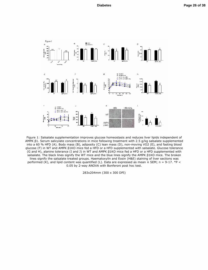
Figure 1: Salsalate supplementation improves glucose homeostasis and reduces liver lipids independent of AMPK β1. Serum salicylate concentrations in mice following treatment with 2.5 g/kg salsalate supplemented into a 60 % HFD (A). Body mass (B), adiposity (C) lean mass (D), non-moving VO2 (E), and fasting blood
glucose (F) in WT and AMPK β1KO mice fed a HFD or a HFD supplemented with salsalate. Glucose tolerance (G and H), alanine tolerance (I and J) in WT and AMPK β1KO mice fed a HFD or a HFD supplemented with salsalate. The black lines signify the WT mice and the blue lines signify the AMPK β1KO mice. The broken
lines signify the salsalate treated groups. Haematoxylin and Eosin (H&E) staining of liver sections was performed (K), and lipid content was quantified (L). Data are expressed as mean ± SEM; n = 9-17. *P <
0.05 by 2-way ANOVA with Bonferoni post hoc test.
283x204mm (300 x 300 DPI)
Page 26 of 38Diabetes
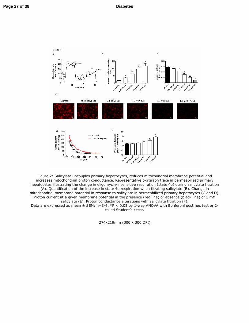
Figure 2: Salicylate uncouples primary hepatocytes, reduces mitochondrial membrane potential and increases mitochondrial proton conductance. Representative oxygraph trace in permeabilized primary
hepatocytes illustrating the change in oligomycin-insensitive respiration (state 4o) during salicylate titration (A). Quantification of the increase in state 4o respiration when titrating salicylate (B). Change in
mitochondrial membrane potential in response to salicylate in permeabilized primary hepatocytes (C and D). Proton current at a given membrane potential in the presence (red line) or absence (black line) of 1 mM
salicylate (E). Proton conductance alterations with salicylate titration (F). Data are expressed as mean ± SEM; n=3-6. *P < 0.05 by 1-way ANOVA with Bonferoni post hoc test or 2-
tailed Student’s t test.
274x219mm (300 x 300 DPI)
Page 27 of 38 Diabetes
Figure 3: In vitro, hepatic de novo lipogenesis is inhibited by salicylate. De novo lipogenesis (from 3H acetate) in primary hepatocytes treated with salicylate at subclinical to clinical concentrations (A).
Representative trace of salicylate-driven mitochondrial uncoupling at subclinical to clinical concentrations (B). Correlation between increase in state 4o respiration and reduction in de novo lipogenesis (C). The effect
of two mitochondrial uncouplers, 2,4 dinitrophenol and carbonyl cyanide-4-(trifluoromethoxy) phenylhydrazone (FCCP) on rates of de novo lipogenesis (D). De novo lipogenesis in primary hepatocytes
derived from WT, AMPK β1KO and ACC-DKI treated with salicylate (E). Data are expressed as mean ± SEM; n = 4-8. *P < 0.05 by 1-way ANOVA and 2-way ANOVA with Bonferoni
post hoc test were used to detect statistical differences.
275x185mm (300 x 300 DPI)
Page 28 of 38Diabetes
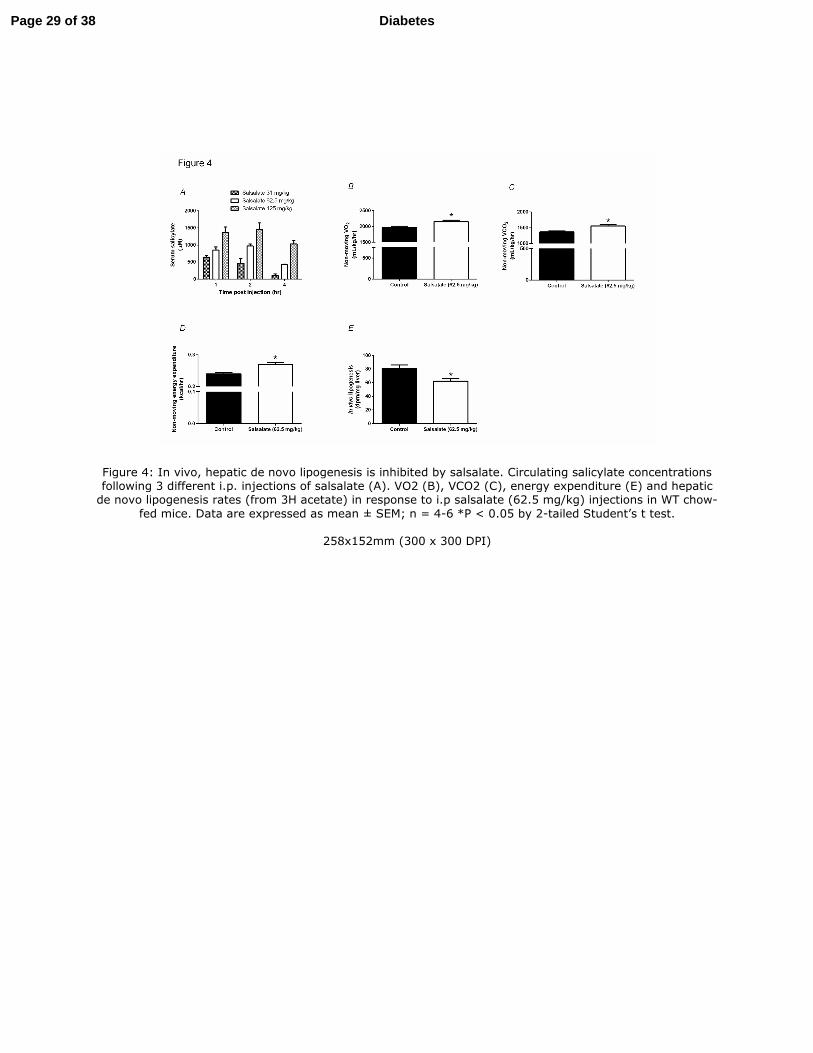
Figure 4: In vivo, hepatic de novo lipogenesis is inhibited by salsalate. Circulating salicylate concentrations following 3 different i.p. injections of salsalate (A). VO2 (B), VCO2 (C), energy expenditure (E) and hepatic de novo lipogenesis rates (from 3H acetate) in response to i.p salsalate (62.5 mg/kg) injections in WT chow-
fed mice. Data are expressed as mean ± SEM; n = 4-6 *P < 0.05 by 2-tailed Student’s t test.
258x152mm (300 x 300 DPI)
Page 29 of 38 Diabetes
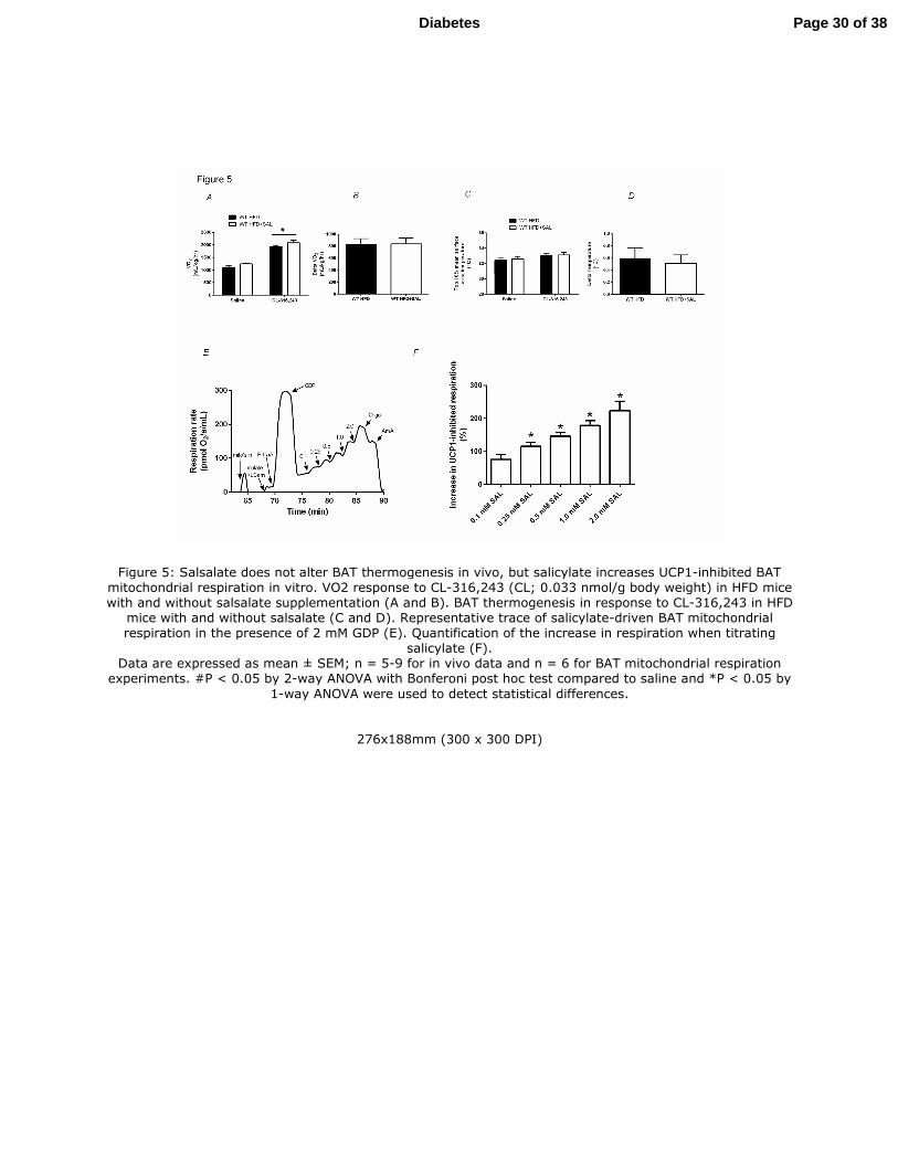
Figure 5: Salsalate does not alter BAT thermogenesis in vivo, but salicylate increases UCP1-inhibited BAT mitochondrial respiration in vitro. VO2 response to CL-316,243 (CL; 0.033 nmol/g body weight) in HFD mice with and without salsalate supplementation (A and B). BAT thermogenesis in response to CL-316,243 in HFD
mice with and without salsalate (C and D). Representative trace of salicylate-driven BAT mitochondrial respiration in the presence of 2 mM GDP (E). Quantification of the increase in respiration when titrating
salicylate (F). Data are expressed as mean ± SEM; n = 5-9 for in vivo data and n = 6 for BAT mitochondrial respiration
experiments. #P < 0.05 by 2-way ANOVA with Bonferoni post hoc test compared to saline and *P < 0.05 by
1-way ANOVA were used to detect statistical differences.
276x188mm (300 x 300 DPI)
Page 30 of 38Diabetes
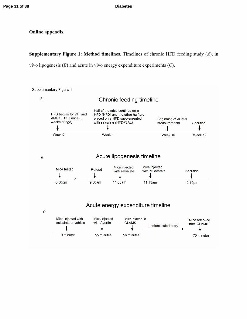
Online appendix
Supplementary Figure 1: Method timelines. Timelines of chronic HFD feeding study (A), in
vivo lipogenesis (B) and acute in vivo energy expenditure experiments (C).
Page 31 of 38 Diabetes
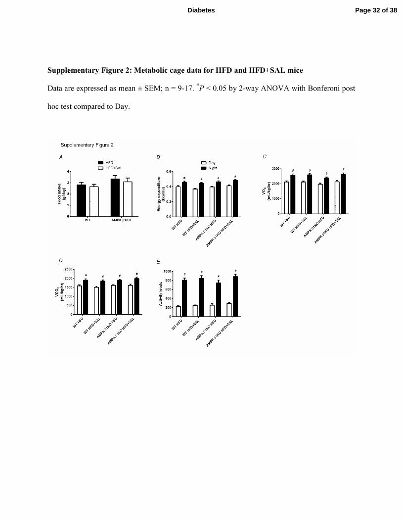
Supplementary Figure 2: Metabolic cage data for HFD and HFD+SAL mice
Data are expressed as mean ± SEM; n = 9-17. #P < 0.05 by 2-way ANOVA with Bonferoni post
hoc test compared to Day.
Page 32 of 38Diabetes
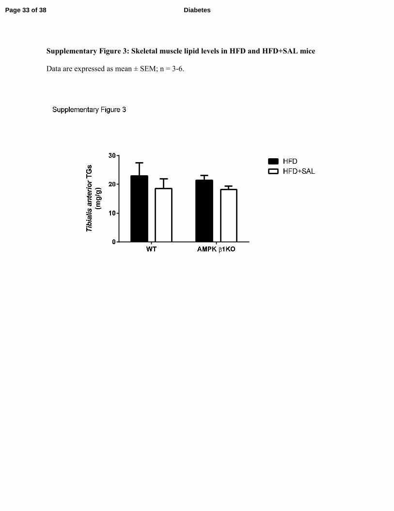
Supplementary Figure 3: Skeletal muscle lipid levels in HFD and HFD+SAL mice
Data are expressed as mean ± SEM; n = 3-6.
Page 33 of 38 Diabetes
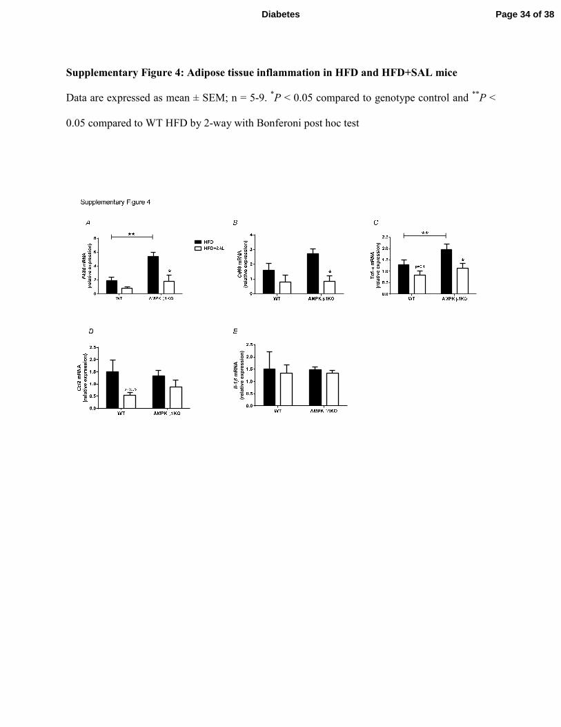
Supplementary Figure 4: Adipose tissue inflammation in HFD and HFD+SAL mice
Data are expressed as mean ± SEM; n = 5-9. *P < 0.05 compared to genotype control and
**P <
0.05 compared to WT HFD by 2-way with Bonferoni post hoc test
Il-1
mRNA
(relative expression)
Page 34 of 38Diabetes
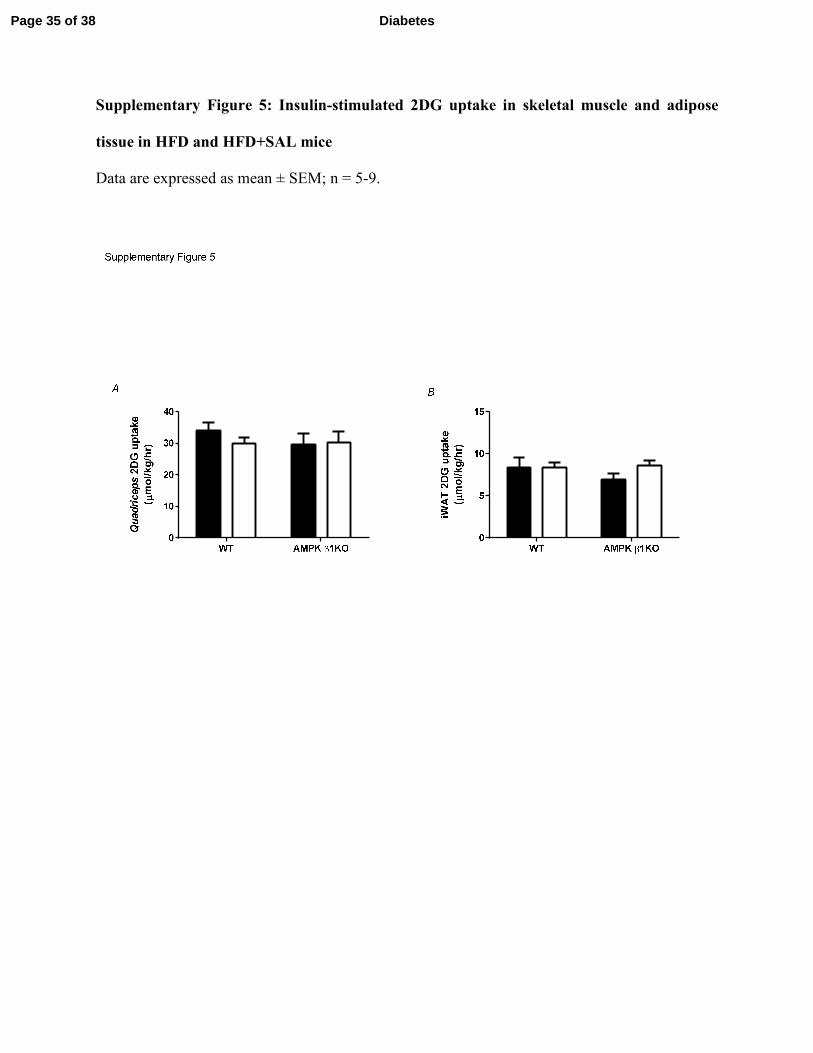
Supplementary Figure 5: Insulin-stimulated 2DG uptake in skeletal muscle and adipose
tissue in HFD and HFD+SAL mice
Data are expressed as mean ± SEM; n = 5-9.
Page 35 of 38 Diabetes
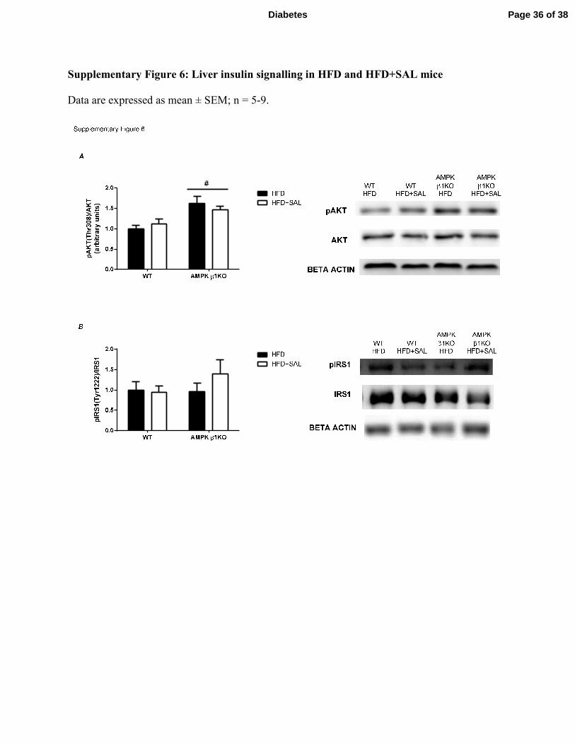
Supplementary Figure 6: Liver insulin signalling in HFD and HFD+SAL mice
Data are expressed as mean ± SEM; n = 5-9.
Page 36 of 38Diabetes
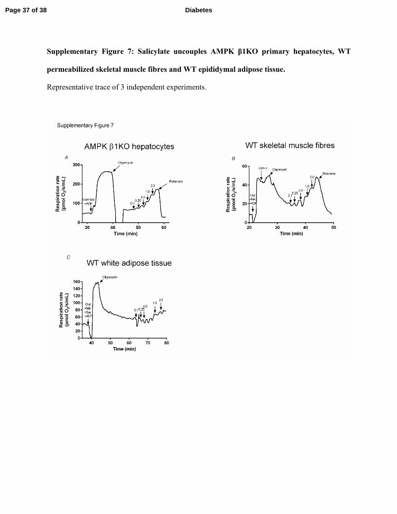
Supplementary Figure 7: Salicylate uncouples AMPK β1KO primary hepatocytes, WT
permeabilized skeletal muscle fibres and WT epididymal adipose tissue.
Representative trace of 3 independent experiments.
Page 37 of 38 Diabetes
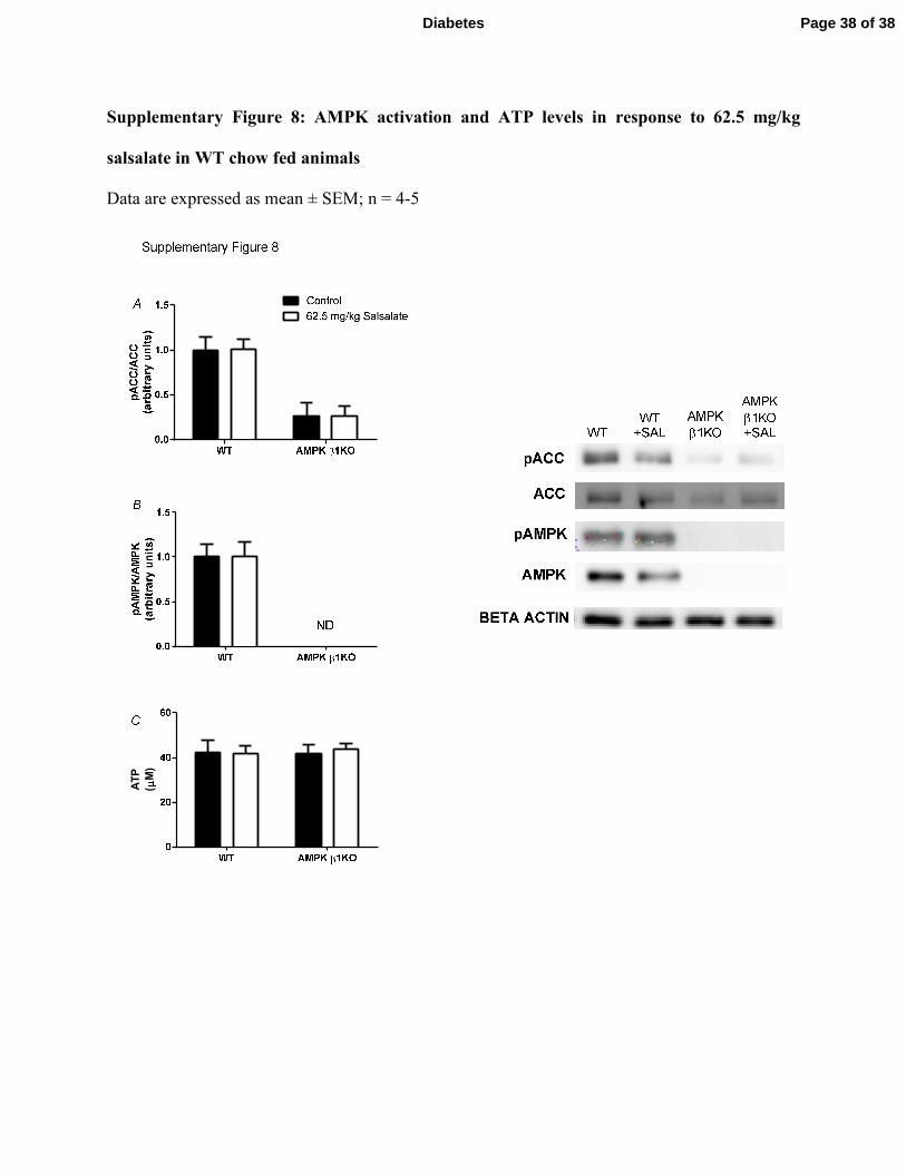
Supplementary Figure 8: AMPK activation and ATP levels in response to 62.5 mg/kg
salsalate in WT chow fed animals
Data are expressed as mean ± SEM; n = 4-5
ATP
(M)
Page 38 of 38Diabetes