Normalization of serum lactic dehydrogenase in β-thalassemia patients following bone marrow...
2
American Journal of Hematology 51:166-167 (1996) Normalization of Serum Lactic Dehydrogenase in p-Thalassemia Patients Following Bone Marrow Transplantation Amos Toren, Reuven Or, Joseph Kapelushnik, Gabriel Chividalli, Memet Aku, Shimon Slavin, and Arnon Nagler Departments of Bone Marrow Transplantation (A.T., R.O., J.K., S.S., A.N.) and Pediatrics (G.C., M.A.), Hadassah University Hospital, Jerusalem, Israel Serum lactic dehydrogenase (LDH) levels are mildly elevated In p-thalassemia major due to Ineffectiveerythropolesls. We reviewed the charts of 15 consecutive thalassemlc children who underwent allogeneic, 1-cell-depletedbone marrow transplantation (BMT) in our department during the last 3 years. Eleven patients had successful engraftment and are alive and well without evidence of disease, according to physical examinations, blood counts, and polymerase chain reaction (PCR) tests, with a median follow-up of 2 years. Two patients died due to transplantation-relatedcomplications, and two rejected the graft and receivedtheir backupautologous marrow. The LDH levels in the transplanted patients gradually decreased from an average of 952 f 155 IUIL 10 days pre-transplant (N = 300-620) to 426 * 56 IUIL at the day of transplantation, and stayed at approximately the same level post-transplant (489 f 55 IUIL). By contrast, the LDH levels reverted to the pre-transplant value in those patients who rejected their marrow. The significance of this clinical observationfor the pathophyslologic mechanismof lntramedullaryhemolysis and Ineffectiveerythropolesls in pthalassemla major is discussed. Q 1996 Wiley-Lirs, Inc. Key words: BMT, hemolysls, LDH, thalassemla INTRODUCTION conditioned with busulfan (16 mgkg), thiothepa (5 mg/ kg), cyclophosphamide (200 mgkg), and total lymphoid irradiation (TLI) (200 cGY/dayX5) or CAMPATH-1 IV (0.2 mgkg/day X 4) and received a T-cell-depleted mar- row (CAMPATH- 1 rat antihuman, CDW52, monoclonal antibody kindly provided by Dr. G. Hale and Dr. H. Waldman, Cambridge UK,) followed by related HLA- matched BMT. Statistical analysis was performed using Student's t-test; P-values are given. Lactic dehydrogenase (LDH) is a known parameter for evaluating liver and lung tissue damage, as well as tumor mass, disease progression, and response to chemotherapy [1,2]. It is also known to be elevated in intravascular hemolysis [3,4]. LDH levels are quite high in P-thalas- semia major as a consequence of ineffective erythropoie- sis [4]. We recently noticed normalization of LDH levels in patients undergoing T-cell-depleted allogeneic bone - - marrow transplantation (BMT) for P-thalissemia and have therefore analyzed the charts of 15 consecutive pa- RESULTS tients, in order to establish and better characterize this phenomenon. Eleven patients underwent successful transplantation. They are alive and well without evidence of disease, Received for publication August 30, 1995; accepted September 19, 1995. MATERIALS AND METHODS Fifteen patients suffering from P-thalassemia major underwent T-cell-depleted ailogeneic BMT in Our depart- merit during the last Address reprint requests to Arnon Nagler, M.D., Department of Bone Marrow Transplantation. Hadassah Universitv Hospital, 91 120 Jerusa- years. The median age was - . years (range 2-1 l), 9 males and 6 females. Patients were 0 1996 Wiley-Llss, Inc. Iem, Israel.
Transcript of Normalization of serum lactic dehydrogenase in β-thalassemia patients following bone marrow...
American Journal of Hematology 51 :166-167 (1996)
Normalization of Serum Lactic Dehydrogenase in p-Thalassemia Patients Following Bone
Marrow Transplantation
Amos Toren, Reuven Or, Joseph Kapelushnik, Gabriel Chividalli, Memet Aku, Shimon Slavin, and Arnon Nagler
Departments of Bone Marrow Transplantation (A.T., R.O., J.K., S.S., A.N.) and Pediatrics (G.C., M.A.), Hadassah University Hospital, Jerusalem, Israel
Serum lactic dehydrogenase (LDH) levels are mildly elevated In p-thalassemia major due to Ineffective erythropolesls. We reviewed the charts of 15 consecutive thalassemlc children who underwent allogeneic, 1-cell-depleted bone marrow transplantation (BMT) in our department during the last 3 years. Eleven patients had successful engraftment and are alive and well without evidence of disease, according to physical examinations, blood counts, and polymerase chain reaction (PCR) tests, with a median follow-up of 2 years. Two patients died due to transplantation-related complications, and two rejected the graft and received their backup autologous marrow. The LDH levels in the transplanted patients gradually decreased from an average of 952 f 155 IUIL 10 days pre-transplant (N = 300-620) to 426 * 56 IUIL at the day of transplantation, and stayed at approximately the same level post-transplant (489 f 55 IUIL). By contrast, the LDH levels reverted to the pre-transplant value in those patients who rejected their marrow. The significance of this clinical observation for the pathophyslologic mechanism of lntramedullary hemolysis and Ineffective erythropolesls in pthalassemla major is discussed. Q 1996 Wiley-Lirs, Inc.
Key words: BMT, hemolysls, LDH, thalassemla
INTRODUCTION conditioned with busulfan (16 mgkg), thiothepa (5 mg/ kg), cyclophosphamide (200 mgkg), and total lymphoid irradiation (TLI) (200 cGY/dayX5) or CAMPATH-1 IV (0.2 mgkg/day X 4) and received a T-cell-depleted mar- row (CAMPATH- 1 rat antihuman, CDW52, monoclonal antibody kindly provided by Dr. G. Hale and Dr. H. Waldman, Cambridge UK,) followed by related HLA- matched BMT. Statistical analysis was performed using Student's t-test; P-values are given.
Lactic dehydrogenase (LDH) is a known parameter for evaluating liver and lung tissue damage, as well as tumor mass, disease progression, and response to chemotherapy [1,2]. It is also known to be elevated in intravascular hemolysis [3,4]. LDH levels are quite high in P-thalas- semia major as a consequence of ineffective erythropoie- sis [4]. We recently noticed normalization of LDH levels in patients undergoing T-cell-depleted allogeneic bone - - marrow transplantation (BMT) for P-thalissemia and have therefore analyzed the charts of 15 consecutive pa- RESULTS tients, in order to establish and better characterize this phenomenon. Eleven patients underwent successful transplantation.
They are alive and well without evidence of disease,
Received for publication August 30, 1995; accepted September 19, 1995.
MATERIALS AND METHODS
Fifteen patients suffering from P-thalassemia major underwent T-cell-depleted ailogeneic BMT in Our depart- merit during the last
Address reprint requests to Arnon Nagler, M.D., Department of Bone Marrow Transplantation. Hadassah Universitv Hospital, 91 120 Jerusa- years. The median age was - .
years (range 2-1 l), 9 males and 6 females. Patients were 0 1996 Wiley-Llss, Inc.
Iem, Israel.
T
Brief Report: LDH Post-BMT in Thalassemia
DISCUSSION
167
The profound and life-threatening anemia in patients with P-thalassemia is ascribed primarily to ineffective erythropoiesis, characterized by intramedullary hemoly- sis, elevated LDH levels, low levels of haptoglobin, hem- opexin, and bizarre-looking red blood cells [4]. The patho- physiology relates directly to the accumulation of unmatched OL chains bearing an obscure toxic effect. Yuan et al. [5 ] found evidence for accelerated programmed cell death (apoptosis) in erythroid precursors in the bone marrow of these patients. They demonstrated the typical ladder pattern of erythroblasts DNA breakdown products typical for apoptosis, explaining why most of these never survive to become mature erythrocytes.
The group of patients we described may indicate that a successful engraftment of marrow resulted in termina- tion of the intramedullary hemolysis typical for P-thalas- semia. In addition, rising levels of LDH post-transplant may serve as a marker for autologous recovery of host bone marrow. LDH may thus serve as an additional sim- ple, reliable, rapid, and economic way to monitor disease status in P-thalassemia major patients post-BMT.
-1 0 D
Day8 post BMT
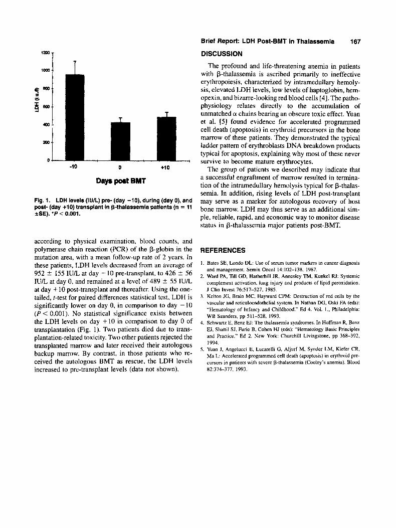
Fig. 1. LDH levels (IUIL) pre- (day -lo), during (day 0), and post- (day +lo) transplant in p-thalassemia patients (n = 11 S E ) . ‘P < 0.001.
according to physical examination, blood counts, and polymerase chain reaction (PCR) of the f3-globin in the mutation area, with a mean follow-up rate of 2 years. In these patients, LDH levels decreased from an average of 952 ? 155 I U L at day - 10 pre-transplant, to 426 ? 56 I U L at day 0, and remained at a level of 489 2 55 IU/L at day + 10 post-transplant and thereafter. Using the one- tailed, I-test for paired differences statistical test, LDH is significantly lower on day 0, in comparison to day - 10 ( P < 0.001). No statistical significance exists between the LDH levels on day +10 in comparison to day 0 of transplantation (Fig. 1). Two patients died due to trans- plantation-related toxicity. Two other patients rejected the transplanted marrow and later received their autologous backup marrow. By contrast, in those patients who re- ceived the autologous BMT as rescue, the LDH levels increased to pre-transplant levels (data not shown).
REFERENCES
1 . Bates SE, Londo DL: Use of serum tumor markers in cancer diagnosis and management, Semin Oncol 14:102-138, 1987.
2. Ward PA, Till GD, Harherhill JR, Annesley TM, Kunkel IU: Systemic complement activation, lung injury and products of lipid peroxidation. J Clin Invest 76517-527. 1985.
3. Kelton JG, Brain MC, Hayward CPM: Destruction of red cells by the vascular and reticuloendothelial system. In Nathan DG, Oski FA (eds): “Hematology of Infancy and Childhood.” Ed 4. Vol. I., Philadelphia: WB Saunders. pp 511-528, 1993.
4. Schwartz E, Benz EJ: The thalassemia syndromes. In Hoffman R, Benz EJ, Shattil SJ, Furie B, Cohen HJ (eds): “Hematology Basic Principles and Practice.” Ed 2. New York: Churchill Livingstone, pp 368-392, 1994.
5 . Yuan J. Angelucci E, Lucarelli G, Aljurf M, Synder LM, Kiefer CR, Ma L: Accelerated programmed cell death (apoptosis) in erythroid pre- cursors in patients with severe p-thalassemia (Cooky’s anemia). Blood 82:374-377, 1993.