NF-κB, iNOS, IL-6, and collagen 1 and 5 expression in ...
10
CORNEA NF-κB, iNOS, IL-6, and collagen 1 and 5 expression in healthy and keratoconus corneal fibroblasts after 0.1% riboflavin UV-A illumination Tim Berger 1 & Nóra Szentmáry 2 & Lorenz Latta 2 & Berthold Seitz 1 & Tanja Stachon 1,2 Received: 26 August 2020 /Revised: 12 December 2020 /Accepted: 18 December 2020 # The Author(s) 2021 Abstract Purpose To analyze the effect of riboflavin UV-A illumination on mRNA and protein expression of healthy (HCFs) and keratoconus human corneal fibroblasts (KC-HCFs), concerning the inflammatory markers NF-κB, iNOS, IL-6, and collagen 1 and 5 (Col 1/Col 5). Methods Keratocytes were isolated from healthy ( n = 3) and keratoconus (KC) corneas (n = 3) and were cultivated in basal medium with 5% fetal calf serum, which resulted in their transformation into human corneal fibroblasts (HCFs/KC-HCFs). Cells underwent 0.1% riboflavin UV-A illumination for 250 s (CXL). NF-κB, iNOS, IL-6, Col 1, and Col 5 expression was investigated by qPCR and Western blot analysis. IL-6 concentration of the cell culture supernatant and cell lysate was determined by ELISA. Results In untreated KC-HCFs, NF-κB(p = 0.0002), iNOS (p = 0.0019), Col 1 (p = 0.0286), and Col 5 (p = 0.0054) mRNA expression was higher and IL-6 expression was lower (p = 0.0057), than in healthy controls. In HCFs, CXL led to an increased NF-κB(p = 0.0286) and IL-6 (p = 0.0057) mRNA expression. The IL-6 concentration in the cell culture supernatant was increased in HCFs (p = 0.0485) and KC-HCFs (p = 0.0485) after CXL. CXL increased intracellular IL-6 concentration only in KC-HCFs (p = 0.0357). In the HCF group (p = 0.0286), an increased Col 1 mRNA expression after CXL could be observed. Conclusion Our study confirmed altered gene expression in untreated KC-HCFs compared to untreated HCFs. Riboflavin UV-A illumination affected gene expression only in HCFs. Increased IL-6 concentration in the cell culture supernatant and cell lysate indicate a secondary inflammatory response of HCFs and KC-HCFs to riboflavin UV-A illumination. Key messages: Untreated KC-HCFs showed an altered expression of genes linked to inflammation (NF- B, iNOS, IL-6) and increased mRNA expression of the collagens (Col 1 and 5) in contrast to untreated HCFs. CXL resulted in an increased gene expression only in HCFs (NF- B, IL-6, Col 1), whereas in KC-HCFs, none of the investigated genes showed expression changes after this treatment. Increased IL-6 concentration in the cell cultures upernatant of HCFs and KC-HCFs and an increased intracellular IL-6 concentration in KC-HCFs indicate an inflammatory response after CXL. This study show that KC-HCFs possess an altered gene and protein expression compared to HCFs before and after CXL. * Tim Berger [email protected] 1 Department of Ophthalmology, Saarland University Medical Center, Kirrberger Str. 100, D-66424 Homburg, Saar, Germany 2 Dr. Rolf M. Schwiete Center for Limbal Stem Cell and Aniridia Research, Saarland University, Homburg, Saar, Germany Graefe's Archive for Clinical and Experimental Ophthalmology https://doi.org/10.1007/s00417-020-05058-z
Transcript of NF-κB, iNOS, IL-6, and collagen 1 and 5 expression in ...
CORNEA
NF-κB, iNOS, IL-6, and collagen 1 and 5 expression in healthyand keratoconus corneal fibroblasts after 0.1% riboflavinUV-A illumination
Tim Berger1 & Nóra Szentmáry2 & Lorenz Latta2 & Berthold Seitz1 & Tanja Stachon1,2
Received: 26 August 2020 /Revised: 12 December 2020 /Accepted: 18 December 2020# The Author(s) 2021
AbstractPurpose To analyze the effect of riboflavin UV-A illumination on mRNA and protein expression of healthy (HCFs) and keratoconushuman corneal fibroblasts (KC-HCFs), concerning the inflammatory markers NF-κB, iNOS, IL-6, and collagen 1 and 5 (Col 1/Col 5).Methods Keratocytes were isolated from healthy (n = 3) and keratoconus (KC) corneas (n = 3) and were cultivated in basal mediumwith 5% fetal calf serum, which resulted in their transformation into human corneal fibroblasts (HCFs/KC-HCFs). Cells underwent0.1% riboflavin UV-A illumination for 250 s (CXL). NF-κB, iNOS, IL-6, Col 1, and Col 5 expression was investigated by qPCR andWestern blot analysis. IL-6 concentration of the cell culture supernatant and cell lysate was determined by ELISA.Results In untreated KC-HCFs, NF-κB (p = 0.0002), iNOS (p = 0.0019), Col 1 (p = 0.0286), and Col 5 (p = 0.0054) mRNAexpression was higher and IL-6 expression was lower (p = 0.0057), than in healthy controls. In HCFs, CXL led to an increasedNF-κB (p = 0.0286) and IL-6 (p = 0.0057) mRNA expression. The IL-6 concentration in the cell culture supernatant wasincreased in HCFs (p = 0.0485) and KC-HCFs (p = 0.0485) after CXL. CXL increased intracellular IL-6 concentration onlyin KC-HCFs (p = 0.0357). In the HCF group (p = 0.0286), an increased Col 1 mRNA expression after CXL could be observed.Conclusion Our study confirmed altered gene expression in untreated KC-HCFs compared to untreated HCFs. Riboflavin UV-Aillumination affected gene expression only in HCFs. Increased IL-6 concentration in the cell culture supernatant and cell lysateindicate a secondary inflammatory response of HCFs and KC-HCFs to riboflavin UV-A illumination.
Key messages:
Untreated KC-HCFs showed an altered expression of genes linked to inflammation (NF- B, iNOS, IL-6) and
increased mRNA expression of the collagens (Col 1 and 5) in contrast to untreated HCFs.
CXL resulted in an increased gene expression only in HCFs (NF- B, IL-6, Col 1), whereas in KC-HCFs, none of
the investigated genes showed expression changes after this treatment.
Increased IL-6 concentration in the cell cultures upernatant of HCFs and KC-HCFs and an increased intracellular
IL-6 concentration in KC-HCFs indicate an inflammatory response after CXL.
This study show that KC-HCFs possess an altered gene and protein expression compared to HCFs before and
after CXL.
* Tim [email protected]
1 Department of Ophthalmology, Saarland University Medical Center,Kirrberger Str. 100, D-66424 Homburg, Saar, Germany
2 Dr. Rolf M. Schwiete Center for Limbal Stem Cell and AniridiaResearch, Saarland University, Homburg, Saar, Germany
Graefe's Archive for Clinical and Experimental Ophthalmologyhttps://doi.org/10.1007/s00417-020-05058-z
Keywords Keratoconus . Cross-linking . Corneal fibroblast . Inflammation . Collagen
Introduction
Keratoconus (KC) is a bilateral, progressive ectatic cor-neal disease, which results in a cone-shaped protrusionof the corneal tissue due to the thinning of the cornealstroma. These morphological changes are associated withreduced visual acuity, progressive myopia, and irregularastigmatism. As part of the progressive stromal thinningin advanced KC, the rupture of Descemet’s membranemay cause an acute hydrops. This stromal edema subse-quently results in corneal scarring, which makes pene-trating keratoplasty in most cases necessary in order toimprove visual acuity [1].
In general, an annual incidence of 2/100,000 and a preva-lence of 54.5/100,000 is assumed [2]. These data are based ona long-term study from 1935 to 1982 and may be outdated dueto progress in diagnostics. A recent study indicates that theannual incidence (13.3/100,000) and the prevalence (265/100,000) could be 5-fold higher than previously reported[3]. Although KC is known to affect all ethnic groups, itsdevelopment shows geographical variability with a higher in-cidence among the Asian population [4].
The etiology of KC has not been clarified so far, but acomplex, multifactorial pathogenesis is assumed. In mostcases, KC occurs as an isolated disease without any evidenceof underlying concomitant diseases. KC shows a higher famil-ial incidence, which could be an indicator of a genetic predis-position. Biochemical alterations associated with increasedproteolytic activity are discussed to be responsible for thedegradation of corneal structural components resulting in thebiomechanical weakening of the cornea. It is also assumedthat a different distribution and lower number of stromal la-mellae in KC is associated with reduced corneal rigidity andthinning, compared to normal corneas. Besides, KC is morefrequent in Down syndrome, Leber’s congenital amaurosis,Ehlers-Danlos syndrome, and osteogenesis imperfecta [5].
Although KC is classified in the common literature as anon-inflammatory disease, there are data referring to potentialunderlying inflammatory components. In a previous study ofour research group, increased nuclear factor kappa B p65(NF-κB) mRNA and protein expression, and an increasedinducible NO synthase (iNOS) mRNA expression could beverified in human keratoconus fibroblasts (KC-HCFs), com-pared to healthy controls, which refers to corneal inflamma-tion in KC [6]. In addition, an increased interleukin-6 (IL-6),tumor necrosis factor-α (TNF-α), and matrix metalloprotease-9 (MMP-9) concentration could be measured in tear fluid ofKC patients, and a higher concentration of these proteins wasassociatedwith a more severe form of KC [7]. Besides, several
studies have demonstrated increased oxidative stress and al-tered metabolism in KC stromal cells [8, 9].
The healthy corneal stroma is rich in collagen 1A1 (Col 1) andcontains a relatively large amount of type 5A1 collagen (Col 5).Corneal collagen fibrils consist of type I collagen molecules thatare integrated into heterotypic fibrils together with type 5 colla-gen. These interactions ensure the proper organization of thecollagen lamellae and regulate the diameter of the collagen fibrilsto maintain corneal transparency [10]. It has been shown that KCis associatedwith altered collagen fibrillar diameter, alterations incollagen distribution, and changes in fibril orientation within thecorneal stroma [11, 12].
Conservative and surgical KC treatment options depend onthe progression of the disease. In the early stages, refractivechanges can be corrected using spectacles or contact lenses.Minimally invasive surgical treatment options include cornealcollagen cross-linking (CXL) and the implantation ofintracorneal ring segments, using these two surgical treatmentoptions either separately or in combination. In advancedstages, either lamellar or penetrating keratoplasty could benecessary [13].
The aim of CXL is to increase the stiffness of the cor-neal tissue by forming further bonds between the collagenfibrils. Therefore, the simultaneous use of riboflavin (vita-min B2) as a photosensitizer and a 370-nm wavelengthUV-A light leads to cross-links between the collagen fi-brils due to a photochemical reaction, resulting in stabili-zation of the current state [14].
Riboflavin is excited by absorbing energy of the UV-Alight (singlet riboflavin), and it is converted into a triplet state.Type I and type II reactions can be distinguished. For the typeII reaction, oxygen is required to form singlet oxygen, anoxygen radical, which react with the carbonyl group of thecollagen [14]. The type I reaction takes place after that theoxygen is exhausted, in which the excited riboflavin (tripletstate) is transferring a hydrogen atom or electron to biomole-cules such as lipids, proteins, and nucleic acids or generatesreactive oxygen species (ROS) like superoxide anion, hydrox-yl radical, or hydrogen peroxide [15]. As a consequence ofthese reactions, the collagen fibrils are cross-linked, and thus,the current stage of the disease is stabilized.
The first clinical results of CXL were published in 2003 byWollensak et al. to conclude that the progression of KC couldbe slowed down [16].
Nowadays, CXL represents an important therapeutic op-tion for KC patients, but the cellular effects are not well un-derstood. In a previous study, we have shown that riboflavinUV-A illumination is associated with increased apoptosis anddecreased viability of keratoconus stromal cells [17].
Graefes Arch Clin Exp Ophthalmol
In the present work, our purpose was to analyze the effects ofriboflavin UV-A illumination on mRNA and protein expressionof healthy (HCFs) and keratoconus human corneal fibroblasts(KC-HCFs), concerning the inflammatory markers NF-κB,iNOS, IL-6, and collagen 1 and 5 (Col 1/Col 5). As a first step,our purpose was to investigate NF-κB, iNOS, IL-6, and Col 1/Col 5 mRNA and protein expression differences between un-treated healthy and KC human corneal fibroblasts. As a secondstep, we aimed to investigate the inflammatory response (NF-κB,iNOS, IL-6) related to the increased free oxygen species forma-tion after riboflavinUV-A illumination in these cells. Thirdly, weaimed to examinewhether a riboflavinUV-A illumination has animpact on Col 1/Col 5 expression.
Materials and methods
Ethics approval and consent to participate
This study was performed in accordance with the Declarationof Helsinki and was approved by the Ethics Committee ofSaarland/Germany (No. 41/18). An informed consent was ob-tained from all participants with KC, before keratoplasty wasperformed.
Cell culture
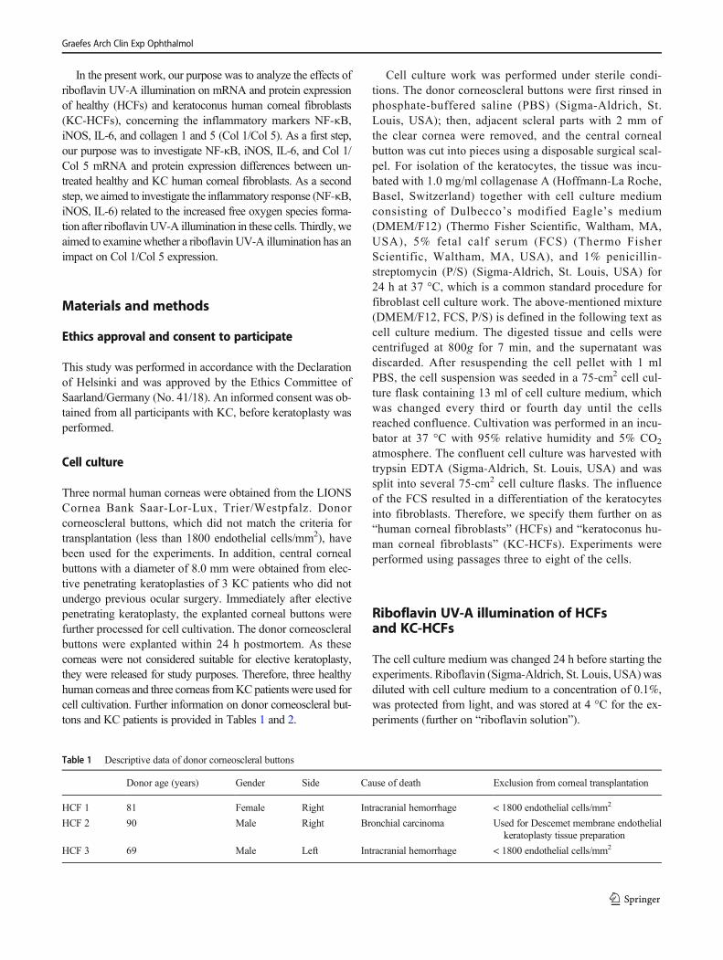
Three normal human corneas were obtained from the LIONSCornea Bank Saar-Lor-Lux, Trier/Westpfalz. Donorcorneoscleral buttons, which did not match the criteria fortransplantation (less than 1800 endothelial cells/mm2), havebeen used for the experiments. In addition, central cornealbuttons with a diameter of 8.0 mm were obtained from elec-tive penetrating keratoplasties of 3 KC patients who did notundergo previous ocular surgery. Immediately after electivepenetrating keratoplasty, the explanted corneal buttons werefurther processed for cell cultivation. The donor corneoscleralbuttons were explanted within 24 h postmortem. As thesecorneas were not considered suitable for elective keratoplasty,they were released for study purposes. Therefore, three healthyhuman corneas and three corneas fromKCpatients were used forcell cultivation. Further information on donor corneoscleral but-tons and KC patients is provided in Tables 1 and 2.
Cell culture work was performed under sterile condi-tions. The donor corneoscleral buttons were first rinsed inphosphate-buffered saline (PBS) (Sigma-Aldrich, St.Louis, USA); then, adjacent scleral parts with 2 mm ofthe clear cornea were removed, and the central cornealbutton was cut into pieces using a disposable surgical scal-pel. For isolation of the keratocytes, the tissue was incu-bated with 1.0 mg/ml collagenase A (Hoffmann-La Roche,Basel, Switzerland) together with cell culture mediumconsisting of Dulbecco’s modified Eagle’s medium(DMEM/F12) (Thermo Fisher Scientific, Waltham, MA,USA), 5% fetal calf serum (FCS) (Thermo FisherScientific, Waltham, MA, USA), and 1% penicillin-streptomycin (P/S) (Sigma-Aldrich, St. Louis, USA) for24 h at 37 °C, which is a common standard procedure forfibroblast cell culture work. The above-mentioned mixture(DMEM/F12, FCS, P/S) is defined in the following text ascell culture medium. The digested tissue and cells werecentrifuged at 800g for 7 min, and the supernatant wasdiscarded. After resuspending the cell pellet with 1 mlPBS, the cell suspension was seeded in a 75-cm2 cell cul-ture flask containing 13 ml of cell culture medium, whichwas changed every third or fourth day until the cellsreached confluence. Cultivation was performed in an incu-bator at 37 °C with 95% relative humidity and 5% CO2
atmosphere. The confluent cell culture was harvested withtrypsin EDTA (Sigma-Aldrich, St. Louis, USA) and wassplit into several 75-cm2 cell culture flasks. The influenceof the FCS resulted in a differentiation of the keratocytesinto fibroblasts. Therefore, we specify them further on as“human corneal fibroblasts” (HCFs) and “keratoconus hu-man corneal fibroblasts” (KC-HCFs). Experiments wereperformed using passages three to eight of the cells.
Riboflavin UV-A illumination of HCFsand KC-HCFs
The cell culture medium was changed 24 h before starting theexperiments. Riboflavin (Sigma-Aldrich, St. Louis, USA)wasdiluted with cell culture medium to a concentration of 0.1%,was protected from light, and was stored at 4 °C for the ex-periments (further on “riboflavin solution”).
Table 1 Descriptive data of donor corneoscleral buttons
Donor age (years) Gender Side Cause of death Exclusion from corneal transplantation
HCF 1 81 Female Right Intracranial hemorrhage < 1800 endothelial cells/mm2
HCF 2 90 Male Right Bronchial carcinoma Used for Descemet membrane endothelialkeratoplasty tissue preparation
HCF 3 69 Male Left Intracranial hemorrhage < 1800 endothelial cells/mm2
Graefes Arch Clin Exp Ophthalmol
As a first step, the cell culture supernatant was removed, andeither 5 ml cell culture medium or 5 ml of the riboflavin solutionwas pipetted into the cell culture flask under the sterile bench.Thereafter, the cell culture was quickly placed in the dark or wasilluminated with 375-nm UV-A light for 250 s (2 J/cm2) in anillumination box. After the immediate removal of the cell culturemedium or the riboflavin solution, the flask was rinsed twicewith 10 ml PBS.We added to each cell culture 10 ml cell culturemedium, and cells were cultured for 24 h or 48 h at 37 °C beforemeasurements. In order to determine the IL-6 concentration inthe cell culture supernatant, 1.5 ml cell culture supernatant wascollected from each flask before harvesting of the cells (24 h or48 h after the experiment) and was stored at − 80 °C.
Then, the cells were harvested with trypsin EDTA andwere stored at − 80 °C until further use.
RNA isolation and cDNA synthesis
RNA was isolated using the RNA Purification Plus Micro Kit(Norgen Biotek Corp., Thorold, Canada), following the instruc-tions provided by the manufacturer. RNA quantity was deter-mined using a UV/VIS spectrophotometer (Analytik Jena AG,Jena, Germany), and the eluted RNA was stored at − 80 °C forfurther use.
The cDNA synthesis was performed using the OneTaq RT-PCR Kit (New England BioLabs, Frankfurt a. M., Germany)according to the instructions provided for the Kit. For cDNAsynthesis, 1 μg of total RNA was used as template for allsamples. The synthesized cDNA was stored at − 20 °C forfurther use.
Quantitative PCR
The reaction mix (total volume: 9 μl) for quantitative PCR(qPCR) consisted of 1 μl of the specific primer solution,5 μl SYBR Green Mix (Qiagen N.V., Venlo, Netherlands),and 3 μl nuclease-free water. The qPCR reactions were per-formed using the QuantStudio 5 real-time PCR system(Thermo Fisher Scientific, Waltham, MA, USA). Sampleswere run in 9 μl volume using 1.5 μl cDNA according tothe manufacturer’s instructions. The amplification conditions(40 cycles) were 95 °C for 10 s, 60 °C for 30 s, and 95 °C for15 s. All samples were measured in duplicate. Values werenormalized to Tata-binding protein (TBP) expression levels asendogenous control gene, using the ΔΔCT method. The foldchange (2ΔΔCT-value) was used for the statistical analysis. A listof primers used for qPCR is summarized in Table 3. NormalHCFs (incubated in the dark with cell culture medium for 250s) were used as controls (fold change = 1). Gene expressionwas measured for predetermined time points (Table 4), whichhave been chosen according to previous measurement seriesof our research group [18].
Protein quantification and Western blot analysis
Cells were lysed in RIPA buffer (Thermo Fisher Scientific,Waltham, MA, USA) 48 h after the experiments, and pro-tein concentration was determined using the Pierce™ BCAProtein Assay Kit (Thermo Fisher Scientific, Waltham,MA, USA). The measurement was performed using theTecan Infinite F50 Absorbance Microplate Reader (Tecan
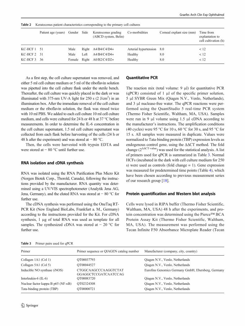
Table 2 Keratoconus patient characteristics corresponding to the primary cell cultures
Patient age (years) Gender Side Keratoconus grading(ABCD system, Belin)
Co-morbidities Corneal explant size (mm) Time fromexplantation tocell cultivation (h)
KC-HCF 1 51 Male Right A4/B4/C4/D4+ Arterial hypertension 8.0 < 12
KC-HCF 2 31 Male Left A4/B4/C4/D4+ Healthy 8.0 < 12
KC-HCF 3 36 Female Right A0/B2/C4/D2+ Healthy 8.0 < 12
Table 3 Primer pairs used for qPCR
Primer Primer sequence or QIAGEN catalog number Manufacturer (company, city, country)
Collagen 1A1 (Col 1) QT00037793 Qiagen N.V., Venlo, Netherlands
Collagen 5A1 (Col 5) QT00044527 Qiagen N.V., Venlo, Netherlands
Inducible NO synthase (iNOS) CTGGCAAGCCCAAGGTCTATGGAGGCTCCGATCAATCCAG
Eurofins Genomics Germany GmbH, Ebersberg, Germany
Interleukin-6 (IL-6) QT00083720 Qiagen N.V., Venlo, Netherlands
Nuclear factor kappa B p65 (NF-κB) QT02324308 Qiagen N.V., Venlo, Netherlands
Tata-binding protein (TBP) QT00000721 Qiagen N.V., Venlo, Netherlands
Graefes Arch Clin Exp Ophthalmol
Group AG, Männedorf, Switzerland) with 560-nm wave-length. Bovine serum albumin (BSA) was used as a stan-dard. The measurements were performed in duplicate. ForWestern blot analysis, samples with 20 μg total proteinwere boiled in a sample buffer for 5 min at 95 °C and wereloaded on a precast 4–12% NuPage™ Bis-Tris SDS Gel(Invitrogen, Waltham, MA, USA). The first well was load-ed with 3 μl Precision Plus Protein™ Dual Color Standard(Bio-Rad Laboratories, Hercules, USA) to determine themolecular weight. NuPAGE™ MOPS SDS RunningBuffer (20×) (Thermo Fisher Scientific, Waltham, MA,USA) was used as an electrophoresis buffer. After proteinseparation, semi-dry blotting on a nitrocellulose membranewas performed using the Trans-Blot Turbo Transfer System(Bio-Rad Laboratories, Hercules, USA), according to thepre-installed blotting protocol for high molecular weightproteins. The membrane was washed three times with15 ml Western Froxx washing solution (BioFroxx GmbH,Einhausen, Germany) for 5 min, which was followed byprimary antibody incubation at 4 °C. A list of antibodiesused for Western blot analysis is summarized in Table 5.The primary antibodies were diluted in a combinedblocking and secondary antibody solution (WesternFroxxanti-Mouse HRP/anti-Rabbit HRP; BioFroxx GmbH,Einhausen, Germany). Calnexin was used as a referenceprotein. After the removal of the antibody solution, themembrane was incubated with 15 ml washing solution threetimes. Protein band detection was performed with the
Western Lightning Plus Chemiluminescence Reagent(PerkinElmer Inc., Waltham, MA, USA). The chemilumi-nescence signal was detected using the ImageQuant LAS400 (GE Healthcare, Chicago, USA). Thereafter, the mem-brane stripping was performed using the Western Froxxstr ipping solut ion (BioFroxx GmbH, Einhausen,Germany).
ELISA
Enzyme-linked immunosorbent assay (ELISA) was used todetermine the IL-6 concentration of the cell culture superna-tant (24 h and 48 h after experiments) and cell lysate (48 hafter experiments).
The detection procedure was based on the sandwichELISA technique’s principle using the Human IL-6Quantikine ELISA Kit (R&D Systems, Minneapolis,USA), according to the provided instructions, at roomtemperature. Measurements were performed in duplicatewith 100 μl of the antigen-containing sample solution(cell culture supernatant or lysate). The quantitative de-tection of the IL-6 concentration was measured by aTecan Infinite F50 Absorbance Microplate Reader usinga standard provided curve. Measured IL-6 concentra-tions were divided by the total protein concentrationsto obtain the IL-6 concentration in picogram per milli-gram protein. The quotient (pg IL-6/mg protein) wasused for further statistical analysis.
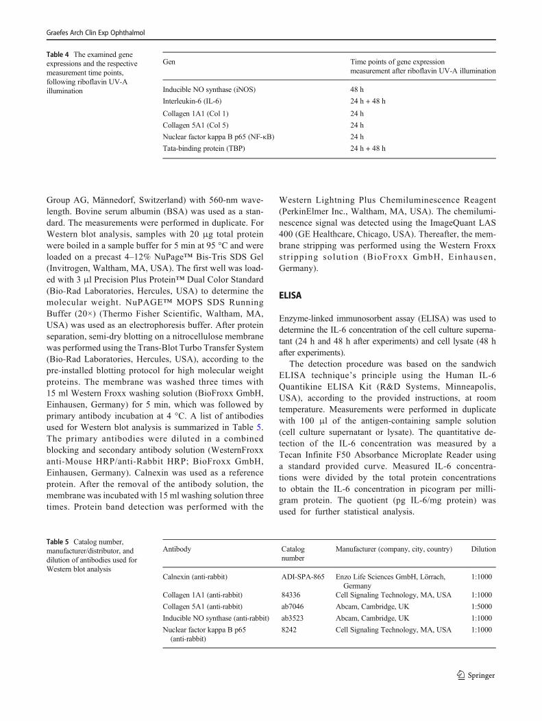
Table 4 The examined geneexpressions and the respectivemeasurement time points,following riboflavin UV-Aillumination
Gen Time points of gene expressionmeasurement after riboflavin UV-A illumination
Inducible NO synthase (iNOS) 48 h
Interleukin-6 (IL-6) 24 h + 48 h
Collagen 1A1 (Col 1) 24 h
Collagen 5A1 (Col 5) 24 h
Nuclear factor kappa B p65 (NF-κB) 24 h
Tata-binding protein (TBP) 24 h + 48 h
Table 5 Catalog number,manufacturer/distributor, anddilution of antibodies used forWestern blot analysis
Antibody Catalognumber
Manufacturer (company, city, country) Dilution
Calnexin (anti-rabbit) ADI-SPA-865 Enzo Life Sciences GmbH, Lörrach,Germany
1:1000
Collagen 1A1 (anti-rabbit) 84336 Cell Signaling Technology, MA, USA 1:1000
Collagen 5A1 (anti-rabbit) ab7046 Abcam, Cambridge, UK 1:5000
Inducible NO synthase (anti-rabbit) ab3523 Abcam, Cambridge, UK 1:1000
Nuclear factor kappa B p65(anti-rabbit)
8242 Cell Signaling Technology, MA, USA 1:1000
Graefes Arch Clin Exp Ophthalmol
Graefes Arch Clin Exp Ophthalmol
Statistical analysis
GraphPad Prism 7.04 was used for the statistical analysis andfor creating the diagrams. All data were expressed as mean ±SEM. Difference between groups was determined usingMann-WhitneyU test. p values < 0.05 were considered statis-tically significant.
Results
qPCR
Gene expression in untreated HCFs and KC-HCFs
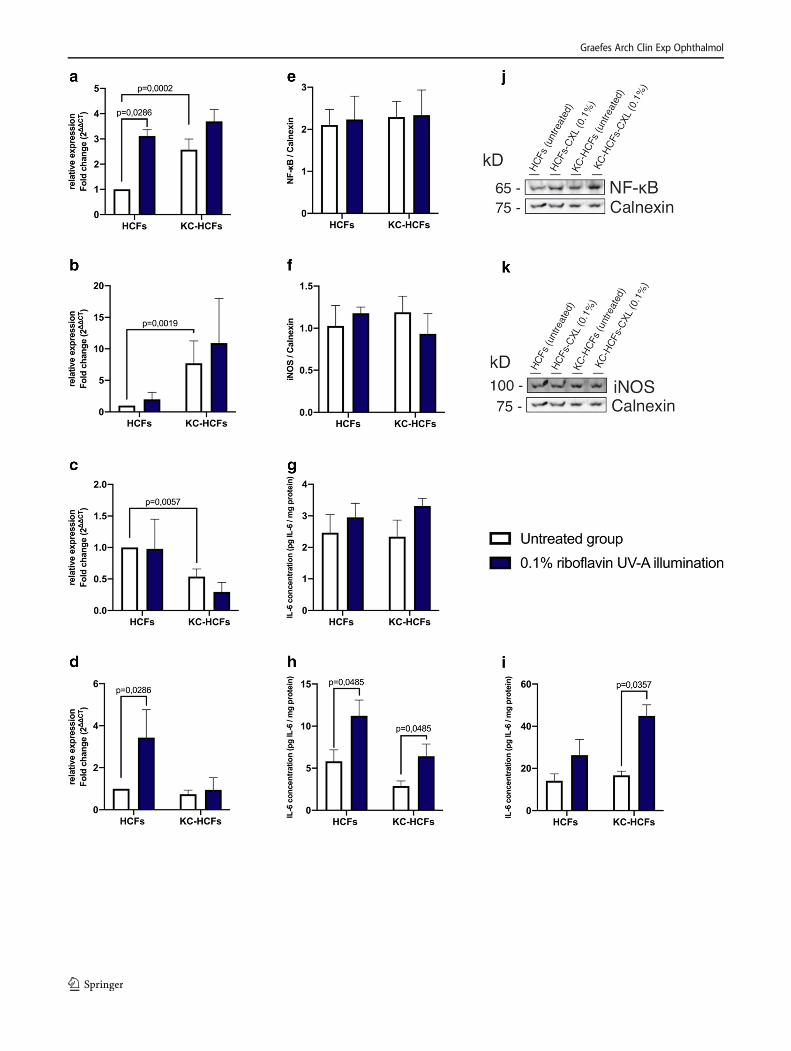
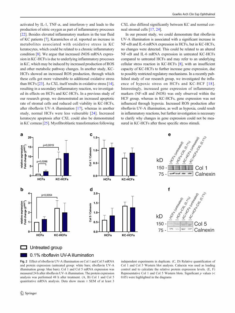
NF-κB and iNOS mRNA expression was higher (p = 0.0002;p = 0.0019) and IL-6 (p = 0.0057) mRNA expression waslower in untreated KC-HCFs than in untreated healthy con-trols (Fig. 1). Col 1 and Col 5 mRNA expression was higher inKC-HCFs than in HCFs (p = 0.0019; p = 0.0054) (Fig. 2).
Gene expression in HCFs and KC-HCFs after riboflavin UV-Aillumination
NF-κB and IL-6 mRNA expression increased (p = 0.0286;p = 0.0286) in HCFs after 0.1% riboflavin UV-A illumina-tion. NF-κB and iNOS mRNA expression remained un-changed in KC-HCFs after treatment (p = 0.2788; p =0.7457) and iNOS mRNA expression did not changethrough 0.1% riboflavin UV-A illumination in HCFs orKC-HCFs (p = 0.3143) (Fig. 1).
Col 1 mRNA expression increased in HCFs following0.1% riboflavin UV-A illumination (p = 0.0286) and Col 5expression remained unchanged in HCFs and KC-HCFs aftertreatment (p = 0.0971; p = 0.4623) (Fig. 2).
Western blot
NF-κB (p = 0.7308), iNOS (p = 0.6282), Col 1 (p = 0.3660),and Col 5 (p = 0.1255) protein expression did not differ be-tween untreated HCFs and KC-HCFs. NF-κB (p = 0.9999; p =0.9999), iNOS (p = 0.9999; p = 0.6667), Col 1 (p = 0.3660; p= 0.5167), and Col 5 (p = 0.8571; p = 0.5714) protein expres-sion remained unchanged in HCFs or KC-HCFs through 0.1%riboflavin UV-A illumination (Figs. 1 and 2).
ELISA
IL-6 concentration in the cell culture supernatant did not differbetween untreated HCFs and KC-HCFs (24 h: p = 0.8785; 48h: p = 0.0650). Twenty-four hours after treatment, IL-6 con-centration in the cell culture supernatant did not change inHCFs and KC-HCFs (p = 0.4970; p = 0.1333) .Nevertheless, 48 h after 0.1% riboflavin UV-A illumination,IL-6 concentration in the cell culture supernatant increased inHCFs (p = 0.0485) and KC-HCFs (p = 0.0485), compared tobaseline values (Fig. 1).
In the cell lysate, IL-6 concentration did not differ betweenuntreated HCFs and KC-HCFs (p = 0.7922). Nevertheless, inthe cell lysate, IL-6 concentration increased in KC-HCFs 48 hafter 0.1% riboflavin UV-A illumination (p = 0.0357). InHCFs, IL-6 concentration in the cell lysate remained un-changed after treatment (p = 0.1667) (Fig. 1).
Discussion
Little is known about the pathogenesis of KC, but along thereported changes in collagen metabolism [11, 12], the suspicionof a chronic inflammatory component is increasingly coming tothe fore. Since cross-linking was first introduced by Wollensaket al. in 2003 [16], it became an indispensable therapeutic optionfor treating ectatic corneal disorders. However, the cell biologicaleffects after CXL were barely investigated. Therefore, in thisstudy, we treated corneal fibroblasts in vitro with riboflavinUV-A illumination, to examine the impact of CXL regardingexpression changes of inflammatory markers NF-κB, iNOS,IL-6, and collagen 1 and 5 in HCFs and KC-HCFs.
We could demonstrate changes in gene expression linkedto inflammation in untreated KC-HCFs. NF-κB is an impor-tant modulator of many cell biological pathways through itsinfluence on more than 500 genes, mainly genes associatedwith inflammatory responses [19]. Both iNOS and IL-6 areinfluenced by NF-κB [20, 21]. Additionally, iNOS is
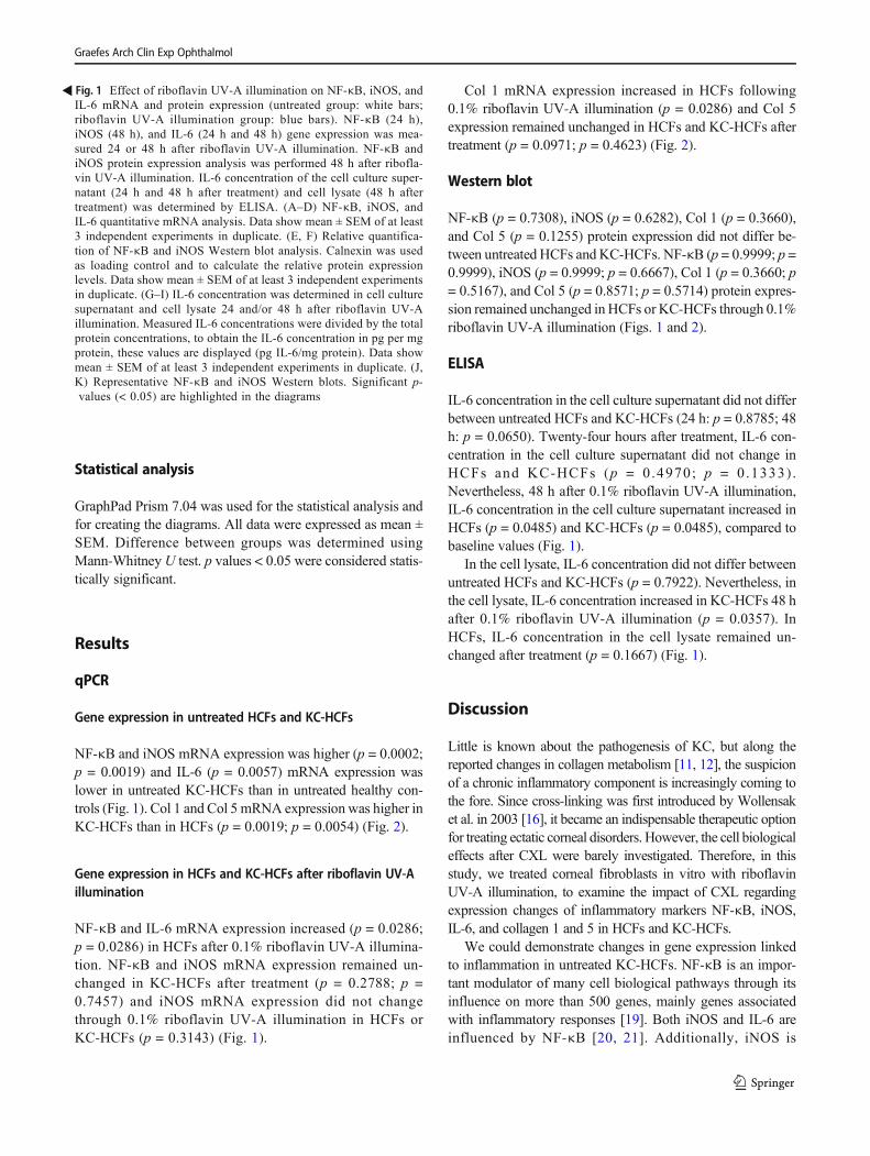
�Fig. 1 Effect of riboflavin UV-A illumination on NF-κB, iNOS, andIL-6 mRNA and protein expression (untreated group: white bars;riboflavin UV-A illumination group: blue bars). NF-κB (24 h),iNOS (48 h), and IL-6 (24 h and 48 h) gene expression was mea-sured 24 or 48 h after riboflavin UV-A illumination. NF-κB andiNOS protein expression analysis was performed 48 h after ribofla-vin UV-A illumination. IL-6 concentration of the cell culture super-natant (24 h and 48 h after treatment) and cell lysate (48 h aftertreatment) was determined by ELISA. (A–D) NF-κB, iNOS, andIL-6 quantitative mRNA analysis. Data show mean ± SEM of at least3 independent experiments in duplicate. (E, F) Relative quantifica-tion of NF-κB and iNOS Western blot analysis. Calnexin was usedas loading control and to calculate the relative protein expressionlevels. Data show mean ± SEM of at least 3 independent experimentsin duplicate. (G–I) IL-6 concentration was determined in cell culturesupernatant and cell lysate 24 and/or 48 h after riboflavin UV-Aillumination. Measured IL-6 concentrations were divided by the totalprotein concentrations, to obtain the IL-6 concentration in pg per mgprotein, these values are displayed (pg IL-6/mg protein). Data showmean ± SEM of at least 3 independent experiments in duplicate. (J,K) Representative NF-κB and iNOS Western blots. Significant p-values (< 0.05) are highlighted in the diagrams
Graefes Arch Clin Exp Ophthalmol
activated by IL-1, TNF-α, and interferon-γ and leads to theproduction of nitric oxygen as part of inflammatory processes[22]. Besides elevated inflammatory markers in the tear fluidof KC patients [7], Karamichos et al. reported an increase inmetabolites associated with oxidative stress in KCkeratocytes, which could be related to a chronic inflammatorycondition [8]. We argue that increased iNOS mRNA expres-sion in KC-HCFs is due to underlying inflammatory processesin KC, whichmay be induced by increased production of ROSand other metabolic pathway changes. In another study, KC-HCFs showed an increased ROS production, through whichthese cells got more vulnerable to additional oxidative stressthan HCFs [23]. As CXL itself results in oxidative stress [14],resulting in a secondary inflammatory reaction, we investigat-ed its effects on HCFs and KC-HCFs. In a previous study ofour research group, we demonstrated an increased apoptoticrate of stromal cells and reduced cell viability in KC-HCFs,after riboflavin UV-A illumination [17], whereas in anotherstudy, normal HCFs were less vulnerable [24]. Increasedkeratocyte apoptosis after CXL could also be demonstratedin KC corneas [25]. Myofibroblastic transformation following
CXL also differed significantly between KC and normal cor-neal stromal cells [17, 24].
In our present study, we could demonstrate that riboflavinUV-A illumination is associated with a significant increase inNF-κB and IL-6 mRNA expression in HCFs, but in KC-HCFs,no changes were detected. This could be related to an alteredNF-κB and IL-6 mRNA expression in untreated KC-HCFscompared to untreated HCFs and may refer to an underlyingcellular stress reaction in KC-HCFs [8], with an insufficientcapacity of KC-HCFs to further increase gene expression, dueto possibly restricted regulatory mechanisms. In a recently pub-lished study of our research group, we investigated the influ-ence of hypoxic stress on HCFs and KC-HCF [18].Interestingly, increased gene expression of inflammatorymarkers (NF-κB and iNOS) was only observed within theHCF group, whereas in KC-HCFs, gene expression was notinfluenced through hypoxia. Increased ROS production afterriboflavin UV-A illumination, as well as hypoxia, could resultin inflammatory reactions, but further investigation is necessaryto clarify why changes in gene expression could not be mea-sured in KC-HCFs after those specific stress stimuli.
Fig. 2 Effect of riboflavin UV-A illumination on Col 1 and Col 5 mRNAand protein expression (untreated group: white bars; riboflavin UV-Aillumination group: blue bars). Col 1 and Col 5 mRNA expression wasmeasured 24 h after riboflavinUV-A illumination. The protein expressionanalysis was performed 48 h after treatment. (A, B) Col 1 and Col 5quantitative mRNA analysis. Data show mean ± SEM of at least 3
independent experiments in duplicate. (C, D) Relative quantification ofCol 1 and Col 5 Western blot analysis. Calnexin was used as loadingcontrol and to calculate the relative protein expression levels. (E, F)Representative Col 1 and Col 5 Western blots. Significant p values (<0.05) were highlighted in the diagrams
Graefes Arch Clin Exp Ophthalmol
Although no IL-6 mRNA expression changes in KC-HCFscould be measured after riboflavin UV-A illumination, IL-6protein amount increased significantly in the cell culture su-pernatant through riboflavin UV-A illumination in both HCFsand KC-HCFs. However, a difference in the IL-6 concentra-tion of the cell culture supernatant could only be observedafter 48 h, indicating that this might be a delayed reaction ofthe corneal fibroblasts after riboflavin UV-A illumination,which should be considered in future studies. Interestingly,riboflavin UV-A illumination resulted in an increased intra-cellular IL-6 concentration only in KC-HCFs, which was notaccompanied by altered mRNA expression. This could indi-cate that KC-HCFs can only partially compensate the inducedstress stimulus and therefore have a significant intracellularstress response.
In this study, corneas with advanced KC stages (BelinKC classification) and corneal scarring were used [26].Most interestingly, we detected an increased Col 1 andCol 5 mRNA expression in KC-HCFs compared toHCFs, which refers to the disturbance of the collagen me-tabolism in KC corneas [11, 12]. In addition, the Col 1 andCol 5 mRNA expression values have shown a larger vari-ability in KC-HCFs than in HCFs. These variable valueswere confirmed through repeat measurements. We suggestthat these differences depend on the individual patient’scornea and in addition, during KC progression, gene ex-pression could also change. It is important to investigatethese results in future studies, for example, by comparingthe mRNA and protein expression of KC corneas with orwithout scars or under consideration of the KC stage.
Furthermore, we investigated whether CXL is associatedwith changes in collagen expression. In this study, we ob-served increased Col 1 mRNA expression only in HCFs with-out significant protein expression changes in HCFs or KC-HCFs following riboflavin UV-A illumination. Another studydescribed a significant Col 1 and Col 5 protein expressiondecrease after 0.1% riboflavin UV-A illumination in KC-HCFs, but not in HCFs, nevertheless, using 3D cell cultures[27]. To which extent these changes could be of cell biologicalor clinical relevance must be critically analyzed.
Finally, it is necessary to mention certain limitations of ourstudy concerning the interpretation of the results. On the onehand, the avascular stroma represents a nutrient-poor tissue.Therefore, in vitro experiments are not equally applicable toin vivo models. Our isolated keratocytes dedifferentiated intofibroblasts during cell cultivation, using FCS, which could prob-ably have a significant impact on gene and protein expression.The results of the 2D culture are also not directly transferable tothe in vivo model, but give a good indication for further studies.In addition, there is no suitable in vivo model or animal model tostudyKC. In summary, we found expression differences betweenuntreated and treated corneal fibroblasts, which is an importantinput for future studies.
Conclusions
In this study, altered mRNA expression of the inflammatorymarkers (NF-κB, iNOS, IL-6), as well as the collagens (Col 1,Col 5), could be detected in untreated KC-HCFs. RiboflavinUV-A illumination resulted in significant gene expressionchanges only in HCFs. Increased IL-6 concentrations in thecell culture supernatant (HCFs and KC-HCFs) and cell lysate(KC-HCFs) indicated an inflammatory response after thistreatment. The effects of cross-linking on gene and proteinexpression have rarely been investigated, and therefore, theresults of this study provide a basis for further researchprojects.
Acknowledgments The work of Dr. Tanja Stachon, Dr. Lorenz Latta,and Prof. Dr. Nóra Szentmáry at the Dr. Rolf M. Schwiete Center forLimbal Stem Cell and Aniridia Research was supported by the Dr. RolfM. Schwiete Foundation.Wewould like to thank Prof. Dr. Veit Flockerzi(Department of Experimental and Clinical Pharmacology, Institute ofExperimental and Clinical Pharmacology and Toxicology, Center forMolecular Signaling (PZMS), Saarland University, Homburg,Germany) for the use of the Western blot visualization system.
Funding information Open Access funding enabled and organized byProjekt DEAL.
Compliance with ethical standards
Conflict of interest The authors declare that they have no conflict ofinterest.
Ethics approval and consent to participate This study was performed inaccordance with the Declaration of Helsinki and was approved by theEthics Committee of Saarland/Germany (No. 41/18). An informed con-sent was obtained from all participants with KC, before keratoplasty wasperformed.
Open Access This article is licensed under a Creative CommonsAttribution 4.0 International License, which permits use, sharing, adap-tation, distribution and reproduction in any medium or format, as long asyou give appropriate credit to the original author(s) and the source, pro-vide a link to the Creative Commons licence, and indicate if changes weremade. The images or other third party material in this article are includedin the article's Creative Commons licence, unless indicated otherwise in acredit line to the material. If material is not included in the article'sCreative Commons licence and your intended use is not permitted bystatutory regulation or exceeds the permitted use, you will need to obtainpermission directly from the copyright holder. To view a copy of thislicence, visit http://creativecommons.org/licenses/by/4.0/.
References
1. Fan Gaskin JC, Patel DV, McGhee CNJ (2014) Acute cornealhydrops in keratoconus - new perspectives. Am J Ophthalmol157:921–928. https://doi.org/10.1016/j.ajo.2014.01.017
2. Rabinowitz YS (1998) Keratoconus. Surv Ophthalmol 42:297–319. https://doi.org/10.1016/S0039-6257(97)00119-7
Graefes Arch Clin Exp Ophthalmol
3. Godefrooij DA, deWit GA, Uiterwaal CS et al (2017) Age-specificincidence and prevalence of keratoconus: a nationwide registrationstudy. Am J Ophthalmol 175:169–172. https://doi.org/10.1016/j.ajo.2016.12.015
4. Cozma I, Atherley C, James N (2005) Influence of ethnic origin onthe incidence of keratoconus and associated atopic disease in Asianand white patients. Eye (Lond) 19:924–925. https://doi.org/10.1038/sj.eye.6701677
5. Romero-Jiménez M, Santodomingo-Rubido J, Wolffsohn JS(2010) Keratoconus: a review. Contact Lens Anterior Eye 33:157–166. https://doi.org/10.1016/j.clae.2010.04.006
6. Stachon T, Latta L, Kolev K et al (2019) Increased NF-κB andiNOS expression in keratoconus keratocytes - hints for an inflam-matory component? Klin Monbl Augenheilkd. https://doi.org/10.1055/a-1002-0100
7. Lema I, Durán JA (2005) Inflammatory molecules in the tears ofpatients with keratoconus. Ophthalmology 112:654–659. https://doi.org/10.1016/j.ophtha.2004.11.050
8. Karamichos D, Hutcheon A, Rich C et al (2014) In vitro modelsuggests oxidative stress involved in keratoconus disease. Sci Rep4:4608. https://doi.org/10.1038/srep04608
9. Karamichos D, Zieske J, Sejersen H et al (2015) Tear metabolitechanges in keratoconus. Exp Eye Res 132:1–8. https://doi.org/10.1016/j.exer.2015.01.007
10. Hassell J, Birk D (2010) The molecular basis of corneal transpar-ency. Exp Eye Res 91:326–335. https://doi.org/10.1016/j.exer.2010.06.021
11. Hayes S, Boote C, Tuft S et al (2007) A study of corneal thickness,shape and collagen organisation in keratoconus usingvideokeratography and X-ray scattering techniques. Exp Eye Res84:423–434. https://doi.org/10.1016/j.exer.2006.10.014
12. Alkanaan A, Barsotti R, Kirat O et al (2019) Collagen fibrils andproteoglycans of peripheral and central stroma of the keratoconuscornea - ultrastructure and 3D transmission electron tomography.Sci Rep 9:1–11. https://doi.org/10.1038/s41598-019-56529-1
13. Parker JS, van Dijk K, Melles GRJ (2015) Treatment options foradvanced keratoconus: a review. Surv Ophthalmol 60:459–480.https://doi.org/10.1016/j.survophthal.2015.02.004
14. Raiskup F, Spoerl E (2013) Corneal crosslinking with riboflavinand ultraviolet A. I. principles. Ocul Surf 11:65–74. https://doi.org/10.1016/j.jtos.2013.01.002
15. Juarranz A, Jaén P, Sanz-Rodriguez F, Cuevas-Santos J (2008)Photodynamic therapy of cancer. Basic principles and applications.Clin Transl Oncol 10:148–154. https://doi.org/10.1007/s12094-008-0172-2
16. Wollensak G, Spoerl E, Seiler T (2003) Riboflavin/ultraviolet-A-induced collagen crosslinking for the treatment of keratoconus. Am
J Ophthalmol 135:620–627. https://doi.org/10.1016/S0002-9394(02)02220-1
17. Song X, Stachon T, Wang J et al (2015) Viability, apoptosis, pro-liferation, activation, and cytokine secretion of human keratoconuskeratocytes after cross-linking. Biomed Res Int 2015:254237.https://doi.org/10.1155/2015/254237
18. Stachon T, Latta L, Seitz B, Szentmáry N (2020) Hypoxic stressincreases NF-κB and iNOSmRNA expression in normal, but not inkeratoconus corneal fibroblasts. Graefe’s Arch Clin ExpOphthalmol. https://doi.org/10.1007/s00417-020-04900-8
19. Gupta SC, Sundaram C, Reuter S, Aggarwal BB (2010) InhibitingNF-κB activation by small molecules as a therapeutic strategy.Biochim Biophys Acta - Gene Regul Mech 1799:775–787.https://doi.org/10.1016/j.bbagrm.2010.05.004
20. Libermann TA, Baltimore D (1990) Activation of interleukin-6gene expression through the NF-kappa B transcription factor. MolCell Biol 10:2327–2334. https://doi.org/10.1128/mcb.10.5.2327
21. Morris KR, Lutz RD, Choi HS et al (2003) Role of the NF-κBsignaling pathway and κB cis-regulatory elements on the IRF-1and iNOS promoter regions in mycobacterial lipoarabinomannaninduction of nitric oxide. Infect Immun 71:1442–1452. https://doi.org/10.1128/IAI.71.3.1442-1452.2003
22. Donnelly LE, Barnes PJ (2002) Expression and regulation of in-ducible nitric oxide synthase from human primary airway epithelialcells. Am J Respir Cell Mol Biol 26:144–151. https://doi.org/10.1165/ajrcmb.26.1.4477
23. Chwa M, Atilano SR, Reddy V et al (2006) Increased stress-induced generation of reactive oxygen species and apoptosis inhuman keratoconus fibroblasts. Invest Ophthalmol Vis Sci 47:1902–1910. https://doi.org/10.1167/iovs.05-0828
24. Stachon T, Wang J, Song X et al (2015) Impact of crosslinking/riboflavin-UVA-photodynamic inactivation on viability, apoptosisand activation of human keratocytes in vitro. J BiomedRes 29:321–325. https://doi.org/10.7555/JBR.29.20130173
25. Messmer E, Meyer P, Herwig-Carl M et al (2012) Morphological andimmunohistochemical changes after corneal cross-linking. Cornea 32:111–117. https://doi.org/10.1097/ICO.0b013e31824d701b
26. Belin MW, Duncan JK (2016) Keratoconus: the ABCD GradingSystem. Klin Monbl Augenheilkd 233:701–707. https://doi.org/10.1055/s-0042-100626
27. Sharif R, Hjortdal J, Sejersen H et al (2017) Human in vitro modelreveals the effects of collagen cross-linking on keratoconus patho-genesis. Sci Rep 7:12517. https://doi.org/10.1038/s41598-017-12598-8
Publisher’s note Springer Nature remains neutral with regard to jurisdic-tional claims in published maps and institutional affiliations.
Graefes Arch Clin Exp Ophthalmol