RESEARCH Open Access Metallothionein 2A inhibits NF-κB ... fileRESEARCH Open Access Metallothionein...
14
RESEARCH Open Access Metallothionein 2A inhibits NF-κB pathway activation and predicts clinical outcome segregated with TNM stage in gastric cancer patients following radical resection Yuanming Pan 1† , Jiaqiang Huang 2† , Rui Xing 1 , Xin Yin 1 , Jiantao Cui 1 , Wenmei Li 1 , Jun Yu 3 and Youyong Lu 1* Abstract Background: Metallothionein 2A (MT2A) as a stress protein, plays a protective role in gastric mucosal barrier. Its role in the development of gastric cancer (GC) is unclear. The mechanism of MT2A will be investigated in gastric tumorigenesis. Methods: MT2A expression was detected in 973 gastric specimens. The biological function was determined through ectopic expressing MT2A in vitro and in vivo. The possible downstream effectors of MT2A were investigated in NF-κB signaling. The protein levels of MT2A, IκB-α and p-IκB-α (ser32/36) expression were analyzed in a subset of 258 patients by IHC staining. The prognostic effects of MT2A, status of IκB-α and TNM stage were evaluated using the Kaplan-Meier method and compared using the log-rank test. Results: Decreased MT2A expression was detected in cell lines and primary tumors of GC. In clinical data, loss of MT2A (MT2A + in Normal (n =171, 76.0%); Intestinal metaplasia (n = 118, 50.8%); GC (n = 684. 22.4%, P < 0.001)) was associated with poor prognosis (P < 0.001), advanced TNM stage (P = 0.05), and down-regulation of IκB-α expression (P < 0.001). Furthermore, MT2A was the independent prognostic signature segregated from the status of IκB-α and pathological features. In addition, MT2A inhibited cell growth through apoptosis and G2/M arrest, which negatively regulated NF-κB pathway through up-regulation of IκB-α and down-regulation of p-IκB-α and cyclin D1 expression. Conclusions: MT2A might play a tumor suppressive activity through inhibiting NF-κB signaling and may be a prognostic biomarker and potential target for individual therapy of GC patients. Keywords: Metallothionein 2A, NF-κB signaling, Gastric cancer, Prognosis, TNM stage Background What’s new Abundance of Metallothionein 2A (MT2A) exhibits in the gastric mucosal barrier, but its role in the progres- sion of gastric cancer (GC) is still unclear. In this ana- lysis of 171 normal gastric tissues, 118 intestinal metaplasia, and 684 primary gastric cancers, decreased MT2A was significantly correlated with poor prognosis. It inhibited the proliferation of GC cells through NF-κB signaling inactivation. These results indicate that MT2A may be an important prognostic marker and therapeutic target for GC. Gastric cancer (GC) is the most commonly diagnosed malignancy and remains a significant burden of cancer in Asia, especially in China [1,2]. Most GC patients undergo- ing surgery are already at an advanced stage and the 5- year survival is varied. Several histological factors have been reported to be prognostic factors of GC, including tumor size, WHO classification, tumor-node-metastasis (TNM) stage system, and differentiation grade [3,4]. How- ever, prognosis of GC patients at the same stage is still inconsistent [5]. Therefore, identification of specific * Correspondence: [email protected] † Equal contributors 1 Laboratory of Molecular Oncology, Key laboratory of Carcinogenesis and Translational Research, Ministry of Education, Peking University Cancer Hospital & Institute, No.52 Fucheng Road, Beijing, Haidian District 100142, People's Republic of China Full list of author information is available at the end of the article © 2013 Pan et al.; licensee BioMed Central Ltd. This is an Open Access article distributed under the terms of the Creative Commons Attribution License (http://creativecommons.org/licenses/by/2.0), which permits unrestricted use, distribution, and reproduction in any medium, provided the original work is properly cited. Pan et al. Journal of Translational Medicine 2013, 11:173 http://www.translational-medicine.com/content/11/1/173
Transcript of RESEARCH Open Access Metallothionein 2A inhibits NF-κB ... fileRESEARCH Open Access Metallothionein...
Pan et al. Journal of Translational Medicine 2013, 11:173http://www.translational-medicine.com/content/11/1/173
RESEARCH Open Access
Metallothionein 2A inhibits NF-κB pathwayactivation and predicts clinical outcomesegregated with TNM stage in gastric cancerpatients following radical resectionYuanming Pan1†, Jiaqiang Huang2†, Rui Xing1, Xin Yin1, Jiantao Cui1, Wenmei Li1, Jun Yu3 and Youyong Lu1*
Abstract
Background: Metallothionein 2A (MT2A) as a stress protein, plays a protective role in gastric mucosal barrier. Itsrole in the development of gastric cancer (GC) is unclear. The mechanism of MT2A will be investigated in gastrictumorigenesis.
Methods: MT2A expression was detected in 973 gastric specimens. The biological function was determinedthrough ectopic expressing MT2A in vitro and in vivo. The possible downstream effectors of MT2A wereinvestigated in NF-κB signaling. The protein levels of MT2A, IκB-α and p-IκB-α (ser32/36) expression were analyzedin a subset of 258 patients by IHC staining. The prognostic effects of MT2A, status of IκB-α and TNM stage wereevaluated using the Kaplan-Meier method and compared using the log-rank test.
Results: Decreased MT2A expression was detected in cell lines and primary tumors of GC. In clinical data, loss ofMT2A (MT2A + in Normal (n =171, 76.0%); Intestinal metaplasia (n = 118, 50.8%); GC (n = 684. 22.4%, P < 0.001)) wasassociated with poor prognosis (P < 0.001), advanced TNM stage (P = 0.05), and down-regulation of IκB-α expression(P < 0.001). Furthermore, MT2A was the independent prognostic signature segregated from the status of IκB-α andpathological features. In addition, MT2A inhibited cell growth through apoptosis and G2/M arrest, which negativelyregulated NF-κB pathway through up-regulation of IκB-α and down-regulation of p-IκB-α and cyclin D1 expression.
Conclusions: MT2A might play a tumor suppressive activity through inhibiting NF-κB signaling and may be aprognostic biomarker and potential target for individual therapy of GC patients.
Keywords: Metallothionein 2A, NF-κB signaling, Gastric cancer, Prognosis, TNM stage
BackgroundWhat’s newAbundance of Metallothionein 2A (MT2A) exhibits inthe gastric mucosal barrier, but its role in the progres-sion of gastric cancer (GC) is still unclear. In this ana-lysis of 171 normal gastric tissues, 118 intestinalmetaplasia, and 684 primary gastric cancers, decreasedMT2A was significantly correlated with poor prognosis.
* Correspondence: [email protected]†Equal contributors1Laboratory of Molecular Oncology, Key laboratory of Carcinogenesis andTranslational Research, Ministry of Education, Peking University CancerHospital & Institute, No.52 Fucheng Road, Beijing, Haidian District 100142,People's Republic of ChinaFull list of author information is available at the end of the article
© 2013 Pan et al.; licensee BioMed Central LtdCommons Attribution License (http://creativecreproduction in any medium, provided the or
It inhibited the proliferation of GC cells through NF-κBsignaling inactivation. These results indicate that MT2Amay be an important prognostic marker and therapeutictarget for GC.Gastric cancer (GC) is the most commonly diagnosed
malignancy and remains a significant burden of cancer inAsia, especially in China [1,2]. Most GC patients undergo-ing surgery are already at an advanced stage and the 5-year survival is varied. Several histological factors havebeen reported to be prognostic factors of GC, includingtumor size, WHO classification, tumor-node-metastasis(TNM) stage system, and differentiation grade [3,4]. How-ever, prognosis of GC patients at the same stage is stillinconsistent [5]. Therefore, identification of specific
. This is an Open Access article distributed under the terms of the Creativeommons.org/licenses/by/2.0), which permits unrestricted use, distribution, andiginal work is properly cited.
Pan et al. Journal of Translational Medicine 2013, 11:173 Page 2 of 14http://www.translational-medicine.com/content/11/1/173
diagnostic markers and therapeutic target would allow re-liable prediction, effective extension of postoperative sur-vival and life quality of patients [6].Cellular stress has been shown to play a role in the mo-
lecular regulation of carcinogenesis [7,8]. Metallothioneins(MTs) as the stress proteins with low molecular weight andrich-cysteine have the ability of a high affinity for metalions and ROS scavengers. MT2A as the main isoform ofMTs plays an important role in gastric mucosal barrier inpatients with gastritis and rodent models [9,10]. Pre-administration of exogenous MT2A or pre-induction ofendogenous MT2A can protect stomach and liver againststress-induced damage and inhibit the formation of stress-induced lipid peroxide, implying a protective effect ofMT2A on stress-induced pathogenesis and a potentialtherapeutic target applied for early prevention [11–13]. Re-cently, MT2A plays an important role in tumorigenesisand progression of multiple carcinomas including GC[14]. The mice loss of MT2A gene predisposed todiethylnitrosamine-induced hepatocarcinogenesis by acti-vating NF-κB target genes, which demonstrates thatMT2A protects mice from hepatocarcinogen-induced liverdamage and carcinogenesis, underscoring its potentialtherapeutic application against hepatocellular cancer [15].Some studies focused on the role of MT2A in the protec-tion against H.pylori-induced gastric injury using MT-nullmice. Himeno, S. discovered that activation of NF-κB andexpression of NF-κB-mediated chemokines in gastric cellswere markedly higher in MT2A-null mice than in wide-type mice [9]. These data imply that MT2A realizes nega-tive control of the transcription factor NF-κB activity, butits role in gastric carcinogenesis is still ambiguous [16–18].Aberrant activation of NF-κB is associated with cell in-
flammation, malignancy, and tumor progression [7,19].The functional activity of NF-κB is inhibited throughbinding to its inhibitor, IκB-α. Activation of NF-κBis resulted from proteasome-mediated degradation ofIκB-α by phosphorylation of the inhibitor (p-IκB-α),which suggests that NF-κB pathway is a potential targetfor individual therapy [20–23].Some evidence indicated that increased MT2A expres-
sion is important for cancer progression, and MT2A isinitially proposed as a proto-oncogene in breast, esopha-geal, prostate, and ovarian cancers, associated with ma-lignancy and poor prognosis [24–27]. In contrast, it isdown-regulated in gastrointestinal tumors and hepato-cellular carcinomas, where MT2A is either inversely cor-related or unrelated to mortality [28,29]. However, thevariation of MT2A and its clinical evaluation remainscontradictory in GC [28,30,31]. These results suggestthat dysregulation of MT2A is involved in tumor patho-genesis, although the exact role is still unclear in GC.Hence, we focused to reveal the co-expression of MT2A
and IκB-α gene correlated with clinical pathological
features and outcomes in a large scale of gastric tumorswith long-term follow-up data. Furthermore, we systemat-ically analyzed the role of MT2A as a stress protein andnegative regulator in NF-κB activation to characterize itsbiological role and molecular mechanism in vitro andin vivo.
MethodsPatient characteristicsGastric tumor samples from 684 GC patients treated atBeijing Cancer Hospital from 1997 to 2007 wereassessed. The patients included 513 men (75.0%) and171 women (25.0%) with an average age at diagnosis of54 years (range, 19 to 81 years). The follow-up periodranged from 1 to 127.2 months (median, 34.2 months).All patients underwent radical resection with curativeintent. None of the patients had received neoadjuvantchemotherapy or radiation therapy prior to surgery. Theinclusion criteria of all the patients is: (a) a distinctiveGC diagnosis based on the sixth edition of the tumor-node-metastasis (TNM) classification of the Inter-national Union against GC, (b) a radical resection,(c) suitable formalin fixed, paraffin embedded tissues.The total of 684 GCs had the clinicopathological data(termed as ‘cohort’). Moreover, a set of 258 eligible sam-ples were collected from the cohort with 10-year follow-up data (termed as ‘subset’). The clinical outcome of thepatients was recorded from the date of surgery to thedate of death. In addition, 118 intestinal metaplasia (IM)and 171 normal gastric tissues were collected from theseGC patients. Histological evaluation was performed bythree senior pathologists. The demographic breakdownof the cohort and subset is listed in SupplementaryTable 1. Besides, 36 paired gastric tumors with adjacentnormal tissues was ground to a powder under liquidnitrogen for RT-PCR and Real-Time PCR of MTisoforms (Additional file 1).
Immunohistochemical analysisIHC staining in tissue array was performed with MT2Aantibody (1:100 dilution; 18-0133, Invitrogen, CA, US),p-IκB-α antibody (1:200 dilution; ab47752, Abcam,Cambridge, UK), IκB-α antibody (1:300 dilution; sc-371,Santa Cruz Biotechnology, CA). For each biomarker, im-ages were scored visually by three pathologists who wereblinded to clinical outcome. Discrepancies were resolvedby consensus. Scores were assigned as a percentage ofpositive staining within each cylinder. The mean percent-age value of the two cores was calculated to represent onetumor (Additional file 1).
Cell linesGastric cancer cell lines BGC823, MGC803, SGC7901and PAMC82 were established in China and purchased
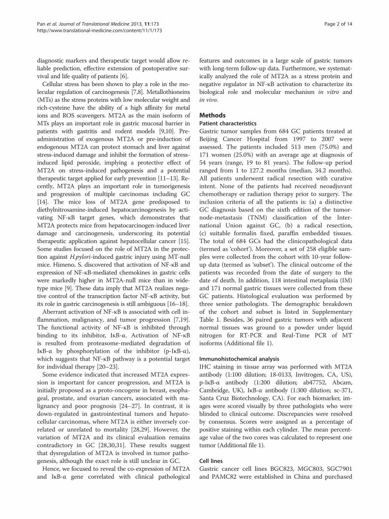
Table 1 Logistic regression analysis of clinicopathologicalfeatures and prognosis in GC
Variates No. of cases(n = 258)
Survival time(month)
P-value
Sex
Male 186 66.4 ± 4.6
Female 72 54.1 ± 4.8 0.604
Age at diagnosis
<60 173 65.0 ± 4.9
≥60 85 59.2 ± 5.6 0.867
TNM stage
I 34 105.5 ± 7.7 I vs. IV < 0.001
II 57 74.0 ± 5.9 II vs. IV < 0.001
III 115 49.1 ± 3.4 III vs. IV 0.010
IV 52 35.8 ± 4.1
Tumor depth
pT1 13 87.2 ± 9.9 pT1 vs. pT4 0.005
pT2 50 89.1 ± 9.1 pT2 vs. pT4 < 0.001
pT3 157 49.1 ± 2.9 pT3 vs. pT4 0.159
pT4 38 40.1 ± 5.3
Lymph node status
pN0 78 89.8 ± 6.2 pN0 vs. pN3 0.001
pN1 115 50.5 ± 3.4 pN1 vs. pN3 0.212
pN2 49 46.6 ± 6.0 pN2 vs. pN3 0.326
pN3 16 47.5 ± 12.6
Distant metastasis
pM0 232 67.5 ± 4.3
pM1 26 27.6 ± 5.8 < 0.001
Degree of differentiation
D1 54 76.0 ± 6.0
D0 204 59.8 ± 4.4 0.011
MT2A expression < 0.001
low 202 53.9 ± 3.4
high 56 86.9 ± 9.2
IκB-α expression
low 164 61.6 ± 4.9
high 94 64.9 ± 5.1 0.248
p-IκB-α expression
low 126 74.1 ± 5.3
high 132 47.7 ± 3.4 0.005
M0: no distant metastasis; M1: distant metastasis;D1: well/moderately-differentiated GCs; D0: poorly-differentiated GCs.
Pan et al. Journal of Translational Medicine 2013, 11:173 Page 3 of 14http://www.translational-medicine.com/content/11/1/173
from the tissue bank of Shanghai (Shanghai, China). Es-pecially, BGC823 cells exhibited high tumorigenecity.MKN45, AGS, N87, RF-1, RF-48, SNU-1, SNU-5and SNU-16 cell lines were purchased from ATCC(American Type Culture Collection, Manassas, US).GES-1, an immortalized human gastric epithelial cellline, was generated by SV40 viral transfection at BeijingCancer Hospital and cultured in DMEM mediumsupplemented with 10% fetal bovine serum (Gibco, Life
technologies, Grand Island, NY, USA) at 37°C in ahumidified atmosphere containing 5% CO2 [32].
MTT and soft agar assayThe cells were seeded into 96-well culture plates, andMTT was added to the cells at 1-5 days. MTT (5 μg/ml)was removed after 4 h incubation, and then dimethylsulfoxide (DMSO) was added to solubilize the formazanproduct. The absorbency at 490 nm/570 nm was assayedby a microplate reader (Bio-rad680 ELISA). Cells werethen incubated for 4 weeks, stained with vital tetrazo-lium dye INT (piodonitrotetrazolium, Invitrogen) todocument the presence or absence of viable cell col-onies. The soft agar was fixed with 100 μl methanol-acetic acid (3:1 vol/vol) and colonies were counted.
Tumorigenicity assayBGC823 cells (5 × 10⁵ cells suspended in 0.1 ml PBS)transfected with MT2A over-expressed vector or emptyvector were injected subcutaneously into the dorsal flankof five 4-week-old female Balb/C nude mice (MT2A-ex-pression clones on the right and vector control cloneson the left. Tumor diameter was measured and docu-mented every 5 days. Tumor volume was calculatedaccording to the formula ab2/2 (a > b). At the end of25 days, all mice were sacrificed and the tumor volumewas measured. Three independent experiments wereperformed and gave the similar results. The animalswere maintained in facilities approved by the associationfor assessment and accreditation of Laboratory AnimalCare in accordance with the current regulations andinternational standards.
Statistical analysisχ2 test statistics and Student’s t-test were used to com-pare pretreatment characteristics of patient cases. Thecancer-related survival was analyzed using the Kaplan–Meier method and compared using log-rank tests. TheSpearman rank test and Fisher’s exact test were appliedto demonstrate clinicopathological correlations. A Coxproportional hazard regression model was used with as-sociated 95% confidence intervals (CIs) and P values. Allstatistical tests were two-sided, and P values of less than0.05 were considered statistically significant. The statis-tical analysis was performed using the statistical packageSPSS (Version 16.0; SPSS Inc, Chicago, IL).
Study approvalAll animal studies were approved by the Ethics Commit-tee of Peking University Cancer Hospital. The use ofhuman tissues and clinical data was according to theguidelines of the hospital and approved by the LocalEthical Committee.
Pan et al. Journal of Translational Medicine 2013, 11:173 Page 4 of 14http://www.translational-medicine.com/content/11/1/173
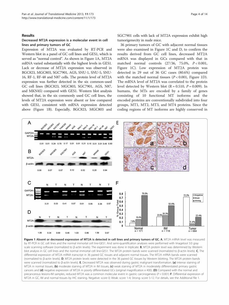
ResultsDecreased MT2A expression is a molecular event in celllines and primary tumors of GCExpression of MT2A was evaluated by RT-PCR andWestern blot in a panel of GC cell lines and GES1, which isserved as “normal control”. As shown in Figure 1A, MT2AmRNA varied substantially with the highest levels in GES1.Lack or decrease of MT2A expression was observed inBGC823, MGC803, SGC7901, AGS, SNU-1, SNU-5, SNU-16, RF-1, RF-48 and N87 cells. The protein level of MT2Aexpression was further detected in the six common-usedGC cell lines (BGC823, MGC803, SGC7901, AGS, N87,and MKN45) compared with GES1. Western blot analysisshowed that, in the six commonly used GC cell lines, thelevels of MT2A expression were absent or low comparedwith GES1, consistent with mRNA expression detectedabove (Figure 1B). Especially, BGC823, MGC803 and
Figure 1 Absent or decreased expression of MT2A is detected in cellby RT-PCR in GC cell lines and the normal immortal cell line-GES1. And semscale scanning software (normalized to β-actin levels). The experiment wasblot analysis in GC cell lines and the normal immortal cell line-GES1. The Mdifferential expression of MT2A mRNA transcript in 36 paired GC tissues an(normalized to β-actin levels). D, MT2A protein levels were detected in thewere scanned (normalized to β-actin levels). E, Decreased MT2A was obserMT2A in normal tissues, (b) moderate staining of MT2A in IM tissues, (c) wcancers and (d) negative expression of MT2A in poorly differentiated GCs (precancerous lesions-IM samples, reduced MT2A was a common molecularMT2A in GC, IM and normal tissues by IHC staining. Negative: score 0; Wea
SGC7901 cells with lack of MT2A expression exhibit hightumorigenecity in nude mice.36 primary tumors of GC with adjacent normal tissues
were also examined in Figure 1C and D, to confirm theresults derived from GC cell lines, decreased MT2AmRNA was displayed in GCs compared with that inmatched normal controls (27/36, 75.0%, P < 0.001,Figure 1C). Low expression of MT2A protein wasdetected in 29 out of 36 GC cases (80.6%) comparedwith the matched normal tissues (P < 0.001, Figure 1D).The mRNA level of MT2A was correlated to the proteinlevel detected by Western blot (R = 0.510, P = 0.009). Inhumans, the MTs are encoded by a family of genesconsisting of 10 functional MT isoforms and theencoded proteins are conventionally subdivided into fourgroups, MT1, MT2, MT3, and MT4 proteins. Since thecoding regions of MT isoforms are highly conserved in
lines and primary tumors of GC. A, MT2A mRNA level was measuredi-quantification analyses were performed with Imagetool 3.0 graydone in triplicate. B, MT2A protein level was determined by WesternT2A protein bands were scanned (normalized to β-actin levels). C, Thed adjacent normal tissues. The MT2A mRNA bands were scanned36 paired GC tissues by Western blotting. The MT2A protein bandsved during gastric malignant transformation. (a) intense staining ofeak staining of MT2A in moderately differentiated primary gastricoriginal magnification × 400). (D) Compared with the normal andevent in gastric carcinogenesis (P < 0.001) F: Differential expression ofk: score 1-4; Strong: score 5-12. For details, see the Additional file 1.
Pan et al. Journal of Translational Medicine 2013, 11:173 Page 5 of 14http://www.translational-medicine.com/content/11/1/173
transcript and protein levels, MT2A is highly homolo-gous with MT1G, MT1E and MT1F (Additional file 1:Figure S1B). Hence, experiments on any individual iso-form should be carefully conducted to ensure that theexact isoform is analyzed. In this study, differential ex-pression of MT1 transcripts was observed in GC celllines and primary tumors of GC with adjacent normalcontrols (n = 36) (Additional file 1: Figures S1A and S2).We authenticated actual MT2A gene down-regulation inGC significantly compared with adjacent normal tissues(27/36, 75%, Figure 1C). There was no difference ofMT1E and MT1F and MT1G mRNA transcripts innormal and tumor samples as well as MT1B mRNAtranscript was not detectable in all cells and tissues(Additional file 1: Figures S1A and S2).To investigate the candidacy of MT2A in gastric
tumorigenesis, we initially characterized the status ofMT2A expression in 171 normal tissues, 118 IM and684 GC samples by IHC staining. The antibody forMT2A (E9 18-0133, Invitrogen) is not specific for MT-2Aonly, and cross-reacts with MT-1 to some extent, even if itis cloned by the full length of MT2A. Since there is nospecific antibody to detect MT2A, so we use the commoncommercial antibody for IHC staining which was pub-lished in most related studies, MT2A expression is classi-fied as low and high expression in normal appearancetissues, IM, and primary GCs. High level of MT2A expres-sion was detected in 153 of 684 (22.4%) GC cases, and60 of 118 (50.8%) IM cases, as well as 130 of 171 (76.0%)normal appearance cases, respectively (Additional file 1:Table S3, P < 0.001), it was consisted with MT2A mRNAtranscripts and protein expression by Western blot. Theexpression of MT2A was classified as negative, weak andstrong cases in 13 (7.6%), 28 (16.4%) and 130 (76.0%) outof 171 normal tissues, respectively; In IM tissues, 27(22.9%), 31 (26.3%) and 60 (50.8%) out of 118 IM caseswere detected respectively; In GC samples, 494 (72.2%),37 (5.4%) and 153 (22.4%) out of 684 GC cases weredetected respectively (Figure 1F, Additional file 1). Therewas a gradually decreased expression of MT2A protein inIM and GC (P < 0.001, Figure 1E and F). It is clear thatMT2A mRNA was highly expressed in GES1 cells andnormal tissues, but was reduced or lost in most GC cellsand tissues. These results suggest that decreased MT2A isa molecular event in tumorigenesis and progression.
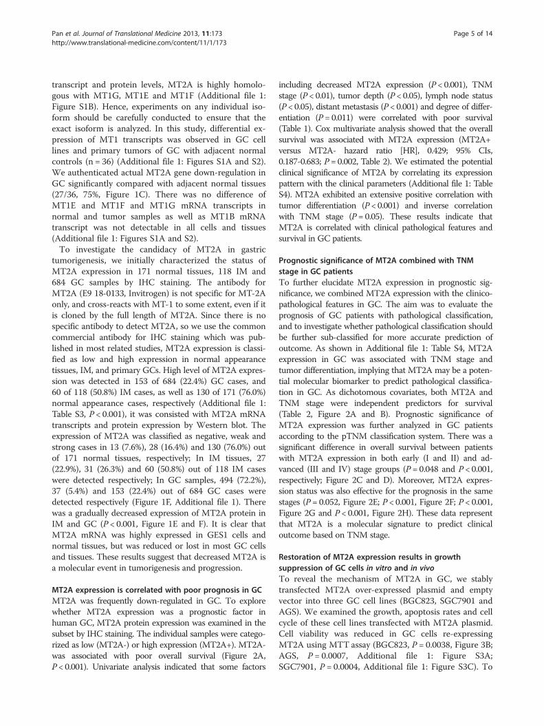
MT2A expression is correlated with poor prognosis in GCMT2A was frequently down-regulated in GC. To explorewhether MT2A expression was a prognostic factor inhuman GC, MT2A protein expression was examined in thesubset by IHC staining. The individual samples were catego-rized as low (MT2A-) or high expression (MT2A+). MT2A-was associated with poor overall survival (Figure 2A,P < 0.001). Univariate analysis indicated that some factors
including decreased MT2A expression (P < 0.001), TNMstage (P < 0.01), tumor depth (P < 0.05), lymph node status(P < 0.05), distant metastasis (P < 0.001) and degree of differ-entiation (P = 0.011) were correlated with poor survival(Table 1). Cox multivariate analysis showed that the overallsurvival was associated with MT2A expression (MT2A+versus MT2A- hazard ratio [HR], 0.429; 95% CIs,0.187-0.683; P = 0.002, Table 2). We estimated the potentialclinical significance of MT2A by correlating its expressionpattern with the clinical parameters (Additional file 1: TableS4). MT2A exhibited an extensive positive correlation withtumor differentiation (P < 0.001) and inverse correlationwith TNM stage (P= 0.05). These results indicate thatMT2A is correlated with clinical pathological features andsurvival in GC patients.
Prognostic significance of MT2A combined with TNMstage in GC patientsTo further elucidate MT2A expression in prognostic sig-nificance, we combined MT2A expression with the clinico-pathological features in GC. The aim was to evaluate theprognosis of GC patients with pathological classification,and to investigate whether pathological classification shouldbe further sub-classified for more accurate prediction ofoutcome. As shown in Additional file 1: Table S4, MT2Aexpression in GC was associated with TNM stage andtumor differentiation, implying that MT2A may be a poten-tial molecular biomarker to predict pathological classifica-tion in GC. As dichotomous covariates, both MT2A andTNM stage were independent predictors for survival(Table 2, Figure 2A and B). Prognostic significance ofMT2A expression was further analyzed in GC patientsaccording to the pTNM classification system. There was asignificant difference in overall survival between patientswith MT2A expression in both early (I and II) and ad-vanced (III and IV) stage groups (P = 0.048 and P < 0.001,respectively; Figure 2C and D). Moreover, MT2A expres-sion status was also effective for the prognosis in the samestages (P = 0.052, Figure 2E; P < 0.001, Figure 2F; P < 0.001,Figure 2G and P < 0.001, Figure 2H). These data representthat MT2A is a molecular signature to predict clinicaloutcome based on TNM stage.
Restoration of MT2A expression results in growthsuppression of GC cells in vitro and in vivoTo reveal the mechanism of MT2A in GC, we stablytransfected MT2A over-expressed plasmid and emptyvector into three GC cell lines (BGC823, SGC7901 andAGS). We examined the growth, apoptosis rates and cellcycle of these cell lines transfected with MT2A plasmid.Cell viability was reduced in GC cells re-expressingMT2A using MTT assay (BGC823, P = 0.0038, Figure 3B;AGS, P = 0.0007, Additional file 1: Figure S3A;SGC7901, P = 0.0004, Additional file 1: Figure S3C). To
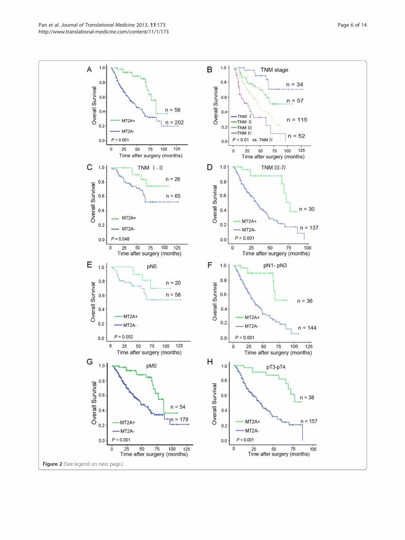
Figure 2 (See legend on next page.)
Pan et al. Journal of Translational Medicine 2013, 11:173 Page 6 of 14http://www.translational-medicine.com/content/11/1/173
(See figure on previous page.)Figure 2 Analysis of combined MT2A with TNM stage to predict clinical outcome in GC. Kaplan–Meier analysis of overall-survival for MT2Aexpression was used according to TNM stage. A, Prognostic value of MT2A expression in GC patients was benefit for predicting the outcome ofpatients, low expression of MT2A was associated with poor overall survival (P < 0.01); B, TNM stage was benefit for the prognostic prediction inGC patients. C D, Prognostic value of MT2A in TNM stage I-II and stage III-IV (P = 0.048, P < 0.001, respectively); E F, Prognostic value of MT2Aexpression in lymph node metastasis or not (P < 0.001, P = 0.052, respectively); G. Prognostic value of MT2A expression in no distant metastasissubgroup (n = 232, P < 0.001); H. Prognostic value of MT2A in pT3-pT4 subgroup (P < 0.001). All data imply that MT2A could be an effectiveprognostic signature.
Pan et al. Journal of Translational Medicine 2013, 11:173 Page 7 of 14http://www.translational-medicine.com/content/11/1/173
further investigate the function of MT2A in GC celllines, we analyzed the effects of MT2A expression oncell proliferation and cell cycle regulation by Annexin-V/PI staining and flow cytometry analysis. More apop-tosis was detected in these GC cells re-expressingMT2A (P < 0.01, respectively, Figure 3F; Additional file 1:Figure S3A and C). Furthermore, G2/M arrest was ob-served in GC cells using ectopic expression of MT2A. Theratios of G2/M phase in MT2A groups were twice higherthan those in the vector groups did (P < 0.01, respectively,Figure 3E and Additional file 1: Figure S3B and D).Further, we plated BGC823 cells repressing MT2A or not
into soft agar, ectopic expression of MT2A was confirmedby western blot analysis first (Figure 3A). After 3 weeks ofculture MT2A expression caused a statistically significantreduction in colony formation (P = 0.0053, Figure 3C).Furthermore, there was a dramatic growth inhibition ofMT2A-re-expressing cells compared with the negative con-trol (P = 0.0027, Figure 3D) in the xenograft models. Thesedata demonstrate that MT2A might play a role in suppress-ing proliferation of GC cells in vitro and in vivo.
MT2A represses NF-κB activity through IκB-α up-regulationIt was reported that the loss of MT2A induced NF-κBsignal activation in transgenetic mice model (MT2A-knock out) [15]. But the role of MT2A as a friend or foein NF-κB signal pathway was still controversial [18,33].In order to interpret the mechanism of MT2A mediated
Table 2 Cox regression analysis of clinicopathologicalfeatures and molecular signatures in GC
Multivariate analysis
Covariate by end point Hazard ratio 95% CI P-value
Age≥ 60 years 1.300 0.840 to 1.934 0.254
Sex 0.823 0.499 to 1.233 0.292
Differentiation 0.753 0.509 to 1.778 0.875
Tumor invasive depth 1.914 1.389 to 2.638 < 0.001
Lymph node status 1.291 1.034 to 1.612 0.024
Distant metastasis 2.595 1.332 to 5.056 0.005
MT2A 0.429 0.187 to 0.683 0.002
IκB-α 0.779 0.524 to 1.311 0.423
p-IκB-α 1.857 1.256 to 2.912 0.003
GC cell growth suppression in vivo and in vitro, we triedto explore the potential relationship between MT2A andNF-κB signaling pathway, and the inner mechanism wasstill unclear in GC. MT2A and the main genes in NF-κBsignaling, such as p65, IκB-α, p-IκB-α and the downstreameffector of NF-κB signaling, cyclin D1 were detected inthis study (Additional file 1). Ectopic expression of MT2Ainduced both mRNA and protein expression of IκB-α inall investigated GC cell lines (BGC823, SGC7901 andAGS), IκB-α mRNA steady-state levels were increased 5.5-fold in BGC823 cells re-expressing MT2A for 48 h. InAGS and SGC7901 cells, we observed a 7.2-fold and 4.3-fold induction of IκB-α mRNA (Figure 4A), the relativeprotein levels were consistent with mRNA transcripts,and the p-IκB-α protein was decreased in those GCcells transfected MT2A vector (Figure 4B). As shown inFigures 4C and D, knockdown of endogenous MT2A inGES1 cells led to down-regulation of IκB-α expression aswell as up-regulation of p-IκB-α and cyclin D1 expression,suggesting a connection between MT2A activity andIκB-α expression. These experiments indicate that MT2Ainduces IκB-α expression in mRNA and protein levels.And MT2A-mediated up-regulation of IκB-α expressionwas further confirmed by immunofluorescence (Figure 5A,Additional file 1). To clarify the mechanism of MT2A onIκB-α expression, we performed luciferase reporter assaysby restructuring of IκB-α promoter region, 5.6-fold up-regulation of IκB-α promoter luciferase activity was foundin MT2A group than that in vector group (negative con-trol) (P = 0.0035, Figure 5B). To investigate the relation-ship between MT2A and IκB-α-mediated NF-κBinactivation, we further studied the effects of MT2A inNF-κB nuclear translocation. As shown in Figure 5C, theDNA binding activity of NF-κB in nuclei was reduced byover-expressed MT2A. The protein levels and transloca-tion of NF-κB main subunit p65 were detected in nuclearand cytoplasmic extracts derived from BGC823 cellstransfected with MT2A or empty vector, and nuclear p65was decreased in MT2A re-expressed group (Figure 5D,Additional file 1). Same results were observed in sectionsfrom BGC823 xenografts (Additional file 1: Figure S5A).Based on the similarity of MT isoform, the specific
primer pairs for different MT isoforms were designed toillustrate whether ectopic expression of MT2A or knock-down of MT2A could affect on other MT isoforms, as
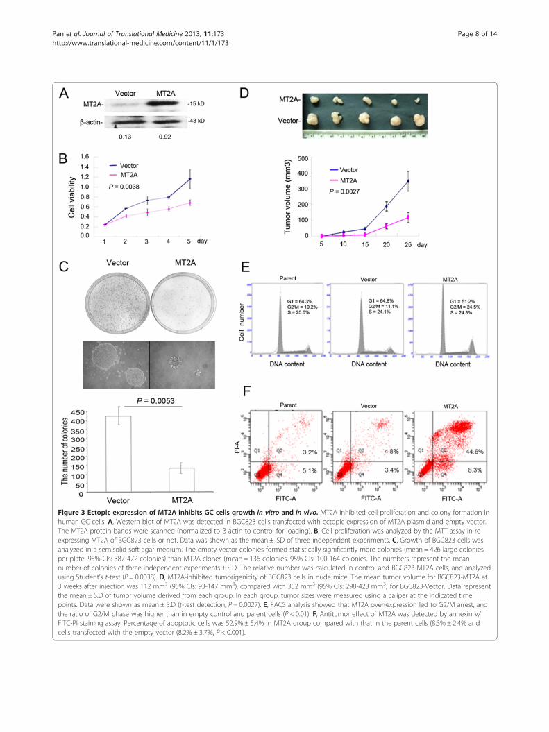
Figure 3 Ectopic expression of MT2A inhibits GC cells growth in vitro and in vivo. MT2A inhibited cell proliferation and colony formation inhuman GC cells. A, Western blot of MT2A was detected in BGC823 cells transfected with ectopic expression of MT2A plasmid and empty vector.The MT2A protein bands were scanned (normalized to β-actin to control for loading). B, Cell proliferation was analyzed by the MTT assay in re-expressing MT2A of BGC823 cells or not. Data was shown as the mean ± .SD of three independent experiments. C, Growth of BGC823 cells wasanalyzed in a semisolid soft agar medium. The empty vector colonies formed statistically significantly more colonies (mean = 426 large coloniesper plate. 95% CIs: 387-472 colonies) than MT2A clones (mean = 136 colonies. 95% CIs: 100-164 colonies. The numbers represent the meannumber of colonies of three independent experiments ± S.D. The relative number was calculated in control and BGC823-MT2A cells, and analyzedusing Student’s t-test (P = 0.0038). D, MT2A-inhibited tumorigenicity of BGC823 cells in nude mice. The mean tumor volume for BGC823-MT2A at3 weeks after injection was 112 mm3 (95% CIs: 93-147 mm3), compared with 352 mm3 (95% CIs: 298-423 mm3) for BGC823-Vector. Data representthe mean ± S.D of tumor volume derived from each group. In each group, tumor sizes were measured using a caliper at the indicated timepoints. Data were shown as mean ± S.D (t-test detection, P = 0.0027). E, FACS analysis showed that MT2A over-expression led to G2/M arrest, andthe ratio of G2/M phase was higher than in empty control and parent cells (P < 0.01). F, Antitumor effect of MT2A was detected by annexin V/FITC-PI staining assay. Percentage of apoptotic cells was 52.9% ± 5.4% in MT2A group compared with that in the parent cells (8.3% ± 2.4% andcells transfected with the empty vector (8.2% ± 3.7%, P < 0.001).
Pan et al. Journal of Translational Medicine 2013, 11:173 Page 8 of 14http://www.translational-medicine.com/content/11/1/173
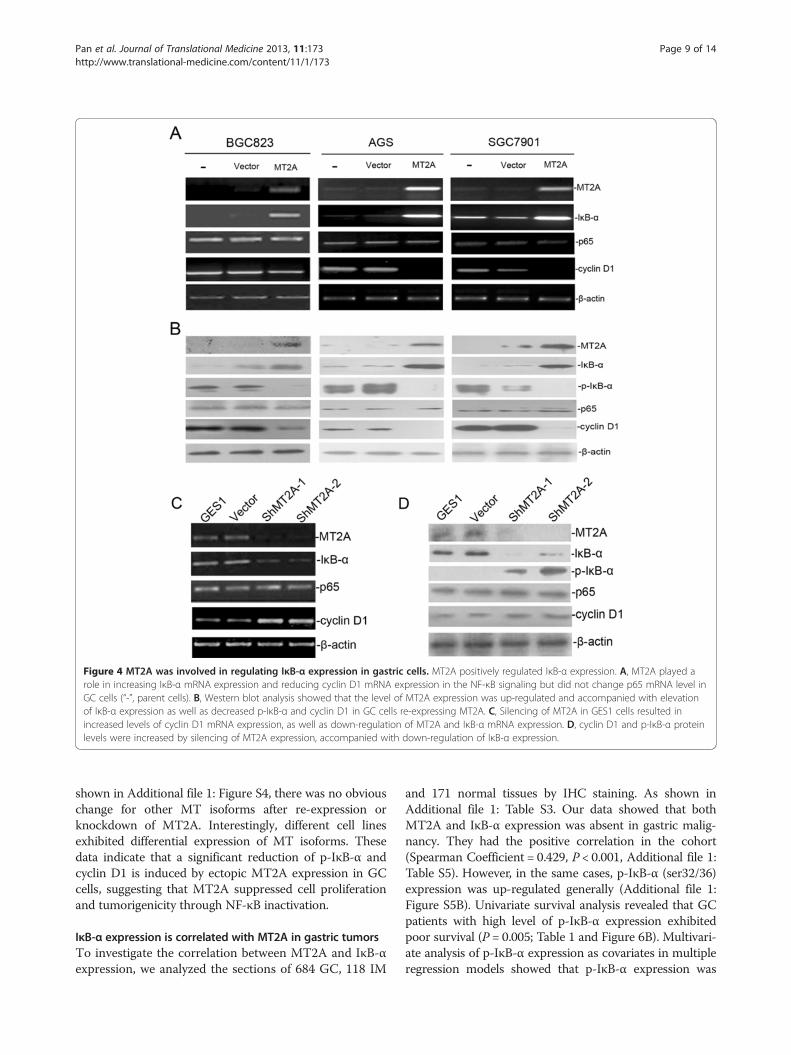
Figure 4 MT2A was involved in regulating IκB-α expression in gastric cells. MT2A positively regulated IκB-α expression. A, MT2A played arole in increasing IκB-α mRNA expression and reducing cyclin D1 mRNA expression in the NF-κB signaling but did not change p65 mRNA level inGC cells (“-”, parent cells). B, Western blot analysis showed that the level of MT2A expression was up-regulated and accompanied with elevationof IκB-α expression as well as decreased p-IκB-α and cyclin D1 in GC cells re-expressing MT2A. C, Silencing of MT2A in GES1 cells resulted inincreased levels of cyclin D1 mRNA expression, as well as down-regulation of MT2A and IκB-α mRNA expression. D, cyclin D1 and p-IκB-α proteinlevels were increased by silencing of MT2A expression, accompanied with down-regulation of IκB-α expression.
Pan et al. Journal of Translational Medicine 2013, 11:173 Page 9 of 14http://www.translational-medicine.com/content/11/1/173
shown in Additional file 1: Figure S4, there was no obviouschange for other MT isoforms after re-expression orknockdown of MT2A. Interestingly, different cell linesexhibited differential expression of MT isoforms. Thesedata indicate that a significant reduction of p-IκB-α andcyclin D1 is induced by ectopic MT2A expression in GCcells, suggesting that MT2A suppressed cell proliferationand tumorigenicity through NF-κB inactivation.
IκB-α expression is correlated with MT2A in gastric tumorsTo investigate the correlation between MT2A and IκB-αexpression, we analyzed the sections of 684 GC, 118 IM
and 171 normal tissues by IHC staining. As shown inAdditional file 1: Table S3. Our data showed that bothMT2A and IκB-α expression was absent in gastric malig-nancy. They had the positive correlation in the cohort(Spearman Coefficient = 0.429, P < 0.001, Additional file 1:Table S5). However, in the same cases, p-IκB-α (ser32/36)expression was up-regulated generally (Additional file 1:Figure S5B). Univariate survival analysis revealed that GCpatients with high level of p-IκB-α expression exhibitedpoor survival (P = 0.005; Table 1 and Figure 6B). Multivari-ate analysis of p-IκB-α expression as covariates in multipleregression models showed that p-IκB-α expression was
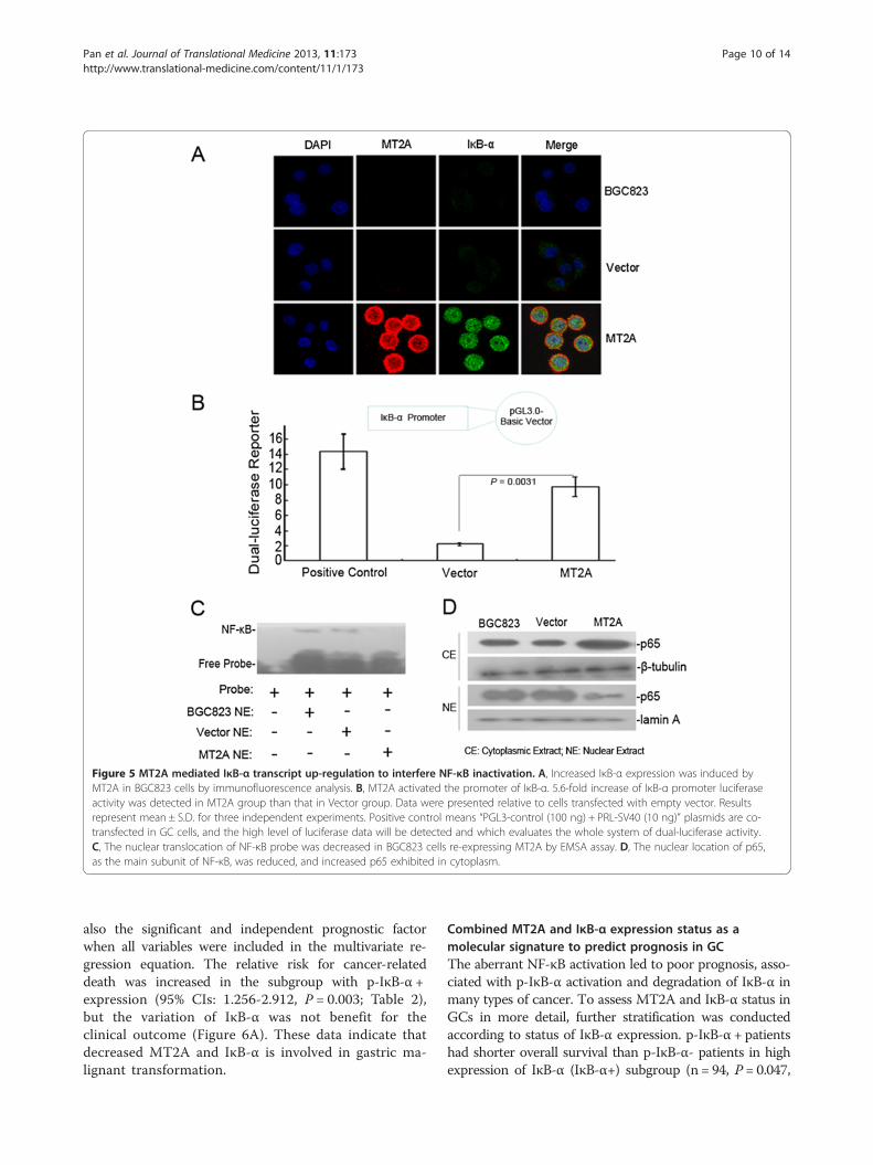
Figure 5 MT2A mediated IκB-α transcript up-regulation to interfere NF-κB inactivation. A, Increased IκB-α expression was induced byMT2A in BGC823 cells by immunofluorescence analysis. B, MT2A activated the promoter of IκB-α. 5.6-fold increase of IκB-α promoter luciferaseactivity was detected in MT2A group than that in Vector group. Data were presented relative to cells transfected with empty vector. Resultsrepresent mean ± S.D. for three independent experiments. Positive control means “PGL3-control (100 ng) + PRL-SV40 (10 ng)” plasmids are co-transfected in GC cells, and the high level of luciferase data will be detected and which evaluates the whole system of dual-luciferase activity.C, The nuclear translocation of NF-κB probe was decreased in BGC823 cells re-expressing MT2A by EMSA assay. D, The nuclear location of p65,as the main subunit of NF-κB, was reduced, and increased p65 exhibited in cytoplasm.
Pan et al. Journal of Translational Medicine 2013, 11:173 Page 10 of 14http://www.translational-medicine.com/content/11/1/173
also the significant and independent prognostic factorwhen all variables were included in the multivariate re-gression equation. The relative risk for cancer-relateddeath was increased in the subgroup with p-IκB-α +expression (95% CIs: 1.256-2.912, P = 0.003; Table 2),but the variation of IκB-α was not benefit for theclinical outcome (Figure 6A). These data indicate thatdecreased MT2A and IκB-α is involved in gastric ma-lignant transformation.
Combined MT2A and IκB-α expression status as amolecular signature to predict prognosis in GCThe aberrant NF-κB activation led to poor prognosis, asso-ciated with p-IκB-α activation and degradation of IκB-α inmany types of cancer. To assess MT2A and IκB-α status inGCs in more detail, further stratification was conductedaccording to status of IκB-α expression. p-IκB-α + patientshad shorter overall survival than p-IκB-α- patients in highexpression of IκB-α (IκB-α+) subgroup (n = 94, P = 0.047,
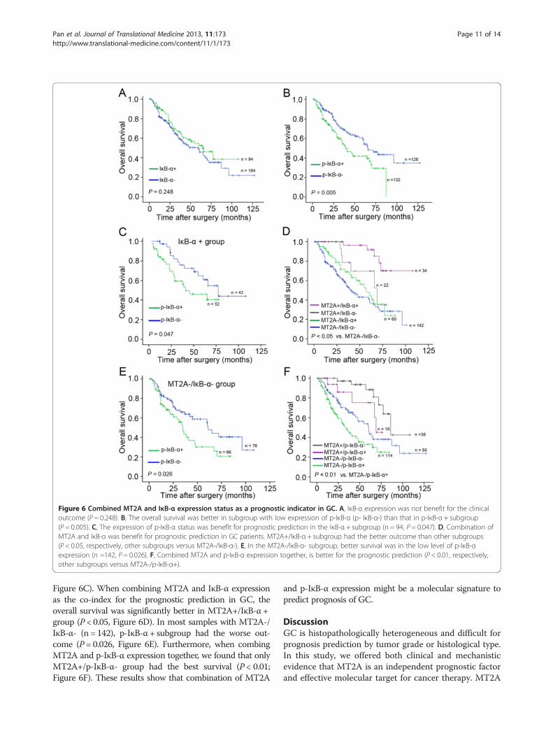
Figure 6 Combined MT2A and IκB-α expression status as a prognostic indicator in GC. A, IκB-α expression was not benefit for the clinicaloutcome (P = 0.248). B, The overall survival was better in subgroup with low expression of p-IκB-α (p- IκB-α-) than that in p-IκB-α + subgroup(P = 0.005). C, The expression of p-IκB-α status was benefit for prognostic prediction in the IκB-α + subgroup (n = 94, P = 0.047). D, Combination ofMT2A and IκB-α was benefit for prognostic prediction in GC patients. MT2A+/IκB-α + subgroup had the better outcome than other subgroups(P < 0.05, respectively, other subgroups versus MT2A-/IκB-α-). E, In the MT2A-/IκB-α- subgroup, better survival was in the low level of p-IκB-αexpression (n =142, P = 0.026). F, Combined MT2A and p-IκB-α expression together, is better for the prognostic prediction (P < 0.01, respectively,other subgroups versus MT2A-/p-IκB-α+).
Pan et al. Journal of Translational Medicine 2013, 11:173 Page 11 of 14http://www.translational-medicine.com/content/11/1/173
Figure 6C). When combining MT2A and IκB-α expressionas the co-index for the prognostic prediction in GC, theoverall survival was significantly better in MT2A+/IκB-α +group (P < 0.05, Figure 6D). In most samples with MT2A-/IκB-α- (n = 142), p-IκB-α + subgroup had the worse out-come (P = 0.026, Figure 6E). Furthermore, when combingMT2A and p-IκB-α expression together, we found that onlyMT2A+/p-IκB-α- group had the best survival (P < 0.01;Figure 6F). These results show that combination of MT2A
and p-IκB-α expression might be a molecular signature topredict prognosis of GC.
DiscussionGC is histopathologically heterogeneous and difficult forprognosis prediction by tumor grade or histological type.In this study, we offered both clinical and mechanisticevidence that MT2A is an independent prognostic factorand effective molecular target for cancer therapy. MT2A
Pan et al. Journal of Translational Medicine 2013, 11:173 Page 12 of 14http://www.translational-medicine.com/content/11/1/173
has been shown to reduce the tumorigenicity in vivo andin vitro, and decrease or loss of MT2A is a critical molecu-lar event in GC cell lines and primary GC tissues. De-creased MT2A was associated with gastric malignanttransformation, as well as poor survival. Re-expressingMT2A significantly inhibited the growth of GC cells.Interestingly, restoration of MT2A led to down-regulationof p-IκB-α and cyclin D1 but to induce IκB-α up-regulation, which was consistent with the typical apoptosisof GC cells resulting from suppression of NF-κB activa-tion, accompanied with G2/M arrest [34-36]. Moreover,cyclin D1 is a therapeutic target in cancer. Its abundancewill lead to oncogenic activation in stomach [37,38].Importantly, the luciferase activity of IκB-α promoter
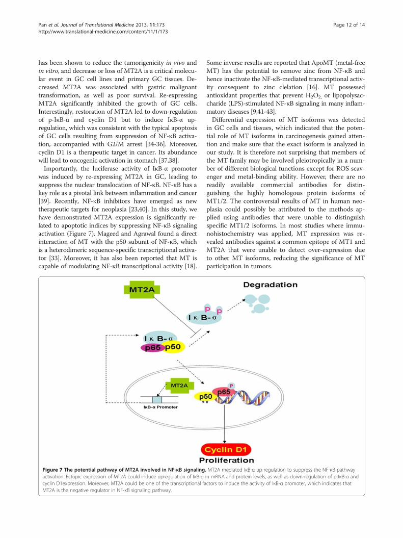
was induced by re-expressing MT2A in GC, leading tosuppress the nuclear translocation of NF-κB. NF-κB has akey role as a pivotal link between inflammation and cancer[39]. Recently, NF-κB inhibitors have emerged as newtherapeutic targets for neoplasia [23,40]. In this study, wehave demonstrated MT2A expression is significantly re-lated to apoptotic indices by suppressing NF-κB signalingactivation (Figure 7). Mageed and Agrawal found a directinteraction of MT with the p50 subunit of NF-κB, whichis a heterodimeric sequence-specific transcriptional activa-tor [33]. Moreover, it has also been reported that MT iscapable of modulating NF-κB transcriptional activity [18].
Figure 7 The potential pathway of MT2A involved in NF-κB signalingactivation. Ectopic expression of MT2A could induce upregulation of IκB-αcyclin D1expression. Moreover, MT2A could be one of the transcriptional faMT2A is the negative regulator in NF-κB signaling pathway.
Some inverse results are reported that ApoMT (metal-freeMT) has the potential to remove zinc from NF-κB andhence inactivate the NF-κB-mediated transcriptional activ-ity consequent to zinc clelation [16]. MT possessedantioxidant properties that prevent H2O2, or lipopolysac-charide (LPS)-stimulated NF-κB signaling in many inflam-matory diseases [9,41-43].Differential expression of MT isoforms was detected
in GC cells and tissues, which indicated that the poten-tial role of MT isoforms in carcinogenesis gained atten-tion and make sure that the exact isoform is analyzed inour study. It is therefore not surprising that members ofthe MT family may be involved pleiotropically in a num-ber of different biological functions except for ROS scav-enger and metal-binding ability. However, there are noreadily available commercial antibodies for distin-guishing the highly homologous protein isoforms ofMT1/2. The controversial results of MT in human neo-plasia could possibly be attributed to the methods ap-plied using antibodies that were unable to distinguishspecific MT1/2 isoforms. In most studies where immu-nohistochemistry was applied, MT expression was re-vealed antibodies against a common epitope of MT1 andMT2A that were unable to detect over-expression dueto other MT isoforms, reducing the significance of MTparticipation in tumors.
. MT2A mediated IκB-α up-regulation to suppress the NF-κB pathwayin mRNA and protein levels, as well as down-regulation of p-IκB-α andctors to induce the activity of IκB-α promoter, which indicates that
Pan et al. Journal of Translational Medicine 2013, 11:173 Page 13 of 14http://www.translational-medicine.com/content/11/1/173
In addition, MT2A suppression is frequently observedin GC, and similar data was reported in hepatocellularand colon cancer [44,45]. Duncan et al reported thatdown-regulation of MT2A expression occurred uponimmortalization, which implies that MT2A is down-regulated when human cells become immortal pheno-types, a key event in tumorigenesis [46].Collectively, down-regulation of MT2A expression is
an independent predictor for clinical outcome. It is con-ceivable that re-expression of MT2A can be consideredas a molecular target in GC for molecular classificationand individual therapy.
Ethics approvalThis study was conducted with the approval of PekingUniversity Cancer Hospital & Institute Review Board.
Additional file
Additional file 1: Table S1. Analysis of clinicopatholigical features andMT2A expression in GCs. Table S2. Oligonucleotide primers for RT-PCR.Table S3. Comparison of MT2A and IκB-α protein expression in tumors,intestinal metaplasia and normal tissues. Table S4. Relationship betweenMT2A and clinical features in GC. Table S5. Analysis of coexpressed MT2Aand IκB-α in GC samples (n = 684). Table S6. Primers for MT isoforms.Figure S1. The similarity of MT1/2 isoforms and differential expression in GCcell lines. Figure S2. Differential expression of MT isoforms in paired GCtissues by RT-PCR and real-time PCR analysis. Figure S3. Ectopic expressionof MT2A induced apoptosis and G2/M arrest in SGC7901 and AGS.Figure S4. The variation of other MT isoforms was detected in GC cellswith re-expressed MT2A or knockdown of MT2A. Figure S5. MT2A andNF-κB signaling were detected in BGC823 xenografts with re-expression ofMT2A and human gastric tissues by IHC staining.
Competing interestsThe authors declare that they have no competing interests.
Authors’ contributionsYP: study concept and design; acquisition of data; analysis and interpretationof data; drafting of the manuscript; final approval of the version to bepublished. JH: study concept and design; analysis and interpretation of data;critical revision of the manuscript for important intellectual content; studysupervision; final approval of the version to be published. RX, XY, JC, WL andJY: study concept and design; technical or material support; analysis andinterpretation of data; critical revision of the manuscript for importantintellectual content; study supervision; final approval of the version to bepublished. YL: study concept and design; analysis and interpretation of data;critical revision of the manuscript for important intellectual content; obtainedfunding; study supervision; final approval of the version to be published. Allauthors read and approved the final manuscript.
AcknowledgementsYuanming Pan and Jiaqiang Huang contributed equally to this work, and wealso thank the tissue bank of Peking University Cancer Hospital & Institute forproviding gastric specimens.
FundingThis work was supported by the National Bio-Tech 863 Program (No.2012AA02A504), The National Key Basic Research Program (973 Program,No. 2004CB518708), and the Beijing Municipal Science & TechnologyCommission (No. D0905001040631).
Author details1Laboratory of Molecular Oncology, Key laboratory of Carcinogenesis andTranslational Research, Ministry of Education, Peking University CancerHospital & Institute, No.52 Fucheng Road, Beijing, Haidian District 100142,People's Republic of China. 2College of Life Sciences & Bioengineering, Schoolof Science, Beijing Jiaotong University, No.3 ShangyuanCun, Beijing, HaidianDistrict 100044, People's Republic of China. 3Institute of Digestive Disease andDepartment of Medicine and Therapeutics, Li Ka Shing Institute of HealthSciences, The Chinese University of Hong Kong, Hong Kong, SAR, China.
Received: 17 March 2013 Accepted: 10 July 2013Published: 19 July 2013
References1. Yang L: Incidence and mortality of gastric cancer in China. World J
Gastroenterol 2006, 12:17–20.2. McCracken M, Olsen M, Chen MS Jr, Jemal A, Thun M, Cokkinides V, Deapen
D, Ward E: Cancer incidence, mortality, and associated risk factors amongAsian Americans of Chinese, Filipino, Vietnamese, Korean, and Japaneseethnicities. CA Canc J Clin 2007, 57:190–205.
3. Mohri Y, Tanaka K, Ohi M, Yokoe T, Miki C, Kusunoki M: Prognosticsignificance of host- and tumor-related factors in patients with gastriccancer. World J Surg 2010, 34:285–290.
4. Kim JS, Kim MA, Oh DY, Lee SH, Kim DW, Im SA, Kim WH, Yang HK, Heo DS,Bang YJ, et al: Increasing nodal ratio is a poor prognostic factor forsurvival in stage III-IV (M0) gastric cancer patients who received curativesurgery followed by adjuvant chemotherapy: a retrospective study. Jpn JClin Oncol 2011, 41:245–252.
5. Park JC, Lee YC, Kim JH, Kim YJ, Lee SK, Hyung WJ, Noh SH, Kim CB:Clinicopathological aspects and prognostic value with respect to age: ananalysis of 3,362 consecutive gastric cancer patients. J Surg Oncol 2009,99:395–401.
6. Liu X, Cai H, Huang H, Long Z, Shi Y, Wang Y: The prognostic significanceof apoptosis-related biological markers in Chinese gastric cancerpatients. PLoS One 2011, 6:e29670.
7. Shibata W, Takaishi S, Muthupalani S, Pritchard DM, Whary MT, Rogers AB,Fox JG, Betz KS, Kaestner KH, Karin M, Wang TC: Conditional deletion ofIkappaB-kinase-beta accelerates helicobacter-dependent gastricapoptosis, proliferation, and preneoplasia. Gastroenterology 2010,138:1022–1034. e1021-1010.
8. Park S, Kim WS, Choi UJ, Han SU, Kim YS, Kim YB, Chung MH, Nam KT, KimDY, Cho SW, Hahm KB: Amelioration of oxidative stress with ensuinginflammation contributes to chemoprevention of H. pylori-associatedgastric carcinogenesis. Antioxid Redox Signal 2004, 6:549–560.
9. Mita M, Satoh M, Shimada A, Okajima M, Azuma S, Suzuki JS, Sakabe K, HaraS, Himeno S: Metallothionein is a crucial protective factor againstHelicobacter pylori-induced gastric erosive lesions in a mouse model.Am J Physiol Gastrointest Liver Physiol 2008, 294:G877–G884.
10. Mitani T, Shirasaka D, Aoyama N, Miki I, Morita Y, Ikehara N, Matsumoto Y,Okuno T, Toyoda M, Miyachi H, et al: Role of metallothionein inHelicobacter pylori-positive gastric mucosa with or without early gastriccancer and the effect on its expression after eradication therapy.J Gastroenterol Hepatol 2008, 23:e334–e339.
11. Tran CD, Huynh H, van den Berg M, van der Pas M, Campbell MA, PhilcoxJC, Coyle P, Rofe AM, Butler RN: Helicobacter-induced gastritis in mice notexpressing metallothionein-I and II. Helicobacter 2003, 8:533–541.
12. Higashimoto M, Isoyama N, Ishibashi S, Inoue M, Takiguchi M, Suzuki S,Ohnishi Y, Sato M: Tissue-dependent preventive effect of metallothioneinagainst DNA damage in dyslipidemic mice under repeated stresses offasting or restraint. Life Sci 2009, 84:569–575.
13. Jiang Y, Kang YJ: Metallothionein gene therapy for chemical-induced liverfibrosis in mice. Mol Ther 2004, 10:1130–1139.
14. Jiang P, Chang L, Pan CS, Qi YF, Tang CS: Protective role of metallothionein instress-induced gastric ulcer in rats. World J Gastroenterol 2005, 11:2739–2743.
15. Majumder S, Roy S, Kaffenberger T, Wang B, Costinean S, Frankel W, BrataszA, Kuppusamy P, Hai T, Ghoshal K, Jacob ST: Loss of metallothioneinpredisposes mice to diethylnitrosamine-induced hepatocarcinogenesisby activating NF-kappaB target genes. Canc Res 2010, 70:10265–10276.
16. Kim CH, Kim JH, Lee J, Ahn YS: Zinc-induced NF-kappaB inhibition can bemodulated by changes in the intracellular metallothionein level. ToxicolAppl Pharmacol 2003, 190:189–196.
Pan et al. Journal of Translational Medicine 2013, 11:173 Page 14 of 14http://www.translational-medicine.com/content/11/1/173
17. Sakurai A, Hara S, Okano N, Kondo Y, Inoue J, Imura N: Regulatory role ofmetallothionein in NF-kappaB activation. FEBS Lett 1999, 455:55–58.
18. Butcher HLKW, Collins O, Zalups RK, Koropatnick J: Metallothioneinmediates the level and activity of nuclear factor kappa B in murinefibroblasts. J Pharmacol Exp Ther 2004, 310:589–598.
19. Karin M: Nuclear factor-kappaB in cancer development and progression.Nature 2006, 441:431–436.
20. Pikarsky E, Porat RM, Stein I, Abramovitch R, Amit S, Kasem S, Gutkovich-Pyest E, Urieli-Shoval S, Galun E, Ben-Neriah Y: NF-kappaB functions as atumour promoter in inflammation-associated cancer. Nature 2004,431:461–466.
21. Dajee M, Lazarov M, Zhang JY, Cai T, Green CL, Russell AJ, Marinkovich MP,Tao S, Lin Q, Kubo Y, Khavari PA: NF-kappaB blockade and oncogenic Rastrigger invasive human epidermal neoplasia. Nature 2003, 421:639–643.
22. Baud VKM: Is NF-kappaB a good target for cancer therapy? Hopes andpitfalls. Nat Rev Drug Discov 2009, 8:33–40.
23. Lee CHJY, Kim SH, Song YS: NF-kappaB as a potential molecular target forcancer therapy. Biofactors 2007, 29:19–35.
24. Jin R, Chow VT, Tan PH, Dheen ST, Duan W, Bay BH: Metallothionein 2Aexpression is associated with cell proliferation in breast cancer.Carcinogenesis 2002, 23:81–86.
25. Kim HG, Kim JY, Han EH, Hwang YP, Choi JH, Park BH, Jeong HG:Metallothionein-2A overexpression increases the expression of matrixmetalloproteinase-9 and invasion of breast cancer cells. FEBS Lett 2011,585:421–428.
26. Lim D, Jocelyn KM, Yip GW, Bay BH: Silencing the Metallothionein-2Agene inhibits cell cycle progression from G1- to S-phase involving ATMand cdc25A signaling in breast cancer cells. Canc Lett 2009, 276:109–117.
27. Rao PS, Jaggi M, Smith DJ, Hemstreet GP, Balaji KC: Metallothionein 2Ainteracts with the kinase domain of PKCmu in prostate cancer. BiochemBiophys Res Commun 2003, 310:1032–1038.
28. Kim JM, Sohn HY, Yoon SY, Oh JH, Yang JO, Kim JH, Song KS, Rho SM, YooHS, Kim YS, et al: Identification of gastric cancer-related genes using acDNA microarray containing novel expressed sequence tags expressedin gastric cancer cells. Clin Canc Res 2005, 11:473–482.
29. Tuccari G, Giuffre G, Arena F, Barresi G: Immunohistochemical detection ofmetallothionein in carcinomatous and normal human gastric mucosa.Histol Histopathol 2000, 15:1035–1041.
30. Janssen AM, van Duijn W, Kubben FJ, Griffioen G, Lamers CB, van KriekenJH, van de Velde CJ, Verspaget HW: Prognostic significance ofmetallothionein in human gastrointestinal cancer. Clin Canc Res 2002,8:1889–1896.
31. Ebert MP, Gunther T, Hoffmann J, Yu J, Miehlke S, Schulz HU, Roessner A,Korc M, Malfertheiner P: Expression of metallothionein II in intestinalmetaplasia, dysplasia, and gastric cancer. Canc Res 2000, 60:1995–2001.
32. Ke Y, Ning T, Wang B: [Establishment and characterization of a SV40transformed human fetal gastric epithelial cell line-GES-1]. ZhonghuaZhong Liu Za Zhi 1994, 16:7–10.
33. Abdel-Mageed AB, Agrawal KC: Activation of nuclear factor kappaB:potential role in metallothionein-mediated mitogenic response. Canc Res1998, 58:2335–2338.
34. Yao J, Duan L, Fan M, Wu X: NF-kappaB signaling pathway is involved ingrowth inhibition, G2/M arrest and apoptosis induced by Trichostatin Ain human tongue carcinoma cells. Pharmacol Res 2006, 54:406–413.
35. Manna SK, Bose JS, Gangan V, Raviprakash N, Navaneetha T, RaghavendraPB, Babajan B, Kumar CS, Jain SK: Novel derivative of benzofuran inducescell death mostly by G2/M cell cycle arrest through p53-dependentpathway but partially by inhibition of NF-kappaB. J Biol Chem 2010,285:22318–22327.
36. Du HP, Shen JK, Yang M, Wang YQ, Yuan XQ, Ma QL, Jin J: 4-Chlorobenzoylberbamine induces apoptosis and G2/M cell cycle arrest through thePI3K/Akt and NF-kappaB signal pathway in lymphoma cells. Oncol Rep2010, 23:709–716.
37. Musgrove EA, Caldon CE, Barraclough J, Stone A, Sutherland RL: Cyclin D asa therapeutic target in cancer. Nat Rev Canc 2011, 11:558–572.
38. Kishimoto I, Mitomi H, Ohkura Y, Kanazawa H, Fukui N, Watanabe M:Abnormal expression of p16(INK4a), cyclin D1, cyclin-dependent kinase4 and retinoblastoma protein in gastric carcinomas. J Surg Oncol 2008,98:60–66.
39. DiDonato JA, Mercurio F, Karin M: NF-kappaB and the link betweeninflammation and cancer. Immunol Rev 2012, 246:379–400.
40. Karin M, Greten FR: NF-kappaB: linking inflammation and immunity tocancer development and progression. Nat Rev Immunol 2005, 5:749–759.
41. Inoue K, Takano H, Shimada A, Wada E, Yanagisawa R, Sakurai M, Satoh M,Yoshikawa T: Role of metallothionein in coagulatory disturbance andsystemic inflammation induced by lipopolysaccharide in mice. FASEB J2006, 20:533–535.
42. Liu AL, Zhang ZM, Zhu BF, Liao ZH, Liu Z: Metallothionein protects bonemarrow stromal cells against hydrogen peroxide-induced inhibition ofosteoblastic differentiation. Cell Biol Int 2004, 28:905–911.
43. Takano H, Inoue K, Yanagisawa R, Sato M, Shimada A, Morita T, Sawada M,Nakamura K, Sanbongi C, Yoshikawa T: Protective role of metallothioneinin acute lung injury induced by bacterial endotoxin. Thorax 2004,59:1057–1062.
44. Tao X, Zheng JM, Xu AM, Chen XF, Zhang SH: Downregulated expressionof metallothionein and its clinicopathological significance inhepatocellular carcinoma. Hepatol Res 2007, 37:820–827.
45. Yan DW, Fan JW, Yu ZH, Li MX, Wen YG, Li DW, Zhou CZ, Wang XL, WangQ, Tang HM, Peng ZH: Downregulation of metallothionein 1F, a putativeoncosuppressor, by loss of heterozygosity in colon cancer tissue. BiochimBiophys Acta 1822, 2012:918–926.
46. Duncan EL, Reddel RR: Downregulation of metallothionein-IIA expressionoccurs at immortalization. Oncogene 1999, 18:897–903.
doi:10.1186/1479-5876-11-173Cite this article as: Pan et al.: Metallothionein 2A inhibits NF-κB pathwayactivation and predicts clinical outcome segregated with TNM stage ingastric cancer patients following radical resection. Journal of TranslationalMedicine 2013 11:173.
Submit your next manuscript to BioMed Centraland take full advantage of:
• Convenient online submission
• Thorough peer review
• No space constraints or color figure charges
• Immediate publication on acceptance
• Inclusion in PubMed, CAS, Scopus and Google Scholar
• Research which is freely available for redistribution
Submit your manuscript at www.biomedcentral.com/submit