Neuromorphic meets neuromechanics, Part II: The … · LATEX2ε 2 pathologies such as dystonia and...
25
Home Search Collections Journals About Contact us My IOPscience Neuromorphic meets neuromechanics, Part II: The role of fusimotor drive This content has been downloaded from IOPscience. Please scroll down to see the full text. Download details: IP Address: 128.125.11.177 This content was downloaded on 24/01/2017 at 15:58 Manuscript version: Accepted Manuscript Jalaleddini et al To cite this article before publication: Jalaleddini et al, 2017, J. Neural Eng., at press: http://dx.doi.org/10.1088/1741-2552/aa59bd This Accepted Manuscript is copyright Copyright 2017 IOP Publishing Ltd During the embargo period (the 12 month period from the publication of the Version of Record of this article), the Accepted Manuscript is fully protected by copyright and cannot be reused or reposted elsewhere. As the Version of Record of this article is going to be / has been published on a subscription basis, this Accepted Manuscript is available for reuse under a CC BY-NC-ND 3.0 licence after a 12 month embargo period. After the embargo period, everyone is permitted to use all or part of the original content in this article for non-commercial purposes, provided that they adhere to all the terms of the licence https://creativecommons.org/licences/by-nc-nd/3.0 Although reasonable endeavours have been taken to obtain all necessary permissions from third parties to include their copyrighted content within this article, their full citation and copyright line may not be present in this Accepted Manuscript version. Before using any content from this article, please refer to the Version of Record on IOPscience once published for full citation and copyright details, as permissions will likely be required. All third party content is fully copyright protected, unless specifically stated otherwise in the figure caption in the Version of Record. When available, you can view the Version of Record for this article at: http://iopscience.iop.org/article/10.1088/1741-2552/aa59bd
Transcript of Neuromorphic meets neuromechanics, Part II: The … · LATEX2ε 2 pathologies such as dystonia and...
Home Search Collections Journals About Contact us My IOPscience
Neuromorphic meets neuromechanics, Part II: The role of fusimotor drive
This content has been downloaded from IOPscience. Please scroll down to see the full text.
Download details:
IP Address: 128.125.11.177
This content was downloaded on 24/01/2017 at 15:58
Manuscript version: Accepted Manuscript
Jalaleddini et al
To cite this article before publication: Jalaleddini et al, 2017, J. Neural Eng., at press:
http://dx.doi.org/10.1088/1741-2552/aa59bd
This Accepted Manuscript is copyright Copyright 2017 IOP Publishing Ltd
During the embargo period (the 12 month period from the publication of the Version of Record of this
article), the Accepted Manuscript is fully protected by copyright and cannot be reused or reposted
elsewhere.
As the Version of Record of this article is going to be / has been published on a subscription basis,
this Accepted Manuscript is available for reuse under a CC BY-NC-ND 3.0 licence after a 12 month embargo
period.
After the embargo period, everyone is permitted to use all or part of the original content in this
article for non-commercial purposes, provided that they adhere to all the terms of the licence
https://creativecommons.org/licences/by-nc-nd/3.0
Although reasonable endeavours have been taken to obtain all necessary permissions from third parties to
include their copyrighted content within this article, their full citation and copyright line may not be
present in this Accepted Manuscript version. Before using any content from this article, please refer to
the Version of Record on IOPscience once published for full citation and copyright details, as
permissions will likely be required. All third party content is fully copyright protected, unless
specifically stated otherwise in the figure caption in the Version of Record.
When available, you can view the Version of Record for this article at:
http://iopscience.iop.org/article/10.1088/1741-2552/aa59bd
Neuromorphic Meets Neuromechanics, Part II: The
Role of Fusimotor Drive
Kian Jalaleddini1, Chuanxin Minos Niu2, Suraj Chakravarthi
Raja3, Won Joon Sohn6, Gerald E. Loeb 4, Terence D.
Sanger3,4,5, Francisco J. Valero-Cuevas1,4
1 Division of Biokinesiology and Physical Therapy, University of Southern California,
USA.2 Department of Rehabilitation, Ruijin Hospital, School of Medicine, Shanghai Jiao
Tong University, Shanghai, China.3 Department of Electrical Engineering, University of Southern California, USA.4 Department of Biomedical Engineering, University of Southern California, USA.
5 Department of Neurology, University of Southern California, USA.
6 Department of Rehabilitation Medicine, Emory University, Atlanta, GA, USA.
E-mail: [email protected]
August 2016
Abstract.
Objective: We studied the fundamentals of muscle afferentation by building a neuro-
mechano-morphic system actuating a cadaveric finger. This system is a faithful
implementation of the stretch reflex circuitry. It allowed the systematic exploration of
the effects of different fusimotor drives to the muscle spindle on the closed-loop stretch
reflex response.
Approach: As in Part I of this work, sensory neurons conveyed proprioceptive
information from muscle spindles (with static and dynamic fusimotor drive) to
populations of α-motor neurons (with recruitment and rate coding properties). The
motor commands were transformed into tendon forces by a Hill-type muscle model
(with activation-contraction dynamics) via brushless DC motors. Two independent
afferented muscles emulated the forces of flexor digitorum profundus and the extensor
indicis proprius muscles, forming an antagonist pair at the metacarpophalangeal joint
of a cadaveric index finger. We measured the physical response to repetitions of bi-
directional ramp-and-hold rotational perturbations for 81 combinations of static and
dynamic fusimotor drives, across four ramp velocities, and three levels of constant
cortical drive to the α-motor neuron pool.
Results: We found that this system produced responses compatible with the
physiological literature. Fusimotor and cortical drives had nonlinear effects on the
reflex forces. In particular, only cortical drive affected the sensitivity of reflex forces to
static fusimotor drive. In contrast, both static fusimotor and cortical drives reduced
the sensitivity to dynamic fusimotor drive. Interestingly, realistic signal-dependent
motor noise emerged naturally in our system without having been explicitly modeled.
Significance:: We demonstrate that these fundamental features of spinal afferentation
sufficed to produce muscle function. As such, our neuro-mechano-morphic system is a
viable platform to study the spinal mechanisms for healthy muscle function — and its
Page 1 of 24 AUTHOR SUBMITTED MANUSCRIPT - JNE-101429.R1
123456789101112131415161718192021222324252627282930313233343536373839404142434445464748495051525354555657585960 A
ccep
ted
Man
uscr
ipt
LATEX2ε 2
pathologies such as dystonia and spasticity. In addition, it is a working prototype of a
robust biomorphic controller for compliant robotic limbs and exoskeletons.
1. INTRODUCTION
The physiology of fusimotor drive encompasses the interaction of γ-motoneurons with
intrafusal muscle fibers. This interaction assists in both set muscle tone and modulation
of stretch reflexes. These mechanisms lie at the heart of many theories of motor
control because they provide the physiological bases for healthy and pathological muscle
function [1]. Yet the functional significance of the specific features and details of
fusimotor drive remain poorly understood.
Fusimotor drive is thought to adjust the gain of the feedback in the feedback
control hypothesis (e.g., [2–4]), or modify the equilibrium of a limb by shifting the
thresholds of stretch reflexes in the equilibrium point hypothesis (e.g., [5, 6]). But
what are the physiological mechanisms by which cortico-spinal commands can set
these gains or thresholds? Stretch reflexes have also been proposed as a mechanism
for sensorimotor pathologies. For instance, spasticity—which is present in multiple
conditions such as cerebral palsy, multiple sclerosis, spinal cord injury, stroke, etc.—, is
thought to come from hyperexcitability of the stretch reflex pathway and other spinal
reflexes (e.g., [7]). But what are the specific spinal, cortical or subcortical pathways,
gains or circuits responsible for these dysfunctional responses? And are they common
across these conditions? Increased fusimotor drive increases spindle afferent activity in
stretched muscles that is thought to contribute to the increased spasticity (e.g., [8]).
But what exactly defines the joint angle and angular velocity thresholds for spastic
responses? Some medications used to manage spasticity are also important in regulating
the fusimotor drive (e.g., [9]), as are some neurotransmitters (e.g., [10]). But, how
exactly do these pharmacological imbalances or re-balances lead to functional changes?
We took to heart Richard Feynman’s idea that ’What I cannot build I do not
understand’ and used the synthetic analytic approach to build a physical implementation
of the closed-loop behavior of the fusimotor drive in the spinal stretch reflex response.
We extended a computational neuromorphic system emulating spinal and transcortical
stretch reflex loops [11], and created a hardware-in-the-loop neuro-mechanomorphic
system capable of controlling robotic and cadaveric fingers in the physical world as
discussed in Part I of our work [12]. In this work — Part II — we present a systematic
application of this neuro-mechano-morphic system to explore the effect of different
combinations of fusimotor drive on the nuances of the reflex force response. To do
so, we replicated the classical neurophysiological paradigm to elicit stretch-reflexes via
ramp-and-hold perturbations of human fingers (see Methods).
Page 2 of 24AUTHOR SUBMITTED MANUSCRIPT - JNE-101429.R1
123456789101112131415161718192021222324252627282930313233343536373839404142434445464748495051525354555657585960 A
ccep
ted
Man
uscr
ipt
LATEX2ε 3
2. METHODS
2.1. Neuro-mechano-morphic System
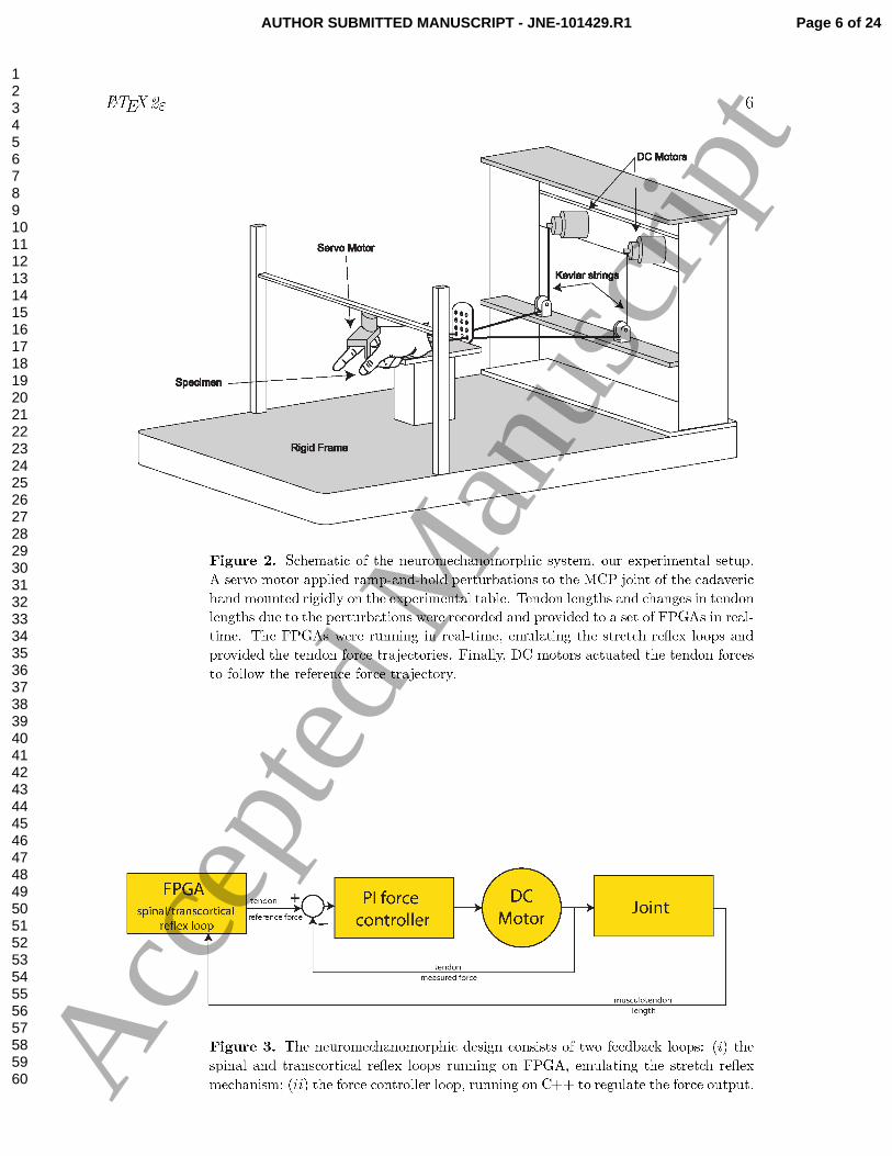
The Neuro-mechano-morphic system shown in Figure 2 was used for the experiments.
This setup has been equipped with state-of-the-art models of populations of muscle
spindle and their γ-motoneuronal drive along with populations of α motor neurons and
their recruitment and rate coding properties, in addition to a mathematical muscle
model — all running in real-time [11, 13]. The aforementioned mathematical models
emulate the final muscle forces and interact with a mechanical plant in real-time,
consisting of a joint - either robotic or cadaveric, tendons and electrical motors ‡. In
Part I of this work, we demonstrate this methodology by successful integration of the
model emulator with a mechanical plant (robotic or cadaveric joint) in real-time [12].
We now present a systematic exploration of the effects of different levels of fusimotor
and cortical drive.
2.1.1. Cadaveric Preparation: To include the actual tendon compliance and
corresponding moment arms, as in our prior work [14, 15], we resected a fresh frozen
cadaver arm at the midforearm level which was dissected the proximal end of the
insertion tendons of the Flexor Digitorum Profundus (FDP) and Extensor Indicis
Proprius (EIP) muscles of the arm. The proximal ends of these tendons were tied
and glued to two high tensile strength para-aramid strings (Kevlar Model 8800K43).
Additionally, the wrist was rigidly mounted to the experimental table using an
external fixator (Agee WristJack, Hand Biomechanics Lab, Inc., Sacramento, CA). The
movements of the proximal and distal interphalangeal joints were also arrested using
finger splints.
2.1.2. Control Loops: Figure 3 shows the schematic of the real-time control loops. The
first is the spinal and transcortical reflex loop which provided the tendon reflex force.
The FPGAs function as controllers and plant was the metacarpophalangeal (MCP) joint
of the cadaveric index finger. Part 1 of this work details the mathematical models
implemented in the controller. The strings that were fastened to the tendons were
connected to DC motors (Faulhaber 3863H024C). The motor driver featured an array
of Western Design LDU-S1 high-current Darlington drivers running on the PCU-S3
Chassis. The rotational movements of the motors were recorded using shaft encoders
‡ A point of clarification: We use the term emulation as distinct from simulation and control. In
our point of view, emulation is the realistic implementation of the mechanisms of interest, running at
the same time-scale as the biological system being studied. By contrast, simulation includes generic
black-box implementations that provide realistic input-output relations but need not implement the
mechanisms of interest in real-time nor be coupled to the real mechanics of the system. Further, the
goal in control theory is to achieve a certain criterion, e.g. stabilizing the system, minimizing the error
between desired and measured signals, etc. The approach expounded in this work does not explicitly
control the stability or simulate the input-output characteristics of the joint. Rather, the system allows
the emergence of behaviors that are analogous to those exhibited by biological systems
Page 3 of 24 AUTHOR SUBMITTED MANUSCRIPT - JNE-101429.R1
123456789101112131415161718192021222324252627282930313233343536373839404142434445464748495051525354555657585960 A
ccep
ted
Man
uscr
ipt
LATEX2ε 4
(HEDS-5500). A custom computer application calculated the musculotendon lengths
from the encoder pulse train and provided them to the FPGA.
The second control loop regulated the tendon forces to closely follow the
reference forces provided by the FPGA. Thus, a Proportional-Integral (PI) controller,
programmed into the app controlled the output forces of the tendons. Load cells
(Interface model SML-10) measured the tendon forces and their outputs were amplified
and low-pass filtered using a signal conditioning module (Transducer Techniques TM0-
1-12 VDC).
A DC Servo motor (Dynammixel RX-28), whose axis of rotation was aligned to
that of the MCP joint, delivered position perturbations to the joint. The servo was
programmed to be much stiffer than the finger joint and the muscle (tendon) force
responses did not significantly change the position of the servo and hence did not stretch
the antagonist muscle. Consequently, the two muscles could be analyzed independently.
2.1.3. Neuromorphic System: Briefly, as described in Part I of our study as well as [16],
a set of three FPGAs (OpalKelly XEM6010-LX150) emulated the stretch reflex loops
for each muscle (total six FPGAs for the antagonist muscle pair). The sensory FPGA
was fed with the static (γs) and dynamic (γd) fusimotor drives (set by user) and the
measured muscle length to emulate an ensemble of 128 muscle spindles (Figure. 1) with
primary (Ia) and secondary (II) afferents. We used the model developed by Mileusnic et
al. which showed a close fit to the afferent firing rates recorded experimentally in a range
of different experimental conditions [17]. This model incorporates the three nonlinear
intrafusal fibers (viz. bag1, bag2, and chain fibers) that contribute to the firing rates
of primary and secondary afferents. The model features realistic temporal properties of
fusimotor activation as well as partial occlusion.
The primary and secondary afferents of the ensemble of muscle spindles made
synaptic connections with the motor neuron pool in the motor FPGA via a
monosynaptic pathway with a delay of 32ms representing a simplified model of the
short latency component of the stretch reflex loop. The spinal projection from secondary
afferents to alpha motor neurons in the model reflects various interneuronal pathways
rather than a monosynaptic loop, which has not been described [1]. Both the primary
and secondary afferents also made synaptic connections to the same motor neuron pool
via the cortical FPGA with a delay of 64 ms and represents a very simplified model of the
long latency, transcortical component of the stretch reflex loop circuitry. The cortical
FPGA emulated 128 neurons which is a clear oversimplification of the real biology.
The cortical neurons had a user-defined tonic drive which functioned as a descending
cortical offset to the motor neuron pool. The motor FPGA emulated models of motor
neuron pool (with recruitment and rate coding) and a skeletal muscle while delivering
the muscle force, EMG and motor neuron pool raster to the real-time custom computer
program over USB 2.0 buses. The total number of neurons emulated for each afferented
muscle was 1152 (total 2304 for the antagonistic muscle pair) which consisted of 256
spindle afferents (primary and secondary), 128 cortical neurons and 768 motor neurons.
Page 4 of 24AUTHOR SUBMITTED MANUSCRIPT - JNE-101429.R1
123456789101112131415161718192021222324252627282930313233343536373839404142434445464748495051525354555657585960 A
ccep
ted
Man
uscr
ipt
LATEX2ε 5
Bag �ber1
Bag �ber2
Chain �ber
Chain �ber
length
velocity
acceleration
Static fusimotor
drive
Dynamic fusimotor
drive
Primary (Ia)
a�erent
Secondary (II)
a�erent
Muscle Spindle
ad
as
Muscle
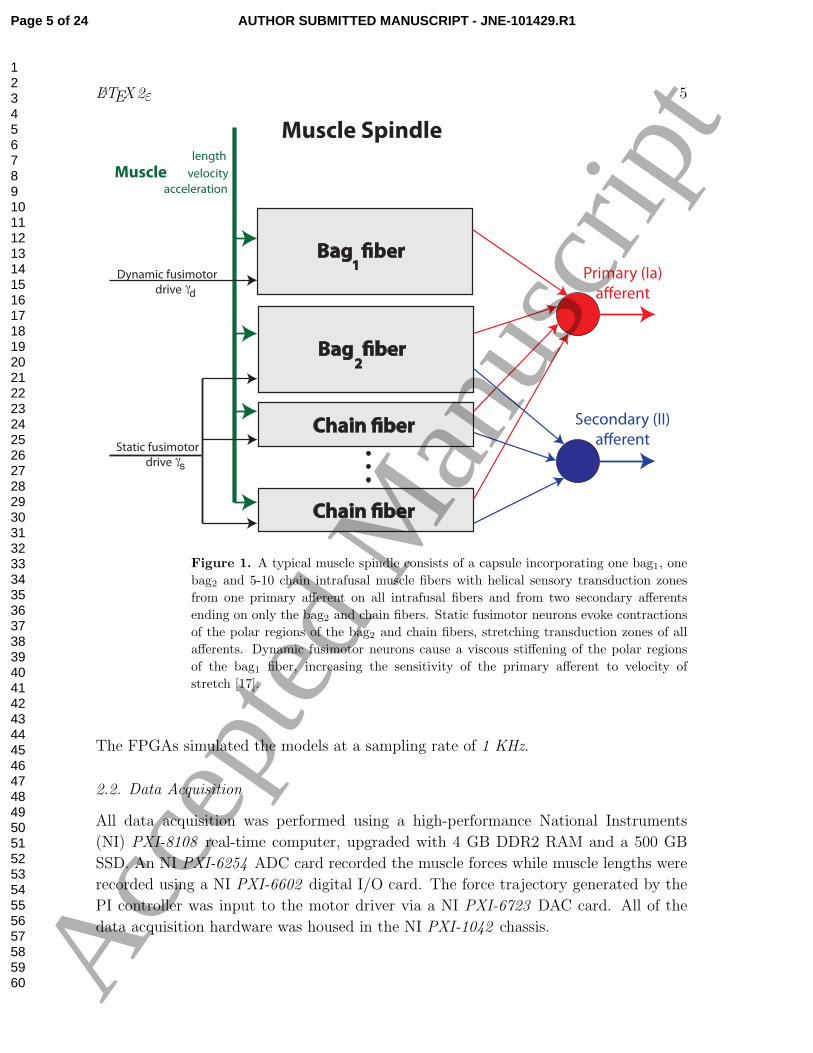
Figure 1. A typical muscle spindle consists of a capsule incorporating one bag1, one
bag2 and 5-10 chain intrafusal muscle fibers with helical sensory transduction zones
from one primary afferent on all intrafusal fibers and from two secondary afferents
ending on only the bag2 and chain fibers. Static fusimotor neurons evoke contractions
of the polar regions of the bag2 and chain fibers, stretching transduction zones of all
afferents. Dynamic fusimotor neurons cause a viscous stiffening of the polar regions
of the bag1 fiber, increasing the sensitivity of the primary afferent to velocity of
stretch [17].
The FPGAs simulated the models at a sampling rate of 1 KHz.
2.2. Data Acquisition
All data acquisition was performed using a high-performance National Instruments
(NI) PXI-8108 real-time computer, upgraded with 4 GB DDR2 RAM and a 500 GB
SSD. An NI PXI-6254 ADC card recorded the muscle forces while muscle lengths were
recorded using a NI PXI-6602 digital I/O card. The force trajectory generated by the
PI controller was input to the motor driver via a NI PXI-6723 DAC card. All of the
data acquisition hardware was housed in the NI PXI-1042 chassis.
Page 5 of 24 AUTHOR SUBMITTED MANUSCRIPT - JNE-101429.R1
123456789101112131415161718192021222324252627282930313233343536373839404142434445464748495051525354555657585960 A
ccep
ted
Man
uscr
ipt
Page 6 of 24AUTHOR SUBMITTED MANUSCRIPT - JNE-101429.R1
123456789101112131415161718192021222324252627282930313233343536373839404142434445464748495051525354555657585960 A
ccep
ted
Man
uscr
ipt
LATEX2ε 7
Read from FPGA
(EMG,spindle,force,etc)
Write to FPGA
(legnth,velocity)
(EMG,spindle,force,etc)
FPGA Thread
Read encoders
Read load cells
Write motor command
Estimate muscle
length & velocity
PID controller update
Wait for next sample time
Controller Thread
Experiment Thread
Control servo motor
(perturbations)
Update fusimotor drive
Log data
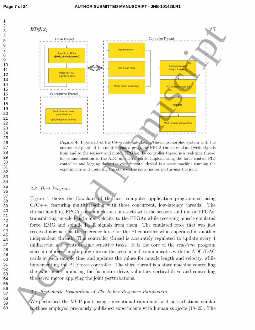
Figure 4. Flowchart of the C++ code interfacing the neuromorphic system with the
anatomical plant. It is a multithreaded program: FPGA thread read and write signals
from and to the sensory and motor FPGAs; the controller thread is a real-time thread
for communication to the ADC and DAC cards, implementing the force control PID
controller and logging data; the experimental thread is a state machine running the
experiments and updating the state of the servo motor perturbing the joint.
2.3. Host Program
Figure 4 shows the flowchart of the host computer application programmed using
C/C++, featuring multithreading with three concurrent, low-latency threads. The
thread handling FPGA communications interacts with the sensory and motor FPGAs,
transmitting muscle length and velocity to the FPGAs while receiving muscle emulated
force, EMG and spindle Ia, II signals from them. The emulated force that was just
received now acts as the reference force for the PI controller which operated in another
independent thread. This controller thread is accurately regulated to update every 1
millisecond and controls time sensitive tasks. It is the core of the real-time program
since it enforces the sampling rate on the system and communicates with the ADC/DAC
cards at each sample time and updates the values for muscle length and velocity, while
implementing the PID force controller. The third thread is a state machine controlling
the experiment, updating the fusimotor drive, voluntary cortical drive and controlling
the servo motor applying the joint perturbations.
2.4. Systematic Exploration of The Reflex Response Parameters
We perturbed the MCP joint using conventional ramp-and-hold perturbations similar
to those employed previously published experiments with human subjects [18–20]. The
Page 7 of 24 AUTHOR SUBMITTED MANUSCRIPT - JNE-101429.R1
123456789101112131415161718192021222324252627282930313233343536373839404142434445464748495051525354555657585960 A
ccep
ted
Man
uscr
ipt
LATEX2ε 8
firing rates of the static (γs) and dynamic (γd) fusimotor neurons were independently
selected from the set {0, 25, 50, · · · , 200} which were all in the range of values validated
in previous work for our spindle model [17]. By selecting values from this grid at
random, it was possible to mitigate the potential confound of presentation order given
that tissue response, such as tendon stiffness, can change over time as the collagen
fibers may reorganize themselves in response to load. We also randomly varied the
velocity of perturbations among the four values {50, 100, 200, 300} (degrees/s) and the
three baseline cortical drive (c) among the three values of {0, 5, 10}% MVC by changing
the baseline firing rate of the cortical neurons. We randomly selected combinations
of parameters exhaustively, with each combination being tested only once. With four
perturbations for each combination, the experiment constituted 9×9×4×3×4 = 3888
ramp-and-hold perturbations.
2.5. Analysis
Given that the stretch reflex loop is sensitive to both the muscle length and velocity,
we analyzed the responses for both the phasic and tonic intervals of stretch [21]. We
segmented the responses per [22, 23]: (i) the phasic interval was 1400 (ms) starting at
the onset of the perturbation; (ii) the tonic interval was 450 (ms) following the phasic
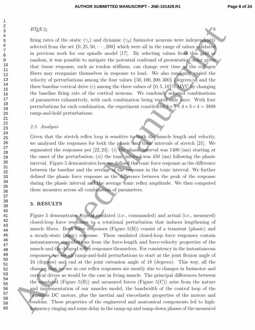
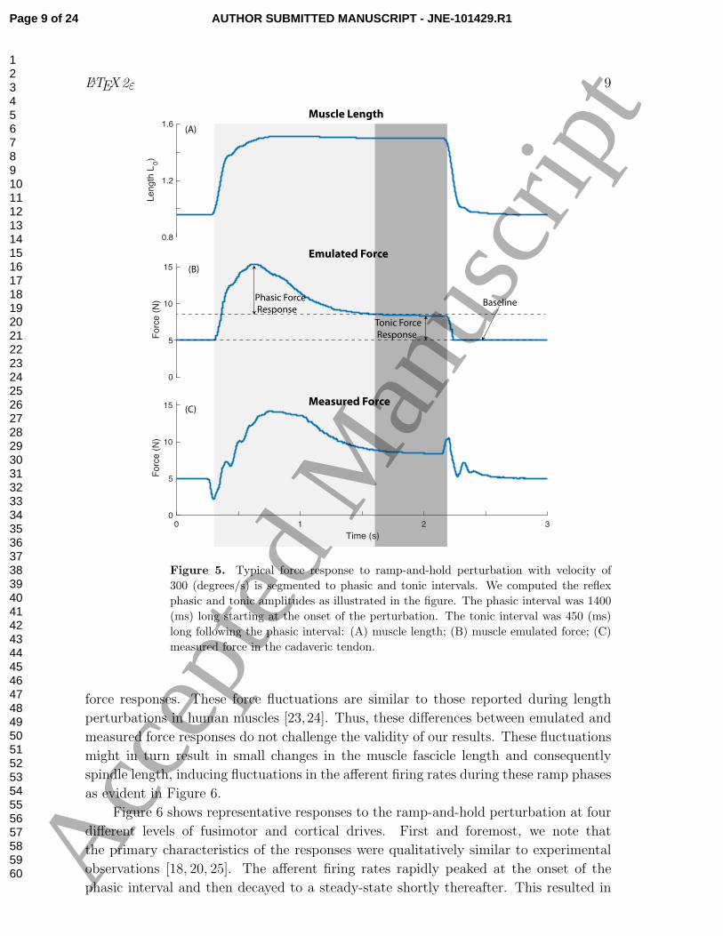
interval. Figure 5 demonstrates how we defined the tonic force response as the difference
between the baseline and the average of the response in the tonic interval. We further
defined the phasic force response as the difference between the peak of the response
during the phasic interval and the average tonic reflex amplitude. We then computed
these measures across all combination of parameters.
3. RESULTS
Figure 5 demonstrates typical emulated (i.e., commanded) and actual (i.e., measured)
closed-loop force responses to a rotational perturbation that induces lengthening of
muscle fibers. Both force responses (Figure 5(B)) consist of a transient (phasic) and
a steady-state (tonic) response. These emulated closed-loop force responses contain
instantaneous contributions from the force-length and force-velocity properties of the
muscle and the delayed reflex responses themselves. For consistency in the instantaneous
responses, we set all ramp-and-hold perturbations to start at the joint flexion angle of
24 (degrees) and end at the joint extension angle of 18 (degrees). This way, all the
changes that we see in our reflex responses are mostly due to changes in fusimotor and
cortical drives as would be the case in living muscle. The principal differences between
the emulated (Figure 5(B)) and measured forces (Figure 5(C)) arise from the nature
and implementation of our muscles model, the bandwidth of the control loop of the
brushless DC motors, plus the inertial and viscoelastic properties of the motors and
tendons. These properties of the engineered and anatomical components led to high-
frequency ringing and some delay in the ramp-up and ramp-down phases of the measured
Page 8 of 24AUTHOR SUBMITTED MANUSCRIPT - JNE-101429.R1
123456789101112131415161718192021222324252627282930313233343536373839404142434445464748495051525354555657585960 A
ccep
ted
Man
uscr
ipt
LATEX2ε 9
0.8
1.2
1.6
Length
L0)
0
5
10
15
Forc
e (
N)
0 1 2 3
Time (s)
0
5
10
15
Forc
e (
N)
Tonic Force
Response
BaselinePhasic Force
Response
(A)
(B)
(C)
Muscle Length
Emulated Force
Measured Force
Figure 5. Typical force response to ramp-and-hold perturbation with velocity of
300 (degrees/s) is segmented to phasic and tonic intervals. We computed the reflex
phasic and tonic amplitudes as illustrated in the figure. The phasic interval was 1400
(ms) long starting at the onset of the perturbation. The tonic interval was 450 (ms)
long following the phasic interval: (A) muscle length; (B) muscle emulated force; (C)
measured force in the cadaveric tendon.
force responses. These force fluctuations are similar to those reported during length
perturbations in human muscles [23,24]. Thus, these differences between emulated and
measured force responses do not challenge the validity of our results. These fluctuations
might in turn result in small changes in the muscle fascicle length and consequently
spindle length, inducing fluctuations in the afferent firing rates during these ramp phases
as evident in Figure 6.
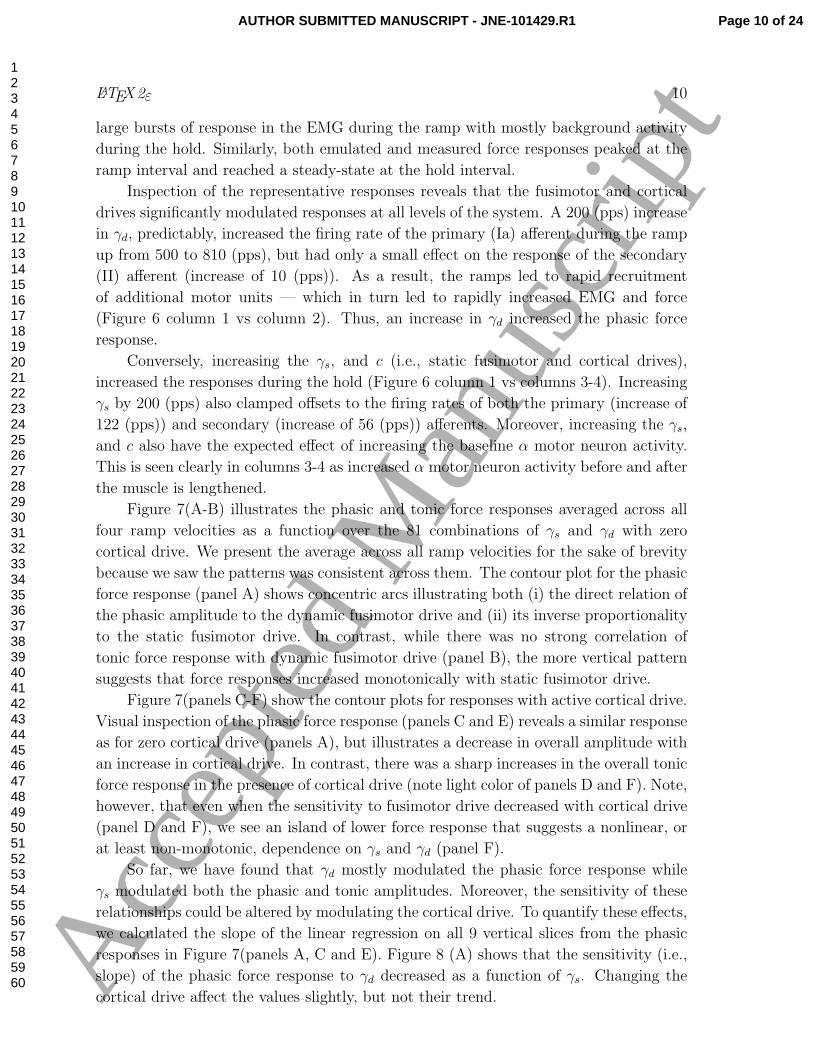
Figure 6 shows representative responses to the ramp-and-hold perturbation at four
different levels of fusimotor and cortical drives. First and foremost, we note that
the primary characteristics of the responses were qualitatively similar to experimental
observations [18, 20, 25]. The afferent firing rates rapidly peaked at the onset of the
phasic interval and then decayed to a steady-state shortly thereafter. This resulted in
Page 9 of 24 AUTHOR SUBMITTED MANUSCRIPT - JNE-101429.R1
123456789101112131415161718192021222324252627282930313233343536373839404142434445464748495051525354555657585960 A
ccep
ted
Man
uscr
ipt
LATEX2ε 10
large bursts of response in the EMG during the ramp with mostly background activity
during the hold. Similarly, both emulated and measured force responses peaked at the
ramp interval and reached a steady-state at the hold interval.
Inspection of the representative responses reveals that the fusimotor and cortical
drives significantly modulated responses at all levels of the system. A 200 (pps) increase
in γd, predictably, increased the firing rate of the primary (Ia) afferent during the ramp
up from 500 to 810 (pps), but had only a small effect on the response of the secondary
(II) afferent (increase of 10 (pps)). As a result, the ramps led to rapid recruitment
of additional motor units — which in turn led to rapidly increased EMG and force
(Figure 6 column 1 vs column 2). Thus, an increase in γd increased the phasic force
response.
Conversely, increasing the γs, and c (i.e., static fusimotor and cortical drives),
increased the responses during the hold (Figure 6 column 1 vs columns 3-4). Increasing
γs by 200 (pps) also clamped offsets to the firing rates of both the primary (increase of
122 (pps)) and secondary (increase of 56 (pps)) afferents. Moreover, increasing the γs,
and c also have the expected effect of increasing the baseline α motor neuron activity.
This is seen clearly in columns 3-4 as increased α motor neuron activity before and after
the muscle is lengthened.
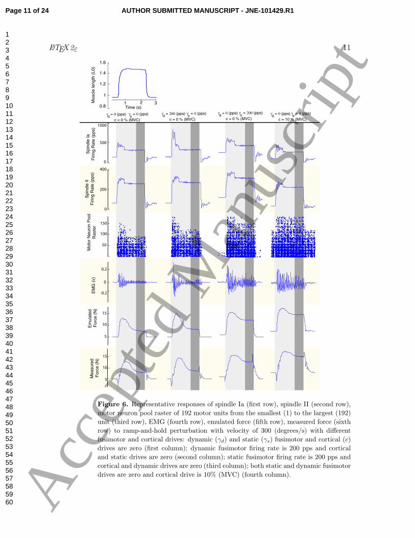
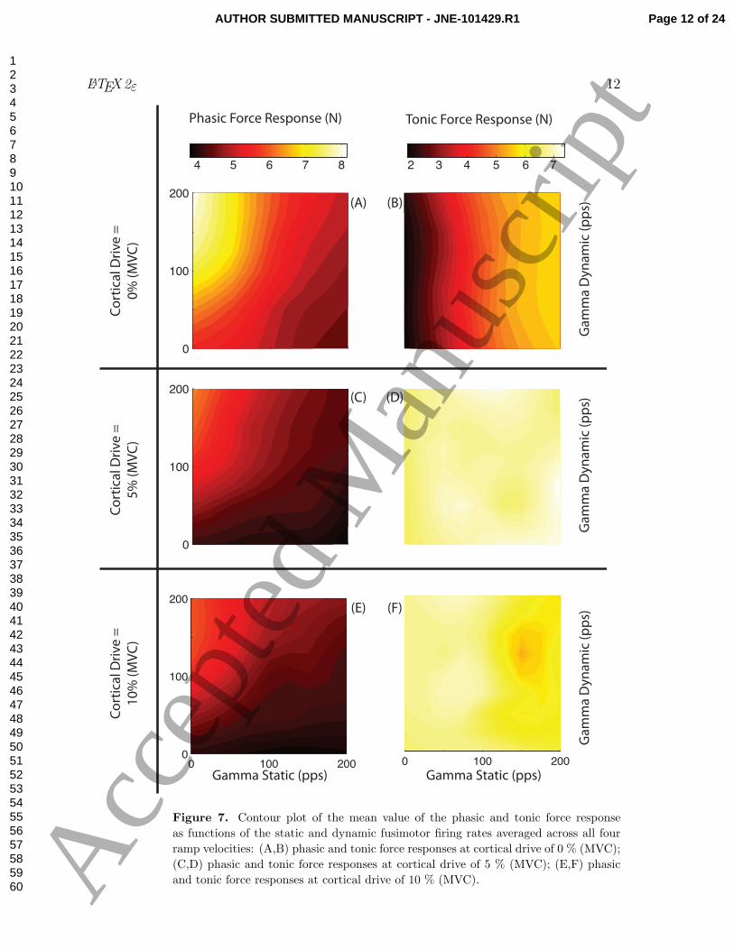
Figure 7(A-B) illustrates the phasic and tonic force responses averaged across all
four ramp velocities as a function over the 81 combinations of γs and γd with zero
cortical drive. We present the average across all ramp velocities for the sake of brevity
because we saw the patterns was consistent across them. The contour plot for the phasic
force response (panel A) shows concentric arcs illustrating both (i) the direct relation of
the phasic amplitude to the dynamic fusimotor drive and (ii) its inverse proportionality
to the static fusimotor drive. In contrast, while there was no strong correlation of
tonic force response with dynamic fusimotor drive (panel B), the more vertical pattern
suggests that force responses increased monotonically with static fusimotor drive.
Figure 7(panels C-F) show the contour plots for responses with active cortical drive.
Visual inspection of the phasic force response (panels C and E) reveals a similar response
as for zero cortical drive (panels A), but illustrates a decrease in overall amplitude with
an increase in cortical drive. In contrast, there was a sharp increases in the overall tonic
force response in the presence of cortical drive (note light color of panels D and F). Note,
however, that even when the sensitivity to fusimotor drive decreased with cortical drive
(panel D and F), we see an island of lower force response that suggests a nonlinear, or
at least non-monotonic, dependence on γs and γd (panel F).
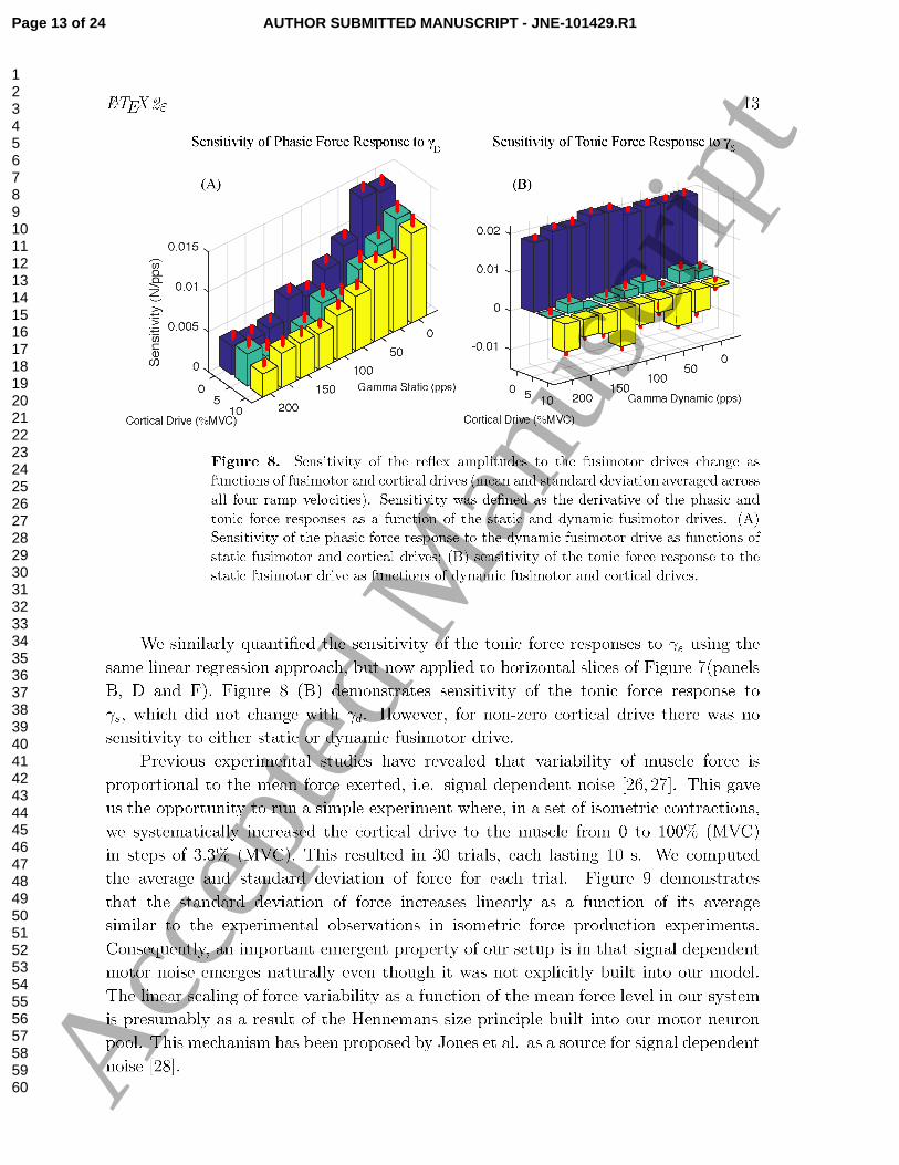
So far, we have found that γd mostly modulated the phasic force response while
γs modulated both the phasic and tonic amplitudes. Moreover, the sensitivity of these
relationships could be altered by modulating the cortical drive. To quantify these effects,
we calculated the slope of the linear regression on all 9 vertical slices from the phasic
responses in Figure 7(panels A, C and E). Figure 8 (A) shows that the sensitivity (i.e.,
slope) of the phasic force response to γd decreased as a function of γs. Changing the
cortical drive affect the values slightly, but not their trend.
Page 10 of 24AUTHOR SUBMITTED MANUSCRIPT - JNE-101429.R1
123456789101112131415161718192021222324252627282930313233343536373839404142434445464748495051525354555657585960 A
ccep
ted
Man
uscr
ipt
LATEX2ε 11
0.8
1
1.2
1.4
1.6
Muscle
length
(L0)
Measure
d
Forc
e (
N)
1 2 3Time (s)
Spin
dle
Ia
Firin
g R
ate
(pps)
Spin
dle
II
Firin
g R
ate
(pps)
EM
G (
v)
Em
ula
ted
Forc
e (
N)
500
1000
200
400
100
50
150
0
0.2
-0.2
0
0
5
10
15
5
10
15
Moto
r N
euro
n P
ool
Raste
r
c = 0 % (MVC)
a����� (pps)d
a����� (pps)s
c = 0 % (MVC)
a������� (pps)d
a����� (pps)s
c = 0 % (MVC)
a����� (pps)d
a������� (pps)s
c = 10 % (MVC)
a����� (pps)d
a����� (pps)s
Figure 6. Representative responses of spindle Ia (first row), spindle II (second row),
motor neuron pool raster of 192 motor units from the smallest (1) to the largest (192)
unit (third row), EMG (fourth row), emulated force (fifth row), measured force (sixth
row) to ramp-and-hold perturbation with velocity of 300 (degrees/s) with different
fusimotor and cortical drives: dynamic (γd) and static (γs) fusimotor and cortical (c)
drives are zero (first column); dynamic fusimotor firing rate is 200 pps and cortical
and static drives are zero (second column); static fusimotor firing rate is 200 pps and
cortical and dynamic drives are zero (third column); both static and dynamic fusimotor
drives are zero and cortical drive is 10% (MVC) (fourth column).
Page 11 of 24 AUTHOR SUBMITTED MANUSCRIPT - JNE-101429.R1
123456789101112131415161718192021222324252627282930313233343536373839404142434445464748495051525354555657585960 A
ccep
ted
Man
uscr
ipt
LATEX2ε 12
0
100
200
0
100
200
4 5 6 7 8 2 3 4 5 6 7
Phasic Force Response (N) Tonic Force Response (N)
Ga
mm
a D
yn
am
ic (
pp
s)
Gamma Static (pps) Gamma Static (pps)
Ga
mm
a D
yn
am
ic (
pp
s)G
am
ma
Dy
na
mic
(p
ps)
Co
rtic
al D
riv
e =
0%
(M
VC
)
Co
rtic
al D
riv
e =
5%
(M
VC
)
Co
rtic
al D
riv
e =
10
% (
MV
C)
(A)
(C)
0 100 2000
100
200
0 100 200
(B)
(D)
(E) (F)
Figure 7. Contour plot of the mean value of the phasic and tonic force response
as functions of the static and dynamic fusimotor firing rates averaged across all four
ramp velocities: (A,B) phasic and tonic force responses at cortical drive of 0 % (MVC);
(C,D) phasic and tonic force responses at cortical drive of 5 % (MVC); (E,F) phasic
and tonic force responses at cortical drive of 10 % (MVC).
Page 12 of 24AUTHOR SUBMITTED MANUSCRIPT - JNE-101429.R1
123456789101112131415161718192021222324252627282930313233343536373839404142434445464748495051525354555657585960 A
ccep
ted
Man
uscr
ipt
Page 13 of 24 AUTHOR SUBMITTED MANUSCRIPT - JNE-101429.R1
123456789101112131415161718192021222324252627282930313233343536373839404142434445464748495051525354555657585960 A
ccep
ted
Man
uscr
ipt
LATEX2ε 14
2 4 6 8 10 12 14 16 18 200.5
1
1.5
2
2.5
3
3.5
4
4.5
5
x 10ï�
Average of Force (N)
Sta
ndard
Devia
tion o
f F
orc
e (
N)
R2=0.95
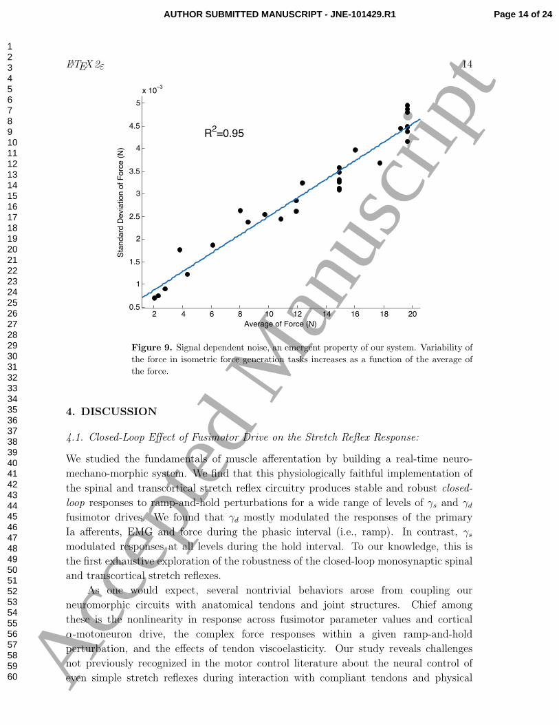
Figure 9. Signal dependent noise, an emergent property of our system. Variability of
the force in isometric force generation tasks increases as a function of the average of
the force.
4. DISCUSSION
4.1. Closed-Loop Effect of Fusimotor Drive on the Stretch Reflex Response:
We studied the fundamentals of muscle afferentation by building a real-time neuro-
mechano-morphic system. We find that this physiologically faithful implementation of
the spinal and transcortical stretch reflex circuitry produces stable and robust closed-
loop responses to ramp-and-hold perturbations for a wide range of levels of γs and γd
fusimotor drives. We found that γd mostly modulated the responses of the primary
Ia afferents, EMG and force during the phasic interval (i.e., ramp). In contrast, γsmodulated responses at all levels during the hold interval. To our knowledge, this is
the first exhaustive exploration of the robustness of the closed-loop monosynaptic spinal
and transcortical stretch reflexes.
As one would expect, several nontrivial behaviors arose from coupling our
neuromorphic circuits with anatomical tendons and joint structures. Chief among
these is the nonlinearity in response across fusimotor parameter values and cortical
α-motoneuron drive, the complex force responses within a given ramp-and-hold
perturbation, and the effects of tendon viscoelasticity. Our study reveals challenges
not previously recognized in the motor control literature about the neural control of
even simple stretch reflexes during interaction with compliant tendons and physical
Page 14 of 24AUTHOR SUBMITTED MANUSCRIPT - JNE-101429.R1
123456789101112131415161718192021222324252627282930313233343536373839404142434445464748495051525354555657585960 A
ccep
ted
Man
uscr
ipt
LATEX2ε 15
environments. These challenges are then amplified when we consider that the nervous
system must control and coordinate the effects of such parameter changes across multiple
muscles — and over time during everyday tasks. Interestingly, we saw that the changes
with fusimotor parameter values were consistent across ramp velocities (Figure 7).
This is not necessarily unexpected because changes in ramp velocity simply induce
a monotonic change in spindle output. However, this work enables a future systematic
exploration of the effect of ramp velocity (e.g., [19]) on reflex responses — as done in
clinics — to understand the role of fusimotor drive on healthy tone and pathologic catch
responses.
As has been previously documented in many experimental studies, the CNS is
capable of adjusting the sensitivity of the stretch reflex loop [29]. In particular, the
sensitivity of the phasic force response to γd decreased with an increase in γs (Figure 8
(A)). This was presumably due to the partial occlusion mechanism modeled into our
muscle spindle. We also observed that the sensitivity slightly decreased with increase in
the cortical drive. Exploring the muscle length data revealed that the baseline of muscle
length was 2.9% shorter and the muscle stretch to ramp-and-hold perturbation of the
joint was 1.9% shorter with background cortical drive. This is likely due to the elastic
nature of the real tendons that stretched under baseline cortical drive and decreased
the muscle length, as well as phasic and tonic tensions. This resulted in shorter muscle
fibers. Afferent firing rate is lower when the muscle is shorter, and this decreased the
phasic force response. Decrease in the phasic force response with increase in tonic
contraction level in normal human has been previously reported in the literature but
had remained difficult to explain [22, 30].
The sensitivity of the tonic force response to γs did not change with increase in γd
(Figure 8(B)). This was because γd is increasing the firing rate of primary Ia afferents
only at the ramp interval, increasing only the phasic force response. However, the
sensitivity of the tonic force response to γs decreased dramatically with increase in
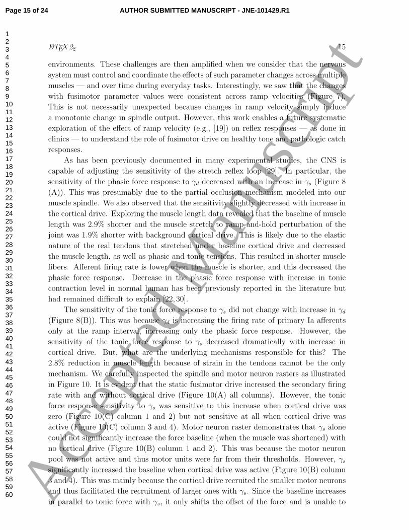
cortical drive. But, what are the underlying mechanisms responsible for this? The
2.8% reduction in muscle length because of strain in the tendons cannot be the only
mechanism. We carefully inspected the spindle and motor neuron rasters as illustrated
in Figure 10. It is evident that the static fusimotor drive increased the secondary firing
rate with and without cortical drive (Figure 10(A) all columns). However, the tonic
force response sensitivity to γs was sensitive to this increase when cortical drive was
zero (Figure 10(C) column 1 and 2) but not sensitive at all when cortical drive was
active (Figure 10(C) column 3 and 4). Motor neuron raster demonstrates that γs alone
could not significantly increase the force baseline (when the muscle was shortened) with
no cortical drive (Figure 10(B) column 1 and 2). This was because the motor neuron
pool was not active and thus motor units were far from their thresholds. However, γssignificantly increased the baseline when cortical drive was active (Figure 10(B) column
3 and 4). This was mainly because the cortical drive recruited the smaller motor neurons
and thus facilitated the recruitment of larger ones with γs. Since the baseline increases
in parallel to tonic force with γs, it only shifts the offset of the force and is unable to
Page 15 of 24 AUTHOR SUBMITTED MANUSCRIPT - JNE-101429.R1
123456789101112131415161718192021222324252627282930313233343536373839404142434445464748495051525354555657585960 A
ccep
ted
Man
uscr
ipt
LATEX2ε 16
50
150
31 2
5
10
15
20
0
200
400
as=0 (pps)
Cortical Drive=
0% (MVC)as=200 (pps)
Cortical Drive=
0% (MVC)as=0 (pps)
Cortical Drive=
10% (MVC)as=200 (pps)
Cortical Drive=
10% (MVC)
(A)
(B)
(C)
Sp
ind
le II
Fir
ing
Ra
te (
pp
s)M
oto
r N
eu
ron
Po
ol
Ra
ste
r
Em
ula
ted
Forc
e (
N)
Time(s)
Figure 10. Sensitivity of tonic force response to γs decreases when there is background
voluntary activity: (A) firing rate of spindle secondary afferent (the most sensitive
afferent to γs) increases with γs; moreover, the firing rate of spindle is slightly decreased
with voluntary, background activity which is due to the shorter muscle length when
the muscle is voluntarily contracting; (B) motorneuron rasters show that the firing
baseline (in the shortening phase of the perturbations) is absent or very small when the
voluntary drive is absent and increases significantly with the voluntary drive, recruiting
the smaller motor units; (C) muscle force shows that tonic force response increases
with γs when voluntary drive is absent. The reflex tonic force is less sensitive γs in the
presence of voluntary drive because both the force baseline and the absolute value of
the force in the hold interval increase with γs.
increase the tonic force response and thus the response becomes insensitive to γs.
One can argue the the large scale trends in reflex response seen in our color maps
(Figure 7) are to be expected and make sense. However, it is important to note that
these properties emerged from interactions among elements that to our knowledge
had not been connected previously. Moreover, it is equally interesting to note that
the departure from linear trends must pose challenges to the nervous system when
controlling afferented muscles. These difficulties are further exacerbated when, as in
the case of tonic response with 10% cortical drive, the response is not monotonic across
the parameter space. This can be seen as islands of high and low activity within the
Page 16 of 24AUTHOR SUBMITTED MANUSCRIPT - JNE-101429.R1
123456789101112131415161718192021222324252627282930313233343536373839404142434445464748495051525354555657585960 A
ccep
ted
Man
uscr
ipt
LATEX2ε 17
colormap. These nonlinearities (or at times departure from monotonicity) likely come
about in part from the nonlinear nature of the spindle model [17] — which to our
knowledge is the most physiologically faithful model in the literature.
4.2. Why Neuromechanomorphic Emulation?
This work extends the work of Sreenivasa et al [31] by using a synthetic neuro-mechano-
morphic system to emulate the stretch reflex loop because our FPGA system can emulate
the populations of neurons and control the mechatronic system in true real-time while
emulating the physiological delays and neuromechanic multi-scale interactions. It is
even capable of operation in hyper-time (365x real-time) to predict long-term changes
of sensorimotor function [13]. This allowed us to use the MCP joint of a cadaveric hand
as the plant to ensure anatomical and physical fidelity. This enables us to perform
realistic simulations and confront the very challenge the central nervous system faces
when actuating actual tendons crossing anatomical joints [14, 15, 32]. A number of
desirable features include: realistic viscoelastic properties of the joint, passive tissues and
tendons, and realistic strain in loaded tendons and their non-constant moment arms [33].
These will result in realistic muscle lengths changes due to external perturbations that
are quite difficult, if not impossible, to model and implement faithfully in computer
simulations [34].
Therefore, this system is not simply doomed to succeed, but represents what is
to our knowledge the first real-world implementation of the fundamental mechanisms
and features of the stretch reflexes when coupled to real anatomical systems. The
real power of this system is to provide a substrate on which to add the complexity
as required to account for able motor function and pathology. Future work will be
able to disambiguate whether and how the response of the system is due to neural or
anatomical elements by systematically comparing this baseline behavior to that arising
when individual elements are replaced by alternative implementations (e.g., changing
muscle models, varying the degree of randomness in the spiking of individual neurons,
changing the synaptic weightings, using inextensible tendons or tendons with varying
degree of elasticity, constant moment arms, etc). Similarly, we will add other known
elements such as Golgi tendon organs, Renshaw cells, Ib interneurons, etc. to understand
their contributions above and beyond the behavior of this basic reflex circuitry.
4.3. Emergent Properties:
It is important to note that because of lack of objective models and limited computation
power, it would be difficult, if not impossible to replicate all behaviors of the
neuromuscular system. However, in a reasonable model, these behaviors should emerge
naturally as a result of interaction among fundamental components of the system. Here
we have observed and documented some of these emergent properties. Examples include
phasic and tonic reflex responses to a ramp-and-hold perturbation, and signal dependent
noise as shown in this paper; as well as maintaining a stable posture following a transient
Page 17 of 24 AUTHOR SUBMITTED MANUSCRIPT - JNE-101429.R1
123456789101112131415161718192021222324252627282930313233343536373839404142434445464748495051525354555657585960 A
ccep
ted
Man
uscr
ipt
LATEX2ε 18
force perturbation to the joint as shown in the companion paper [12]. In future, we will
explore other emergent properties of the system (see scientific and clinical implications
below).
4.4. Limitations of the Study:
Numerical simulations are promising tools to understand healthy function, disease and
role of therapeutic intervention [34]. In the context of afferented muscles [17,35], several
different mechanisms can potentially contribute to pathological muscle tone such as
fusimotor drive of the muscle spindle, long-latency reflex response, inward currents,
presynaptic modulation of the spindle Ia-motorneuron, interneuronal modulation of the
spindle II-motorneuron projections, biasing of the various motor units with respect
to their threshold and frequency-modulation nonlinearities, etc. [29]. In this paper,
we chose to study the effect of fusimotor drive as the first step to understand the
sufficient pathways involved to emulate healthy and pathologic responses. Our hardware
limitation of being able to implement only 128 pairs of spindle afferents per muscle
(natural muscles have many more) has the consequence that the aggregate strength of
the afferent drive to the motor neuron pool is weaker than in its biological counterpart.
Therefore, we added a scaling factor in the form of an offset and gain to increase firing
rates to a level that was able to appropriately drive their respective motor neuron pools.
This is why the firing rates for the spindle afferents in Figure 6 are higher than those
seen in spindle afferents which rarely fire faster than 250pps [17,36]. However, we believe
that this does not affect the validity of our results, and will not be necessary once we
are able to implement thousands of spindle afferents.
While a model of the glutamatergic ribbon synapse from a hair cell in the
cochlea was used in our work [37], there is ample evidence to support the role of
serotonin synapses in the modulation of spinal reflexes and in the production of
rhythmic locomotion [38, 39]. This makes the inclusion of models for these synaptic
neurotransmitter a logical next step.
We used a conventional model of motor neuron pool consisted of 128 × 6 = 768
motor units for each muscle whose parameters were adjusted according to [40] and a
simple Hill-type model of skeletal muscle [41]. It is trivial that the results might depend
on the properties of the motor neuron pool such as motor unit firing rate, recruitment
strategy, etc. We also believe that the choice of muscle model plays a significant role in
the reflex responses (see scientific implications below). In addition, our muscle model
does not yet consider the aponeuroses of these muscles, which may have more significant
effects for the long intrinsic muscles [42]. Future work will test whether and how different
muscle models affect force responses, including tremor and other force oscillations [43].
4.5. Scientific Implications:
Signal dependent noise was an emerging property of our system. This opens the
possibility to study other types of motor variability such as physiologic and pathologic
Page 18 of 24AUTHOR SUBMITTED MANUSCRIPT - JNE-101429.R1
123456789101112131415161718192021222324252627282930313233343536373839404142434445464748495051525354555657585960 A
ccep
ted
Man
uscr
ipt
LATEX2ε 19
tremor as they have been attributed to physiological mechanisms including the stretch
reflex loops [44]. The system we have built is an enabling platform to study the
properties of tremor and its mechanical and neural mechanisms. As such, the neural
connectivity across different motor unit pools can be changed to begin to, for example,
understand the nature of cortico-muscular or musculo-muscular coherence, tremor and
clonus in health and disease [45, 46].
We validated the notion that the fusimotor drive can significantly modulate the
stretch reflex response. But again, the main benefit of this work was to quantify the
details of those responses, and how they are affected by both neural (e.g., fusimotor
and cortical commands), physiological (e.g., muscle spindle structure and function),
and mechanical (e.g., tendon compliance, moment arms, etc) properties. In future, it
is within our goals to emulate and compare against data from pathologic responses in
patients with disrupted reflexes, to find the range of fusimotor drive that is sufficient
to mimic these behaviors. More specifically, we are interested to mimic spasticity and
compare with clinical tests such as Modified Ashworth Scale [7, 47].
Regarding muscle function, we used a simple Hill-type muscle model with force-
length and force-velocity curves that does not consider the dynamics of slow versus fast
twitch fibers, post-activation potentiation, or dynamics of activation versus de-activation
[48]. We have begun investigating other muscle models that are capable of explaining
more complicated physiological phenomena as demonstrated in our companion paper
[12]. By implementing a library of muscle models, our neuro-mechano-morphic setup
can provide a benchmarking system to compare them in closed-loop (i.e. with muscle
afferentation). This is critical in understanding the mechanisms of muscle function,
for instance, the choice of muscle model in replicating nonlinear properties of joint
impedance [49] or in stability of the joint in response to perturbations or in voluntary
movement. Therefore, the selection of the muscle model is a known and unavoidable
limitation of this work that merits further investigation by a systematic comparison for
a variety of muscle models proposed in the literature — whose behavior when coupled
with a closed-loop neuromorphic system remains unknown [50].
Lastly, this study reinforces the notion that the time-critical coordination of
afferented muscles undergoing eccentric contractions during smooth and accurate
voluntary movements remains poorly understood [51, 52], [53, 54]. Sherrington
highlighted this at the birth of motor neuroscience as the problem of excitation-inhibition
(refined now to include α−γ coactivation [55]) being fundamental to motor control [56].
Our recent theoretical and modeling work added details to the notion that controlling
natural and compliant limb function requires careful orchestration of the obligatory
changes in the lengths of all muscles in response to the rotation of a few joints—which
is an overdetermined problem that is the opposite of redundant [51–53]. Our findings
compound the difficulties posed by the need for appropriate α − γ coactivation by
the fact that the reflex response to eccentric contractions is sensitive to multiple factors
including the details of the tendon excursion and its associated departure from linearity,
the physiological response of the muscle spindles, etc. This underscores the longstanding
Page 19 of 24 AUTHOR SUBMITTED MANUSCRIPT - JNE-101429.R1
123456789101112131415161718192021222324252627282930313233343536373839404142434445464748495051525354555657585960 A
ccep
ted
Man
uscr
ipt
LATEX2ε 20
difficulties in our understanding of motor control using simplified models [34, 57]. This
work aims to provide a physiologically realistic platform to bring these issues to light by
confronting the very challenge the central nervous system faces: using multiple afferented
muscles simultaneously to pull on viscoelastic tendons that act on anatomical joints to
interact with the physical world.
4.6. Clinical Implications:
Our goal is to understand how the musculotendon mechanics interact with spinal
mechanisms to produce able and pathologic muscle function. This is significant in
clinical research as a number of neuromuscular pathologies that change the muscle
tone arise from presumed alterations of the spinal reflex mechanisms (e.g., changes
in fusimotor drive, faulty α−γ coactivation) or compromised musculotendon structures
(e.g., muscle contractures, sarcopenia, increase in motor unit innervation numbers).
Even if still in the early stages of its development, our neuro-mechano-morphic system
provides framework, test bed and reality check to quantify how presumed disruptions
of these known mechanisms lead to pathologies, for example, in dystonia or spinal cord
injury, which can be sufficiently described by simply tuning the offset and gain of the
spinal or the transcortical loop [11]. Modeling other motor centers of the brain —
such as the basal ganglia and the primary motor cortex — may also allow us to model
hyperkinetic pathologies [58]. The system can also allow us to verify the extent to which
available or proposed interventions can improve these conditions [59–61].
5. ACKNOWLEDGEMENT
This project is supported by Fonds de Recherche du Quebec- Nature et Technologies to
K. Jalaleddini, National Natural Science Foundation of China (Grant No. 81501570) to
C.M. Niu, and the Youth Eastern Scholar program at Shanghai Institutions of Higher
Learning (Award No. QD2015007) to C.M. Niu. The authors are grateful for support
from the James S McDonnell Foundation to T.D. Sanger. Research reported in this
publication was supported by the National Institute of Arthritis and Musculoskeletal and
Skin Diseases of the National Institutes of Health under Awards Number R01 AR050520
and R01 AR052345 to F.J. Valero-Cuevas, the National Institute of Neurological
Disorders and Stroke award R01 NS069214 to T.D. Sanger. The contents of this
endeavor is solely the responsibility of the authors and does not necessarily represent
the official views of the National Institutes of Health.
The authors thank Dr. Emily Lawrence, Dr. Nina R. Lightdale-Miric, Victor Bar-
radas and Christoff Sulzenbacher for their help in preparation of the cadaveric specimen
for the help during the data collection.
[1] E. Pierrot-Deseilligny and D. Burke, The circuitry of the human spinal cord: its role in motor
control and movement disorders. Cambridge University Press, 2005.
Page 20 of 24AUTHOR SUBMITTED MANUSCRIPT - JNE-101429.R1
123456789101112131415161718192021222324252627282930313233343536373839404142434445464748495051525354555657585960 A
ccep
ted
Man
uscr
ipt
LATEX2ε 21
[2] A. Prochazka, “Proprioceptive feedback and movement regulation,” Comprehensive Physiology,
2010.
[3] G. Goodwin, M. Hulliger, and P. Matthews, “The effects of fusimotor stimulation during small
amplitude stretching on the frequency-response of the primary ending of the mammalian muscle
spindle.,” The Journal of physiology, vol. 253, no. 1, p. 175, 1975.
[4] E. J. Hwang and R. Shadmehr, “Internal models of limb dynamics and the encoding of limb state,”
Journal of neural engineering, vol. 2, no. 3, p. S266, 2005.
[5] A. G. Feldman, “Once more on the equilibrium-point hypothesis (λ model) for motor control,”
Journal of motor behavior, vol. 18, no. 1, pp. 17–54, 1986.
[6] E. Bizzi, A. Polit, and P. Morasso, “Mechanisms underlying achievement of final head position,”
Journal of Neurophysiology, vol. 39, no. 2, pp. 435–444, 1976.
[7] M. Adams and A. Hicks, “Spasticity after spinal cord injury,” Spinal cord, vol. 43, no. 10, pp. 577–
586, 2005.
[8] J. Nielsen, C. Crone, and H. Hultborn, “The spinal pathophysiology of spasticity–from a basic
science point of view,” Acta Physiologica, vol. 189, no. 2, pp. 171–180, 2007.
[9] J.-M. Gracies, P. Nance, E. Elovic, J. McGuire, and D. M. Simpson, “Traditional pharmacological
treatments for spasticity part ii: General and regional treatments,” Muscle & Nerve, vol. 20,
no. S6, pp. 92–120, 1997.
[10] K. Wei, J. I. Glaser, L. Deng, C. K. Thompson, I. H. Stevenson, Q. Wang, T. G. Hornby, C. J.
Heckman, and K. P. Kording, “Serotonin affects movement gain control in the spinal cord,” The
Journal of Neuroscience, vol. 34, no. 38, pp. 12690–12700, 2014.
[11] W. J. Sohn, C. M. Niu, and T. D. Sanger, “Increased long-latency reflex activity as a sufficient
explanation for childhood hypertonic dystonia: a neuromorphic emulation study,” Journal of
neural engineering, vol. 12, no. 3, p. 036010, 2015.
[12] C. M. Niu, K. Jalaleddini, W. J. Sohn, J. Rocamora, T. D. Sanger, and F. J. Valero-Cuevas,
“Neuromorphic meets neuromechanics part i: The methodology,” Journal of Neural Engineering,
In Review.
[13] C. M. Niu, S. K. Nandyala, and T. D. Sanger, “Emulated muscle spindle and spiking afferents
validates vlsi neuromorphic hardware as a testbed for sensorimotor function and disease,”
Frontiers in computational neuroscience, vol. 8, 2014.
[14] J. L. Pearlman, S. S. Roach, and F. J. Valero-Cuevas, “The fundamental thumb-tip force vectors
produced by the muscles of the thumb,” Journal of Orthopaedic Research, vol. 22, no. 2, pp. 306–
312, 2004.
[15] F. J. Valero-Cuevas, J. D. Towles, and V. R. Hentz, “Quantification of fingertip force reduction
in the forefinger following simulated paralysis of extensor and intrinsic muscles,” Journal of
Biomechanics, vol. 33, no. 12, pp. 1601 – 1609, 2000.
[16] C. M. Niu, S. Nandyala, W. J. Sohn, and T. Sanger, “Multi-scale hyper-time hardware emulation
of human motor nervous system based on spiking neurons using fpga,” in Advances in Neural
Information Processing Systems, pp. 37–45, 2012.
[17] M. P. Mileusnic, I. E. Brown, N. Lan, and G. E. Loeb, “Mathematical models of proprioceptors.
i. control and transduction in the muscle spindle,” Journal of neurophysiology, vol. 96, no. 4,
pp. 1772–1788, 2006.
[18] B. B. Edin and A. Vallbo, “Dynamic response of human muscle spindle afferents to stretch,”
Journal of Neurophysiology, vol. 63, no. 6, pp. 1297–1306, 1990.
[19] A. Jobin and M. F. Levin, “Regulation of stretch reflex threshold in elbow flexors in children
with cerebral palsy: a new measure of spasticity,” Developmental Medicine & Child Neurology,
vol. 42, no. 8, pp. 531–540, 2000.
[20] D. G. Kamper and W. Z. Rymer, “Quantitative features of the stretch response of extrinsic finger
muscles in hemiparetic stroke,” Muscle & nerve, vol. 23, no. 6, pp. 954–961, 2000.
[21] A. R. Radulescu, “Mechanisms explaining transitions between tonic and phasic firing in neuronal
populations as predicted by a low dimensional firing rate model,” PloS one, vol. 5, no. 9,
Page 21 of 24 AUTHOR SUBMITTED MANUSCRIPT - JNE-101429.R1
123456789101112131415161718192021222324252627282930313233343536373839404142434445464748495051525354555657585960 A
ccep
ted
Man
uscr
ipt
LATEX2ε 22
p. e12695, 2010.
[22] E. Toft, T. Sinkjaer, S. Andreassen, and K. Larsen, “Mechanical and electromyographic responses
to stretch of the human ankle extensors,” Journal of Neurophysiology, vol. 65, no. 6, pp. 1402–
1410, 1991.
[23] R. Powers, D. Campbell, and W. Rymer, “Stretch reflex dynamics in spastic elbow flexor muscles,”
Annals of neurology, vol. 25, no. 1, pp. 32–42, 1989.
[24] C. Lariviere, D. Ludvig, R. Kearney, H. Mecheri, J.-M. Caron, and R. Preuss, “Identification of
intrinsic and reflexive contributions to low-back stiffness: medium-term reliability and construct
validity,” Journal of Biomechanics, vol. 48, no. 2, pp. 254 – 261, 2015.
[25] M. M. Mirbagheri, L. Alibiglou, M. Thajchayapong, and W. Z. Rymer, “Muscle and reflex changes
with varying joint angle in hemiparetic stroke,” Journal of neuroengineering and rehabilitation,
vol. 5, no. 1, p. 1, 2008.
[26] A. F. d. C. Hamilton, K. E. Jones, and D. M. Wolpert, “The scaling of motor noise with muscle
strength and motor unit number in humans,” Experimental Brain Research, vol. 157, no. 4,
pp. 417–430, 2004.
[27] R. M. Enoka, R. A. Burnett, A. E. Graves, K. W. Kornatz, and D. H. Laidlaw, “Task-and age-
dependent variations in steadiness,” Progress in brain research, vol. 123, pp. 389–395, 1999.
[28] K. E. Jones, A. F. d. C. Hamilton, and D. M. Wolpert, “Sources of signal-dependent noise during
isometric force production,” Journal of neurophysiology, vol. 88, no. 3, pp. 1533–1544, 2002.
[29] R. B. Stein and C. Capaday, “The modulation of human reflexes during functional motor tasks,”
Trends in neurosciences, vol. 11, no. 7, pp. 328–332, 1988.
[30] M. M. Mirbagheri, H. Barbeau, and R. E. Kearney, “Intrinsic and reflex contributions to human
ankle stiffness: variation with activation level and position,” Experimental Brain Research,
vol. 135, no. 4, pp. 423–436, 2000.
[31] M. Sreenivasa, K. Ayusawa, and Y. Nakamura, “Modeling and identification of a realistic spiking
neural network and musculoskeletal model of the human arm, and an application to the stretch
reflex,” IEEE Transactions on Neural Systems and Rehabilitation Engineering, vol. 24, no. 5,
pp. 591–602, 2016.
[32] J. J. Kutch and F. J. Valero-Cuevas, “Challenges and new approaches to proving the existence of
muscle synergies of neural origin,” PLoS Comput. Biol, vol. 8, no. 5, 2012.
[33] M. U. Kurse, H. Lipson, and F. J. Valero-Cuevas, “Extrapolatable analytical functions for tendon
excursions and moment arms from sparse datasets,” Biomedical Engineering, IEEE Transactions
on, vol. 59, no. 6, pp. 1572–1582, 2012.
[34] F. J. Valero-Cuevas, H. Hoffmann, M. U. Kurse, J. J. Kutch, and E. A. Theodorou, “Computational
models for neuromuscular function,” Biomedical Engineering, IEEE Reviews in, vol. 2, pp. 110–
135, 2009.
[35] M. P. Mileusnic and G. E. Loeb, “Mathematical models of proprioceptors. ii. structure and function
of the golgi tendon organ,” Journal of Neurophysiology, vol. 96, no. 4, pp. 1789–1802, 2006.
[36] M. Dimitriou, “Human muscle spindle sensitivity reflects the balance of activity between
antagonistic muscles,” The Journal of Neuroscience, vol. 34, no. 41, pp. 13644–13655, 2014.
[37] E. Glowatzki and P. A. Fuchs, “Transmitter release at the hair cell ribbon synapse,” Nature
neuroscience, vol. 5, no. 2, pp. 147–154, 2002.
[38] B. J. Schmidt and L. M. Jordan, “The role of serotonin in reflex modulation and locomotor rhythm
production in the mammalian spinal cord,” Brain research bulletin, vol. 53, no. 5, pp. 689–710,
2000.
[39] S. Grillner, “The motor infrastructure: from ion channels to neuronal networks,” Nature Reviews
Neuroscience, vol. 4, no. 7, pp. 573–586, 2003.
[40] A. J. Fuglevand, D. A. Winter, and A. E. Patla, “Models of recruitment and rate coding
organization in motor-unit pools,” Journal of neurophysiology, vol. 70, no. 6, pp. 2470–2488,
1993.
[41] R. Shadmehr and S. P. Wise, The computational neurobiology of reaching and pointing: a
Page 22 of 24AUTHOR SUBMITTED MANUSCRIPT - JNE-101429.R1
123456789101112131415161718192021222324252627282930313233343536373839404142434445464748495051525354555657585960 A
ccep
ted
Man
uscr
ipt
LATEX2ε 23
foundation for motor learning. MIT press, 2005.
[42] F. E. Zajac, “How musculotendon architecture and joint geometry affect the capacity of muscles to
move and exert force on objects: a review with application to arm and forearm tendon transfer
design,” The Journal of hand surgery, vol. 17, no. 5, pp. 799–804, 1992.
[43] C. M. Laine, A. Nagamori, and F. J. Valero-Cuevas, “The dynamics of voluntary force production
in afferented muscle influence involuntary tremor,” Frontiers in Computational Neuroscience,
vol. 10, no. 86, 2016.
[44] C. M. Laine, E. Martinez-Valdes, D. Falla, F. Mayer, and D. Farina, “Motor neuron pools of
synergistic thigh muscles share most of their synaptic input,” The Journal of Neuroscience,
vol. 35, no. 35, pp. 12207–12216, 2015.
[45] C. Laine, S. Yavuz, and D. Farina, “Task-related changes in sensorimotor integration influence
the common synaptic input to motor neurones,” Acta Physiologica, vol. 211, no. 1, pp. 229–239,
2014.
[46] T. W. Boonstra, “The potential of corticomuscular and intermuscular coherence for research on
human motor control,” Frontiers in human neuroscience, vol. 7, 2013.
[47] R. W. Bohannon and M. B. Smith, “Interrater reliability of a modified ashworth scale of muscle
spasticity,” Physical therapy, vol. 67, no. 2, pp. 206–207, 1987.
[48] E. J. Cheng, I. E. Brown, and G. E. Loeb, “Virtual muscle: a computational approach to
understanding the effects of muscle properties on motor control,” Journal of neuroscience
methods, vol. 101, no. 2, pp. 117–130, 2000.
[49] K. Jalaleddini, T. E. Sobhani, and R. Kearney, “A subspace approach to the structural
decomposition and identification of ankle joint dynamic stiffness,” IEEE transactions on bio-
medical engineering, 2016.
[50] F. Valero-Cuevas and H. Lipson, “A computational environment to simulate complex tendinous
topologies,” in Engineering in Medicine and Biology Society, 2004. IEMBS’04. 26th Annual
International Conference of the IEEE, vol. 2, pp. 4653–4656, IEEE, 2004.
[51] F. J. Valero-Cuevas, Fundamentals of Neuromechanics. Springer, 2016.
[52] J. M. Inouye and F. J. Valero-Cuevas, “Muscle synergies heavily influence the neural control of
arm endpoint stiffness and energy consumption,” PLoS Comput Biol, vol. 12, no. 2, p. e1004737,
2016.
[53] F. Valero-Cuevas, B. Cohn, H. Yngvason, and E. Lawrence, “Exploring the high-dimensional
structure of muscle redundancy via subject-specific and generic musculoskeletal models,” Journal
of biomechanics, 2015.
[54] K. G. Keenan, V. J. Santos, M. Venkadesan, and F. J. Valero-Cuevas, “Maximal voluntary fingertip
force production is not limited by movement speed in combined motion and force tasks,” The
Journal of Neuroscience, vol. 29, no. 27, pp. 8784–8789, 2009.
[55] A. Vallbo, “Discharge patterns in human muscle spindle afferents during isometric voluntary
contractions,” Acta Physiologica Scandinavica, vol. 80, no. 4, pp. 552–566, 1970.
[56] C. S. Sherrington, “Reflex inhibition as a factor in the co-ordination of movements and postures,”
Quarterly Journal of Experimental Physiology, vol. 6, no. 3, pp. 251–310, 1913.
[57] G. Raphael, G. A. Tsianos, and G. E. Loeb, “Spinal-like regulator facilitates control of a two-
degree-of-freedom wrist,” The Journal of Neuroscience, vol. 30, no. 28, pp. 9431–9444, 2010.
[58] T. D. Sanger, D. Chen, D. L. Fehlings, M. Hallett, A. E. Lang, J. W. Mink, H. S. Singer, K. Alter,
H. Ben-Pazi, E. E. Butler, et al., “Definition and classification of hyperkinetic movements in
childhood,” Movement Disorders, vol. 25, no. 11, pp. 1538–1549, 2010.
[59] V. R. Hentz and C. Leclercq, Surgical rehabilitation of the upper limb in tetraplegia. WB Saunders
London, 2002.
[60] E. L. Air, J. L. Ostrem, T. D. Sanger, and P. A. Starr, “Deep brain stimulation in children:
experience and technical pearls: Clinical article,” Journal of Neurosurgery: Pediatrics, vol. 8,
no. 6, pp. 566–574, 2011.
[61] H. B. Hayes, A. Jayaraman, M. Herrmann, G. S. Mitchell, W. Z. Rymer, and R. D. Trumbower,
Page 23 of 24 AUTHOR SUBMITTED MANUSCRIPT - JNE-101429.R1
123456789101112131415161718192021222324252627282930313233343536373839404142434445464748495051525354555657585960 A
ccep
ted
Man
uscr
ipt
LATEX2ε 24
“Daily intermittent hypoxia enhances walking after chronic spinal cord injury a randomized
trial,” Neurology, vol. 82, no. 2, pp. 104–113, 2014.
Page 24 of 24AUTHOR SUBMITTED MANUSCRIPT - JNE-101429.R1
123456789101112131415161718192021222324252627282930313233343536373839404142434445464748495051525354555657585960 A
ccep
ted
Man
uscr
ipt