Myostatin Signals through a Transforming Growth Factor ²-Like
13
MOLECULAR AND CELLULAR BIOLOGY, Oct. 2003, p. 7230–7242 Vol. 23, No. 20 0270-7306/03/$08.000 DOI: 10.1128/MCB.23.20.7230–7242.2003 Copyright © 2003, American Society for Microbiology. All Rights Reserved. Myostatin Signals through a Transforming Growth Factor -Like Signaling Pathway To Block Adipogenesis A. Rebbapragada, 1 H. Benchabane, 2,3 J. L. Wrana, 2,3 A. J. Celeste, 4 and L. Attisano 1 * Department of Biochemistry 1 and Department of Medical Genetics and Microbiology, 2 University of Toronto, Toronto M5S 1A8, and Program in Molecular Biology and Cancer, Samuel Lunenfeld Research Institute, Mount Sinai Hospital, Toronto M5G 1X5, 3 Canada, and Wyeth Research, Cambridge, Massachusetts 02140 4 Received 21 October 2002/Returned for modification 14 May 2003/Accepted 14 July 2003 Myostatin, a transforming growth factor (TGF-) family member, is a potent negative regulator of skeletal muscle growth. In this study we characterized the myostatin signal transduction pathway and examined its effect on bone morphogenetic protein (BMP)-induced adipogenesis. While both BMP7 and BMP2 activated transcription from the BMP-responsive I-BRE-Lux reporter and induced adipogenic differentiation, myostatin inhibited BMP7- but not BMP2-mediated responses. To dissect the molecular mechanism of this antagonism, we characterized the myostatin signal transduction pathway. We showed that myostatin binds the type II Ser/ Thr kinase receptor. ActRIIB, and then partners with a type I receptor, either activin receptor-like kinase 4 (ALK4 or ActRIB) or ALK5 (TRI), to induce phosphorylation of Smad2/Smad3 and activate a TGF--like signaling pathway. We demonstrated that myostatin prevents BMP7 but not BMP2 binding to its receptors and that BMP7-induced heteromeric receptor complex formation is blocked by competition for the common type II receptor, ActRIIB. Thus, our results reveal a strikingly specific antagonism of BMP7-mediated processes by myostatin and suggest that myostatin is an important regulator of adipogenesis. Mesenchymal stem cell differentiation is generally thought to be initiated by the inductive action of specific growth factors, and abundant evidence demonstrates that transforming growth factor (TGF-) superfamily members can profoundly regu- late these processes (12, 17, 18, 34, 58). For instance, TGF- can inhibit adipogenesis and myogenesis while bone morpho- genetic proteins (BMPs) can promote chondrogenesis, osteo- genesis, and adipogenesis. Myostatin (previously known as growth and differentiation factor 8 [GDF8]) is a key critical regulator of skeletal muscle development (26). Myostatin-null mice display widespread increases in muscle mass (36) and decreased body fat accumulation (28, 38), and inhibition of myostatin with blocking antibodies increases muscle mass (8). Myostatin function appears to be well conserved, since muta- tions in the myostatin gene have been identified in the double- muscled Belgium Blue and Piedmontese cattle breeds (37). Consistent with this, myostatin mRNA is first expressed in somites, in the myotome layer that gives rise to skeletal muscle (36), and is highly expressed in skeletal muscle at later devel- opmental stages and in adults and has been detected in both fetal and adult heart and in adipose tissue (36, 50). Of note, systemic administration of myostatin to adult mice results in a cachexia-like syndrome that is associated with a profound loss of both muscle and fat (64). Since decreased fat accumulation is observed both in knock-out mice that lack myostatin and in wild-type adult mice in which myostatin has been systemically administered (28, 38), it appears that myostatin may play dis- tinct physiological roles during early development and during adult homeostatic processes. Like all TGF- superfamily members, myostatin is synthe- sized in a precursor form that is proteolytically cleaved to release a C-terminal mature ligand (34). Within this mature re- gion, myostatin is most closely related to mammalian GDF11/ BMP11 (90% identity), Drosophila myoglianin, and Caenorhab- ditis elegans unc-129 (11, 31, 35). Interestingly, the pro-domain can antagonize the biological activity of the mature ligand and overexpression of this protein in transgenic mice results in increased muscle mass (54, 60). TGF- superfamily members signal through heteromeric re- ceptor complexes composed of two homodimers each of type I and type II serine/threonine kinase receptors (33). A large body of evidence indicates that certain TGF- superfamily members, including TGF-, activins, and BMP7, initiate sig- naling by first directly binding to the type II receptor, which leads to the recruitment of an appropriate type I receptor. A variation on this theme is observed for BMP2 or BMP4 and the highly related Drosophila ligand, decapentaplegic (dpp). Here, the ligand can bind directly to the type I receptor but forma- tion of a high-affinity receptor complex requires the presence of a type II receptor (9, 23, 24, 30, 32, 41, 45, 49, 53, 59). While the TGF- receptors TRII and TRI (also known as ALK5) are currently thought to be specific for TGF-, other Ser/Thr kinase receptors display more promiscuous behavior. For in- stance, the type II receptors ActRII and ActRIIB can associate with the type I receptor ALK4 to mediate activin or nodal signals, with ALK7 to mediate nodal signals, and with ALK2, ALK3, or ALK6 to propagate BMP signals (33, 47, 61). In all cases thus far examined, once a receptor complex is formed, the type II receptor phosphorylates the type I receptor in the highly conserved juxtamembrane region known as the GS do- main. This activated type I receptor then propagates the signal by phosphorylating members of the Smad family of intracellu- lar mediators (5, 33, 40). * Corresponding author. Mailing address: Department of Biochem- istry, Medical Sciences Bldg. Rm. 6336, University of Toronto, To- ronto, ON M5S 1A8, Canada. Phone: (416) 946-3129. Fax: (416) 978- 3954. E-mail: [email protected]. 7230 Downloaded from https://journals.asm.org/journal/mcb on 23 November 2021 by 126.79.56.108.
Transcript of Myostatin Signals through a Transforming Growth Factor ²-Like
MOLECULAR AND CELLULAR BIOLOGY, Oct. 2003, p. 7230–7242 Vol. 23, No. 200270-7306/03/$08.00�0 DOI: 10.1128/MCB.23.20.7230–7242.2003Copyright © 2003, American Society for Microbiology. All Rights Reserved.
Myostatin Signals through a Transforming Growth Factor �-LikeSignaling Pathway To Block Adipogenesis
A. Rebbapragada,1 H. Benchabane,2,3 J. L. Wrana,2,3 A. J. Celeste,4 and L. Attisano1*Department of Biochemistry1 and Department of Medical Genetics and Microbiology,2 University of Toronto,
Toronto M5S 1A8, and Program in Molecular Biology and Cancer, Samuel Lunenfeld Research Institute,Mount Sinai Hospital, Toronto M5G 1X5,3 Canada, and Wyeth Research,
Cambridge, Massachusetts 021404
Received 21 October 2002/Returned for modification 14 May 2003/Accepted 14 July 2003
Myostatin, a transforming growth factor � (TGF-�) family member, is a potent negative regulator of skeletalmuscle growth. In this study we characterized the myostatin signal transduction pathway and examined itseffect on bone morphogenetic protein (BMP)-induced adipogenesis. While both BMP7 and BMP2 activatedtranscription from the BMP-responsive I-BRE-Lux reporter and induced adipogenic differentiation, myostatininhibited BMP7- but not BMP2-mediated responses. To dissect the molecular mechanism of this antagonism,we characterized the myostatin signal transduction pathway. We showed that myostatin binds the type II Ser/Thr kinase receptor. ActRIIB, and then partners with a type I receptor, either activin receptor-like kinase 4 (ALK4or ActRIB) or ALK5 (T�RI), to induce phosphorylation of Smad2/Smad3 and activate a TGF-�-like signalingpathway. We demonstrated that myostatin prevents BMP7 but not BMP2 binding to its receptors and thatBMP7-induced heteromeric receptor complex formation is blocked by competition for the common type IIreceptor, ActRIIB. Thus, our results reveal a strikingly specific antagonism of BMP7-mediated processes bymyostatin and suggest that myostatin is an important regulator of adipogenesis.
Mesenchymal stem cell differentiation is generally thoughtto be initiated by the inductive action of specific growth factors,and abundant evidence demonstrates that transforming growthfactor � (TGF-�) superfamily members can profoundly regu-late these processes (12, 17, 18, 34, 58). For instance, TGF-�can inhibit adipogenesis and myogenesis while bone morpho-genetic proteins (BMPs) can promote chondrogenesis, osteo-genesis, and adipogenesis. Myostatin (previously known asgrowth and differentiation factor 8 [GDF8]) is a key criticalregulator of skeletal muscle development (26). Myostatin-nullmice display widespread increases in muscle mass (36) anddecreased body fat accumulation (28, 38), and inhibition ofmyostatin with blocking antibodies increases muscle mass (8).Myostatin function appears to be well conserved, since muta-tions in the myostatin gene have been identified in the double-muscled Belgium Blue and Piedmontese cattle breeds (37).Consistent with this, myostatin mRNA is first expressed insomites, in the myotome layer that gives rise to skeletal muscle(36), and is highly expressed in skeletal muscle at later devel-opmental stages and in adults and has been detected in bothfetal and adult heart and in adipose tissue (36, 50). Of note,systemic administration of myostatin to adult mice results in acachexia-like syndrome that is associated with a profound lossof both muscle and fat (64). Since decreased fat accumulationis observed both in knock-out mice that lack myostatin and inwild-type adult mice in which myostatin has been systemicallyadministered (28, 38), it appears that myostatin may play dis-tinct physiological roles during early development and duringadult homeostatic processes.
Like all TGF-� superfamily members, myostatin is synthe-sized in a precursor form that is proteolytically cleaved torelease a C-terminal mature ligand (34). Within this mature re-gion, myostatin is most closely related to mammalian GDF11/BMP11 (90% identity), Drosophila myoglianin, and Caenorhab-ditis elegans unc-129 (11, 31, 35). Interestingly, the pro-domaincan antagonize the biological activity of the mature ligand andoverexpression of this protein in transgenic mice results inincreased muscle mass (54, 60).
TGF-� superfamily members signal through heteromeric re-ceptor complexes composed of two homodimers each of type Iand type II serine/threonine kinase receptors (33). A largebody of evidence indicates that certain TGF-� superfamilymembers, including TGF-�, activins, and BMP7, initiate sig-naling by first directly binding to the type II receptor, whichleads to the recruitment of an appropriate type I receptor. Avariation on this theme is observed for BMP2 or BMP4 and thehighly related Drosophila ligand, decapentaplegic (dpp). Here,the ligand can bind directly to the type I receptor but forma-tion of a high-affinity receptor complex requires the presenceof a type II receptor (9, 23, 24, 30, 32, 41, 45, 49, 53, 59). Whilethe TGF-� receptors T�RII and T�RI (also known as ALK5)are currently thought to be specific for TGF-�, other Ser/Thrkinase receptors display more promiscuous behavior. For in-stance, the type II receptors ActRII and ActRIIB can associatewith the type I receptor ALK4 to mediate activin or nodalsignals, with ALK7 to mediate nodal signals, and with ALK2,ALK3, or ALK6 to propagate BMP signals (33, 47, 61). In allcases thus far examined, once a receptor complex is formed,the type II receptor phosphorylates the type I receptor in thehighly conserved juxtamembrane region known as the GS do-main. This activated type I receptor then propagates the signalby phosphorylating members of the Smad family of intracellu-lar mediators (5, 33, 40).
* Corresponding author. Mailing address: Department of Biochem-istry, Medical Sciences Bldg. Rm. 6336, University of Toronto, To-ronto, ON M5S 1A8, Canada. Phone: (416) 946-3129. Fax: (416) 978-3954. E-mail: [email protected].
7230
Dow
nloa
ded
from
http
s://j
ourn
als.
asm
.org
/jour
nal/m
cb o
n 23
Nov
embe
r 20
21 b
y 12
6.79
.56.
108.
The specificity of biological responses is determined by thetype I receptor, which targets specific Smad proteins andthereby initiates distinct intracellular signaling cascades (5, 33,40). For instance, T�RI (ALK5), ALK4, and ALK7 phosphor-ylate Smad2 and Smad3 and thereby transduce TGF-�-likesignals for TGF-�s, activins, and nodals. In contrast, BMP-likeligands, such as BMP2, BMP4, BMP7, and GDF5 activate thetype I receptors ALK2, ALK3, and/or ALK6, which phosphor-ylate Smad1, Smad5, and Smad8 and thereby generate BMP-specific responses. Once phosphorylated, these receptor-regu-lated Smads (R-Smads) associate with a common Smad,Smad4. The R-Smad/Smad4 complex then translocates to thenucleus, associates with one of many potential DNA-bindingpartners and thereby positively or negatively regulates thetranscription of target genes (5, 33, 40).
TGF-�-like and BMP-like ligands utilize distinct pathwaysto mediate their biological effects, and growing evidence indi-cates that these two pathways can antagonize each other’sactivities (5, 33, 40, 55). BMP7 and BMP2 can induce adipo-genesis, and here we demonstrate that myostatin can potentlyantagonize BMP7- but not BMP2-induced differentiation. Todecipher the mechanism underlying the biological role of myo-statin, we have identified the myostatin receptors and its intra-cellular signaling pathway. We demonstrate that myostatin ef-ficiently binds the type II receptor ActRIIB and forms aheteromeric complex with the type I receptor ALK4 or ALK5,thereby inducing phosphorylation of Smad2 and Smad3 toactivate a TGF-�-like signaling pathway. This represents thefirst demonstration that ALK5 can mediate signals for a ligandother than TGF-�. Furthermore, we reveal that myostatin spe-cifically antagonizes BMP7 but not BMP2 by competing forbinding to the type II receptor. These findings suggest thatdifferential antagonism of BMPs by myostatin may be an im-portant mechanism underlying the control of mesenchymal celldifferentiation in development and homeostasis.
MATERIALS AND METHODS
Cell culture and differentiation assays. C3H 10T1/2 and 3T3-L1 cells wereobtained from the American Type Culture Collection (Manassas, Va.). Fordifferentiation assays, C3H 10T1/2 (between passages 10 and 15) and 3T3-L1cells were plated at 2,000 cells/cm2 in Dulbecco’s modified Eagle’s mediumcontaining high glucose supplemented with 10% fetal bovine serum (FBS) andantibiotics on day 0. Growth medium was replaced on day 2 with mediumcontaining 10% FBS and combinations of myostatin, BMP7, or BMP2, and cellswere cultured for an additional 6 to 10 days. Lipid accumulation was visualizedwith the Oil Red O stain (Sigma) as described previously (43). Briefly, cellmonolayers were washed once with cold phosphate-buffered saline, fixed for 1 hin 10% neutral formalin buffer, and stained for 1 h in Oil Red O solution. Stainedcells were washed briefly with 70% ethanol, and excess stain was removed bywashing in water.
Transcriptional activation and RNA interference assays. C3H 10T1/2, 3T3-L1,and HepG2 cells were transiently transfected by using Lipofectamine (Life Tech-nologies, Gaithersburg, Md.), Superfect (Qiagen, Valencia, Calif.), or the cal-cium phosphate-precipitation method, respectively (19). RIB L17 and DR 27cells were transfected by the DEAE-dextran method as described previously (4).The next day, the cells were washed with medium containing 0.2% FBS andtreated overnight with the indicated ligands. Cell lysates were harvested 48 hposttransfection, and luciferase activity was normalized to �-galactosidase activ-ity. The 3TP-Lux, A3-Lux, SBE4-Lux, ALK4, ALK5, and T�RII constructs weredescribed previously (16, 57, 62). Small interfering RNAs were purchased assingle-strand oligonucleotides, deprotected, and annealed as specified by themanufacturer (Dharmacon Research Inc., Lafayette, Colo.). The oligonucleotidesequences for the ActRIIB-specific siRNA are 5�AACTTCTGCAACGAGCGCTTC3� and 5�AAGAAGCGCUCGUUGCAGAAG3�, and the irrelevant scram-
bled siRNAs are 5� AAGGGCAAGACGAGCGGGAAG3� and 5�AACUUCCCGCUCGUCUUGCCC3�.
Smad phosphorylation. Subconfluent cell monolayers were preincubated inlow-serum medium for 3 h and then treated with ligand for 1 h. The cells werewashed once with cold phosphate-buffered saline and lysed in lysis buffer (50 mMTris-HCl, 150 mM NaCl, 1 mM EDTA, 0.5% Triton X-100, 1 mM dithiothreitol,10% [vol/vol] glycerol) containing phosphatase and protease inhibitors as de-scribed previously (19). Cell lysates were either immunoprecipitated with anti-Smad2/Smad3 goat polyclonal antibody (Upstate Biotechnology Inc., LakePlacid, N.Y.) or directly separated by sodium dodecyl sulfate-polyacrylamide gelelectrophoresis (SDS-PAGE), transferred to nitrocellulose membranes, and sub-jected to immunoblotting with anti-phospho-Smad1, anti-phospho-Smad2 (Up-state Biotechnology), or anti-phospho Smad2/Smad3 (Santa Cruz Biotechnology,Inc., Santa Cruz, Calif.). Total Smad1, Smad2, and Smad3 levels were detectedwith rabbit anti-Smad1 (32), mouse anti-Smad2 (Transduction Laboratories,Lexington, Ky.), or rabbit anti-Smad3 (Zymed Laboratories, San Francisco,Calif.) antibodies, and proteins were visualized by chemiluminescence.
RNA isolation and PCR analysis. Total cellular RNA was extracted from cellmonolayers with Trizol reagent (Invitrogen, Carlsbad, Calif.). First-strand cDNAwas reverse transcribed from 2 �g of total RNA with either the 3� specific primeror random hexanucleotides with RevertAid H Minus Moloney murine leukemiavirus M-MuLV reverse transcriptase (MBI Fermentas, Hanover, Md.) as spec-ified by the manufacturer. Primer sequences and conditions for PCR were asdescribed previously for peroxisome proliferator-activated receptor �2 (PPAR�2),CCAAT enhancer binding protein � (C/EBP�), glyceraldehyde 3-phosphate de-hydrogen (GPDH) (15), lipoprotein lipase (LPL) (25), adipose tissue-specificsecretory factor (22), and hypoxanthine phosphoribosyltransferase (HPRT) (52).PCR was performed within the linear range of amplification for each primer pair.
For quantitative PCR (Q-PCR) of ActRIIB transcripts, cDNA was reversetranscribed from DNase I-treated RNA and amplified using Brilliant SYBRGreen Q-PCR master mix as specified by the manufacturer (Stratagene, CedarCreek, Tex.), using an ABI Prism 7700 sequence detection system (AppliedBiosystems, Inc., Foster City, Calif.). Oligonucleotide primers were designedusing Primer Express software (Applied Biosystems, Inc.) and were as follows:ActRIIB, 5�CCCCCACCTTCCCCATTAC3� and 5�CCAAATCTTCCCCTTGCTTTC3�; ActRII, 5�CCCATGGGCAGGTTGGTA3� and 5�ATGCGTCCCTTTGGAAGTTTATAG3�; and HPRT, 5�AAACAATGCAAACTTGCTTTCC3� and 5�GGTCCTTTTCACCAGCAAGCT3�. The oligonucleotides were val-idated to amplify the desired target within a linear range of template cDNAconcentrations. Data from triplicate cDNA standards were exported from theABI Prism 7700 SDS software into a Microsoft Excel spreadsheet, and theTrendline option was utilized to construct a relative standard curve. The inputamount of ActRIIB or ActRII transcript in the test samples was calculated withthe equation of the standard curve and normalized to the amount of HPRT pertotal RNA in each sample. All samples were assayed for ActRIIB, ActRII, andHPRT in triplicate in two independent experiments.
Affinity labeling. Receptor constructs as described previously (4, 19, 57) weretransiently transfected into COS-1 cells by the DEAE-dextran method. Purifiedmyostatin was labeled with 125I as described previously (54). COS-1 cells wereaffinity labeled with 1 ng of [125I]myostatin per ml with or without 500 ng ofunlabeled myostatin per ml for 4 h at 4°C, the receptors were cross-linked to theligand with BS3 reagent (Pierce), and cells were lysed as described previously(54). Total-cell lysates were separated by SDS-PAGE, and 125I-bound ligand wasvisualized by autoradiography. For BMPs, transfected COS-1 cells were affinitylabeled with 2 nM [125I]BMP7 or 2 nM [125I]BMP4 and cross-linked with dis-uccinimidyl suberate (Pierce, Rockford, Ill.) as described previously (57). Lysateswere subjected to immunoprecipitation with anti-HA monoclonal antibodies(12CA5; Roche Diagnostics), and receptor-ligand complexes were visualized bySDS-PAGE and either autoradiography or phosphorimaging.
RESULTS
Myostatin inhibits mesenchymal cell differentiation to adi-pocytes. BMPs play an important role in the determination anddifferentiation of mesenchymal progenitors along a variety oflineages, including that of adipogenic pathways (58). For in-stance, both BMP2 and BMP7 can induce adipogenic conver-sion of the pluripotent mesenchymal precursor cells, C3H10T1/2 (1, 3, 56). Since exogenous myostatin can induce fat lossin vivo (28, 64), we examined the effect of myostatin on C3H
VOL. 23, 2003 MYOSTATIN BLOCKS BMP7-INDUCED ADIPOGENESIS 7231
Dow
nloa
ded
from
http
s://j
ourn
als.
asm
.org
/jour
nal/m
cb o
n 23
Nov
embe
r 20
21 b
y 12
6.79
.56.
108.
10T1/2 differentiation in the presence and absence of BMP.We first confirmed by reverse transcription-PCR analysis thatundifferentiated cells express all of the relevant receptors in-cluding the type II receptors T�RII, ActRII, ActRIIB, andBMPRII and the type I receptors, ALK2, ALK3, ALK4, andALK5, although they did not express ALK6 (data not shown).
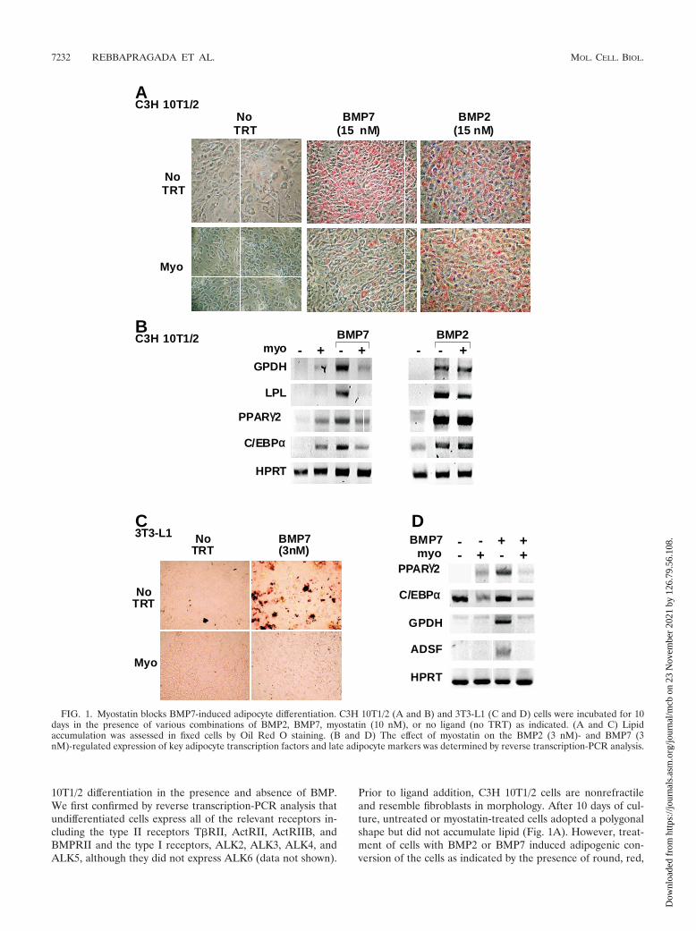
Prior to ligand addition, C3H 10T1/2 cells are nonrefractileand resemble fibroblasts in morphology. After 10 days of cul-ture, untreated or myostatin-treated cells adopted a polygonalshape but did not accumulate lipid (Fig. 1A). However, treat-ment of cells with BMP2 or BMP7 induced adipogenic con-version of the cells as indicated by the presence of round, red,
NoTRT
Myo
BMP7(3nM)
NoTRT
NoTRT
Myo
BMP7(15 nM)
BMP2(15 nM)
NoTRT
A
BC3H 10T1/2
myo - - - -+ + +BMP7 BMP2
PPARγγ2
C/EBPα
GPDH
HPRT
LPL
C D
ADSF
HPRT
C/EBPα
PPARγ2
GPDH
BMP7 - - + +-myo + +-
3T3-L1
C3H 10T1/2
FIG. 1. Myostatin blocks BMP7-induced adipocyte differentiation. C3H 10T1/2 (A and B) and 3T3-L1 (C and D) cells were incubated for 10days in the presence of various combinations of BMP2, BMP7, myostatin (10 nM), or no ligand (no TRT) as indicated. (A and C) Lipidaccumulation was assessed in fixed cells by Oil Red O staining. (B and D) The effect of myostatin on the BMP2 (3 nM)- and BMP7 (3nM)-regulated expression of key adipocyte transcription factors and late adipocyte markers was determined by reverse transcription-PCR analysis.
7232 REBBAPRAGADA ET AL. MOL. CELL. BIOL.
Dow
nloa
ded
from
http
s://j
ourn
als.
asm
.org
/jour
nal/m
cb o
n 23
Nov
embe
r 20
21 b
y 12
6.79
.56.
108.
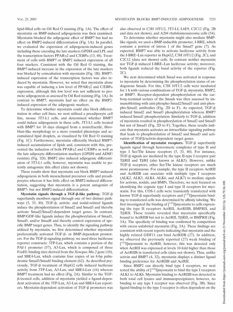
lipid-filled cells on Oil Red O staining (Fig. 1A). The effect ofmyostatin on BMP-induced adipogenesis was then examined.Myostatin blocked the adipogenic effect of BMP7 but had noeffect on BMP2-induced differentiation (Fig. 1A). In parallel,we evaluated the expression of adipogenesis-induced genesincluding those encoding the late markers GPDH and LPL andthe transcription factors PPAR�2 and C/EBP� (13, 48). Treat-ment of cells with BMP7 or BMP2 induced expression of allfour markers. Consistent with the Oil Red O staining, theBMP7-induced increase in the expression of the late markerswas blocked by coincubation with myostatin (Fig. 1B). BMP7-induced expression of the transcription factors was also re-duced by myostatin. However, we noted that myostatin alonewas capable of inducing a low level of PPAR�2 and C/EBP�expression, although this low level was not sufficient to pro-mote adipogenesis as assessed by Oil Red staining (Fig. 1A). Incontrast to BMP7, myostatin had no effect on the BMP2-induced expression of the adipogenic markers.
To determine whether myostatin could also block differen-tiation in other cell lines, we next utilized a preadipocyte cellline, mouse 3T3-L1 cells, and determined whether BMP7could induce adipogenesis in these cells. 3T3-L1 cells treatedwith BMP7 for 10 days changed from a nonrefractile, fibro-blast-like morphology to a more rounded phenotype and ac-cumulated lipid droplets, as visualized by Oil Red O staining(Fig. 1C). Furthermore, myostatin efficiently blocked BMP7-induced accumulation of lipids and, consistent with this, pre-vented the induction of both PPAR�2 and C/EBP� as well asthe late adipocyte differentiation markers (GPDH and ADSF/resistin) (Fig. 1D). BMP2 also induced adipogenic differenti-ation of 3T3-L1 cells; however, myostatin was unable to po-tently antagonize this effect (data not shown).
These results show that myostatin can block BMP7-inducedadipogenesis in both mesenchymal precursor cells and preadi-pocytes whereas it has little effect on BMP2-induced differen-tiation, suggesting that myostatin is a potent antagonist ofBMP7- but not BMP2-induced differentiation.
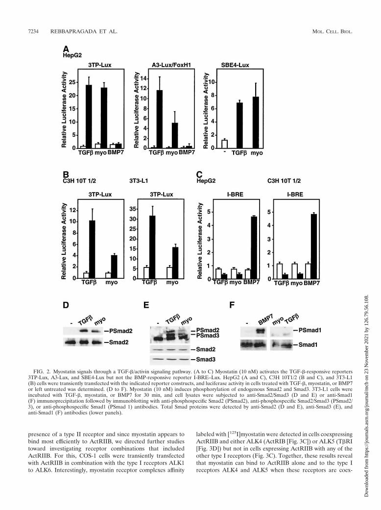
Myostatin signals through a TGF-�-like pathway. TGF-�superfamily members signal through one of two distinct path-ways (5, 33, 40). TGF-�, activin- and nodal-related ligandsinduce the phosphorylation of Smad2 and Smad3 and therebyactivate Smad2/Smad3-dependent target genes. In contrast,BMP/GDF-like ligands induce the phosphorylation of Smad1,Smad5, and/or Smad8 and thereby control expression of spe-cific BMP target genes. Thus, to identify the signaling pathwayutilized by myostatin, we first determined whether myostatinpreferentially activated TGF-�- or BMP-dependent promot-ers. For the TGF-� signaling pathway, we used three luciferasereporter constructs: 3TP-Lux, which contains a portion of thePAI-1 promoter (57), A3-Lux, which is composed of threeFoxH1 binding sites derived from the Xenopus Mix.2 gene (10),and SBE4-Lux, which contains four copies of an 8-bp palin-dromic Smad3/Smad4 binding element (62). As described pre-viously, TGF-� treatment of HepG2 cells induced luciferaseactivity from 3TP-Lux, A3-Lux, and SBE4-Lux (16) whereasBMP7 treatment had no effect (Fig. 2A). Similar to the TGF-�-treated cells, addition of myostatin yielded a ligand-depen-dent activation of the 3TP-Lux, A3-Lux and SBE4-Lux report-ers. Myostatin-dependent activation of TGF-� promoters was
also observed in C3H 10T1/2, 3T3-LI, L6E9, C2C12 (Fig. 2Band data not shown), and A204 rhabdomyosarcoma cells (54).
To determine whether myostatin might also mediate BMP-like signals, we used a BMP-inducible promoter, I-BRE, whichcontains a portion of intron 1 of the Smad7 gene (7). Asexpected, BMP7 was able to activate luciferase activity fromthe I-BRE–Lux reporter in HepG2, C3H 10T1/2 (Fig. 2C), andC2C12 (data not shown) cells. In contrast neither myostatinnor TGF-� induced I-BRE–Lux luciferase activity; moreover,both ligands reduced the basal activity of the reporter (Fig.2C).
We next determined which Smad was activated in responseto myostatin by determining the phosphorylation status of en-dogenous Smads. For this, C3H 10T1/2 cells were incubatedfor 1 h with various combinations of TGF-�, myostatin, BMP2,or BMP7. Receptor-dependent phosphorylation on the car-boxy-terminal serines of the Smads was then assessed by im-munoblotting with anti-phospho-Smad2/Smad3 and anti-phos-pho-Smad1 antibodies (Fig. 2D to F). As expected, TGF-�induced Smad2 and Smad3 phosphorylation whereas BMP7induced Smad1 phosphorylation. Similarly to TGF-�, additionof myostatin resulted in phosphorylation of Smad2 and Smad3but not of Smad1 (Fig. 2D to F). Together, these results indi-cate that myostatin activates an intracellular signaling pathwaythat leads to phosphorylation of Smad2 and Smad3 and acti-vation of TGF�/activin-dependent target genes.
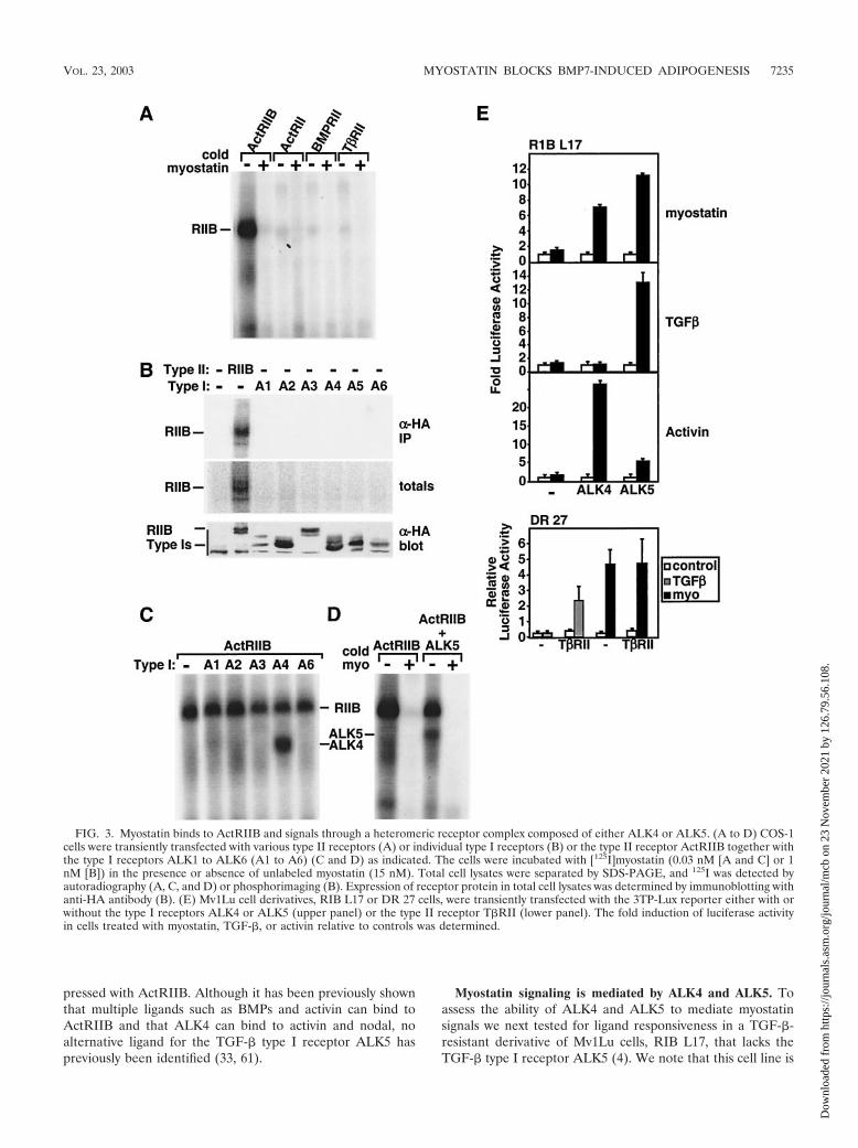
Identification of myostatin receptors. TGF-� superfamilyligands signal through heteromeric complexes of type II andtype I Ser/Thr kinase receptors (5, 33, 40). For instance,TGF-� signals are mediated by the type II-type I receptor pairT�RII and T�RI (also known as ALK5). However, unlikeTGF-� receptors, other Ser/Thr kinase receptors are muchmore promiscuous. For example, the type II receptors ActRIIand ActRIIB can associate with multiple type I receptors(ALK2, ALK3, ALK4, ALK6, and ALK7) to mediate signalsfor activins, nodals, and BMPs. Therefore, we next focused onidentifying the cognate type I and type II receptors for myo-statin. For this, COS-1 cells were transiently transfected withknown TGF-� superfamily receptors and [125I]myostatin bind-ing to transfected cells was determined by affinity labeling. Wefirst investigated the binding of [125I]myostatin to cells express-ing the type II receptors ActRII, ActRIIB, BMPRII, andT�RII. These results revealed that myostatin specificallybound to ActRIIB but not to ActRII, T�RII, or BMPRII (Fig.3A). The specificity of binding was confirmed by competitionwith excess unlabeled myostatin (Fig. 3A). These findings areconsistent with recent reports indicating that myostatin and thehighly related GDF11 can bind ActRIIB (27). In addition,we observed the previously reported (27) weak binding of[125I]myostatin to ActRII; however, this was detected onlywhen ActRII was expressed at levels 10-fold higher than thoseof ActRIIB in transfected cells (data not shown). Thus, unlikeactivin and BMP7 (4, 32), myostatin displays a distinct ligandbinding preference for ActRIIB and ActRII.
Since BMP2 can directly bind type I receptors, we nexttested the ability of [125I]myostatin to bind the type I receptorsALK1 to ALK6. Myostatin binding to ActRIIB was detected inboth total cell lysates and immunoprecipitates; however, nobinding to any type I receptor was observed (Fig. 3B). Sinceligand binding to the type I receptor is often dependent on the
VOL. 23, 2003 MYOSTATIN BLOCKS BMP7-INDUCED ADIPOGENESIS 7233
Dow
nloa
ded
from
http
s://j
ourn
als.
asm
.org
/jour
nal/m
cb o
n 23
Nov
embe
r 20
21 b
y 12
6.79
.56.
108.
presence of a type II receptor and since myostatin appears tobind most efficiently to ActRIIB, we directed further studiestoward investigating receptor combinations that includedActRIIB. For this, COS-1 cells were transiently transfectedwith ActRIIB in combination with the type I receptors ALK1to ALK6. Interestingly, myostatin receptor complexes affinity
labeled with [125I]myostatin were detected in cells coexpressingActRIIB and either ALK4 (ActRIB [Fig. 3C]) or ALK5 (T�RI[Fig. 3D]) but not in cells expressing ActRIIB with any of theother type I receptors (Fig. 3C). Together, these results revealthat myostatin can bind to ActRIIB alone and to the type Ireceptors ALK4 and ALK5 when these receptors are coex-
FIG. 2. Myostatin signals through a TGF-�/activin signaling pathway. (A to C) Myostatin (10 nM) activates the TGF-�-responsive reporters3TP-Lux, A3-Lux, and SBE4-Lux but not the BMP-responsive reporter I-BRE–Lux. HepG2 (A and C), C3H 10T1/2 (B and C), and 3T3-L1(B) cells were transiently transfected with the indicated reporter constructs, and luciferase activity in cells treated with TGF-�, myostatin, or BMP7or left untreated was determined. (D to F). Myostatin (10 nM) induces phosphorylation of endogenous Smad2 and Smad3. 3T3-L1 cells wereincubated with TGF-�, myostatin, or BMP7 for 30 min, and cell lysates were subjected to anti-Smad2/Smad3 (D and E) or anti-Smad1(F) immunoprecipitation followed by immunoblotting with anti-phosphospecific Smad2 (PSmad2), anti-phosphospecific Smad2/Smad3 (PSmad2/3), or anti-phosphospecific Smad1 (PSmad 1) antibodies. Total Smad proteins were detected by anti-Smad2 (D and E), anti-Smad3 (E), andanti-Smad1 (F) antibodies (lower panels).
7234 REBBAPRAGADA ET AL. MOL. CELL. BIOL.
Dow
nloa
ded
from
http
s://j
ourn
als.
asm
.org
/jour
nal/m
cb o
n 23
Nov
embe
r 20
21 b
y 12
6.79
.56.
108.
pressed with ActRIIB. Although it has been previously shownthat multiple ligands such as BMPs and activin can bind toActRIIB and that ALK4 can bind to activin and nodal, noalternative ligand for the TGF-� type I receptor ALK5 haspreviously been identified (33, 61).
Myostatin signaling is mediated by ALK4 and ALK5. Toassess the ability of ALK4 and ALK5 to mediate myostatinsignals we next tested for ligand responsiveness in a TGF-�-resistant derivative of Mv1Lu cells, RIB L17, that lacks theTGF-� type I receptor ALK5 (4). We note that this cell line is
FIG. 3. Myostatin binds to ActRIIB and signals through a heteromeric receptor complex composed of either ALK4 or ALK5. (A to D) COS-1cells were transiently transfected with various type II receptors (A) or individual type I receptors (B) or the type II receptor ActRIIB together withthe type I receptors ALK1 to ALK6 (A1 to A6) (C and D) as indicated. The cells were incubated with [125I]myostatin (0.03 nM [A and C] or 1nM [B]) in the presence or absence of unlabeled myostatin (15 nM). Total cell lysates were separated by SDS-PAGE, and 125I was detected byautoradiography (A, C, and D) or phosphorimaging (B). Expression of receptor protein in total cell lysates was determined by immunoblotting withanti-HA antibody (B). (E) Mv1Lu cell derivatives, RIB L17 or DR 27 cells, were transiently transfected with the 3TP-Lux reporter either with orwithout the type I receptors ALK4 or ALK5 (upper panel) or the type II receptor T�RII (lower panel). The fold induction of luciferase activityin cells treated with myostatin, TGF-�, or activin relative to controls was determined.
VOL. 23, 2003 MYOSTATIN BLOCKS BMP7-INDUCED ADIPOGENESIS 7235
Dow
nloa
ded
from
http
s://j
ourn
als.
asm
.org
/jour
nal/m
cb o
n 23
Nov
embe
r 20
21 b
y 12
6.79
.56.
108.
also insensitive to activin due to extremely low levels of theactivin type I receptor ALK4 (4). Treatment of cells withTGF-� or activin failed to activate 3TP-Lux reporter activity inthe absence of coexpressed type I receptors (Fig. 3E). How-ever, transient coexpression of ALK5 restored TGF-�- but notactivin-dependent activation of luciferase activity whereas co-transfection of ALK4 restored activin but not TGF-� signaling(Fig. 3E), consistent with the known binding specificity of thesetype I receptors. Addition of myostatin also failed to activatethe 3TP-Lux reporter in control cells; however, coexpression ofeither ALK4 or ALK5 resulted in myostatin-dependent acti-vation of 3TP-Lux reporter activity (Fig. 3E). Unlike for RIBL17 cells, myostatin signaling was not impaired in the activin-sensitive but TGF-�-resistant Mv1Lu derivative, DR 27, whichlacks the TGF-� type II receptor but expresses normal type Ireceptors (Fig. 3E). Furthermore, coexpression of T�RII re-stored TGF-� responsiveness to these cells but had no effecton myostatin-dependent signaling. Together with the ligandbinding assays, our results reveal that myostatin signals throughheteromeric receptor complexes composed of ActRIIB to-gether with either ALK4 or ALK5 to induce Smad2 and Smad3phosphorylation and activate a TGF-�-like signaling pathway.
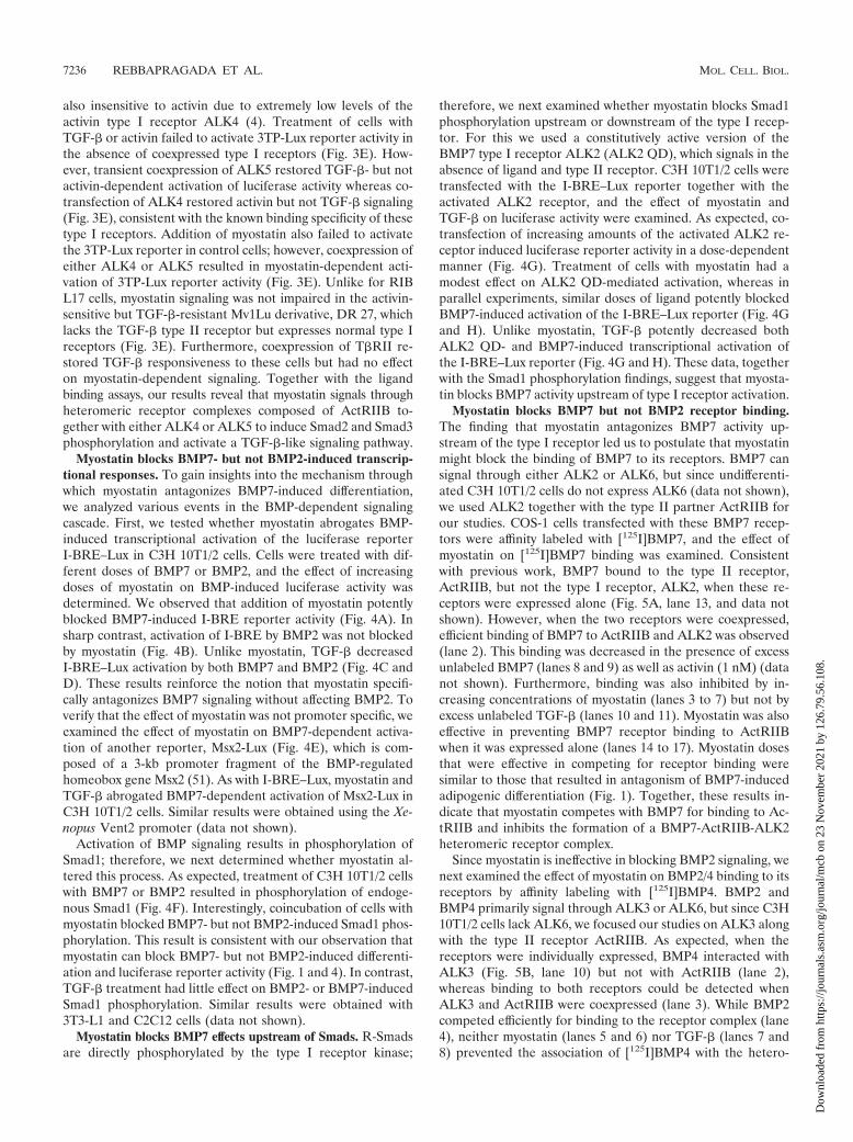
Myostatin blocks BMP7- but not BMP2-induced transcrip-tional responses. To gain insights into the mechanism throughwhich myostatin antagonizes BMP7-induced differentiation,we analyzed various events in the BMP-dependent signalingcascade. First, we tested whether myostatin abrogates BMP-induced transcriptional activation of the luciferase reporterI-BRE–Lux in C3H 10T1/2 cells. Cells were treated with dif-ferent doses of BMP7 or BMP2, and the effect of increasingdoses of myostatin on BMP-induced luciferase activity wasdetermined. We observed that addition of myostatin potentlyblocked BMP7-induced I-BRE reporter activity (Fig. 4A). Insharp contrast, activation of I-BRE by BMP2 was not blockedby myostatin (Fig. 4B). Unlike myostatin, TGF-� decreasedI-BRE–Lux activation by both BMP7 and BMP2 (Fig. 4C andD). These results reinforce the notion that myostatin specifi-cally antagonizes BMP7 signaling without affecting BMP2. Toverify that the effect of myostatin was not promoter specific, weexamined the effect of myostatin on BMP7-dependent activa-tion of another reporter, Msx2-Lux (Fig. 4E), which is com-posed of a 3-kb promoter fragment of the BMP-regulatedhomeobox gene Msx2 (51). As with I-BRE–Lux, myostatin andTGF-� abrogated BMP7-dependent activation of Msx2-Lux inC3H 10T1/2 cells. Similar results were obtained using the Xe-nopus Vent2 promoter (data not shown).
Activation of BMP signaling results in phosphorylation ofSmad1; therefore, we next determined whether myostatin al-tered this process. As expected, treatment of C3H 10T1/2 cellswith BMP7 or BMP2 resulted in phosphorylation of endoge-nous Smad1 (Fig. 4F). Interestingly, coincubation of cells withmyostatin blocked BMP7- but not BMP2-induced Smad1 phos-phorylation. This result is consistent with our observation thatmyostatin can block BMP7- but not BMP2-induced differenti-ation and luciferase reporter activity (Fig. 1 and 4). In contrast,TGF-� treatment had little effect on BMP2- or BMP7-inducedSmad1 phosphorylation. Similar results were obtained with3T3-L1 and C2C12 cells (data not shown).
Myostatin blocks BMP7 effects upstream of Smads. R-Smadsare directly phosphorylated by the type I receptor kinase;
therefore, we next examined whether myostatin blocks Smad1phosphorylation upstream or downstream of the type I recep-tor. For this we used a constitutively active version of theBMP7 type I receptor ALK2 (ALK2 QD), which signals in theabsence of ligand and type II receptor. C3H 10T1/2 cells weretransfected with the I-BRE–Lux reporter together with theactivated ALK2 receptor, and the effect of myostatin andTGF-� on luciferase activity were examined. As expected, co-transfection of increasing amounts of the activated ALK2 re-ceptor induced luciferase reporter activity in a dose-dependentmanner (Fig. 4G). Treatment of cells with myostatin had amodest effect on ALK2 QD-mediated activation, whereas inparallel experiments, similar doses of ligand potently blockedBMP7-induced activation of the I-BRE–Lux reporter (Fig. 4Gand H). Unlike myostatin, TGF-� potently decreased bothALK2 QD- and BMP7-induced transcriptional activation ofthe I-BRE–Lux reporter (Fig. 4G and H). These data, togetherwith the Smad1 phosphorylation findings, suggest that myosta-tin blocks BMP7 activity upstream of type I receptor activation.
Myostatin blocks BMP7 but not BMP2 receptor binding.The finding that myostatin antagonizes BMP7 activity up-stream of the type I receptor led us to postulate that myostatinmight block the binding of BMP7 to its receptors. BMP7 cansignal through either ALK2 or ALK6, but since undifferenti-ated C3H 10T1/2 cells do not express ALK6 (data not shown),we used ALK2 together with the type II partner ActRIIB forour studies. COS-1 cells transfected with these BMP7 recep-tors were affinity labeled with [125I]BMP7, and the effect ofmyostatin on [125I]BMP7 binding was examined. Consistentwith previous work, BMP7 bound to the type II receptor,ActRIIB, but not the type I receptor, ALK2, when these re-ceptors were expressed alone (Fig. 5A, lane 13, and data notshown). However, when the two receptors were coexpressed,efficient binding of BMP7 to ActRIIB and ALK2 was observed(lane 2). This binding was decreased in the presence of excessunlabeled BMP7 (lanes 8 and 9) as well as activin (1 nM) (datanot shown). Furthermore, binding was also inhibited by in-creasing concentrations of myostatin (lanes 3 to 7) but not byexcess unlabeled TGF-� (lanes 10 and 11). Myostatin was alsoeffective in preventing BMP7 receptor binding to ActRIIBwhen it was expressed alone (lanes 14 to 17). Myostatin dosesthat were effective in competing for receptor binding weresimilar to those that resulted in antagonism of BMP7-inducedadipogenic differentiation (Fig. 1). Together, these results in-dicate that myostatin competes with BMP7 for binding to Ac-tRIIB and inhibits the formation of a BMP7-ActRIIB-ALK2heteromeric receptor complex.
Since myostatin is ineffective in blocking BMP2 signaling, wenext examined the effect of myostatin on BMP2/4 binding to itsreceptors by affinity labeling with [125I]BMP4. BMP2 andBMP4 primarily signal through ALK3 or ALK6, but since C3H10T1/2 cells lack ALK6, we focused our studies on ALK3 alongwith the type II receptor ActRIIB. As expected, when thereceptors were individually expressed, BMP4 interacted withALK3 (Fig. 5B, lane 10) but not with ActRIIB (lane 2),whereas binding to both receptors could be detected whenALK3 and ActRIIB were coexpressed (lane 3). While BMP2competed efficiently for binding to the receptor complex (lane4), neither myostatin (lanes 5 and 6) nor TGF-� (lanes 7 and8) prevented the association of [125I]BMP4 with the hetero-
7236 REBBAPRAGADA ET AL. MOL. CELL. BIOL.
Dow
nloa
ded
from
http
s://j
ourn
als.
asm
.org
/jour
nal/m
cb o
n 23
Nov
embe
r 20
21 b
y 12
6.79
.56.
108.
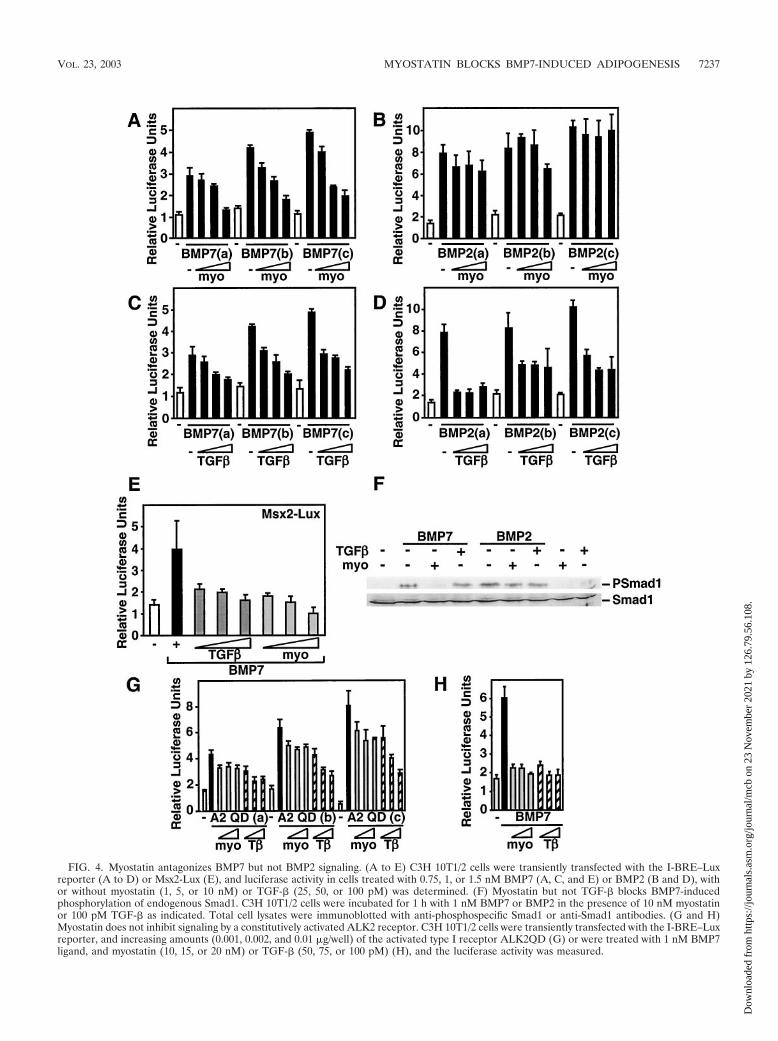
FIG. 4. Myostatin antagonizes BMP7 but not BMP2 signaling. (A to E) C3H 10T1/2 cells were transiently transfected with the I-BRE–Luxreporter (A to D) or Msx2-Lux (E), and luciferase activity in cells treated with 0.75, 1, or 1.5 nM BMP7 (A, C, and E) or BMP2 (B and D), withor without myostatin (1, 5, or 10 nM) or TGF-� (25, 50, or 100 pM) was determined. (F) Myostatin but not TGF-� blocks BMP7-inducedphosphorylation of endogenous Smad1. C3H 10T1/2 cells were incubated for 1 h with 1 nM BMP7 or BMP2 in the presence of 10 nM myostatinor 100 pM TGF-� as indicated. Total cell lysates were immunoblotted with anti-phosphospecific Smad1 or anti-Smad1 antibodies. (G and H)Myostatin does not inhibit signaling by a constitutively activated ALK2 receptor. C3H 10T1/2 cells were transiently transfected with the I-BRE–Luxreporter, and increasing amounts (0.001, 0.002, and 0.01 �g/well) of the activated type I receptor ALK2QD (G) or were treated with 1 nM BMP7ligand, and myostatin (10, 15, or 20 nM) or TGF-� (50, 75, or 100 pM) (H), and the luciferase activity was measured.
VOL. 23, 2003 MYOSTATIN BLOCKS BMP7-INDUCED ADIPOGENESIS 7237
Dow
nloa
ded
from
http
s://j
ourn
als.
asm
.org
/jour
nal/m
cb o
n 23
Nov
embe
r 20
21 b
y 12
6.79
.56.
108.
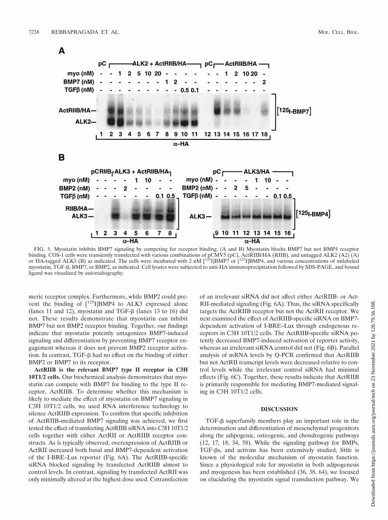
meric receptor complex. Furthermore, while BMP2 could pre-vent the binding of [125I]BMP4 to ALK3 expressed alone(lanes 11 and 12), myostatin and TGF-� (lanes 13 to 16) didnot. These results demonstrate that myostatin can inhibitBMP7 but not BMP2 receptor binding. Together, our findingsindicate that myostatin potently antagonizes BMP7-inducedsignaling and differentiation by preventing BMP7 receptor en-gagement whereas it does not prevent BMP2 receptor activa-tion. In contrast, TGF-� had no effect on the binding of eitherBMP2 or BMP7 to its receptor.
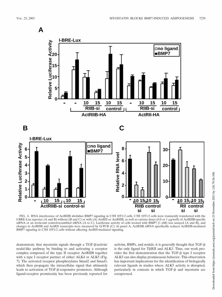
ActRIIB is the relevant BMP7 type II receptor in C3H10T1/2 cells. Our biochemical analysis demonstrates that myo-statin can compete with BMP7 for binding to the type II re-ceptor, ActRIIB. To determine whether this mechanism islikely to mediate the effect of myostatin on BMP7 signaling inC3H 10T1/2 cells, we used RNA interference technology tosilence ActRIIB expression. To confirm that specific inhibitionof ActRIIB-mediated BMP7 signaling was achieved, we firsttested the effect of transfecting ActRIIB siRNA into C3H 10T1/2cells together with either ActRII or ActRIIB receptor con-structs. As is typically observed, overexpression of ActRIIB orActRII increased both basal and BMP7-dependent activationof the I-BRE–Lux reporter (Fig. 6A). The ActRIIB-specificsiRNA blocked signaling by transfected ActRIIB almost tocontrol levels. In contrast, signaling by transfected ActRII wasonly minimally altered at the highest dose used. Cotransfection
of an irrelevant siRNA did not affect either ActRIIB- or Act-RII-mediated signaling (Fig. 6A). Thus, the siRNA specificallytargets the ActRIIB receptor but not the ActRII receptor. Wenext examined the effect of ActRIIB-specific siRNA on BMP7-dependent activation of I-BRE–Lux through endogenous re-ceptors in C3H 10T1/2 cells. The ActRIIB-specific siRNA po-tently decreased BMP7-induced activation of reporter activity,whereas an irrelevant siRNA control did not (Fig. 6B). Parallelanalysis of mRNA levels by Q-PCR confirmed that ActRIIBbut not ActRII transcript levels were decreased relative to con-trol levels while the irrelevant control siRNA had minimaleffects (Fig. 6C). Together, these results indicate that ActRIIBis primarily responsible for mediating BMP7-mediated signal-ing in C3H 10T1/2 cells.
DISCUSSION
TGF-� superfamily members play an important role in thedetermination and differentiation of mesenchymal progenitorsalong the adipogenic, osteogenic, and chondrogenic pathways(12, 17, 18, 34, 58). While the signaling pathway for BMPs,TGF-�s, and activins has been extensively studied, little isknown of the molecular mechanism of myostatin function.Since a physiological role for myostatin in both adipogenesisand myogenesis has been established (36, 38, 64), we focusedon elucidating the myostatin signal transduction pathway. We
FIG. 5. Myostatin inhibits BMP7 signaling by competing for receptor binding. (A and B) Myostatin blocks BMP7 but not BMP4 receptorbinding. COS-1 cells were transiently transfected with various combinations of pCMV5 (pC), ActRIIB/HA (RIIB), and untagged ALK2 (A2) (A)or HA-tagged ALK3 (B) as indicated. The cells were incubated with 2 nM [125I]BMP7 or [125I]BMP4, and various concentrations of unlabeledmyostatin, TGF-�, BMP7, or BMP2, as indicated. Cell lysates were subjected to anti-HA immunoprecipitation followed by SDS-PAGE, and boundligand was visualized by autoradiography.
7238 REBBAPRAGADA ET AL. MOL. CELL. BIOL.
Dow
nloa
ded
from
http
s://j
ourn
als.
asm
.org
/jour
nal/m
cb o
n 23
Nov
embe
r 20
21 b
y 12
6.79
.56.
108.
demonstrate that myostatin signals through a TGF-�/activin/nodal-like pathway by binding to and activating a receptorcomplex composed of the type II receptor ActRIIB togetherwith a type I receptor partner of either ALK4 or ALK5 (Fig.7). The activated receptor phosphorylates Smad2 and Smad3,which then propagate the intracellular signal that ultimatelyleads to activation of TGF-�-responsive promoters. Althoughligand-receptor promiscuity has been previously reported for
activins, BMPs, and nodals, it is generally thought that TGF-�is the only ligand for T�RII and ALK5. Thus, our work pro-vides the first demonstration that the TGF-� type I receptorALK5 can also display promiscuous behavior. This observationhas important implications for the identification of biologicallyrelevant ligands in studies where ALK5 activity is disrupted,particularly in contexts in which TGF-� and myostatin arecoexpressed.
0
1
2
3
4
5
6no ligandBMP7
- 1510 10 15ActRIIB-si control-si
I-BRE-Lux
B
0
5
10
15
20
AI-BRE-Lux
no ligandBMP7
- -
ActRIIB-HARIIB-si control
ActRII-HA
-
0
2
4
6
8
- 1510 10 15RIIB
sicontrol
si
Rel
ativ
e R
NA
leve
l
C
1510 1510 1510 1510RIIB-si control
Rel
ativ
e L
uci
fera
se A
ctiv
ity
Rel
ativ
e L
uci
fera
se A
ctiv
ity
- 1510 10 15RIIsi
controlsi
10
20
30
0
FIG. 6. RNA interference of ActRIIB abolishes BMP7 signaling in C3H 10T1/2 cells. C3H 10T1/2 cells were transiently transfected with theI-BRE-Lux reporter (A and B) without (B and C) or with (A) ActRII or ActRIIB, as well as various doses (0.8 or 1 �g/well) of ActRIIB-specificsiRNA or an irrelevant control/scrambled siRNA (A to C). Luciferase activity of cells treated with BMP7 (1 nM) was assayed (A and B), andchanges in ActRIIB and ActRII transcripts were measured by Q-PCR (C). In panel A, ActRIIB siRNA specifically reduces ActRIIB-mediatedBMP7 signaling in C3H 10T1/2 cells without affecting ActRII-mediated signaling.
VOL. 23, 2003 MYOSTATIN BLOCKS BMP7-INDUCED ADIPOGENESIS 7239
Dow
nloa
ded
from
http
s://j
ourn
als.
asm
.org
/jour
nal/m
cb o
n 23
Nov
embe
r 20
21 b
y 12
6.79
.56.
108.
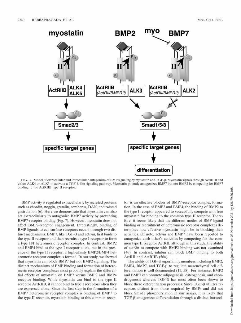
BMP activity is regulated extracellularly by secreted proteinssuch as chordin, noggin, gremlin, cereberus, DAN, and twistedgastrulation (6). Here we demonstrate that myostatin can alsoact extracellularly to antagonize BMP7 activity by preventingBMP7-receptor binding (Fig. 7). However, myostatin does notaffect BMP2-receptor engagement. Interestingly, binding ofBMP ligands to cell surface receptors occurs through two dis-tinct mechanisms. BMP7, like TGF-� and activin, first binds tothe type II receptor and then recruits a type I receptor to forma type II/I heteromeric receptor complex. In contrast, BMP2and BMP4 bind to the type I receptor alone, but in the pres-ence of the type II receptor, a high-affinity BMP2/BMP4 het-eromeric receptor complex is formed. In our study, we showedthat myostatin can block BMP7 but not BMP2 signaling. Thedistinct mechanisms of BMP binding and formation of hetero-meric receptor complexes most probably explain the differen-tial effects of myostatin on BMP7 versus BMP2 and BMP4receptor binding. While myostatin can bind to the type IIreceptor ActRIIB, it cannot bind to type I receptors when theyare expressed alone. Since the first step in the formation of aBMP7 heteromeric receptor complex is binding of BMP7 tothe type II receptor, myostatin binding to this common recep-
tor is an effective blocker of BMP7-receptor complex forma-tion. In the case of BMP2 and BMP4, the binding of BMP2 tothe type I receptor appeared to successfully compete with freemyostatin for binding to the common type II receptor. There-fore, it seems likely that the different modes of BMP ligandbinding or recruitment of heteromeric receptor complexes de-termines how effective myostatin might be in blocking theiractivities. Of note, activin and BMP7 have been reported toantagonize each other’s activities by competing for the com-mon type II receptor ActRII, although in this study, the abilityof activin to compete with BMP2 binding was not examined(46). In contrast, inhibin can block BMP binding to bothActRII and ActRIIB (56a).
The ability of TGF-� superfamily members including BMP2,BMP4, BMP7, and TGF-� to regulate mesenchymal cell dif-ferentiation is well documented (17, 58). For instance, BMP2and BMP7 can promote adipogenesis, osteogenesis, and chon-drogenesis whereas TGF-� has most often been shown toblock these differentiation processes. Since TGF-� utilizes re-ceptors distinct from those required by BMPs and did notblock Smad1 phosphorylation in our assays, it is likely thatTGF-� antagonizes differentiation through a distinct intracel-
FIG. 7. Model of extracellular and intracellular antagonism of BMP signaling by myostatin and TGF-�. Myostatin signals through ActRIIB andeither ALK4 or ALK5 to activate a TGF-�-like signaling pathway. Myostatin potently antagonizes BMP7 but not BMP2 by competing for BMP7binding to the ActRIIB type II receptor.
7240 REBBAPRAGADA ET AL. MOL. CELL. BIOL.
Dow
nloa
ded
from
http
s://j
ourn
als.
asm
.org
/jour
nal/m
cb o
n 23
Nov
embe
r 20
21 b
y 12
6.79
.56.
108.
lular mechanism. As myostatin and TGF-� utilize similar in-tracellular signaling cascades, it would seem that both ligandsshould be able to utilize this common intracellular mechanismto inhibit differentiation, and perhaps myostatin does so. How-ever, the ability of myostatin to block BMP7 extracellularlyprovides for an important distinction. Cells exposed to bothBMP7 and TGF-� would activate both Smad1 and Smad2,which are opposing pathways, and the final determination ofcell fate may depend on the relative balance between the twosignals. In contrast, by blocking Smad1 phosphorylation as wellas activating the Smad2 pathway, myostatin provides for a pureTGF-�-like signal and may thereby have a more potent orperhaps distinct effect on differentiation. Presumably, this dis-tinction would be lost in BMP-independent differentiation pro-cesses. Interestingly, there are many examples in the literaturethat demonstrate that TGF-� effects on differentiation, pre-sumably through this intracellular pathway, can vary signifi-cantly depending on external factors such as cell type, densityof plating, length of treatment, and medium composition (forexample see references 14, 20, 21, and 63). The mechanism ofthis intracellular block remains unclear, although the possibil-ities include competition for limiting quantities of the commonSmad, Smad4 (55), Smad-dependent regulation of specifictranscriptional events (2, 29), and TGF-�-mediated activationof distinct Smad pathways through distinct ALKs (44). Therelative contributions of myostatin-mediated extracellular ver-sus intracellular competition in vivo remain to be determined;however, a more detailed understanding of the intracellularpathway of antagonism is required before this can be exam-ined.
In addition to ActRIIB, both BMP2 and BMP7 signalthrough the type II receptors ActRII and BMPRII. Similarly,activin can bind equivalently to ActRII and ActRIIB. In con-trast, myostatin displays a marked preference for ActRIIB overActRII (27; also see above), which is consistent with the ideathat there are distinct functions for these two related receptors.In the context of BMP receptor antagonism, binding of myo-statin primarily to ActRIIB suggests that myostatin wouldbe an ineffective extracellular inhibitor of BMP7 signalingthrough the other type II receptors. While ActRIIB contrib-utes significantly to BMP7-induced responses in our modelsystem (C3H 10T1/2 cells), other lineages may preferentiallysignal through ActRII or BMPRII. Therefore, a receptor-de-pendent differential ability of myostatin to block BMP7 activitymay be particularly important in certain biological contexts,where changes in expression patterns of these type II receptorsmay determine whether BMP7 signals and associated differ-entiation events are negatively regulated by myostatin. Consis-tent with this possibility, ActRII and ActRIIB display bothcommon and distinct expression patterns in the developingchicken limb (39, 42). Although extremely speculative, it maybe that BMP-dependent patterning of bone and cartilaginouselements within the limb bud may be refined by myostatin,which is expressed in the myotome layer of adjacent somitesthat migrate into the limb to form the musculature (36). Bydifferentially regulating BMPs and by functioning only in thecontext of appropriate receptor combinations, myostatin mayprovide for exquisite control of complex differentiation pro-cesses. Although BMPs have been implicated in the control ofmuscle growth and positioning during embryonic development,
their importance in adult muscle or fat homeostasis is notcurrently known. However, it is intriguing to speculate thatmuscle mass or fat accumulation in adults might also be con-trolled by a balance between myostatin and BMP. If so, it willbe important to consider how systemic administration of myo-statin might alter BMP-dependent events in adults.
ACKNOWLEDGMENTS
We thank K. Cho (Xvent2), S. Ghosh (Msx2-Lux), and B. Vogelstein(SBE4-Lux) for plasmids.
This work was supported by grants to L.A. and J.L.W. from theCanadian Institute for Health Research (CIHR). H.B. is the recipientof a studentship from NSERC and the Heart and Stroke Foundationof Canada. L.A. and J.L.W. are CIHR Investigators.
REFERENCES
1. Aherns, M., T. Ankenbauer, D. Schroder, A. Hollnagel, H. Mayer, and G.Gross. 1993. Expression of human bone morphogenetic proteins-2 or -4 inmurine mesenchymal progenitor C3H 10T1/2 cells induces differentiationinto distinct mesenchymal cell lineages. DNA Cell Biol. 12:871–880.
2. Alliston, T., L. Choy, P. Ducy, G. Karsenty, and R. Derynck. 2001. TGF-�-induced repression of CBFA1 by Smad3 decreases cbfa1 and osteocalcinexpression and inhibits osteoblast differentiation. EMBO J. 20:2254–2272.
3. Asahina, I., T. K. Sampath, and P. V. Hauschka. 1996. Human osteogenicprotein-1 induces chondroblastic, osteoblastic, and/or adipocytic differenti-ation of clonal murine target cells. Exp. Cell Res. 222:38–47.
4. Attisano, L., J. Carcamo, F. Ventura, F. M. B. Weis, J. Massague, and J. L.Wrana. 1993. Identification of human activin and TGF-� type I receptorsthat form heteromeric kinase complexes with type II receptors. Cell 75:671–680.
5. Attisano, L., and J. L. Wrana. 2002. Signal transduction by the TGF-�superfamily. Science 296:1646–1647.
6. Balemans, W., and W. Van Hul. 2002. Extracellular regulation of BMPsignaling in vertebrates: a cocktail of modulators. Dev. Biol. 250:231–250.
7. Benchabane, H., and J. L. Wrana. 2003. GATA and Smad1-dependentenhancers in the Smad7 gene differentially interpret BMP gradients. Mol.Cell. Biol. 23:6646–6661.
8. Bogdanovich, S., T. O. Krag, E. R. Barton, L. D. Morris, L. A. Whittemore,R. S. Ahima, and T. S. Khurana. 2002. Functional improvement of dystro-phic muscle by myostatin blockade. Nature 420:418–421.
9. Brummel, T., V. Twombly, G. Marques, J. L. Wrana, S. Newfeld, L. Attisano,J. Massague, M. B. O’Connor, and W. M. Gelbart. 1994. Characterizationand relationship of dpp receptors encoded by the saxophone and thick veinsgenes in Drosophila. Cell 78:251–261.
10. Chen, X., M. J. Rubock, and M. Whitman. 1996. A transcriptional partnerfor MAD proteins in TGF-� signalling. Nature 383:691–696.
11. Colavita, A., S. Krishna, H. Zheng, R. W. Padgett, and J. G. Culotti. 1998.Pioneer axon guidance by UNC-129, a C. elegans TGF-�. Science 281:706–709.
12. Derynck, R., and L. Choy. 1998. Transforming growth factor-� and its re-ceptors, p. 593–636. In A. W. Thomson (ed.), The cytokine handbook, 3rded. Academic Press, Inc., Orlando, Fla.
13. Fajas, L., J. C. Fruchart, and J. Auwerx. 1998. Transcriptional control ofadipogenesis. Curr. Opin. Cell Biol. 10:165–173.
14. Filvaroff, E. H., R. Ebner, and R. Derynck. 1994. Inhibition of myogenicdifferentiation in myoblasts expressing a truncated type II TGF-beta recep-tor. Development 120:1085–1095.
15. Hansen, J. B., R. K. Petersen, B. M. Larsen, J. Bartkova, J. Alsner, and K.Kristiansen. 1999. Activation of peroxisome proliferator-activated receptorgamma bypasses the function of the retinoblastoma protein in adipocytedifferentiation. J. Biol. Chem. 274:2386–2393.
16. Hayashi, H., S. Abdollah, Y. Qiu, J. Cai, Y.-Y. Xu, B. W. Grinnell, M. A.Richardson, J. N. Topper, M. A. Gimbrone, Jr., J. L. Wrana, and D. Falb.1997. The MAD-related protein Smad7 associates with the TGF� receptorand functions as an antagonist of TGF� signaling. Cell 89:1165–1173.
17. Hoffman, A., and G. Gross. 2001. BMP signaling pathways in cartilage andbone formation. Crit. Rev. Eukaryot. Gene Expr. 11:23–45.
18. Hogan, B. L. 1996. Bone morphogenetic proteins in development. Curr.Opin. Genet. Dev. 7:856.
19. Hoodless, P. A., T. Haerry, S. Abdollah, M. Stapleton, M. B. O’Connor, L.Attisano, and J. L. Wrana. 1996. MADR1, a MAD-related protein thatfunctions in BMP2 signalling pathways. Cell 85:489–500.
20. Ignotz, R. A., and J. Massague. 1985. Type � transforming growth factorcontrols the adipogenic differentiation of 3T3 fibroblasts. Proc. Natl. Acad.Sci. USA 82:8530–8534.
21. Katagiri, T., A. Yamaguchi, M. Komaki, E. Abe, N. Takahashi, T. Ikeda, V.Rosen, J. M. Wozney, A. Fujisawa-Sehara, and T. Suda. 1994. Bone mor-
VOL. 23, 2003 MYOSTATIN BLOCKS BMP7-INDUCED ADIPOGENESIS 7241
Dow
nloa
ded
from
http
s://j
ourn
als.
asm
.org
/jour
nal/m
cb o
n 23
Nov
embe
r 20
21 b
y 12
6.79
.56.
108.
phogenetic protein-2 converts the differentiation pathway of C2C12 myo-blasts into the osteoblast lineage. J. Cell Biol. 127:1755–1766.
22. Kim, K. H., K. Lee, Y. S. Moon, and H. S. Sul. 2001. A cysteine-rich adiposetissue-specific secretory factor inhibits adipocyte differentiation. J. Biol.Chem. 276:11252–11256.
23. Kirsch, T., J. Nickel, and W. Sebald. 2000. BMP-2 antagonists emerge fromalterations in the low-affinity binding epitope for receptor BMPR-II. EMBOJ. 19:3314–3324.
24. Koenig, B. B., J. S. Cook, D. H. Wolsing, J. Ting, J. P. Tiesman, P. E. Correa,C. A. Olson, A. L. Pecquet, F. Ventura, R. A. Grant, G.-X. Chen, J. L. Wrana,J. Massague, and J. S. Rosenbaum. 1994. Characterization and cloning of areceptor for BMP-2 and BMP-4 from NIH 3T3 cells. Mol. Cell. Biol. 14:5961–5974.
25. Lapsys, N. M., A. D. Kriketos, M. Lim-Fraser, A. M. Poynten, A. Lowy, S. M.Furler, D. J. Chisholm, and G. J. Cooney. 2000. Expression of genes involvedin lipid-metabolism correlate with peroxisome proliferator-activated recep-tor gamma expression in human skeletal muscle. J. Clin. Endocrinol. Metab.85:4293–4297.
26. Lee, S.-J., and A. C. McPherron. 1999. Myostatin and the control of skeletalmuscle mass. Curr. Op. Genet. Dev. 9:604–607.
27. Lee, S.-J., and A. C. McPherron. 2001. Regulation of myostatin activity andmuscle growth. Proc. Natl. Acad. Sci. USA 98:9306–9311.
28. Lin, J., H. B. Arnold, M. A. Della-Fera, M. J. Azain, D. L. Hartzell, and C. A.Baile. 2002. Myostatin knockout in mice increases myogenesis and decreasesadipogenesis. Biochem. Biophys. Res. Commun. 291:701–706.
29. Liu, D., B. L. Black, and R. Derynck. 2001. TGF-� inhibits muscle differen-tiation through functional repression of myogenic transcription factors bySmad3. Genes Dev. 15:2950–2966.
30. Liu, F., F. Ventura, J. Doody, and J. Massague. 1995. Human type IIreceptor for bone morphogenetic proteins (BMPs): extension of the two-kinase receptor model to the BMPs. Mol. Cell. Biol. 15:3479–3486.
31. Lo, P. C. H., and M. Frasch. 1999. Sequence and expression of myoglianin,a novel Drosophila gene of the TGF-� superfamily. Mech. Dev. 86:171–175.
32. Macıas-Silva, M., P. A. Hoodless, S. J. Tang, M. Buchwald, and J. L. Wrana.1998. Specific activation of Smad1 signalling pathways by the BMP7 type Ireceptor, ALK 2. J. Biol. Chem. 273:25628–25636.
33. Massague, J. 1998. TGF-� signal transduction. Annu. Rev. Biochem. 67:753–791.
34. Massague, J. 1990. The transforming growth factor-� family. Annu. Rev.Cell Biol. 6:597–641.
35. McPherron, A. C., A. M. Lawler, and S.-J. Lee. 1999. Regulation of anterior/posterior patterning of the axial skeleton by growth/differentiation factor 11.Nat. Genet. 22:260–264.
36. McPherron, A. C., A. M. Lawler, and S. J. Lee. 1997. Regulation of skeletalmuscle mass in mice by a new TGF-beta superfamily member. Nature 387:83–90.
37. McPherron, A. C., and S.-J. Lee. 1997. Double muscling in cattle due tomutations in the myostatin gene. Proc. Natl. Acad. Sci. USA 94:12457–12461.
38. McPherron, A. C., and S. J. Lee. 2002. Suppresion of body fat accumulationin myostatin-deficient mice. J. Clin. Investig. 109:595–601.
39. Merino, R., D. Macias, Y. Ganan, J. Rodriguez-Leon, A. N. Economides, C.Rodriguez-Esteban, J. C. Izpisua-Belmonte, and J. M. Hurle. 1999. Controlof digit formation by activin signalling. Development 126:2161–2170.
40. Moustakas, A., S. Souchelnytskyi, and C.-H. Heldin. 2001. Smad regulationin TGF-� signal transduction. J. Cell Sci. 114:4359–4369.
41. Nohno, T., T. Ishikawa, T. Saito, K. Hosokawa, S. Noji, D. H. Wolsing, andJ. S. Rosenbaum. 1995. Identification of a human type II receptor for bonemorphogenetic protein-4 that forms differential heteromeric complexes withbone morphogenetic protein type I receptors. J. Biol. Chem. 270:22522–22526.
42. Nohno, T., S. Noji, E. Koyama, F. Myokai, H. Ohuchi, K. Nishikawa, S.Sumitomo, S. Taniguchi, and T. Saito. 1993. Expression patterns of theactivin receptor IIA and IIB genes during chick limb development. Prog.Clin. Biol. Res. 383B:705–714.
43. Novikoff, A. B., P. M. Novikoff, O. M. Rosen, and C. S. Rubin. 1980. Or-ganelle relationships in cultured 3T3-L1 preadipocytes. J. Cell Biol. 87:180–196.
44. Oh, S. P., T. Seki, K. A. Goss, T. Imamura, Y. Yi, P. K. Donahoe, L. Li, K.Miyazono, P. ten Dijke, S. Kim, and E. Li. 2000. Activin receptor-like kinase
1 modulates transforming growth factor-beta 1 signaling in the regulation ofangiogenesis. Proc. Natl. Acad. Sci. USA 97:2626–2631.
45. Penton, A., Y. Chen, K. Staehling-Hampton, J. L. Wrana, L. Attisano, J.Szidonya, A. Cassill, J. Massague, and F. M. Hoffmann. 1994. Identificationof two bone morphogenetic protein type I receptors in Drosophila andevidence that Brk25D is a decapentaplegic receptor. Cell 78:239–250.
46. Piek, E., M. Afrakhte, K. Sampath, E. J. Van Zoelen, C.-H. Heldin, and P.ten Dijke. 1999. Functional antagonism between activin and osteogenic pro-tein-1 in human embryonal carcinoma cells. J. Cell. Physiol. 180:141–149.
47. Reissmann, E., H. Jornvall, A. Blokzijl, O. Andersson, C. Chang, G. Min-chiotti, M. G. Persico, C. F. Ibanez, and A. H. Brivanlou. 2001. The orphanreceptor ALK7 and the activin receptor ALK4 mediate signaling by nodalproteins during vertebrate development. Genes Dev. 15:2010–2022.
48. Rosen, E. D., and B. M. Spiegelman. 2000. Molecular recognition of adipo-genesis. Annu. Rev. Cell Dev. Biol. 16:145–171.
49. Rosenzweig, B. L., T. Imamura, T. Okadome, G. N. Cox, H. Yamashita, P.ten Dijke, C. H. Heldin, and K. Miyazono. 1995. Cloning and characteriza-tion of a human type II receptor for bone morphogenetic proteins. Proc.Natl. Acad. Sci. USA 92:7632–7636.
50. Sharma, M., R. Kambadur, K. G. Matthews, W. G. Somers, G. P. Devlin,J. V. Conaglen, P. J. Fowke, and J. J. Bass. 1999. Myostatin, a transforminggrowth factor-� superfamily member, is expressed in heart muscle and isupregulated in cardiomyocytes after infarct. J. Cell. Physiol. 180:1–9.
51. Sirard, C., S. Kim, C. Mirtsos, P. Tadich, P. A. Hoodless, A. Itie, R. Maxson,J. L. Wrana, and T. W. Mak. 2000. Targeted disruption in murine cellsreveals variable requirement for Smad4 in transforming growth factor �-re-lated signaling. J. Biol. Chem. 275:2063–2070.
52. Steuerwald, N., J. Cohen, R. J. Herrera, and C. A. Brenner. 2000. Quanti-fication of mRNA in single oocytes and embryos by real-time rapid cyclefluorescence monitored RT-PCR. Mol. Hum. Reprod. 6:448–453.
53. ten Dijke, P., H. Yamashita, T. K. Sampath, A. H. Reddi, M. Estevez, D. L.Riddle, H. Ichijo, C.-H. Heldin, and K. Miyazono. 1994. Identification oftype I receptors for osteogenic protein-1 and bone morphogenetic protein-4.J. Biol. Chem. 269:16985–16988.
54. Thies, R. S., T. Chen, M. V. Davies, K. N. Tomkinson, A. A. Pearson, Q. A.Shakey, and N. M. Wolfman. 2001. GDF-8 propeptide binds to GDF-8 andantagonizes biological activity by inhibiting GDF-8 receptor binding. GrowthFactors 19:251–259.
55. von Bubnoff, A., and K. W. Cho. 2001. Intracellular BMP signaling regulationin vertebrates: pathway or network? Dev. Biol. 239:1–14.
56. Wang, E. A., D. I. Israel, S. Kelly, and D. P. Luxenberg. 1993. Bone mor-phogenetic protein-2 causes commitment and differentiation in C3H10T1/2and 3T3 cells. Growth Factors 9:57–71.
56a.Wiater, E., and W. Vale. 2003. Inhibin is an antagonist of bone morphoge-netic protein signaling. J. Biol. Chem. 278:7934–7941.
57. Wrana, J. L., L. Attisano, J. Carcamo, A. Zentella, J. Doody, M. Laiho, X.-F.Wang, and J. Massague. 1992. TGF� signals through a heteromeric proteinkinase receptor complex. Cell 71:1003–1014.
58. Yamaguchi, A. 1995. Regulation of differentiation pathway of skeletal mes-enchymal cells in cell lines by transforming growth factor-beta superfamily.Semin. Cell. Biol. 6:165–173.
59. Yamashita, H., P. ten Dijke, D. Huylebroeck, T. K. Sampath, M. Andries,J. C. Smith, C.-H. Heldin, and K. Miyazono. 1995. Osteogenic protein-1binds to activin type II receptors and induces certain activin-like effects.J. Cell Biol. 130:217–226.
60. Yang, J., T. Ratovitski, J. P. Brady, M. B. Solomon, K. D. Wells, and R. J.Wall. 2001. Expression of myostatin pro domain results in muscular trans-genic mice. Mol. Reprod. Dev. 60:351–361.
61. Yeo, C.-Y., and M. Whitman. 2001. Nodal signals to Smads through cripto-dependent and cripto-independent mechanisms. Mol. Cell 7:949–957.
62. Zawel, L., J. L. Dai, P. Buckhaults, S. Zhou, K. W. Kinzler, B. Vogelstein,and S. E. Kern. 1998. Human Smad3 and Smad4 are sequence-specifictranscription activators. Mol. Cell 1:611–617.
63. Zentella, A., and J. Massague. 1992. TGF-� induces myoblast differentiationin the presence of mitogens. Proc. Natl. Acad. Sci. USA 89:5176–5180.
64. Zimmers, T. A., M. V. Davies, L. G. Koniaris, P. Haynes, A. F. Esquela, K. N.Tomkinson, A. C. McPherron, N. M. Wolfman, and S.-J. Lee. 2002. Induc-tion of cachexia in mice by systemically administered myostatin. Science296:1486–1488.
7242 REBBAPRAGADA ET AL. MOL. CELL. BIOL.
Dow
nloa
ded
from
http
s://j
ourn
als.
asm
.org
/jour
nal/m
cb o
n 23
Nov
embe
r 20
21 b
y 12
6.79
.56.
108.