MYASTHENIA GRAVIS: DIAGNOSIS AND TREATMENT · developed grade >III, as opposed to 31.3% of patients...
59
Kleopas A. Kleopa, MD Kleopas A. Kleopa, MD Neurology Clinics Neurology Clinics and and Neuroscience Laboratory Neuroscience Laboratory The The Cyprus Cyprus Institute Institute of of Neurology Neurology and and Genetics Genetics MYASTHENIA GRAVIS: DIAGNOSIS AND TREATMENT
Transcript of MYASTHENIA GRAVIS: DIAGNOSIS AND TREATMENT · developed grade >III, as opposed to 31.3% of patients...
Kleopas A. Kleopa, MDKleopas A. Kleopa, MD
Neurology Clinics Neurology Clinics and and
Neuroscience LaboratoryNeuroscience Laboratory
TheThe
CyprusCyprus
InstituteInstitute
ofof
NeurologyNeurology
andand
GeneticsGenetics
MYASTHENIA GRAVIS: DIAGNOSIS AND TREATMENT
What
is
myasthenia?myasthenia (= αδυναμία
μυών) gravis (=σοβαρή)
•
Chronic, autoimmune
disease
that
affects
the neuromuscular
junction, causing
muscle
weakness
and
fatigability
• Not
inherited
(but
influenced
by
genetic
factors)
• Not
contageous
•
Exact
cause
remains
unknown, but
possible mechanisms
have
been
revealed
•
Rare
disease
(frequency
< 1: 2000) but
with
potentially serious
progression
(“myasthenia gravis”) if
not
treated
correctly
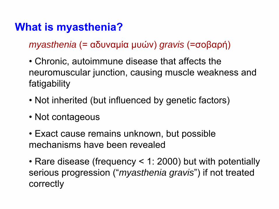
Organs of the immunesystem and their role
Thymus gland
Maturation of T- lymphocytes
Maturation of B- lymphocytes
Bone marrow
Activation of mature Τ& Β-
lymphocytes
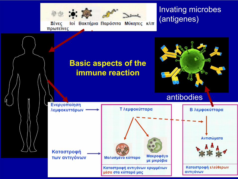
Basic aspects of the immune reaction
Invating microbes (antigenes)
antibodies
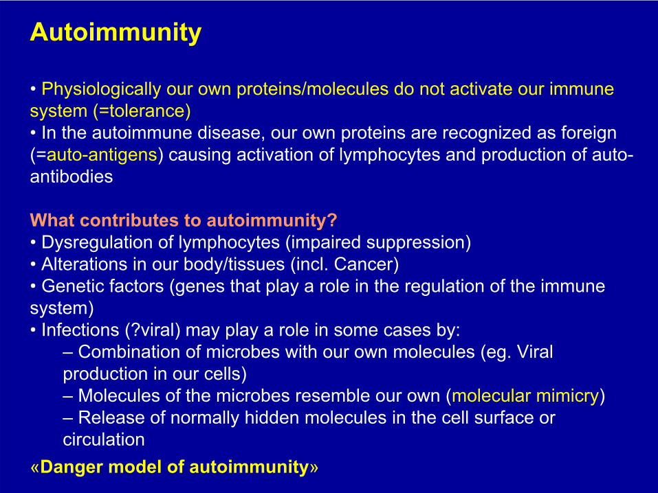
Autoimmunity
•
Physiologically
our
own
proteins/molecules
do
not
activate
our
immune system
(=tolerance)
•
In
the
autoimmune
disease, our
own
proteins
are
recognized
as
foreign (=auto-antigens) causing
activation
of
lymphocytes
and
production
of
auto-
antibodies
What
contributes
to
autoimmunity? •
Dysregulation
of
lymphocytes
(impaired
suppression)
•
Alterations
in
our
body/tissues
(incl. Cancer)•
Genetic
factors
(genes
that
play
a role
in
the
regulation
of
the
immune
system) • Infections (?viral) may play a role in some cases by:
–
Combination
of
microbes
with
our
own
molecules
(eg. Viral production
in
our
cells)
–
Molecules
of
the
microbes
resemble
our
own
(molecular
mimicry)–
Release
of
normally
hidden
molecules
in
the
cell
surface
or
circulation«Danger
model
of
autoimmunity»
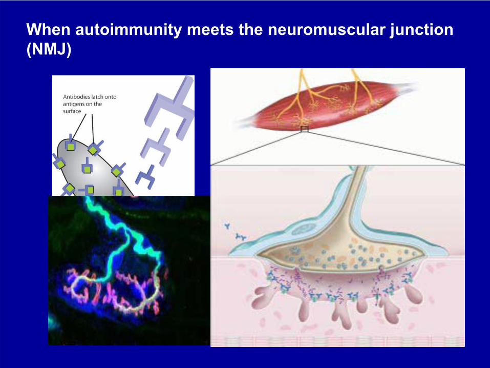
When
autoimmunity
meets
the
neuromuscular
junction (NMJ)
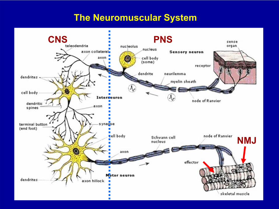
The Neuromuscular System
CNS PNS
NΜJ
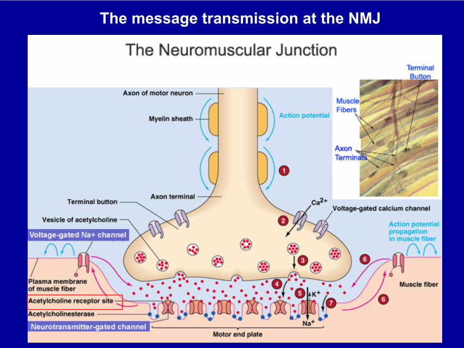
The message transmission at the NMJ
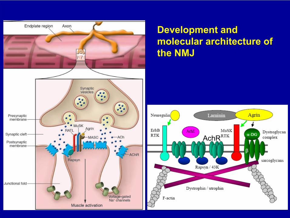
Development and molecular architecture of the NMJ
AchR
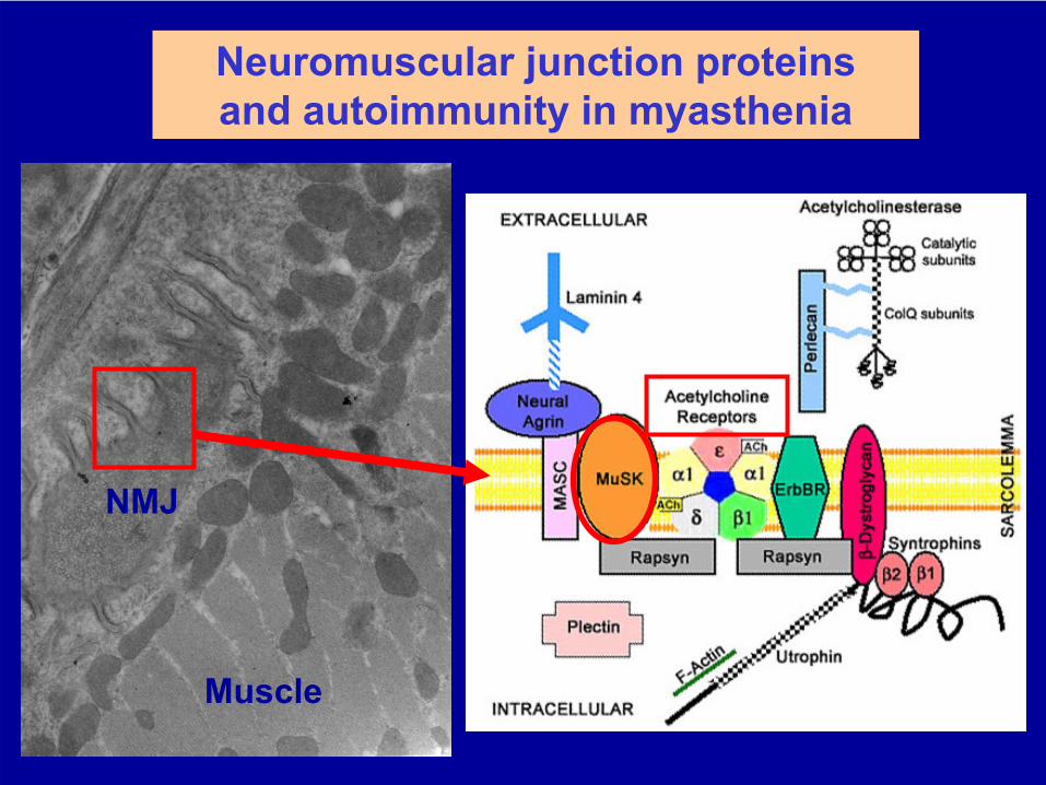
Neuromuscular junction proteinsand autoimmunity in myasthenia
Muscle
NMJ
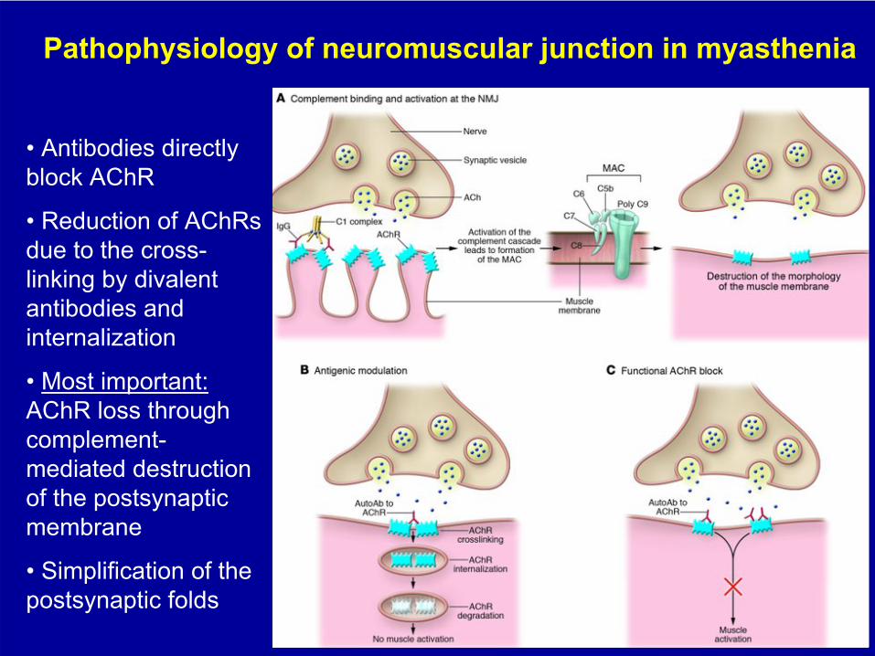
•
Antibodies
directly block
AChR
•
Reduction
of
AChRs due
to
the
cross-
linking
by
divalent antibodies
and
internalization
•
Most
important: AChR
loss
through
complement- mediated
destruction
of
the
postsynaptic membrane
•
Simplification
of
the postsynaptic
folds
Pathophysiology
of neuromuscular junction in myasthenia
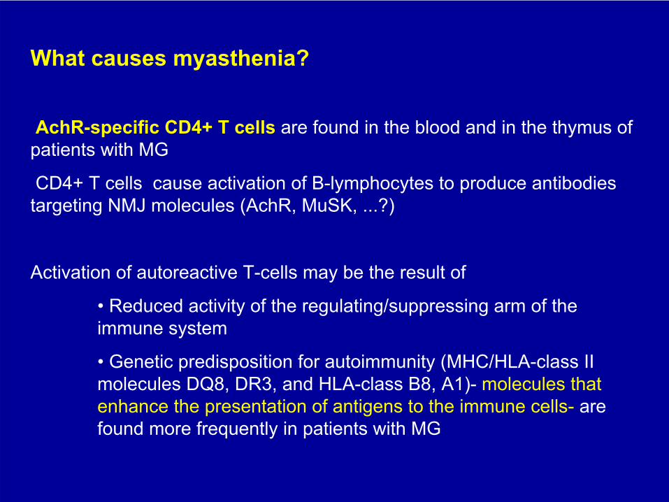
What causes myasthenia?
AchR-specific
CD4+ T cells
are found in the blood and in the thymus of patients with MG
CD4+ T cells
cause activation of Β-lymphocytes to produce antibodies targeting NMJ molecules (AchR, MuSK, ...?)
Activation of autoreactive T-cells may be the result of
•
Reduced activity of the regulating/suppressing arm of the immune system
•
Genetic predisposition for autoimmunity (MHC/HLA-class
II molecules
DQ8, DR3, and
HLA-class
B8, A1)-
molecules that
enhance the presentation of antigens to the immune cells-
are found more frequently in patients with MG
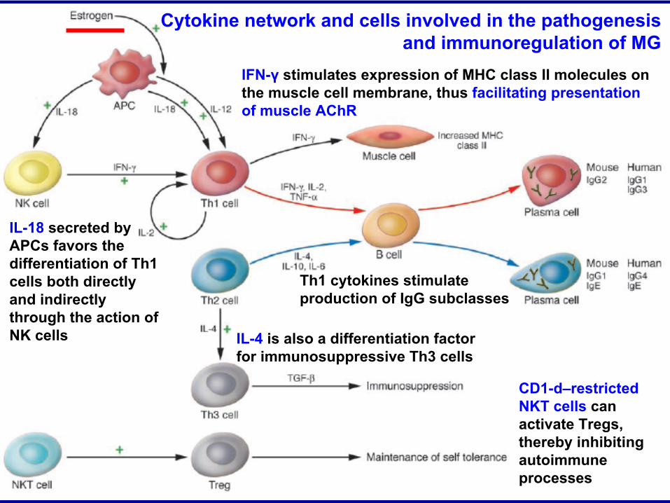
Cytokine network and cells involved in the pathogenesis and immunoregulation of MG
IL-4 is also a differentiation factor for immunosuppressive Th3 cells
Th1 cytokines stimulate production of IgG subclasses
IFN-γ
stimulates expression of MHC class II molecules on the muscle cell membrane, thus facilitating presentation of muscle AChR
IL-18 secreted by APCs favors the differentiation of Th1 cells both directly and indirectly through the action of NK cells
CD1-d–restricted NKT cells
can activate Tregs, thereby inhibiting autoimmune processes
0
10
20
30
40
50
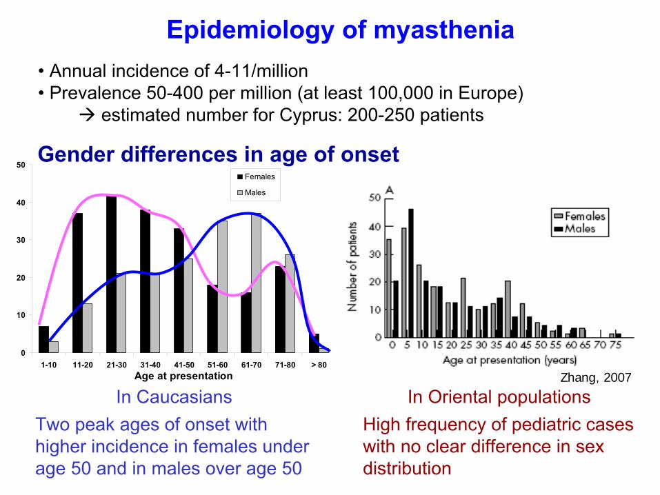
1-10 11-20 21-30 31-40 41-50 51-60 61-70 71-80 > 80Age at presentation
Females
Males
In CaucasiansTwo peak ages of onset with higher incidence in females under age 50 and in males over age 50
In Oriental populationsHigh frequency of pediatric cases with no clear difference in sex distribution
Zhang, 2007
Gender differences in age of onset
• Annual
incidence of 4-11/million• Prevalence 50-400 per million (at least 100,000 in Europe)
estimated number for Cyprus: 200-250 patients
Epidemiology
of
myasthenia
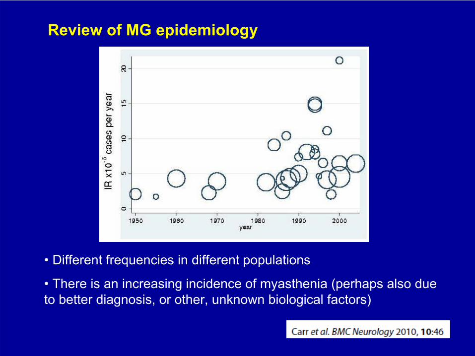
• Different frequencies in different populations
•
There is an increasing incidence of myasthenia (perhaps also due to better diagnosis, or other, unknown biological factors)
Review of MG epidemiology
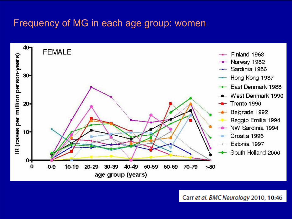
Frequency of MG in each age group: women
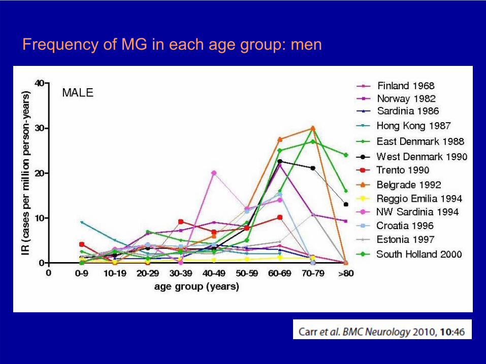
Frequency of MG in each age group: men
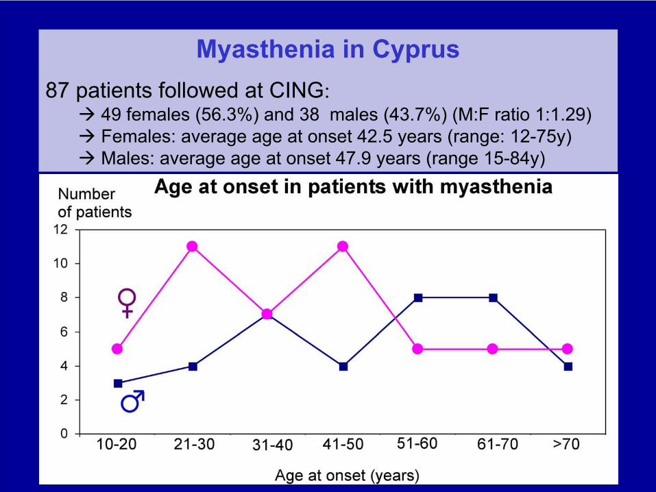
Myasthenia in Cyprus87 patients followed at CING:
49
females (56.3%) and 38 males (43.7%) (M:F ratio 1:1.29) Females: average age at onset 42.5 years (range: 12-75y) Males: average age at onset 47.9 years (range 15-84y)
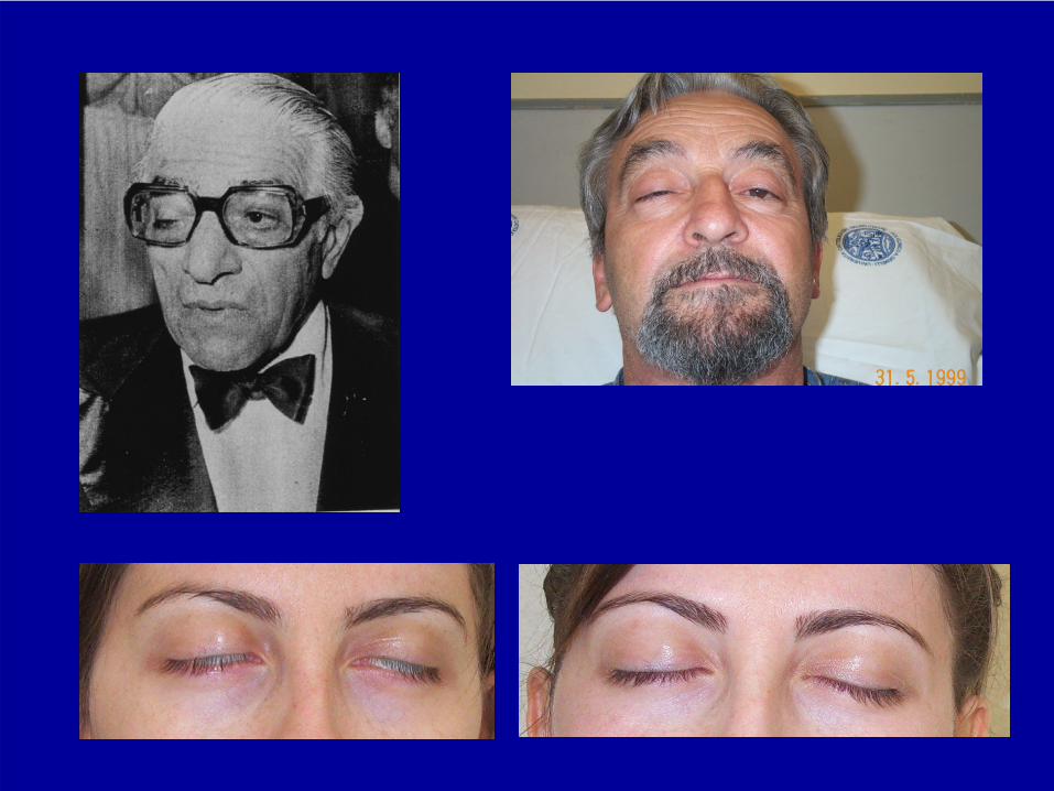
Clinical features of myasthenia
Painless voluntary muscle weakness with fatigability
Typical pattern of weakness in most cases: diplopia, ptosis
(asymmetric, fatigues with upgaze), dysarthria
(nasal
speech), dysphagia, dysphonia, dyspnea
(exertional), proximal limb (arms>legs), facial and neck muscle weakness
Fluctuating, chronic
course with remissions and relapses
Deterioration
of fatigability towards the end of the day
and with repetitive exercise, improvement with rest Not typical in myasthenia:
•
“generalized fatigue”•
reflex and sensory abnormalities
•
elevated serum CK level •
muscle atrophy is rare (but more frequent in MuSK-Ab+ patients)
and restricted to single muscles
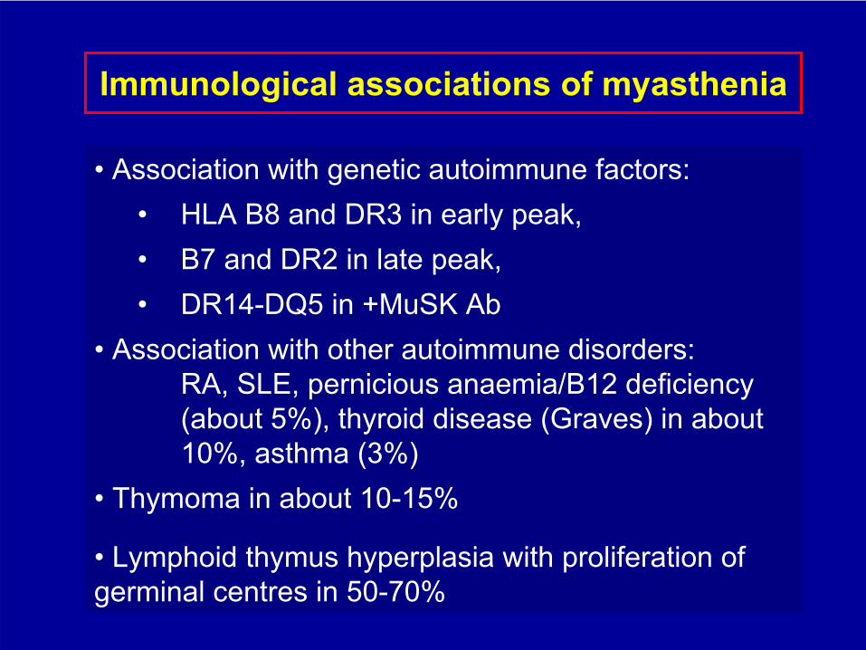
• Association with genetic autoimmune factors:• HLA B8 and DR3 in early peak, • B7 and DR2 in late peak, • DR14-DQ5 in +MuSK
Ab
•
Association with other autoimmune disorders: RA, SLE, pernicious anaemia/B12 deficiency (about 5%), thyroid disease (Graves) in about 10%, asthma (3%)
• Thymoma
in about 10-15%
•
Lymphoid
thymus hyperplasia with proliferation of germinal centres in 50-70%
Immunological associations of myasthenia
Utility
of
Tensilon
test• Only
useful in
patients
with
objective, measurable, findings
• Rarely
helpful
in
the
diagnostic
evaluation
of
equivocal
cases
of
MG • Sensitivity for
MG is
low
(60%) compared
to
other
diagnostic
tests
•
False positive results
in
patients
with
LEMS, ALS, GBS, or
even
localized, intracranial
mass
lesions
• In anti-MuSK+ MG often negative and can even increase weakness Rarely used due to the availability of more sensitive methods!
Edrophonium
(Tensilon) Test
•
Tensilon
(2 mg 8mg
i.v) inhibits acetylcholin-esterase
• Works within 3-45 sec, for a few minutes
•
Have atropin
available
to
reverse hemodynamic effects
–caution in elderly!
before after
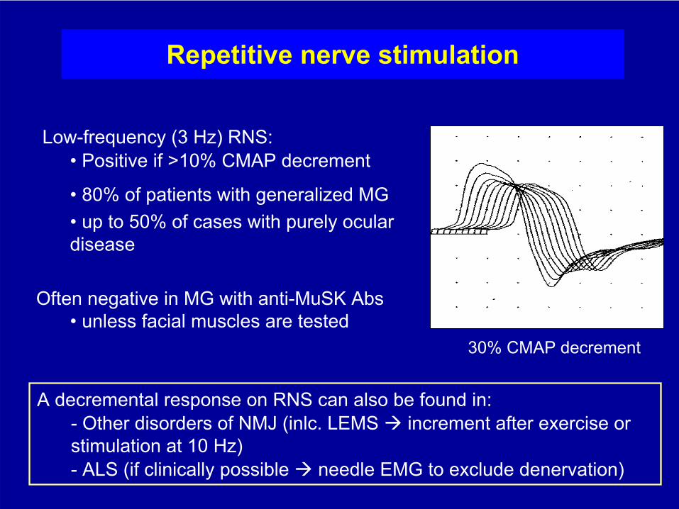
Repetitive nerve stimulation
30% CMAP decrement
A decremental
response on RNS
can also be found in:-
Other disorders of NMJ (inlc. LEMS
increment
after
exercise
or
stimulation
at
10 Hz)- ALS
(if
clinically
possible
needle
EMG to exclude denervation)
Low-frequency (3 Hz) RNS:• Positive
if
>10% CMAP decrement
• 80% of patients with generalized MG•
up to 50% of cases with purely ocular
disease
Often negative in MG with anti-MuSK
Abs• unless facial muscles are tested
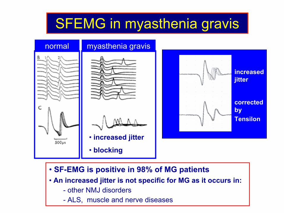
SFEMG in myasthenia gravis
-
• SF-EMG is positive in 98% of MG patients• An increased jitter is not specific for MG as it occurs in:
- other NMJ disorders - ALS, muscle and nerve diseases
normal myasthenia gravis
• increased jitter
• blocking
increased jitter
corrected by Tensilon
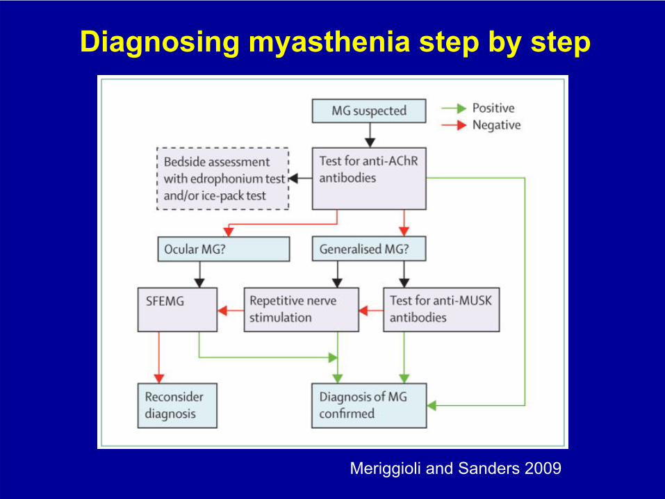
Diagnosing myasthenia step by step
Meriggioli
and
Sanders
2009
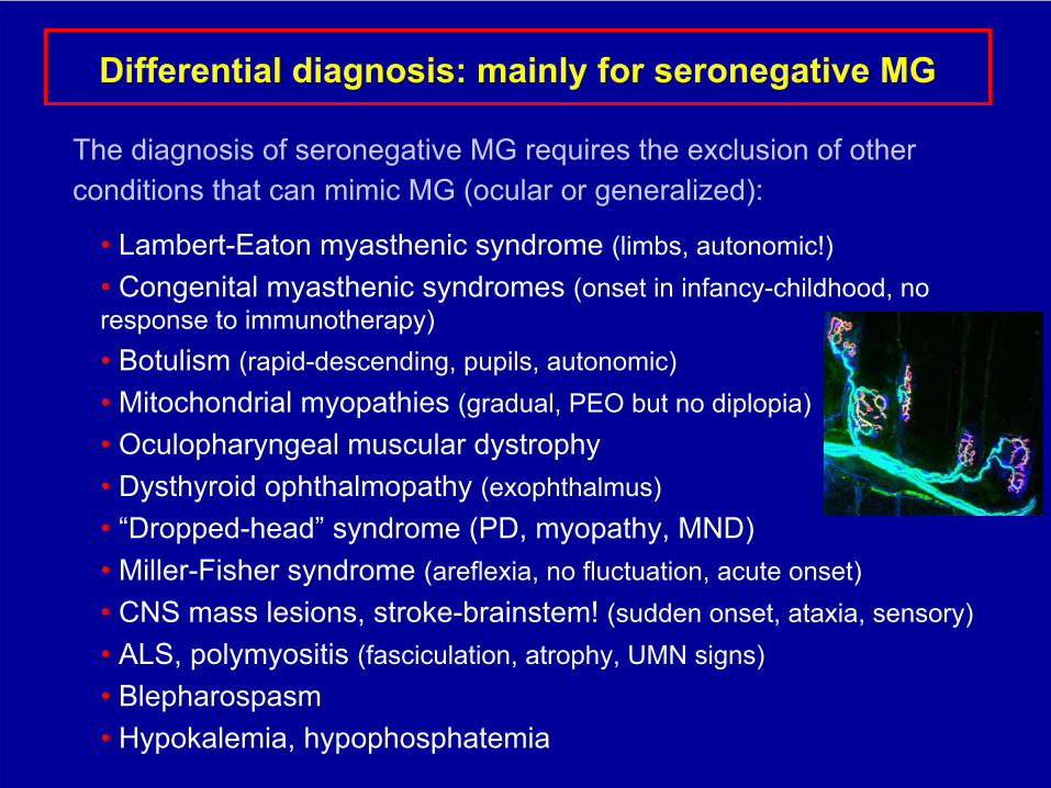
Differential diagnosis: mainly for
seronegative
MG
The diagnosis of seronegative
MG requires the exclusion of other conditions that can mimic MG
(ocular
or
generalized):
• Lambert-Eaton myasthenic
syndrome (limbs, autonomic!)•
Congenital myasthenic
syndromes (onset in
infancy-childhood, no response to immunotherapy)• Botulism
(rapid-descending, pupils, autonomic)
• Mitochondrial myopathies
(gradual, PEO but no diplopia)• Oculopharyngeal
muscular
dystrophy
• Dysthyroid
ophthalmopathy
(exophthalmus)• “Dropped-head”
syndrome (PD, myopathy, MND)
• Miller-Fisher syndrome (areflexia, no fluctuation, acute onset)• CNS mass lesions, stroke-brainstem!
(sudden onset, ataxia, sensory)
• ALS, polymyositis
(fasciculation, atrophy, UMN signs)• Βlepharospasm• Ηypokalemia, hypophosphatemia
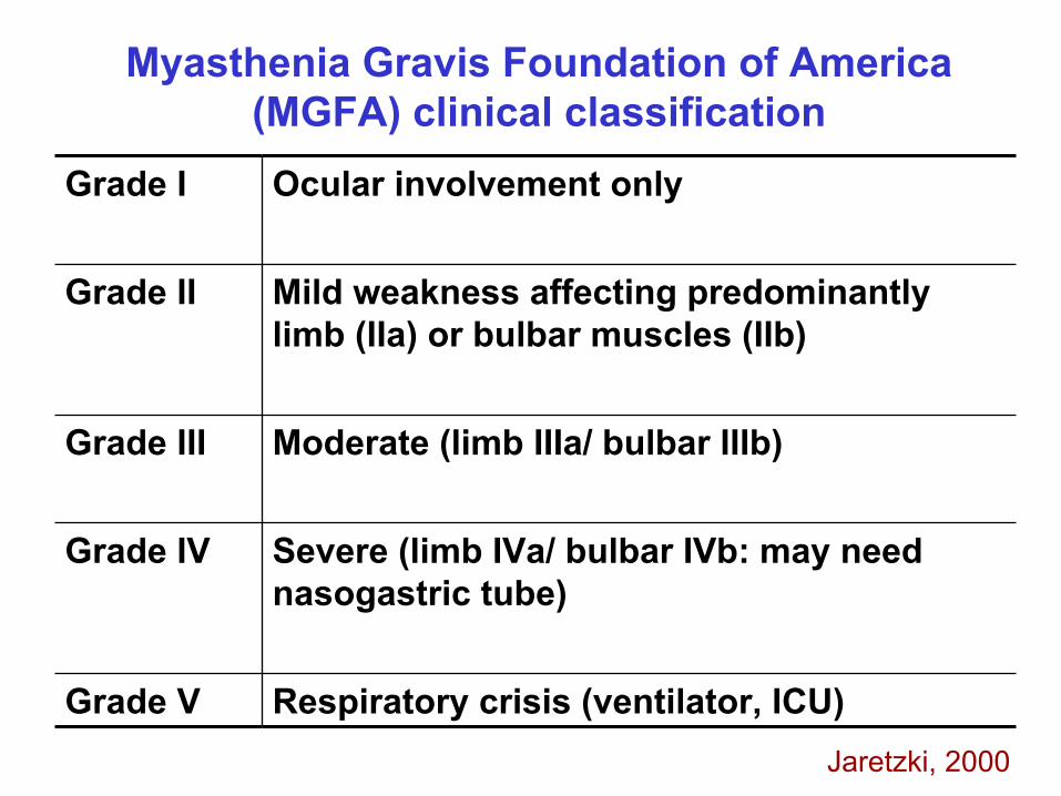
Grade I Ocular
involvement
only
Grade II Mild weakness affecting
predominantly limb (IIa) or bulbar muscles (IIb)
Grade III Moderate (limb IIIa/ bulbar IIIb)
Grade IV Severe (limb IVa/ bulbar IVb: may
need nasogastric
tube)
Grade V Respiratory crisis (ventilator, ICU)
Myasthenia
Gravis
Foundation
of
America (MGFA) clinical classification
Jaretzki, 2000
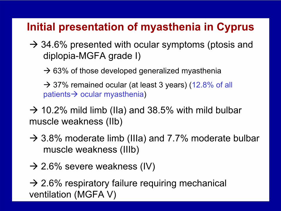
Initial presentation of myasthenia in Cyprus 34.6% presented with ocular symptoms (ptosis
and
diplopia-MGFA grade
I) 63% of those developed generalized myasthenia
37% remained ocular
(at least 3 years) (12.8% of all patients ocular
myasthenia)
10.2% mild limb (IIa) and 38.5% with mild bulbar muscle weakness (IIb)
3.8% moderate limb (IIIa) and 7.7% moderate bulbar muscle weakness (IIIb)
2.6% severe weakness (IV)
2.6% respiratory failure requiring mechanical ventilation (MGFA V)
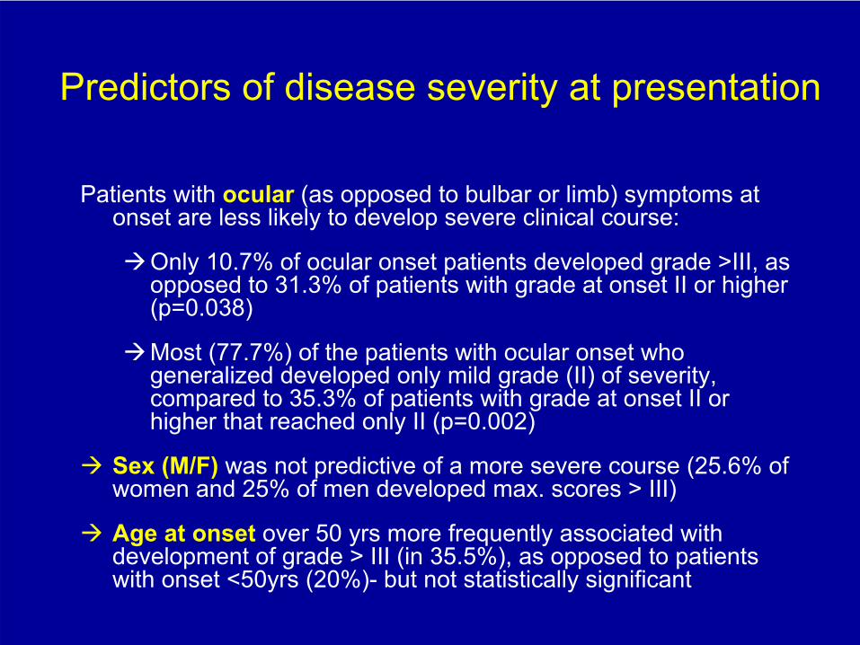
Predictors of
disease severity
at presentation
Patients
with
ocular
(as
opposed
to
bulbar
or
limb) symptoms at onset
are less likely to develop
severe clinical
course:
Only
10.7% of
ocular onset patients
developed grade
>III,
as opposed
to
31.3% of
patients
with
grade
at
onset
II or
higher
(p=0.038)
Most
(77.7%) of
the
patients
with
ocular
onset
who generalized
developed
only
mild
grade
(II) of
severity,
compared
to
35.3% of
patients
with
grade
at
onset
II or higher
that
reached
only
II (p=0.002)
Sex (M/F)
was not predictive of a more severe course (25.6% of women and 25% of men developed max. scores > III)
Age at onset
over 50 yrs more frequently associated with development of grade
> III
(in 35.5%), as opposed to patients
with onset <50yrs (20%)-
but
not
statistically
significant
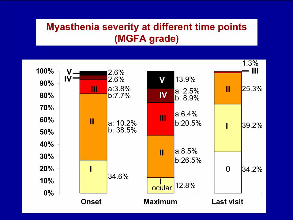
Myasthenia severity at different time points (MGFA grade)
0%10%20%30%40%50%60%70%80%90%
100%
1 2 3Onset Maximum Last visit
0
I
II
III
V
IVIII
II
II
II
IIIIVV
34.2%
39.2%
25.3%
1.3%
12.8%ocular
a:6.4%b:20.5%
a: 2.5%13.9%
b: 8.9%
a:8.5%b:26.5%
a:3.8%
a: 10.2%
2.6%
b: 38.5%
34.6%
b:7.7%
2.6%
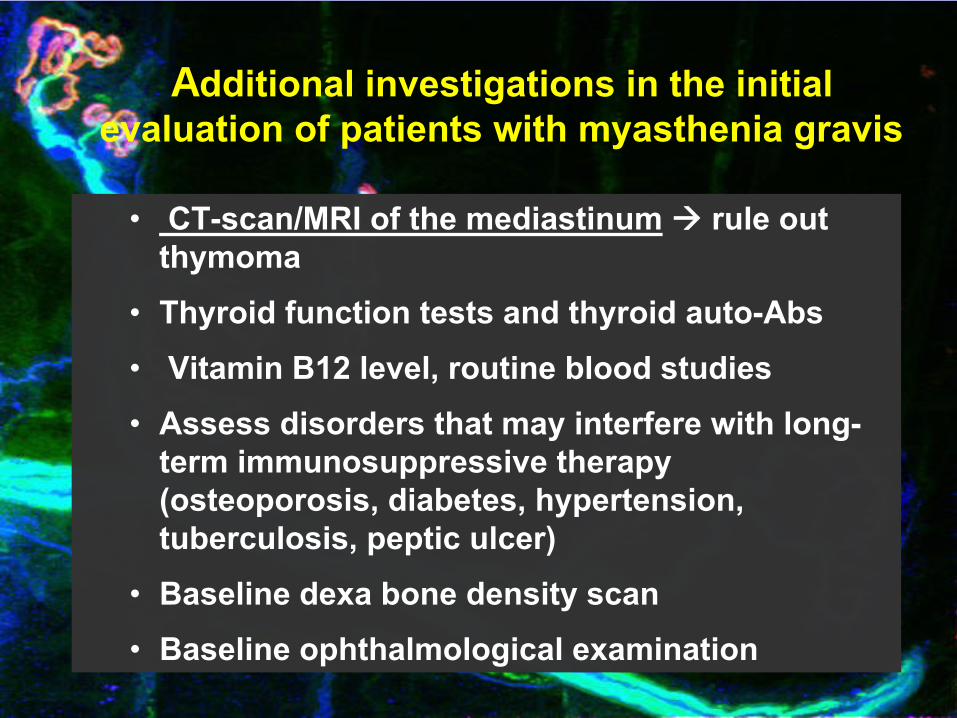
•
CT-scan/MRI of the mediastinum
rule
out thymoma
•
Thyroid function tests and thyroid auto-Abs
•
Vitamin
B12 level, routine blood studies
•
Assess
disorders
that may interfere with long- term immunosuppressive therapy
(osteoporosis, diabetes, hypertension, tuberculosis, peptic ulcer)
•
Baseline
dexa
bone
density
scan
•
Baseline
ophthalmological
examination
Additional
investigations
in
the
initial evaluation
of
patients
with
myasthenia
gravis
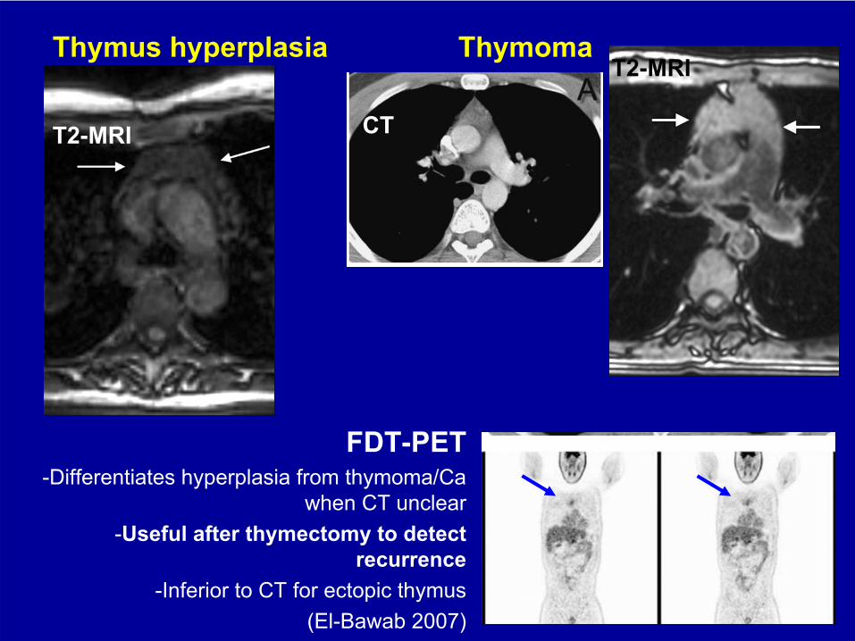
Thymus
hyperplasia
T2-MRI
ThymomaT2-MRI
CT
FDT-PET-Differentiates
hyperplasia
from
thymoma/Ca when
CT unclear-Useful
after
thymectomy
to
detect recurrence
-Inferior
to
CT for
ectopic
thymus(El-Bawab
2007)
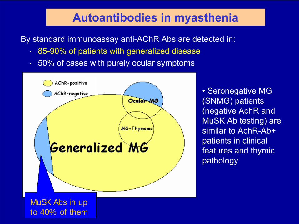
MuSK Abs in up to 40% of them
By standard immunoassay anti-AChR
Abs are detected in:•
85-90% of patients with generalized disease•
50% of cases with purely ocular symptoms
Autoantibodies in myasthenia
•
Seronegative
MG (SNMG) patients (negative AchR
and
MuSK
Ab
testing) are similar to AchR-Ab+ patients in clinical features and thymic
pathology
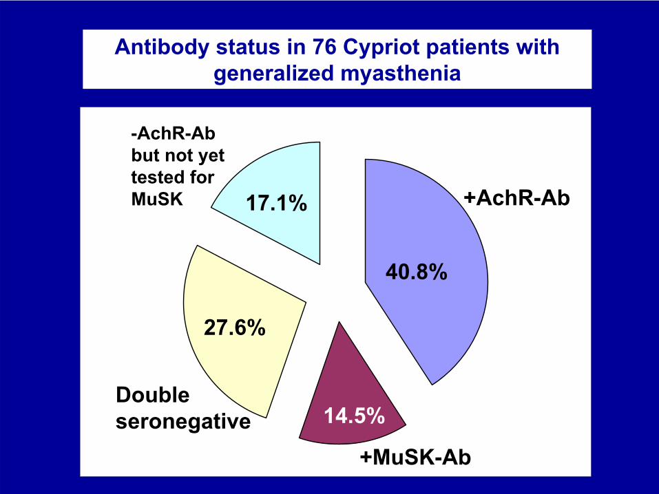
Antibody status in 76 Cypriot patients with generalized myasthenia
+MuSK-Ab
14.5%
+AchR-Ab
+MuSK-Ab
Double seronegative
-AchR-Ab but not yet
tested for MuSK
40.8%
14.5%
27.6%
17.1%
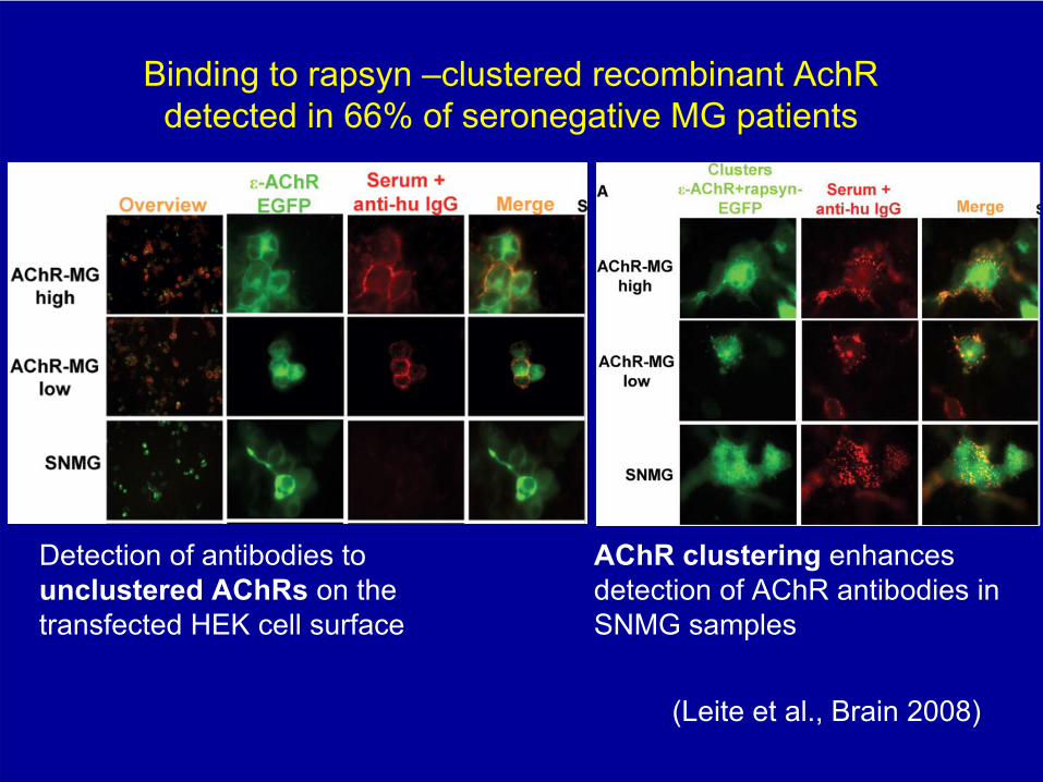
Binding to rapsyn
–clustered recombinant AchR detected in 66% of seronegative
MG patients
(Leite
et al., Brain 2008)
AChR
clustering
enhances detection
of
AChR
antibodies
in
SNMG samples
Detection
of
antibodies
to unclustered
AChRs
on
the
transfected
HEK cell
surface
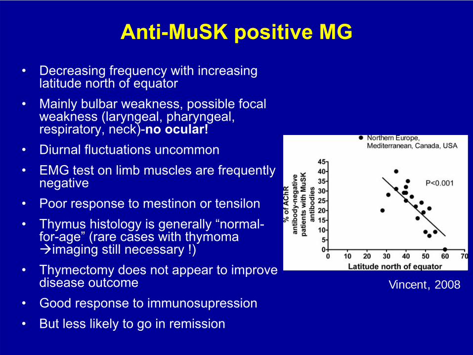
Anti-MuSK
positive MG•
Decreasing
frequency
with
increasing
latitude
north
of
equator•
Mainly
bulbar
weakness, possible
focal
weakness
(laryngeal, pharyngeal, respiratory, neck)-no
ocular!
•
Diurnal
fluctuations uncommon•
EMG test on limb muscles are frequently negative
•
Poor
response to mestinon
or
tensilon•
Thymus histology is generally “normal-
for-age”
(rare
cases
with
thymoma imaging
still
necessary
!)
•
Thymectomy
does not appear to improve disease outcome
•
Good
response
to
immunosupression•
But
less
likely
to
go
in
remission
Vincent, 2008
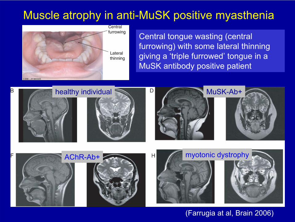
Muscle atrophy in anti-MuSK
positive myasthenia
(Farrugia
at al, Brain 2006)
healthy individual MuSK-Ab+
AChR-Ab+ myotonic
dystrophy
Central tongue wasting (central furrowing) with some lateral thinning giving a ‘triple furrowed’
tongue in a
MuSK
antibody positive patient
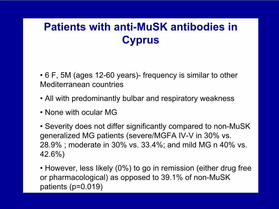
Patients with anti-MuSK
antibodies in Cyprus
•
6 F, 5M
(ages 12-60 years)-
frequency is similar to other Mediterranean countries
• All with predominantly bulbar and respiratory weakness
• None with ocular MG
•
Severity does not differ significantly compared to non-MuSK generalized MG patients (severe/MGFA IV-V
in 30% vs.
28.9% ; moderate in 30% vs. 33.4%; and mild MG n 40% vs. 42.6%)
•
However, less likely (0%) to go in remission
(either
drug
free or
pharmacological) as
opposed
to
39.1% of
non-MuSK
patients
(p=0.019)
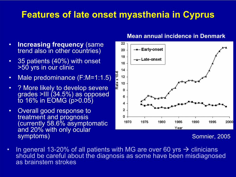
Features of late onset myasthenia in Cyprus
•
Increasing
frequency
(same trend
also
in
other
countries)
•
35 patients (40%) with onset >50 yrs
in
our
clinic
•
Male predominance (F:M=1:1.5)•
? More likely to
develop severe
grades
>III
(34.5%)
as
opposed to
16% in
EOMG (p>0.05)
•
Overall good response to treatment and prognosis (currently 58.6% asymptomatic and 20% with only ocular symptoms)
Mean annual incidence in Denmark
Somnier, 2005
•
In general 13-20% of all patients with MG are over 60
yrs clinicians should be careful about the diagnosis as some have been misdiagnosed as brainstem strokes
■
Individualized■
Choice
of
medications
and
their
dose
should
not
and
cannot
be
the
same
in
all
patients-
each
may
have
different
circumstances■
MG treatment
is
not
static-
it
may
need
changes
of
medications
and
doses
during
the
course
of
the
illness
■
The
choice
of
medications
and
their
dose
depends
on
the
type
and severity
of
MG, and
the
profile
of
the
patient
(life
style, family
planning,
age, co-morbidities, other
medications)
■
In
most
patients
a combination
of
medications
will
be
needed
at
some point
■
The
side
effects
and
the
efficacy of
the
same
medication
may
differ from
patient
to
patient
!
■
Close
follow-up
and
good
collaboration
between
patient
and
doctor
is essential
for
success!
Basic
principles
of
myasthenia
treatment
Current Treatment of MyastheniaCholinesterase Inhibitors (Pyridostigmine) •
Symptomatic therapy:
Increases
ACh
availability only a minority will
respond as monotherapy!
Thymectomy•
In patients with thymic
hyperplasia
removes the possible site of auto-
sensitization against AChR
and a relevant site of antibody production•
In patients with thymoma/Ca
removes a potentially invasive tumor
Immunosuppressive therapy•
Inhibits lymphocyte proliferation and antibody production
Short-term, rapid
onset
therapies•
Plasma-exchange (PE): Removes serum antibodies and cytokines
•
IVIG (2 g/kg in 2-5 days):
Interfere with T-cell activation, Ab
production and activity
Useful in myasthenic
crisis (PE may have a more rapid effect), patients refractory to immunosupression, or when quick response needed
Thymectomy
in
MG patients without thymoma
•
Thymectomized
patients appear to have a better prognosis in terms
of drug-free remission rate (2.1 higher than in unthymectomized
cases)
(Gronseth, 2001)
•
Most
experts
consider
thymectomy
to
be
a therapeutic
option
in anti-AChR-positive,
generalised
MG with
onset
<
50 years, in
experienced centers
•
Generally
not
recommended
in
late
onset, ocular, or
MuSK-Ab+ cases, and
questionable
in
seronegative
•
Benefit
of thymectomy
not
definitely
proven:–
Lack of randomized studies and standardized outcome measures
–
Presence of confounding factors (disease duration before surgery, associated treatment, different surgical procedures)
•
A multicenter randomized trial (MGTX study) to establish the effect of thymectomy
in non-thymomatous
patients is currently underway
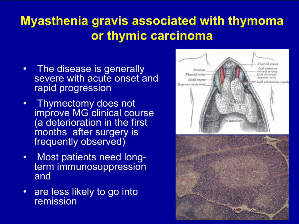
Myasthenia gravis associated with thymoma or
thymic
carcinoma
•
The disease is generally severe with acute onset and rapid progression
•
Thymectomy
does not improve MG clinical course (a deterioration in the first months after surgery is frequently observed)
•
Most patients need long- term immunosuppression and
•
are less likely to go into remission
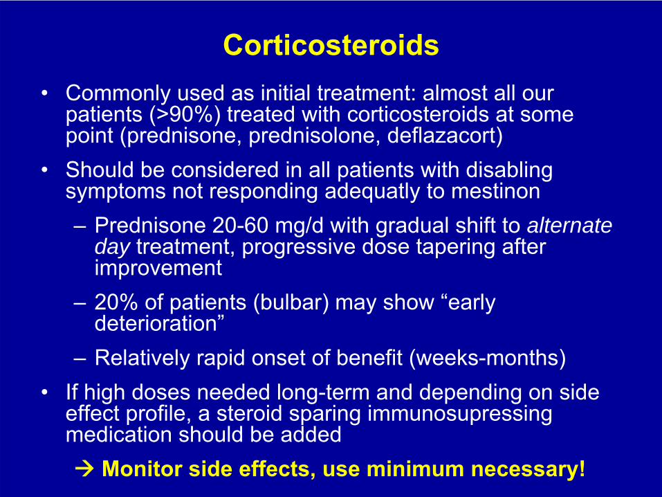
Corticosteroids•
Commonly used as initial treatment: almost all our patients (>90%) treated with corticosteroids at some point (prednisone, prednisolone, deflazacort)
•
Should
be
considered
in
all
patients
with
disabling symptoms
not
responding
adequatly
to
mestinon
–
Prednisone 20-60 mg/d with gradual shift to alternate day treatment, progressive dose tapering after improvement
–
20% of patients (bulbar) may show “early deterioration”
–
Relatively
rapid
onset
of
benefit
(weeks-months)•
If
high
doses
needed
long-term
and
depending
on
side
effect
profile, a steroid
sparing
immunosupressing medication
should
be
added
Monitor side effects, use minimum necessary!
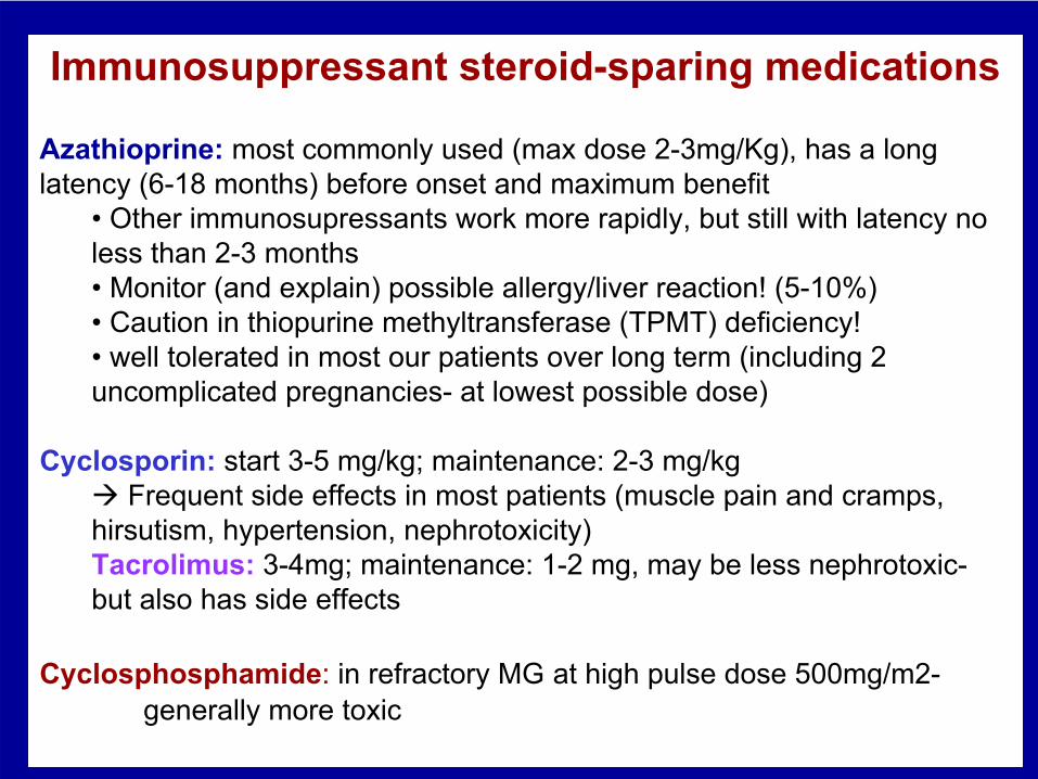
Immunosuppressant steroid-sparing medications
Azathioprine: most
commonly
used
(max
dose
2-3mg/Kg), has
a long latency
(6-18 months) before
onset
and
maximum
benefit
•
Other
immunosupressants
work
more
rapidly, but
still
with
latency
no less
than
2-3 months
• Monitor (and explain) possible allergy/liver
reaction! (5-10%)• Caution in thiopurine
methyltransferase
(TPMT) deficiency!
•
well tolerated in most our patients over long term (including 2 uncomplicated pregnancies-
at
lowest
possible
dose)
Cyclosporin:
start 3-5 mg/kg; maintenance: 2-3 mg/kg Frequent side effects in
most
patients
(muscle pain and cramps,
hirsutism, hypertension, nephrotoxicity) Tacrolimus:
3-4mg; maintenance: 1-2 mg, may be less nephrotoxic-
but also has
side effects
Cyclosphosphamide:
in refractory MG
at high pulse dose 500mg/m2- generally more toxic
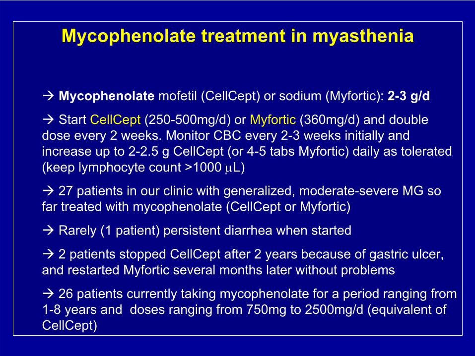
Mycophenolate
treatment in myasthenia
Mycophenolate
mofetil
(CellCept) or sodium (Myfortic): 2-3 g/d
Start CellCept
(250-500mg/d) or Myfortic
(360mg/d) and double dose every 2 weeks. Monitor CBC every 2-3 weeks initially and increase up to 2-2.5 g CellCept
(or 4-5 tabs Myfortic) daily as tolerated
(keep lymphocyte count >1000 L)
27
patients in our clinic with generalized, moderate-severe MG so far treated with mycophenolate
(CellCept
or Myfortic)
Rarely (1 patient) persistent diarrhea when started
2 patients stopped CellCept
after 2 years because of gastric ulcer, and restarted Myfortic
several months later without problems
26
patients currently taking mycophenolate
for a period ranging from 1-8 years and doses ranging from 750mg to 2500mg/d (equivalent of
CellCept)
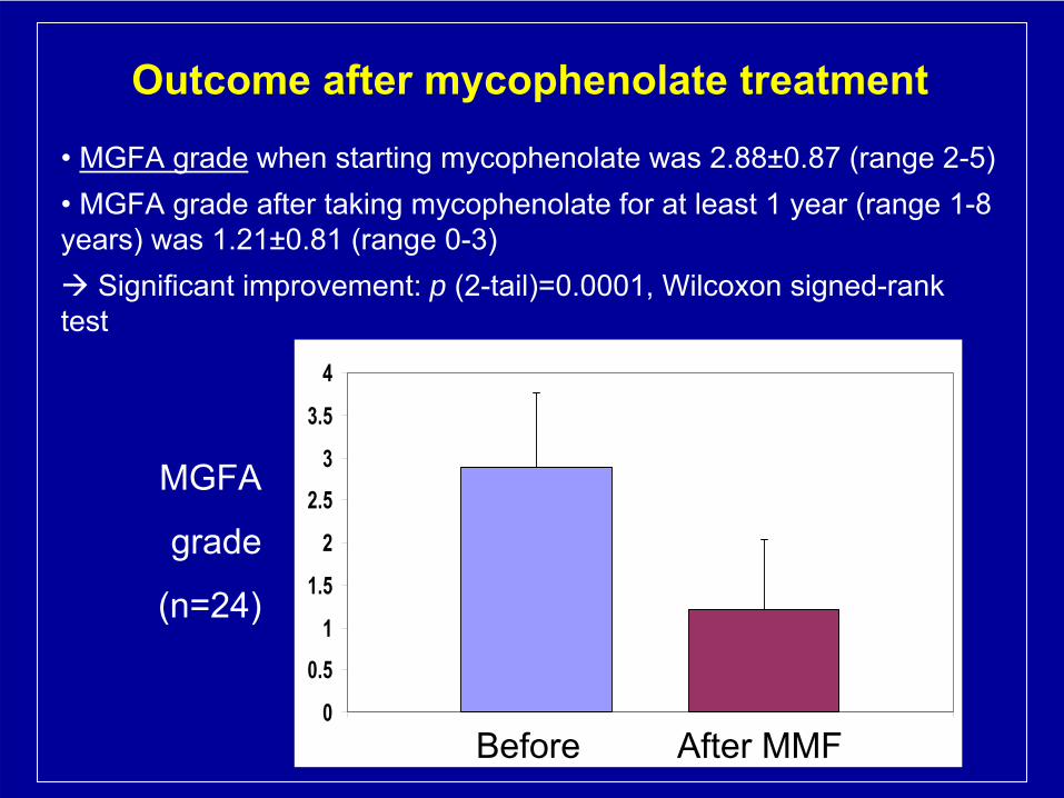
Outcome after mycophenolate
treatment
• MGFA grade
when starting mycophenolate
was 2.88±0.87 (range 2-5) •
MGFA grade after taking mycophenolate
for at least 1 year (range 1-8
years) was 1.21±0.81 (range 0-3) Significant improvement: p (2-tail)=0.0001, Wilcoxon
signed-rank
test
0
0.51
1.5
2
2.53
3.5
4
1Before After MMF
MGFA
grade
(n=24)
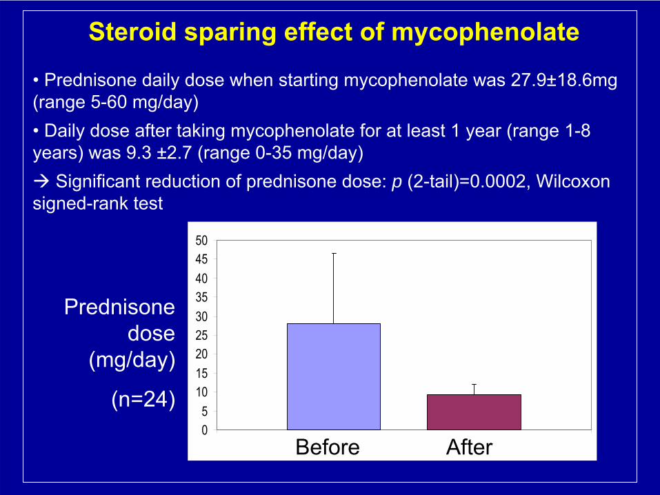
Steroid
sparing
effect
of mycophenolate
•
Prednisone daily dose when starting mycophenolate
was 27.9±18.6mg (range 5-60 mg/day) •
Daily dose
after taking mycophenolate
for at least 1 year (range 1-8
years) was 9.3 ±2.7 (range 0-35 mg/day) Significant reduction of prednisone dose: p (2-tail)=0.0002, Wilcoxon
signed-rank test
05
101520253035404550
1Before After
Prednisone dose
(mg/day)
(n=24)
Mycophenolate: conclusions from CING
experience
•
Mycophenolate
appears to be safe for patients with myasthenia gravis and compares favourably with other standard medications regarding side effect profile
•
Effective for refractory patients: reduces exacerbations, IVIG/PE dependency, and allows significant reduction of steroids
•
Mycophenolate
should be considered also
as the first choice of immunosupression
for moderate-severe MG, or patients
refractory to steroid treatment, or
patients
requiring
fast immunosuppression
(as
oposed
to
AZT) as these patients
appear to benefit most from this medication
• Contraindicated
in
pregnancy
(or
planning
of
it)
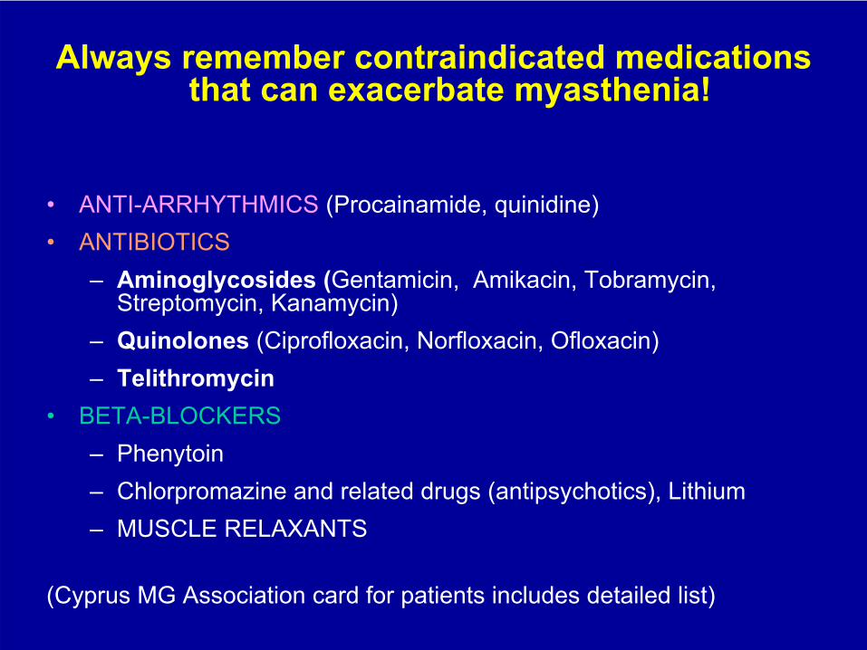
Always remember contraindicated medications that can exacerbate myasthenia!
•
ANTI-ARRHYTHMICS
(Procainamide, quinidine)•
ANTIBIOTICS–
Aminoglycosides
(Gentamicin, Amikacin, Tobramycin,
Streptomycin, Kanamycin)–
Quinolones
(Ciprofloxacin, Norfloxacin, Ofloxacin)
–
Telithromycin•
BETA-BLOCKERS–
Phenytoin
–
Chlorpromazine
and
related
drugs
(antipsychotics), Lithium–
MUSCLE RELAXANTS
(Cyprus
MG Association card for patients
includes
detailed
list)
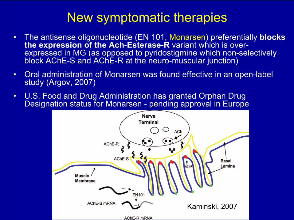
New symptomatic therapies•
The antisense oligonucleotide
(EN 101, Monarsen) preferentially blocks
the expression of the Ach-Esterase-R
variant which is over- expressed in MG
(as
opposed
to
pyridostigmine
which
non-selectively
block AChE-S and AChE-R at the neuro-muscular junction)
•
Oral administration of Monarsen
was found effective in an open-label study (Argov, 2007)
•
U.S. Food
and
Drug
Administration
has
granted
Orphan
Drug Designation
status
for
Monarsen
-
pending
approval
in
Europe
Kaminski, 2007
New immunosuppressive therapiesMonoclonal antibodies
(mAbs)
Humanized and chimeric
mAbs
are designed to interact with specific antigens
Rituximab
is directed against CD20, expressed on pre-B and B cells
–
In uncontrolled reports, it proved effective in both AChR-MG and MuSK-MG.
–
Therapeutic effect in autoimmune diseases associated with B cell depletion
without significant changes in Ab
levels (Kessel, 2008;
Liossis, 2008)
Anti-cytokine treatmentsTNF-a inhibitors have been used in single cases (Infliximab)
(Kakouliku, 2007) and in a small prospective trial (Etanercept) (Rowin, 2008)-
worsening in some patients!!!
•
Therapeutic effect was counterbalanced by frequent side effects
C5 Inhibition (Eculizumab) is a possible treatment for acute phases of anti-AChR
positive MG
–
Under investigation: anti-IL-6 antibodies (block EAMG)
The future of myasthenia treatment: targeting the pathological antibodies and their production without general immunosuppression
Emerging Antigen-specific
treatments for MG•
Selective recognition and elimination of nicotinic acetylcholine receptor-
reactive B cells
by a recombinant fusion protein AChR-Fc
in myasthenia gravis in vitro (Chang
et
al., J Neuroimmunol
2010)
•
In vivo adsorption of autoantibodies in myasthenia gravis using Nanodisc-incorporated acetylcholine receptor
(Sheng
et
al., Exp
Neurol
2010)
•
AchR-transferrin
fusion protein
(traps
and
destroys
AchR
antibodies) (Keefe
et
al., Autoimmunity
2010)
•
Specific immunotherapy of experimental myasthenia gravis
with human muscle AChR
constructs: diverting autoantibody production away
from pathologically relevant specificities directed at epitopes
on the extracellular surface of muscle AChRs
toward pathologically irrelevant
epitopes
on the cytoplasmic
domain (Luo
et
al., Ann
Neurol
2010)
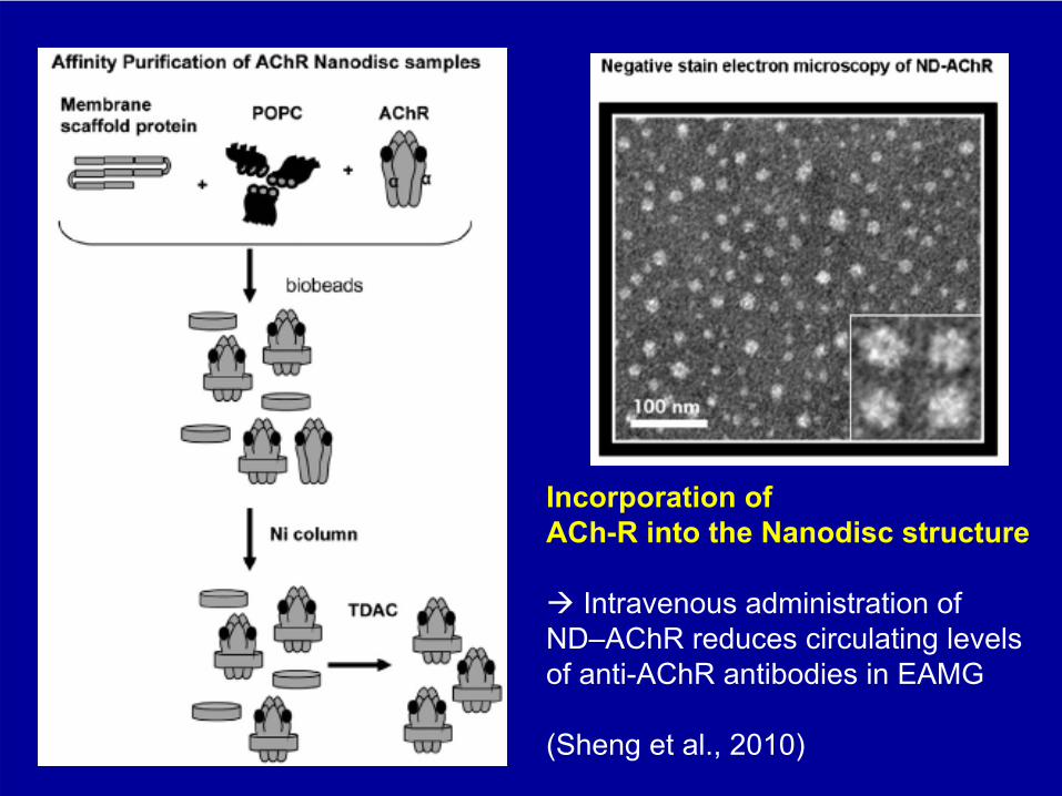
Incorporation ofACh-R into the Nanodisc structure
Intravenous administration of ND–AChR reduces circulating levels of anti-AChR antibodies in EAMG
(Sheng et al., 2010)
Importance of care provision for patients with myasthenia in a specialized center
Unsatisfactory outcomes in myasthenia gravis: influence by care providers.Dunand
M
et al., Lausanne,Switzerland. J Neurol.
2010 Mar;257(3):338-43
Factors leading to unsatisfactory outcome (UO)
in MG:41 patients with autoimmune MG were followed prospectively
•
UO in 54% related to under-treatment (41%),
poor treatment compliance (23%),
infections
(23%), and adverse drug effects (13%)•
When care was provided by neuromuscular (NM) specialists, patients had significantly better follow-up scores (P = 0.01)•
At final assessment UO rates were 7% and significantly better in
patients treated by NM specialists, compared to other physicians where UO
rates reached 27%•
Nearly two-thirds of the UOs
could have been prevented
by appropriate therapeutic adjustments
and improved compliance•
The differential UO rates at follow-up, their dependency on the degree to which the management was specialized and their correlation with final outcomes suggest that specialized MG care improves outcomes
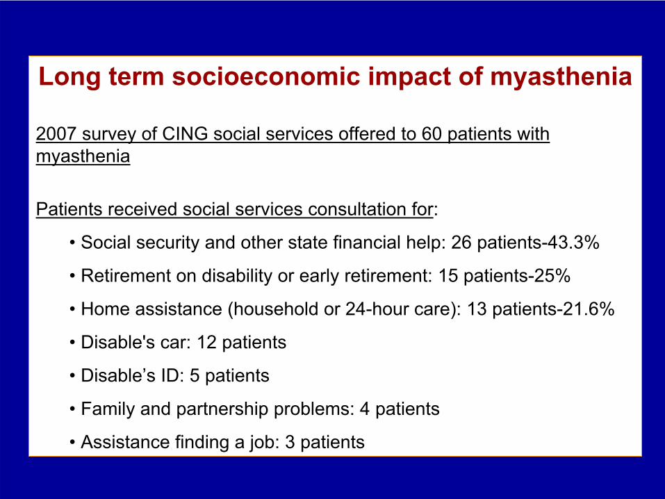
Long term socioeconomic impact of myasthenia
2007 survey of CING social services offered to 60 patients with myasthenia
Patients received social services consultation for:
• Social security and other state financial help: 26 patients-43.3%
• Retirement on disability or early retirement: 15 patients-25%
• Home assistance (household or 24-hour care): 13 patients-21.6%
• Disable's car: 12 patients
• Disable’s ID: 5 patients
• Family and partnership problems: 4 patients
• Assistance finding a job: 3 patients
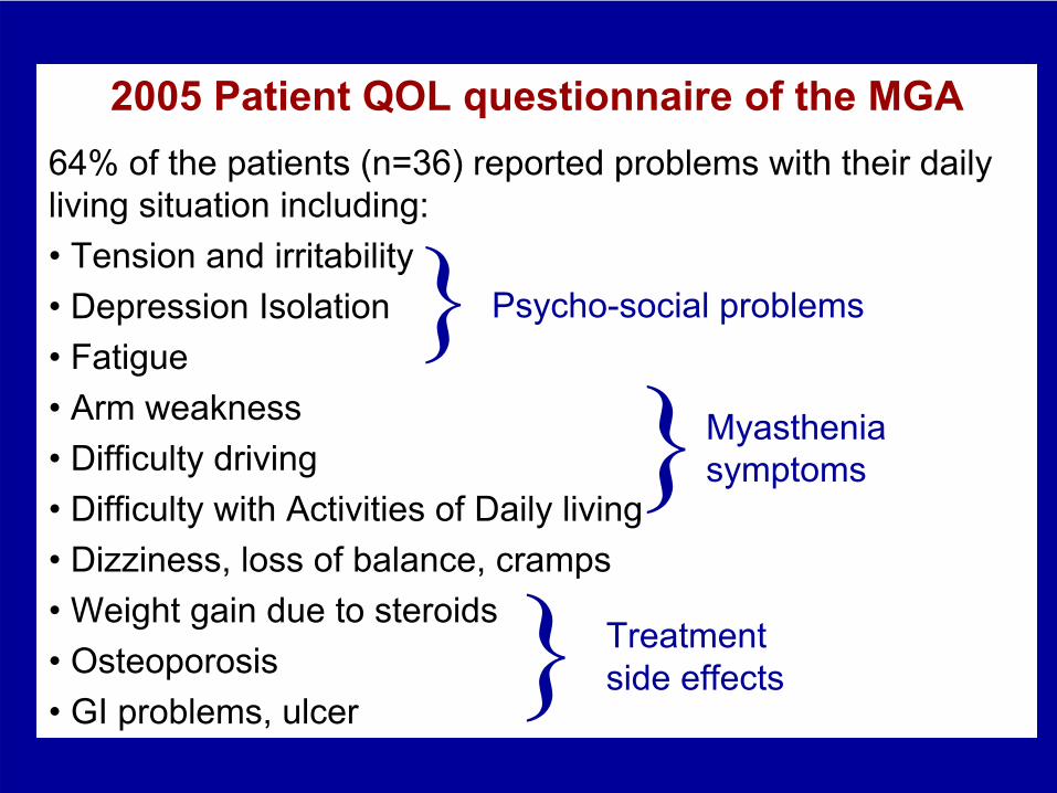
2005 Patient QOL questionnaire of the MGA64%
of the patients (n=36) reported problems with their daily
living situation including:• Tension and irritability• Depression
Isolation
• Fatigue• Arm weakness• Difficulty driving• Difficulty with Activities of Daily living• Dizziness, loss of balance, cramps• Weight gain due to steroids• Osteoporosis• GI problems, ulcer
Treatment side effects
Myasthenia symptoms}
Psycho-social problems}
}
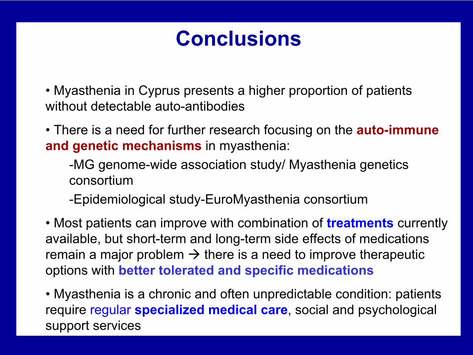
Conclusions
•
Myasthenia in Cyprus presents a higher proportion of patients without detectable auto-antibodies
•
There is a need for further research focusing on the auto-immune and
genetic
mechanisms
in myasthenia:
-MG genome-wide association study/ Myasthenia
genetics consortium
-Epidemiological study-EuroMyasthenia
consortium
•
Most patients can improve with combination of treatments currently available, but short-term and long-term side effects of medications remain a major problem there is a need to improve therapeutic options with better tolerated
and specific
medications
•
Myasthenia is a chronic and often unpredictable condition: patients require regular specialized medical care, social and psychological support services
ACKNOWLEDGEMENTS
Ασθενείς
και
Σύνδεσμος
Ασθενών
με
Μυασθένεια/Cyprus MG Association
MGMyasthenia
Gravis
Association
THANK YOU