Neuroanatomical assessment of the integrin β3 mouse model ...
ARTICLE
Received 18 Feb 2013 | Accepted 5 Aug 2013 | Published 29 Aug 2013
Modulation of b-catenin function maintains mouseepiblast stem cell and human embryonic stem cellself-renewalHoon Kim1,*, Jun Wu1,*,w, Shoudong Ye1,*, Chih-I Tai1, Xingliang Zhou1, Hexin Yan1, Ping Li1, Martin Pera1,2
& Qi-Long Ying1
Wnt/b-catenin signalling has a variety of roles in regulating stem cell fates. Its specific role in
mouse epiblast stem cell self-renewal, however, remains poorly understood. Here we show
that Wnt/b-catenin functions in both self-renewal and differentiation in mouse epiblast stem
cells. Stabilization and nuclear translocation of b-catenin and its subsequent binding to T-cell
factors induces differentiation. Conversely, retention of stabilized b-catenin in the cytoplasm
maintains self-renewal. Cytoplasmic retention of b-catenin is effected by stabilization of
Axin2, a downstream target of b-catenin, or by genetic modifications to b-catenin that
prevent its nuclear translocation. We also find that human embryonic stem cell and mouse
epiblast stem cell fates are regulated by b-catenin through similar mechanisms. Our results
elucidate a new role for b-catenin in stem cell self-renewal that is independent of its
transcriptional activity and will have broad implications in understanding the molecular
regulation of stem cell fate.
DOI: 10.1038/ncomms3403
1 Department of Cell and Neurobiology, Eli and Edythe Broad Center for Regenerative Medicine and Stem Cell Research at USC, Keck School of Medicine,University of Southern California, Los Angeles, California 90033, USA. 2 Department of Anatomy and Neuroscience, The Walter and Eliza Hall Institute ofMedical Research, Florey Neurosciences Institutes, University of Melbourne, Melbourne, Victoria 3010, Australia. * These authors contributed equally to thiswork. w Present address: Salk Institute for Biological Studies, 2880 Torrey Pines Scenic Drive, La Jolla, California 92037, USA. Correspondence and requestsfor materials should be addressed to Q.-L.Y. (email: [email protected]).
NATURE COMMUNICATIONS | 4:2403 | DOI: 10.1038/ncomms3403 | www.nature.com/naturecommunications 1
& 2013 Macmillan Publishers Limited. All rights reserved.
Mouse epiblast stem cells (EpiSCs) are pluripotent stemcells derived from post-implantation epiblasts, andrequire the cytokines fibroblast growth factor 2 (FGF2)
and activin A for self-renewal1,2. Mouse EpiSCs cultured inFGF2/activin survive poorly upon single-cell dissociation, andtherefore are routinely passaged as small clumps. The lowerviability of dissociated EpiSCs suggests that signalling pathwaysother than FGF2/activin might be involved in regulating EpiSCself-renewal. Although inhibition of Rho-associated kinase hasbeen shown to protect cells from the apoptotic effects of single-cell dissociation3, it has no obvious effect in promoting self-renewal. We focused on Wnt/b-catenin as a candidate pathwayfor supporting EpiSC expansion, as it has been shown to have acentral and conserved role in controlling cell proliferation andlineage specification during early embryogenesis4,5, and has alsobeen implicated in promoting self-renewal of various types ofstem cells6–10.
In the absence of Wnt ligand, b-catenin, the key mediator of thecanonical Wnt/b-catenin pathway, is phosphorylated by glycogensynthase kinase 3 (GSK3), leading to proteasome-mediateddegradation of b-catenin. When Wnt ligand binds to its receptorcomplex, composed of frizzled and low-density-lipoprotein-receptor-related protein 5 or 6, the canonical Wnt/b-cateninpathway is activated, leading to the inhibition of GSK3 and thestabilization of b-catenin. Stabilized b-catenin then translocates tothe nucleus, where it interacts with T-cell factors (TCFs) toregulate gene expression. Activation of Wnt/b-catenin signallingproduces diverse and sometimes opposite outcomes in differentcell types, and it has therefore been proposed that Wnt/b-cateninmight regulate cell fates in a context- and cell type-dependentmanner11. How activation of the same Wnt/b-catenin signal yieldsdisparate effects in different cell types, however, remains poorlyunderstood. Here we reveal a mechanism by which Wnt/b-cateninregulates stem cell fates. Wnt/b-catenin signalling promotesmouse EpiSC self-renewal when stabilized b-catenin is retainedin the cytoplasm, and induces differentiation if b-catenin trans-locates into the nucleus and binds TCFs. Wnt/b-catenin alsoregulates human ESC fate through a mechanism similar to that inmouse EpiSCs. This supports the notion that human ESCs aremore closely related to mouse EpiSCs than to mouse ESCs, whichin a b-catenin-mediated self-renewal context are able to maintainself-renewal only when b-catenin binds TCFs9,12.
ResultsSmall molecules maintains EpiSC self-renewal. Previously, weshowed that two small-molecule inhibitors (2i), CHIR99021(CHIR) and PD0325901, could efficiently maintain mouse ESCself-renewal independent of LIF/STAT3 signalling9. CHIRstabilizes b-catenin through inhibition of GSK3, andPD0325901 suppresses the mitogen-activated protein kinasepathway. To ascertain whether this inhibitor-based system isalso capable of maintaining self-renewal in EpiSCs, weadministered CHIR with or without PD0325901 and found thatEpiSCs rapidly differentiated or died in both cases(Supplementary Fig. S1a). We therefore reasoned that if CHIRinduces EpiSC differentiation through stabilization of b-catenin,de-stabilization of b-catenin might promote EpiSC self-renewal.We tested this hypothesis by administering the tankyrase inhi-bitor XAV939 to mouse EpiSC cultures. XAV939-mediatedinhibition of tankyrase stabilizes Axin, leading to the formation ofthe b-catenin destruction complex, composed of GSK3, Axin andadenomatous polyposis coli (APC)13. In the presence of XAV939,mouse EpiSCs remained undifferentiated for B1 week, butdifferentiated after passaging (Supplementary Fig. S1b). Surpris-ingly, dual administration of CHIR and XAV939 (‘CHIR/XAV’
hereafter) allowed long-term maintenance of undifferentiatedEpiSCs without exogenous growth factors or cytokines (Fig. 1a).EpiSCs cultured in CHIR/XAV could be routinely passaged bysingle-cell dissociation and replating onto gelatin-coated dishes,and could be cryo-preserved and recovered at high efficiency bystandard techniques.
We compared the clonogenicity of EpiSCs cultured in differentconditions. Approximately 13% of individual EpiSCs plated ontogelatin-coated 96-well plates and cultured in CHIR/XAV formedmorphologically undifferentiated colonies. This colony formationfrequency is approximately six times greater than that of EpiSCscultured in FGF2/activin (Supplementary Fig. S1c,d). EpiSCcolonies that formed in CHIR/XAV were readily expanded toestablish stable cell lines. The high propagation efficiency inCHIR/XAV prompted us to test the derivation of EpiSC lines denovo in this condition. As expected, in the presence of CHIR/XAV, EpiSCs were readily derived from embryonic day (E) 5.75embryos of CD1 and 129SvE mice (Fig. 1b and SupplementaryFig. S1e). EpiSCs were also established from E7.5 Sprague–Dawley and Dark Agouti rat embryos using CHIR/XAV(Supplementary Fig. S1f,g).
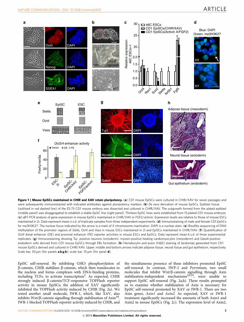
To determine whether the cells derived and maintained in theCHIR/XAV condition retain an EpiSC identity, we examinedtheir molecular and epigenetic profile and tested their differentia-tion potential. These cells expressed Oct4 and Sox2, the keypluripotency genes, and Fgf5, a post-implantation epiblast-specific marker1,2. Their expression of Rex1, Nr0b1 and Stella,markers for the pre-implantation epiblast and primordial germcells, was significantly lower than that of ESCs (Fig. 1c).Immunostaining for tri-methylated H3 lysine 27 (me3H3K27)on the X chromosome showed a large nuclear focus in eachfemale EpiSC maintained in CHIR/XAV, indicating X chromo-some silencing (Fig. 1d). In EpiSCs maintained in CHIR/XAV,the Oct4 promoter was unmethylated, while promoter regions ofStella and Vasa, specific markers for ESCs and primordial germcells, were heavily methylated (Fig. 1e). EpiSCs maintained inCHIR/XAV showed strong activity in the Oct4-proximalenhancer, which is preferentially active in EpiSCs, in contrastwith ESCs, which mainly exhibit activity in the distalenhancer14,15 (Fig. 1f). EpiSCs readily formed embryoid bodies(EBs) in suspension culture upon withdrawal of CHIR/XAV anddifferentiated into cell types representative of all three embryonicgerm layers (Fig. 1g). We injected CD1 mouse EpiSCs derivedand maintained in CHIR/XAV into two SCID mice. Teratomascontaining tissues of all three embryonic germ layers were formedin both mice (Fig. 1h). We also tested the chimera formationability of these CD1 EpiSCs by injecting them into C57BL/6mouse blastocysts. No chimeras ensued from 58 blastocystsinjected, an outcome consistent with previous observations1,2.
To further establish the identity of EpiSCs maintained inCHIR/XAV, we performed whole-genome microarray analyses.EpiSCs derived and grown in CHIR/XAV or FGF2/activinexhibited similar gene expression patterns; these patterns weredistinct from those of mouse ESCs (Supplementary Fig. S2a).Notably, expression of some ESC-specific genes, including Dppa2,Dppa4 and Dppa5a16,17, was upregulated, whereas expression ofthe differentiation-associated genes Eomes and Nodal wasdownregulated in EpiSCs maintained in CHIR/XAV comparedwith EpiSCs in FGF2/activin (Supplementary Fig. S2b). Theseresults suggest that although EpiSCs in CHIR/XAV exhibit keyEpiSC features, they might be developmentally closer to ESCsthan to EpiSCs grown in FGF2/activin.
EpiSC self-renewal in CHIR/XAV is mediated by Axin2. Next,we investigated the mechanism by which CHIR/XAV promotes
ARTICLE NATURE COMMUNICATIONS | DOI: 10.1038/ncomms3403
2 NATURE COMMUNICATIONS | 4:2403 | DOI: 10.1038/ncomms3403 | www.nature.com/naturecommunications
& 2013 Macmillan Publishers Limited. All rights reserved.
EpiSC self-renewal. By inhibiting GSK3 phosphorylation ofb-catenin, CHIR stabilizes b-catenin, which then translocates tothe nucleus and forms complexes with DNA-binding proteins,including TCFs, to activate transcription5. As expected, CHIRstrongly induced b-catenin/TCF-responsive TOPFlash reporteractivity in mouse EpiSCs; the addition of XAV significantlyinhibited the TOPFlash activity induced by CHIR (Fig. 2a). Wetested another small molecule, IWR-1, which, like XAV, alsoinhibits Wnt/b-catenin signalling through stabilization of Axin18.IWR-1 blocked TOPFlash reporter activity induced by CHIR, and
the simultaneous presence of these inhibitors promoted EpiSCself-renewal. In contrast, IWP-2 and Pyrvinium, two smallmolecules that inhibit Wnt/b-catenin signalling through Axinstabilization-independent mechanisms18,19, were unable tosupport EpiSC self-renewal (Fig. 2a,b). These results promptedus to examine whether stabilization of Axin is necessary forEpiSC self-renewal promoted by XAV or IWR-1. There are twoAxin genes, Axin1 and Axin2. As expected, XAV or IWR-1treatment significantly increased the amounts of both Axin1 andAxin2 in mouse EpiSCs (Fig. 2c). The expression level of Axin2,
Oct4
Nanog
SSEA1
DAPI
DAPI
DAPI p10
Day 2outgrowth
30
25
20
46C ESCsCD1 EpiSCs(CHIR/XAV)CD1 EpiSCs(Activin A/FGF2) Blue: DAPI
Green: me3H3K27
XX
XY
2.0
Rel
ativ
e ex
pres
sion
46C
ES
Cs=
1
1.5
1.0
0.5
0.0
Oct4Rex
1Sox
2Ste
lla
Nr0b1
Fgf5
Tuj1
Myosin
Gata4ESC-
2i
Fire
fly/r
enill
a
EpiSC-
CHIR/X
AV
EpiSC-
FGF2/ac
tivin
DAPI
DAPI
DAPIGut epithelium (endoderm)
Neural tissue (ectoderm)
Adipose tissue (mesoderm)
Vector DE PE60
40
Oct3/4 enhancer activity
20
0
Vasa
Oct4
Stella
EpiSC ESC
Figure 1 | Mouse EpiSCs maintained in CHIR and XAV retain pluripotency. (a) CD1 mouse EpiSCs were cultured in CHIR/XAV for seven passages and
were subsequently immunostained with indicated antibodies against pluripotency markers. (b) De novo derivation of mouse EpiSCs. Epiblast tissue
(outlined in red dashed line) of the E5.75 CD1 mouse embryo was dissected and cultured in CHIR/XAV. The outgrowth formed from the plated epiblast
(middle panel) was disaggregated to establish a stable EpiSC line (right panel). Thirteen EpiSC lines were established from 13-plated CD1 mouse embryos.
(c) qRT–PCR analysis of gene expression in mouse EpiSCs maintained in CHIR/XAV or FGF2/activin. Expression levels are relative to those of mouse ESCs
maintained in 2i. Data represent mean±s.d. of triplicate samples from three independent experiments. (d) Immunostaining of male and female CD1 EpiSCs
for me3H3K27. The nuclear focus indicated by the arrow is a mark of X chromosome inactivation. DAPI is a nuclear stain. (e) Bisulfite sequencing of DNA
methylation of the promoter regions of Stella, Oct4 and Vasa in mouse ESCs maintained in 2i and EpiSCs maintained in CHIR/XAV. (f) Quantification of
Oct4 distal enhancer (DE) and proximal enhancer (PE) reporter activities in mouse ESCs and EpiSCs. Data represent mean±s.d. of three experimental
replicates. (g) Immunostaining showing Tuj- positive neurons (ectoderm), myosin-positive beating cardiomyocytes (mesoderm) and Gata4-positive
endoderm cells derived from CD1 mouse EpiSCs through EBs formation. (h) Hematoxylin and eosin (H&E) staining of teratomas generated from CD1
mouse EpiSCs derived and cultured in CHIR/XAV. Upper, middle and bottom arrows indicate adipose tissue, neural tissue and gut epithelium, respectively.
Scale bar, 50mm (for panels a,b,g,h); scale bar, 10mm (for panel d).
NATURE COMMUNICATIONS | DOI: 10.1038/ncomms3403 ARTICLE
NATURE COMMUNICATIONS | 4:2403 | DOI: 10.1038/ncomms3403 | www.nature.com/naturecommunications 3
& 2013 Macmillan Publishers Limited. All rights reserved.

but not Axin1, was also elevated by CHIR treatment (Fig. 2c), anoutcome consistent with previous findings that Axin2 is a directdownstream target of Wnt/b-catenin signalling20. As expected,the use of CHIR with either XAV or IWR-1 further increased thequantity of Axin2 protein in EpiSCs (Fig. 2c). To determinewhether Axin mediates EpiSC self-renewal in CHIR/XAV orCHIR/IWR-1, we designed short hairpin RNAs (shRNAs) toknockdown Axin1 and Axin2. Interestingly, knockdown ofAxin2, but not Axin1, impaired the self-renewal-promoting effectof CHIR/IWR-1 (Fig. 2d–f). To further test the influence of Axinon self-renewal, we established mouse EpiSCs overexpressingAxin1 (Axin1-EpiSCs) or Axin2 (Axin2-EpiSCs) in the FGF2/activin condition (Fig. 2g). Addition of CHIR failed to induce
TOPFlash activity in Axin2-EpiSCs (Supplementary Fig. S3).CHIR alone was sufficient to support robust and long-termexpansion of undifferentiated Axin2-EpiSCs following theremoval of FGF2/activin. In contrast, Axin1-EpiSCs rapidly dif-ferentiated in the presence of CHIR after the removal of FGF2/activin (Fig. 2h). Taken together, these results suggest that Axin2is the key mediator of EpiSC self-renewal promoted by CHIR/IWR-1 or CHIR/XAV.
EpiSC self-renewal mediated by Axin2 is b-Catenin-dependent.Next, we investigated how Axin2 mediates EpiSC self-renewal.We first sought to determine whether b-catenin is required for
Flag
α-Tubulin
Vecto
r
Clone1
Clone2
Clone3
Clone1
Clone2
Clone3
Axin2
shRNA
Axin1
shRNA
CHIR
No tre
atm
ent
XAVIW
R1IW
P2
Pyrvin
ium
CHIR/X
AV
CHIR/IW
R1
CHIR/IW
P2
CHIR/P
yrvin
ium
Scram
ble
Axin2
100
48
Axin1
0.0
0.5
1.0
1.5 UndifferentiatedPartly differentiatedDifferentiated
Per
cent
age
of c
olon
ies
0
1
2Fire
fly/r
enill
ano
trea
tmen
t=1
35
10
15 TOP Flash
CHIR/IWR1 CHIR/IWP2 CHIR/Pyrvinium
Vector Clone1 Clone2 Clone3
Clone1 Clone2 Clone3
Axin1
Axin1
shRNA
Axin2
shRNA
Scram
ble
No tre
atm
ent
CHIRXAV
CHIR/X
AV
CHIR/W
R1
IWR1
α-Tubulin
β-Catenin
Axin2
Axin1 135
100
7548 0.0
1.5
0.5
1.0
Rel
ativ
e ex
pres
sion
scra
mbl
e=1
Axin1
shRNA
Axin2
shRNA
Scram
ble
Scram
ble
Axin2
Figure 2 | Stabilization of Axin2 mediates self-renewal of EpiSCs maintained in CHIR and XAV or CHIR and IWR-1. (a) TOPFlash assay in CD1 mouse
EpiSCs treated with the indicated inhibitors for 12 h. Data represent mean±s.d. of three biological replicates. (b) Representative phase contrast images
of CD1 mouse EpiSCs cultured in the indicated conditions for 7 days. (c) Western blot analysis of CD1 mouse EpiSCs treated with the indicated inhibitors for 12 h.
(d) qRT–PCR analysis of Axin1 and Axin2 mRNA levels in CD1 mouse EpiSCs stably transfected with Axin1 shRNA or Axin2 shRNA. Data represent mean±s.d.
of three biological replicates. (e) Colony assay on CD1 mouse EpiSCs stably transfected with scramble, Axin1 or Axin2 shRNAs. Cells were plated onto a
six-well plate at a density of 5,000 cells per well and cultured in CHIR/IWR-1. Colonies were counted 7 days after plating. Data represent the total combined
numbers of colonies from two independent experiments in which each shRNA-transfected group was cultured in one well of a six-well plate. (f) Representative
phase contrast images of CD1 mouse EpiSCs stably transfected with the indicated shRNAs and cultured in CHIR/IWR-1 for 7 days. (g) Western blot
analysis of CD1 mouse EpiSCs overexpressing Flag-tagged Axin1 or Axin2. (h) Representative phase contrast images of CD1 mouse EpiSCs overexpressing Axin1
or Axin2 and cultured in CHIR for 7 days. Axin2-overexpressing EpiSCs could be continually passaged in CHIR alone. Scale bar, 50mm (for panels b,f,h).
ARTICLE NATURE COMMUNICATIONS | DOI: 10.1038/ncomms3403
4 NATURE COMMUNICATIONS | 4:2403 | DOI: 10.1038/ncomms3403 | www.nature.com/naturecommunications
& 2013 Macmillan Publishers Limited. All rights reserved.
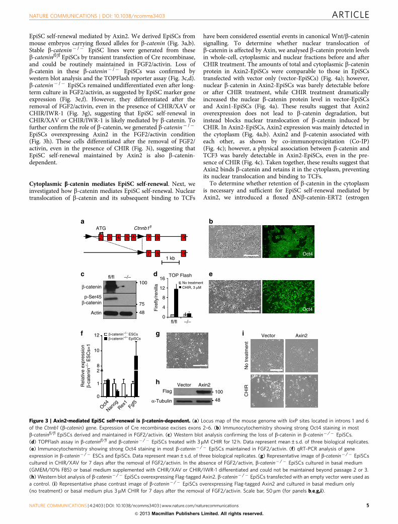
EpiSC self-renewal mediated by Axin2. We derived EpiSCs frommouse embryos carrying floxed alleles for b-catenin (Fig. 3a,b).Stable b-catenin� /� EpiSC lines were generated from theseb-cateninfl/fl EpiSCs by transient transfection of Cre recombinase,and could be routinely maintained in FGF2/activin. Loss ofb-catenin in these b-catenin� /� EpiSCs was confirmed bywestern blot analysis and the TOPFlash reporter assay (Fig. 3c,d).b-catenin� /� EpiSCs remained undifferentiated even after long-term culture in FGF2/activin, as suggested by EpiSC marker geneexpression (Fig. 3e,f). However, they differentiated after theremoval of FGF2/activin, even in the presence of CHIR/XAV orCHIR/IWR-1 (Fig. 3g), suggesting that EpiSC self-renewal inCHIR/XAV or CHIR/IWR-1 is likely mediated by b-catenin. Tofurther confirm the role of b-catenin, we generated b-catenin� /�
EpiSCs overexpressing Axin2 in the FGF2/activin condition(Fig. 3h). These cells differentiated after the removal of FGF2/activin, even in the presence of CHIR (Fig. 3i), suggesting thatEpiSC self-renewal maintained by Axin2 is also b-catenin-dependent.
Cytoplasmic b-catenin mediates EpiSC self-renewal. Next, weinvestigated how b-catenin mediates EpiSC self-renewal. Nucleartranslocation of b-catenin and its subsequent binding to TCFs
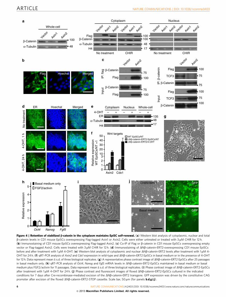
have been considered essential events in canonical Wnt/b-cateninsignalling. To determine whether nuclear translocation ofb-catenin is affected by Axin, we analysed b-catenin protein levelsin whole-cell, cytoplasmic and nuclear fractions before and afterCHIR treatment. The amounts of total and cytoplasmic b-cateninprotein in Axin2-EpiSCs were comparable to those in EpiSCstransfected with vector only (vector-EpiSCs) (Fig. 4a); however,nuclear b-catenin in Axin2-EpiSCs was barely detectable beforeor after CHIR treatment, while CHIR treatment dramaticallyincreased the nuclear b-catenin protein level in vector-EpiSCsand Axin1-EpiSCs (Fig. 4a). These results suggest that Axin2overexpression does not lead to b-catenin degradation, butinstead blocks nuclear translocation of b-catenin induced byCHIR. In Axin2-EpiSCs, Axin2 expression was mainly detected inthe cytoplasm (Fig. 4a,b). Axin2 and b-catenin associated witheach other, as shown by co-immunoprecipitation (Co-IP)(Fig. 4c); however, a physical association between b-catenin andTCF3 was barely detectable in Axin2-EpiSCs, even in the pre-sence of CHIR (Fig. 4c). Taken together, these results suggest thatAxin2 binds b-catenin and retains it in the cytoplasm, preventingits nuclear translocation and binding to TCFs.
To determine whether retention of b-catenin in the cytoplasmis necessary and sufficient for EpiSC self-renewal mediated byAxin2, we introduced a floxed DNb-catenin-ERT2 (estrogen
4
ATGa
c
f g
h
i
d e
b
32 4 5 6 7
7
8
81 kb
9
9
Ctnnb1fl
fl/fl –/–100
β-catenin
p-Ser45β-catenin
Actin 48
75
0
0
1
2
Rel
ativ
e ex
pres
sion
β-ca
teni
n–/–
ES
Cs=
1
8
10
12
Oct4
Nanog
Rex1
Fgf5
β-catenin–/– ESCsβ-catenin–/– EpiSCs
8
12
16
fl/fl –/–
TOP Flash
No treatmentCHIR, 3 μM
Fire
fly/r
enill
a
FlagVector Axin2
100
48
CH
IR
Vector Axin2
Oct4
Oct4
No
trea
tmen
t
α-Tubulin
Figure 3 | Axin2-mediated EpiSC self-renewal is b-catenin-dependent. (a) Locus map of the mouse genome with loxP sites located in introns 1 and 6
of the Ctnnb1 (b-catenin) gene. Expression of Cre recombinase excises exons 2–6. (b) Immunocytochemistry showing strong Oct4 staining in most
b-cateninfl/fl EpiSCs derived and maintained in FGF2/activin. (c) Western blot analysis confirming the loss of b-catenin in b-catenin� /� EpiSCs.
(d) TOPFlash assay in b-cateninfl/fl and b-catenin� /� EpiSCs treated with 3mM CHIR for 12 h. Data represent mean±s.d. of three biological replicates.
(e) Immunocytochemistry showing strong Oct4 staining in most b-catenin� /� EpiSCs maintained in FGF2/activin. (f) qRT–PCR analysis of gene
expression in b-catenin� /� ESCs and EpiSCs. Data represent mean±s.d. of three biological replicates. (g) Representative image of b-catenin� /� EpiSCs
cultured in CHIR/XAV for 7 days after the removal of FGF2/activin. In the absence of FGF2/activin, b-catenin� /� EpiSCs cultured in basal medium
(GMEM/10% FBS) or basal medium supplemented with CHIR/XAV or CHIR/IWR-1 differentiated and could not be maintained beyond passage 2 or 3.
(h) Western blot analysis of b-catenin� /� EpiSCs overexpressing Flag-tagged Axin2. b-catenin� /� EpiSCs transfected with an empty vector were used as
a control. (i) Representative phase contrast image of b-catenin� /� EpiSCs overexpressing Flag-tagged Axin2 and cultured in basal medium only
(no treatment) or basal medium plus 3mM CHIR for 7 days after the removal of FGF2/activin. Scale bar, 50mm (for panels b,e,g,i).
NATURE COMMUNICATIONS | DOI: 10.1038/ncomms3403 ARTICLE
NATURE COMMUNICATIONS | 4:2403 | DOI: 10.1038/ncomms3403 | www.nature.com/naturecommunications 5
& 2013 Macmillan Publishers Limited. All rights reserved.
Vecto
r
Axin1
Axin2
Vecto
r
Vecto
r
Axin1
Axin2
Axin2
Vecto
r
Axin2
Vecto
r
Axin1
Axin2
Vecto
r
Axin1
Axin2
Vecto
r
Axin1
Axin2
100
48
β-Catenin
α-Tubulin
β-Catenin
α-Tubulin
H4
No treatment No treatmentCHIR
Cytoplasm Nucleus
17
48
100100
100
100100
100
100
TCF3
TCF3
Flag
FlagFlag Hoechst Merged
ER
No
trea
tmen
t4-
OH
T, 1
h4-
OH
T, 2
4 h
Basal medium only
FGF2/activin5
4
Rel
ativ
e ex
pres
sion
3
2
1
0Oct4 Nanog Fgf5
Hoechst Merged Cytoplasm
4-OHT
α-Tubulin
ER
H4
Wnt targetsWT EpiSCs/NTΔNβ-catenin-ERT2 EpiSCs/NTΔNβ-catenin-ERT2/4-OHT
– – –+ +1354817
+
Nucleus Whole-cell
Flag
Flag
IP: β
-cat
enin
IP: f
lag
Inpu
t
Inpu
tβ-Catenin
β-Catenin
β-Catenin
β-Catenin
75
75
75
75
75
CHIR
Flag
Whole-cell
Rel
ativ
e ex
pres
sion
WT
Epi
SC
s/N
T =
1
Basal medium
CHIR/IWR-1 GFP
GFP
0123
10
20
30
40
50
Axin2 Cdx1
Figure 4 | Retention of stabilized b-catenin in the cytoplasm maintains EpiSC self-renewal. (a) Western blot analysis of cytoplasmic, nuclear and total
b-catenin levels in CD1 mouse EpiSCs overexpressing Flag-tagged Axin1 or Axin2. Cells were either untreated or treated with 3mM CHIR for 12 h.
(b) Immunostaining of CD1 mouse EpiSCs overexpressing Flag-tagged Axin2. (c) Co-IP of Flag or b-catenin in CD1 mouse EpiSCs overexpressing empty
vector or Flag-tagged Axin2. Cells were treated with 3 mM CHIR for 12 h. (d) Immunostaining of DNb-catenin-ERT2-overexpressing CD1 mouse EpiSCs
before and after treatment with 1 mM 4-OHT. (e) Western blot analysis of cytoplasmic and nuclear DNb-catenin-ERT2 levels after treatment with 1 mM 4-
OHT for 24 h. (f) qRT–PCR analysis of Axin2 and Cdx1 expression in wild-type and DNb-catenin-ERT2 EpiSCs in basal medium or in the presence of 4-OHT
for 12 h. Data represent mean±s.d. of three biological replicates. (g) A representative phase contrast image of DNb-catenin-ERT2 EpiSCs after 25 passages
in basal medium only. (h) qRT–PCR analysis of Oct4, Nanog and Fgf5 mRNA levels in DNb-catenin-ERT2-EpiSCs maintained in basal medium or basal
medium plus FGF2/activin for 11 passages. Data represent mean±s.d. of three biological replicates. (i) Phase contrast image of DNb-catenin-ERT2-EpiSCs
after treatment with 1mM 4-OHT for 24 h. (j) Phase contrast and fluorescent images of floxed DNb-catenin-ERT2-EpiSCs cultured in the indicated
conditions for 7 days after Cre-recombinase-mediated excision of the DNb-catenin-ERT2 transgene. GFP expression was driven by the constitutive CAG
promoter after excision of the floxed DNb-catenin-ERT2-STOP cassette. Scale bar, 50mm (for panels b,d,g,i,j).
ARTICLE NATURE COMMUNICATIONS | DOI: 10.1038/ncomms3403
6 NATURE COMMUNICATIONS | 4:2403 | DOI: 10.1038/ncomms3403 | www.nature.com/naturecommunications
& 2013 Macmillan Publishers Limited. All rights reserved.
ligand-binding domain) transgene into mouse EpiSCs. DNb-catenin-ERT2 is a fusion protein containing an N-terminallytruncated, stabilized b-catenin and a mutant ERT2 (ref. 21). Inthe absence of 4-hydroxytamoxifen (4-OHT), DNb-catenin-ERT2remains in the cytoplasm. Administration of 4-OHT results inthe translocation of DNb-catenin-ERT2 into the nucleus, asconfirmed by immunocytochemistry staining and immuno-blotting (Fig. 4d,e), and activates the Wnt/b-catenin signallingpathway, as indicated by the increased expression of Wnt/b-catenin targets Axin2 and Cdx1 (Fig. 4f). EpiSCs overexpressingDNb-catenin-ERT2 were expanded continuously for more than25 passages in basal medium without addition of exogenouscytokines or small molecules, while retaining an EpiSC identity(Fig. 4g,h). The addition of 4-OHT resulted in rapid differentia-tion, even in the presence of IWR-1 (Fig. 4i). These results arelikely attributable to the presence of the DNb-catenin-ERT2transgene, as its excision by Cre recombinase was associated withreversion to a wild-type EpiSC-like phenotype (Fig. 4j). Collec-tively, these results suggest that retention of stabilized b-cateninin the cytoplasm is necessary and sufficient for EpiSC self-renewalmediated by Axin2, and that nuclear b-catenin induces EpiSCdifferentiation.
To further elaborate the role of b-catenin in EpiSCs, wegenerated a DNb-catenin mutant containing two point mutations,at A295 and I296 (referred to as A295W/I296W hereafter). Thesepoint mutations render b-catenin unable to bind TCFs as wellas Axin and APC22. b-catenin� /� EpiSCs overexpressingDNb-catenin or DNb-catenin/A295W/I296W were establishedin FGF2/activin. Although DNb-catenin/A295W/I296W waspresent in the cytoplasm and nucleus of DNb-catenin/A295W/I296W-expressing cells (Supplementary Fig. S4a), these cellsexhibited no TOPFlash activity even in the presence of CHIR,whereas DNb-catenin cells exhibited a high level of TOPFlashactivity in the absence and presence of CHIR (SupplementaryFig. S4b). b-catenin� /� EpiSCs overexpressing DNb-cateninrapidly differentiated after the removal of FGF2/activin. Incontrast, b-catenin� /� EpiSCs overexpressing DNb-catenin/A295W/I296W could be continuously expanded in basal mediumwithout overt differentiation (Supplementary Fig. S4c). Asnuclear b-catenin is mainly associated with TCFs, our resultssuggest that EpiSC differentiation induced by nuclear b-catenin islikely mediated by b-catenin–TCF binding; nonetheless, theinteraction of b-catenin with Axin and APC conceivably mightalso contribute to the observed effects.
A recent report has suggested that b-catenin forms a complexwith Oct4 and E-cadherin in the membrane and this complex isinvolved in regulating ESC self-renewal and differentiation23.Next, we investigated whether membrane-bound b-catenin alsohas a role in the maintenance of EpiSCs. b-catenin is recruited tothe cell membrane mainly through binding to E-cadherin24. Weconverted E-cadherin� /� mouse ESCs to EpiSCs under theFGF2/activin condition. These E-cadherin� /� EpiSCs could beexpanded in CHIR/IWR-1, and retained an EpiSC identity(Supplementary Fig. S4d,e), indicating that membrane-boundb-catenin is likely not required for EpiSC self-renewal mediatedby Axin2 and b-catenin.
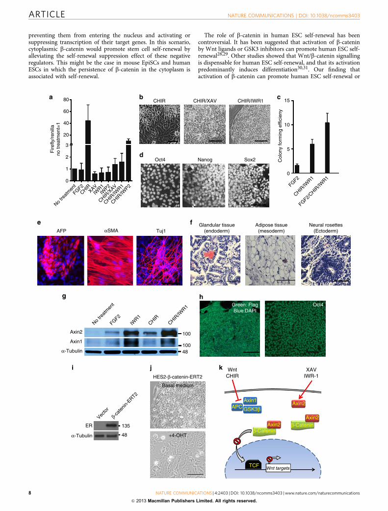
Modulating b-catenin function maintains human ESC self-renewal. As human ESCs share defining features with mouseEpiSCs2,25,26, we tested whether modulating b-catenin functioncan also promote human ESC self-renewal. As was the case inmouse EpiSCs, TOPFlash reporter activity in human ESCs wasstrongly induced by CHIR; addition of either XAV or IWR-1abolished this TOPFlash activity, while IWP-2 only partiallysuppressed such activity (Fig. 5a). Next, we examined the effect of
CHIR/XAV and CHIR/IWR-1 on human ESC self-renewal.CHIR induced differentiation of human ESCs. In contrast,administration of CHIR with either XAV or IWR-1 resulted inrobust self-renewal of human ESCs (Fig. 5b). We found thatCHIR/IWR-1 is more effective than CHIR/XAV in promotinghuman ESC self-renewal, especially in feeder- and FGF2-freeconditions; therefore, we focused on CHIR/IWR-1 for our humanESC study. Supplementation of conventional human ESC med-ium with CHIR/IWR-1 allowed robust propagation of HumanESCs; moreover, the clonogenicity of human ESCs cultured inCHIR/IWR-1 was significantly greater than that in the FGF2condition (Fig. 5c). Human ESCs maintained in CHIR/IWR-1express pluripotency markers Oct4, Nanog and Sox2 (Fig. 5d),and retain the ability to differentiate into cells of all three germlayers, both in vitro and in vivo (Fig. 5e,f). These results indicatethat CHIR/IWR-1 mediates similar self-renewal responses inhuman ESCs and mouse EpiSCs.
Next, we investigated whether CHIR/IWR-1 maintains humanESC self-renewal through a mechanism similar to that in mouseEpiSCs. IWR-1 treatment significantly increased the amounts ofboth Axin1 and Axin2 in human ESCs. CHIR induced theexpression of Axin2, but not Axin1, and combined use of CHIRwith IWR-1 further increased Axin2 protein level (Fig. 5g), anoutcome similar to what we observed in mouse EpiSCs. Toconfirm whether Axin2 also mediates human ESC self-renewal,we stably introduced an Axin2 transgene into human ESCs(Axin2-hESC). As expected, CHIR, administrated alone, couldsupport stable and long-term self-renewal of Axin2-hESCs(Fig. 5h).
Finally, to determine whether cytoplasmic b-catenin can alsopromotes human ESC self-renewal, we introduced the DNb-catenin-ERT2 transgene into human ESCs (Fig. 5i). As expected,human ESCs overexpressing DNb-catenin-ERT2 could be con-tinually passaged without overt differentiation, whereas additionof 4-OHT induced rapid differentiation (Fig. 5j). These resultssuggest that human ESC self-renewal and mouse EpiSC self-renewal are supported by a similar mechanism: an increased levelof cytoplasmic b-catenin and the prevention of b-catenin–TCFinteraction.
DiscussionOur study demonstrates that Wnt/b-catenin signalling canpromote self-renewal or differentiation of mouse EpiSCs andhuman ESCs. The retention of stabilized b-catenin in the cyto-plasm maintains mouse EpiSC and human ESC self-renewal,whereas nuclear translocation of b-catenin and subsequentbinding to TCFs induces differentiation (Fig. 5k). Our findingthat cytoplasmic and nuclear b-catenin pools are both involved inregulating cell fates might provide a rational explanation for someof the diverse and sometimes opposite effects of Wnt/b-cateninobserved in different contexts. More importantly, our studyreveals a new functional avenue of the canonical Wnt/b-cateninpathway, which current dogma depicts as being functionallydefined by nuclear translocation of b-catenin and its subsequentbinding to TCFs.
The gene regulatory effects of Wnt/b-catenin signalling areinitiated upon the binding of b-catenin to TCFs in the nucleus. Sohow is cytoplasmic b-catenin involved in regulating cell fates?One possible model of regulation is suggested by the interactionof cytoplasmic b-catenin with cadherin, a-catenin and actinfilaments27, as these interactions have been shown to havemultiple and important roles in regulating cellular organization,cell adhesion and signal transduction from cell surface to thenucleus. Another possibility is that cytoplasmic b-catenin mighthold negative regulators of self-renewal in the cytoplasm,
NATURE COMMUNICATIONS | DOI: 10.1038/ncomms3403 ARTICLE
NATURE COMMUNICATIONS | 4:2403 | DOI: 10.1038/ncomms3403 | www.nature.com/naturecommunications 7
& 2013 Macmillan Publishers Limited. All rights reserved.
preventing them from entering the nucleus and activating orsuppressing transcription of their target genes. In this scenario,cytoplasmic b-catenin would promote stem cell self-renewal byalleviating the self-renewal suppression effect of these negativeregulators. This might be the case in mouse EpiSCs and humanESCs in which the persistence of b-catenin in the cytoplasm isassociated with self-renewal.
The role of b-catenin in human ESC self-renewal has beencontroversial. It has been suggested that activation of b-cateninby Wnt ligands or GSK3 inhibitors can promote human ESC self-renewal28,29. Other studies showed that Wnt/b-catenin signallingis dispensable for human ESC self-renewal, and that its activationpredominantly induces differentiation30,31. Our finding thatactivation of b-catenin can promote human ESC self-renewal or
WntCHIR
XAVIWR-1
Axin1Axin2
Axin2Axin2
TCF Wnt targets
β-Cateninβ-Catenin
GSK3βAPC
80
60
40
20
Fire
fly/r
enill
ano
trea
tmen
t=1
3
2
1
0
Axin2
No tre
atm
ent
FGF2IW
R1CHIR
CHIR/IW
R1
Axin1
α-Tubulin
Glandular tissue(endoderm)
Adipose tissue(mesoderm)
Neural rosettes(Ectoderm)AFP Tuj1αSMA
No tre
atm
ent
FGF2CHIR
XAVIW
R1IW
P2
CHIR/X
AV
CHIR/IW
R1
CHIR/IW
P2
100
10048
Green: FlagBlue:DAPI
Oct4
HES2-β-catenin-ERT2
Basal medium
+4-OHT
ER 135
48α-Tubulin
Vecto
r
β-cat
enin-
ERT2
Oct4
CHIR CHIR/XAV CHIR/IWR1
Nanog Sox2
0
FGF2
CHIR/IW
R1
FGF2/CHIR
/IWR1
5
10
15
Col
ony
form
ing
effic
ieny
ARTICLE NATURE COMMUNICATIONS | DOI: 10.1038/ncomms3403
8 NATURE COMMUNICATIONS | 4:2403 | DOI: 10.1038/ncomms3403 | www.nature.com/naturecommunications
& 2013 Macmillan Publishers Limited. All rights reserved.
differentiation, and that the respective outcome is dictated bywhether b-catenin translocates into the nucleus, provides arational explanation for earlier, seemingly paradoxical results. Wealso found that FGF2 and CHIR/IWR-1 act synergistically topromote human ESC self-renewal (Fig. 5c). This is noteworthybecause in mouse ESCs, LIF and CHIR/PD can also indepen-dently promote self-renewal, yet there is a synergistic effect whenthe two are combined32,33. Understanding how these differentpathways work independently or synergistically to maintain stemcell self-renewal will advance our efforts to better control stemcell fate, which is critical to the future of regenerative medicine.
MethodsSmall-molecule inhibitors and cytokines. The following small-molecule inhibi-tors and cytokines were used at the indicated final concentrations: CHIR99021(3mM), PD0325901 (1 mM), XAV939 (Sigma, 2 mM), IWR-1 (Sigma, 2.5 mM), IWP-2 (Stemgent, 2.5 mM), Pyrvinium (Sigma, 100 nM), recombinant human FGF2(PeproTech, 10 ng ml� 1), and recombinant human activin A (PeproTech,10 ng ml� 1). CHIR99021 and PD0325901 were synthesized in the Division ofSignal Transduction Therapy, University of Dundee, UK.
Culture media for mouse and rat EpiSCs, and human ESCs. The basal mediumfor mouse EpiSC culture is the conventional mouse ESC medium, which consists ofGMEM (Sigma) supplemented with 10% fetal bovine serum (FBS) (Hyclone),2 mM L-glutamine (Invitrogen), 1 mM sodium pyruvate (Invitrogen), 1% non-essential amino acids (Invitrogen) and 0.1 mM b-mercaptoethanol. Mouse EpiSCswere derived and maintained in the basal medium supplemented with FGF2/activin, CHIR/XAV or CHIR/IWR-1. The basal medium for rat EpiSC culture isN2B27, which was prepared by mixing 500 ml of DMEM/F12 (Invitrogen) with500 ml of neurobasal medium (Invitrogen) and adding 5 ml of N2 (Invitrogen),10 ml of B27 (Invitrogen), 5 ml of glutamax (Invitrogen), and 1 ml of 0.1 Mb-mercaptoethanol (Sigma)34. The basal medium for human ESC culture consistsof Knockout DMEM/F12 supplemented with 20% Knockout serum replacement(KSR) (Invitrogen), 1% non-essential amino acids, 2 mM L-glutamine and 0.1 mMb-mercaptoethanol. Human ESCs were cultured in the basal mediumsupplemented with FGF2, CHIR/XAV or CHIR/IWR-1.
Derivation and propagation of EpiSCs. Post-implantation epiblasts were isolatedfrom E5.75 mouse embryos or E7.5 rat embryos and dissociated into small clumpswith 0.05% trypsin35. Epiblast fragments were placed into four-well plates pre-coated with 0.1% gelatin (for mouse epiblasts) or pre-seeded with g-irradiatedmouse embryonic fibroblasts (MEFs) (for rat epiblasts) and cultured in either theFGF2/activin or the CHIR/XAV conditions. Emerging EpiSCs were trypsinized andexpanded every 2–3 days at a subculture ratio of 1:4. Animal experiments wereperformed according to the investigator’s protocols approved by the University ofSouthern California Institutional Animal Care and Use Committee.
Human ESC culture. H1, H9, HES2 and HES3 human ESC lines were kindlyprovided by the University of Southern California Stem Cell Core Facility. HumanESCs were routinely maintained on g-irradiated MEF feeders in human ESC basalmedium supplemented with 10 ng ml� 1 FGF2. For culture in the CHIR/IWR-1 orCHIR/XAV condition, human ESCs were plated onto dishes pre-coated with
Matrigel (BD Biosciences) or pre-seeded with MEFs and cultured in the basalmedium supplemented with 3 mM CHIR99021, 2.5 mM IWR-1 or 2 mM XAV939.For passaging, human ESCs were dissociated into single cells with 0.05% trypsin orsmall clumps with the Calcium Trypsin KSR(CTK) solution every 2–4 days36, andreplated into the CHIR/IWR-1 or CHIR/XAV condition. To evaluate the colony-forming efficiency of human ESCs cultured in FGF2, CHIR/IWR-1 or FGF2/CHIR/IWR-1 condition, cells were trypsinized and passed through 40 mm cell strainer(BD Biosciences) to obtain single-cell suspension. Cells were then counted andseeded at a density of 1,000 cells per well onto six-well plates pre-seeded withMEFs. After 7 days, cells were fixed with 4% paraformaldehyde and stainedfor alkaline phosphatase (AP) using the Vector Blue Substrate kit (Vectorlaboratories). Colony-forming efficiencies were calculated as the number ofAP-positive colonies formed divided by the number of cells plated.
Generation of b-catenin� /� mouse EpiSCs and ESCs. b-cateninfl/fl
EpiSCs were derived from B6.129-Ctnnb1tm2Kem/KnwJ mice (The JacksonLaboratory) that possess loxP sites located in introns 1 and 6 of the Ctnnb1 (b-catenin) gene37.b-cateninfl/fl EpiSCs were derived and maintained in the FGF2/activin condition.b-catenin� /� ESCs were generated from b-cateninfl/fl ESCs by transienttransfection of the pCAG-Cre-IRES-Puro plasmid using Lipofectamine(Invitrogen). Transfectants were selected for 7 days in GMEM/10% FBS mediumsupplemented with 10 ng ml� 1 LIF, 1 mM PD0325901 and 1 mg ml� 1 puromycin.Puromycin-resistant ESC colonies were picked and expanded in theLIFþ PD0325901 condition (LIF alone was not sufficient to maintain self-renewalof b-catenin� /� ESCs). Loss of b-catenin in b-catenin� /� ESCs was confirmedby western blot analysis. b-catenin� /� EpiSCs were generated from b-cateninfl/fl
EpiSCs by transient transfection of the pCAG-Cre-IRES-Puro plasmid or from b-catenin� /� ESCs by culturing them in FGF2/activin condition38,39. b-catenin� /�
EpiSCs were routinely maintained in the FGF2/activin condition.
Construction of b-catenin mutant plasmids. pcDNA3-human b-cateninand pcDNA3-human DNb-catenin plasmids40 (Addgene) were double-digestedwith BamH1 and Not1. Full-length and DNb-catenin fragments were collected andligated into the pCAG-IRES-hygro vector. The A295W and I296W pointmutations22 were introduced into full-length b-catenin and the DNb-cateninmutant by PCR-driven overlap extension41 using the two PCR primer pairs shownin Supplementary Table S1. Floxed DNb-catenin-ERT2 plasmid was constructedby insertion of the DNb-catenin-ERT2 cassette into the pCAG-loxP-IRES-pac-STOP-loxP-EGFP-pA vector42. Full-length b-catenin or b-catenin mutantswere transfected into mouse ESCs, mouse EpiSCs and human ESCs byelectroporation. Drug-resistant colonies were picked and expanded to establishstable cell lines.
Construction of Axin expression plasmids and Axin shRNA. Mouse Axin1 andAxin2 open reading frames were amplified by PCR from CD1 EpiSCs using KODHot Start DNA Polymerase (EMD). Axin1 and Axin2 open reading frames werethen cloned into the PiggyBac transposon vector and verified by DNA sequencing.For RNA interference of Axin1 and Axin2 in EpiSCs, shRNA constructs designedto target 21 base-pair gene-specific regions in Axin1 and Axin2 were cloned intopLKO.1-TRC vector (Addgene). The targeted sequences were as follows: Axin1,GCCACAGAAATTTGCTGAAGA; Axin2, GGTTTGCTTGTAATGGGTTCA.For overexpression of Axin1 or Axin2 in EpiSCs, 2 mg transposon vector wasco-transfected with 2 mg PiggyBac-Axin1 or PiggyBac-Axin2 into EpiSCs usingLipofectamine LTX and Plus reagent (Invitrogen) according to the manufacturer’s
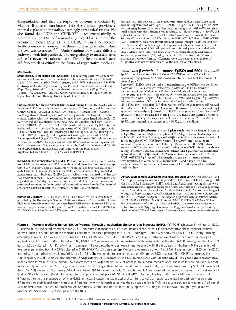
Figure 5 | b-catenin mediates human ESC self-renewal through a mechanism similar to that in mouse EpiSCs. (a) TOPFlash assay in H9 human ESCs
subjected to the indicated treatments for 24 h. Data represent mean±s.d. of three biological replicates. (b) Representative phase contrast images
of H9 human ESCs cultured in the indicated conditions for three passages (CHIR) or 11 passages (CHIR/XAV and CHIR/IWR-1). (c) Colony-forming
efficiency assay of H9 human ESCs cultured in FGF2, CHIR/IWR-1 or FGF2/CHIR/IWR-1 conditions. Data represent mean±s.d. of three biological
replicates. (d) H9 human ESCs cultured in CHIR/IWR-1 for 11 passages were immunostained with the indicated antibodies. (e) EBs were generated from H9
human ESCs cultured in CHIR/IWR-1 for 11 passages. The outgrowths of EBs were immunostained with the indicated antibodies. (f) H&E staining of
teratomas generated from H9 ESCs cultured in CHIR/IWR-1 for 20 passages. (g) Western blot analysis of Axin1 and Axin2 expression in HES3 human ESCs
treated with the indicated cytokines/inhibitors for 24 h. (h) Immunofluorescence images of H9 human ESCs (passage 5 in CHIR) overexpressing
Flag-tagged Axin2. (i) Western blot analysis of DNb-catenin-ERT2 expression in HES2 human ESCs with ER antibody. (j) Top panel: (a) representative
phase contrast image of HES2 human ESCs overexpressing DNb-catenin-ERT2 at passage six in basal medium only. These cells were cultured in basal
medium only for more than 15 passages and remained morphologically undifferentiated. Bottom panel: 3 days after treatment with 1 mM 4-OHT, nearly all
the HES2-DNb-catenin-ERT2 human ESCs differentiated. (k) Model of mouse EpiSC and human ESC self-renewal mediated by b-catenin. In the absence of
Wnt or GSK3 inhibitor, a b-catenin destruction complex, containing Axin1, GSK3 and APC is formed, leading to the degradation of b-catenin and
differentiation. In the presence of Wnt or GSK3 inhibitor, b-catenin is stabilized and can initiate cellular responses related to both self-renewal and
differentiation. Stabilized b-catenin induces differentiation when it translocates into the nucleus and binds TCFs to activate downstream targets. Addition of
XAV or IWR-1 stabilizes Axin2. Stabilized Axin2 binds b-catenin and retains it in the cytoplasm, resulting in self-renewal through a yet unknown
mechanism. Scale bar, 50mm (for panels b,d,e,f,h,j).
NATURE COMMUNICATIONS | DOI: 10.1038/ncomms3403 ARTICLE
NATURE COMMUNICATIONS | 4:2403 | DOI: 10.1038/ncomms3403 | www.nature.com/naturecommunications 9
& 2013 Macmillan Publishers Limited. All rights reserved.
instructions. Twenty four hours after transfection, 1 mg ml� 1 puromycin wasadded to the cell culture medium to select for transfected colonies. For RNAiexperiments, pLKO.1-TRC-based lentiviral vectors were transfected with packagingplasmids pMD2.G and psPAX2 into 293FT cells (Invitrogen) using LipofectamineLTX and Plus reagent. Virus-containing supernatant was collected 48 h aftertransfection. EpiSCs were incubated in the virus supernatant supplemented with8 mg ml� 1 polybrene (Sigma) for 24 h. Supernatant was then replaced with freshCHIR/IWR-1 medium supplemented with 1 mg ml� 1 puromycin to select fortransfected cells.
qRT–PCR. Total RNA was extracted with the RNeasy Mini Kit (Qiagen). cDNAwas synthesized with 1 ng of total RNA, using the QuantiTech Rev. TranscriptionKit (Qiagen). qRT–PCR was performed with Power SYBR Green PCR Master Mix(Applied Biosystems) according to the manufacturer’s instructions. Signals weredetected with an ABI7900HT Real-Time PCR System (Applied Biosystems). Therelative expression level was determined by the 2-DCT method and normalizedagainst GAPDH. The primers used for qRT–PCR are shown in SupplementaryTable S2.
Teratoma formation of mouse EpiSCs and human ESCs. Mouse EpiSCs andhuman ESCs maintained in CHIR/XAV or CHIR/IWR-1 conditions were testedfor their ability to form teratomas in immunodeficient SCID mice. Colonieswere dissociated into small-cell clumps with CTK solution and cells wereresuspended in PBS at a concentration of 1� 107 cells per ml. Five hundredmicroliters of cell suspension was subcutaneously injected into right and left flankof 12-week-old NOD SCID mice (Charles River). Tumors were allowed to developfor 8 weeks. Teratomas were removed and fixed in 4% paraformaldehyde for48 h, followed by paraffin embedding, sectioning and staining with hematoxylinand eosin (H&E).
In vitro differentiation of mouse EpiSCs and human ESCs. In vitro mouse EpiSCand human ESC differentiation was induced by formation of EBs. Mouse EpiSC- orhuman ESC-derived EBs were plated onto gelatin-coated dishes and cultured inGMEM/10% FBS medium. Spontaneously beating cardiomyocytes appeared after2 weeks in culture. Neural differentiation of mouse EpiSCs and human ESCs wasinduced by culturing them on 0.1% gelatin-coated dishes in serum-free N2B27medium43,44.
Western blot and Co-IP. Western blotting was performed according to a standardprotocol. Nuclear and cytoplasmic proteins were extracted using NE-PER Nuclearprotein Extraction Kit (Thermo). For Co-IP, cell extracts were prepared usingNonidet P-40 lysis buffer (50 mM Tris–HCl, pH 7.5, 150 mM NaCl, 0.5% NonidetP-40, 1 mM EDTA, 10% glycerol, 1 mM Na3VO4, 50 mM NaF and protease inhi-bitors). The supernatant was collected and incubated with either anti-b-catenin oranti-Flag antibody for 2 h at 4 �C following incubation with protein A/G Plus-Agarose (Santa Cruz) for 1 h. The beads were then washed five times with lysisbuffer and resuspended in SDS sample buffer. Primary antibodies used include thefollowing: b-catenin (BD Bioscience, 1:2,000), phospho-Ser45 b-catenin (9564, Cellsignalling, 1:500), Axin1 (AF3287, R&D, 1:1,000), Axin 2 (M-20, Santa Cruz,1:200), histone H4 (2592, Cell signalling, 1:1,000), ERa (MC-20, Santa Cruz,1:1,000), TCF3 (M-20, Santa Cruz, 1:1,000), actin (C-11, Santa Cruz, 1:1,000), Flag(F3165, Sigma, 1:2,000), a-tubulin (B-5-1-2, Invitrogen, 1:2,000). Full western blotimages are shown in Supplementary Fig. S5.
Immunostaining. Immunostaining was performed according to a standard pro-tocol. Primary antibodies used include the following: Oct4 (C-10, Santa Cruz,1:200), Sox2 (Y-17, Santa Cruz, 1:200), SSEA-1 (480, Santa Cruz, 1:200), GATA4(G-4, Santa Cruz, 1:200), Nanog (R&D Systems, 1:200), bIII-tubulin (Invitrogen,1:2,000), Myosin (MF-20, DSHB, 1:5), AFP (mouse monoclonal, Sigma) and aSMA(mouse monoclonal, Dako). Alexa Flour fluorescent secondary antibodies (Invi-trogen) were used at a 1:2,000 dilution. Nuclei were visualized with DAPI orHoechst. AP staining was performed with an alkaline phosphatase kit (Sigma)according to the manufacturer’s instructions.
Promoter-enhancer reporter assay. For quantifying relative Oct3/4 enhanceractivities, pGL3-Oct4 DE and pGL3-Oct4 PE plasmids (gifts from Hans Scholer’slab) were co-transfected with the Renilla vector, using the Amaxa Transfection Kit(Lonza). Dual Luciferase Assay (Promega) was performed the following dayaccording to the manufacturer’s instructions. For quantifying relative b-catenin/Tcftranscriptional activity, pGL2-SuperTOP plasmid (gift from Randall Moon) wasco-transfected with the Renilla vector and assayed accordingly.
Bisulfite sequencing. Genomic DNA was extracted with the QIAamp DNA MiniKit (Qiagen). Approximately 500 ng DNA from each sample was treated with theEZ DNA methylation kit (ZYMO) to convert the unmethylated C’s to U’s. Thepromoter regions of Oct4, Stella and Vasa were amplified with primer sets(Supplementary Table S3)17 using the Expand High-fidelity PCR system (Roche),
cloned into the pCR-BluntII-TOPO vector (Invitrogen) and sequenced with theT7-promoter primer.
DNA microarray analysis. Total RNA was extracted with the RNeasy Mini Kit(Qiagen). RNA was amplified, labeled and hybridized to the GeneChip MouseGene 1.0 ST Array according to standard Affymetrix protocols. A DNA microarraywas performed at the University of California, Los Angeles DNA Microarray corefacility. The data analysis was performed using Partek Microarray Software.
References1. Brons, I. G. et al. Derivation of pluripotent epiblast stem cells from mammalian
embryos. Nature 448, 191–195 (2007).2. Tesar, P. J. et al. New cell lines from mouse epiblast share defining features with
human embryonic stem cells. Nature 448, 196–199 (2007).3. Watanabe, K. et al. A ROCK inhibitor permits survival of dissociated human
embryonic stem cells. Nat. Biotechnol. 25, 681–686 (2007).4. van Amerongen, R. & Nusse, R. Towards an integrated view of Wnt signalling
in development. Development 136, 3205–3214 (2009).5. Logan, C. Y. & Nusse, R. The Wnt signalling pathway in development and
disease. Annu. Rev. Cell Dev. Biol. 20, 781–810 (2004).6. Clevers, H. Wnt/b-catenin signalling in development and disease. Cell 127,
469–480 (2006).7. Nusse, R. Wnt signalling and stem cell control. Cell Res. 18, 523–527 (2008).8. ten Berge, D. et al. Embryonic stem cells require Wnt proteins to
prevent differentiation to epiblast stem cells. Nat. Cell Biol. 13, 1070–1075(2011).
9. Ying, Q. L. et al. The ground state of embryonic stem cell self-renewal. Nature453, 519–523 (2008).
10. Hasegawa, K. et al. Wnt signalling orchestration with a small molecule DYRKinhibitor provides long-term xeno-free human pluripotent cell expansion.Stem Cells Transl. Med. 1, 18–28 (2012).
11. Sokol, S. Y. Maintaining embryonic stem cell pluripotency with Wnt signalling.Development 138, 4341–4350 (2011).
12. Wray, J. et al. Inhibition of glycogen synthase kinase-3 alleviates Tcf3repression of the pluripotency network and increases embryonic stem cellresistance to differentiation. Nat. Cell Biol. 13, 838–845 (2011).
13. Huang, S. M. et al. Tankyrase inhibition stabilizes axin and antagonizes Wntsignalling. Nature 461, 614–620 (2009).
14. Yeom, Y. I. et al. Germline regulatory element of Oct-4 specific for thetotipotent cycle of embryonal cells. Development 122, 881–894 (1996).
15. Bao, S. et al. Epigenetic reversion of post-implantation epiblast to pluripotentembryonic stem cells. Nature 461, 1292–1295 (2009).
16. Maldonado-Saldivia, J. et al. Dppa2 and Dppa4 are closely linked SAP motifgenes restricted to pluripotent cells and the germ line. Stem Cells 25, 19–28(2007).
17. Han, D. W. et al. Epiblast stem cell subpopulations represent mouse embryos ofdistinct pregastrulation stages. Cell 143, 617–627 (2010).
18. Chen, B. et al. Small molecule-mediated disruption of Wnt-dependentsignalling in tissue regeneration and cancer. Nat. Chem. Biol. 5, 100–107(2009).
19. Thorne, C. A. et al. Small-molecule inhibition of Wnt signalling throughactivation of casein kinase 1a. Nat. Chem. Biol. 6, 829–836 (2010).
20. Jho, E. H. et al. Wnt/b-catenin/Tcf signalling induces the transcription ofAxin2, a negative regulator of the signalling pathway. Mol. Cell Biol. 22,1172–1183 (2002).
21. Lo Celso, C., Prowse, D. M. & Watt, F. M. Transient activation of beta-cateninsignalling in adult mouse epidermis is sufficient to induce new hair follicles butcontinuous activation is required to maintain hair follicle tumours.Development 131, 1787–1799 (2004).
22. Graham, T. A., Weaver, C., Mao, F., Kimelman, D. & Xu, W. Crystal structureof a b-catenin/Tcf complex. Cell 103, 885–896 (2000).
23. Faunes, F. et al. A membrane-associated beta-catenin/Oct4 complex correlateswith ground-state pluripotency in mouse embryonic stem cells. Development140, 1171–1183 (2013).
24. Orsulic, S., Huber, O., Aberle, H., Arnold, S. & Kemler, R. E-cadherin bindingprevents beta-catenin nuclear localization and beta-catenin/LEF-1-mediatedtransactivation. J. Cell Sci. 112(Pt 8): 1237–1245 (1999).
25. Hanna, J. et al. Human embryonic stem cells with biological and epigeneticcharacteristics similar to those of mouse ESCs. Proc. Natl Acad. Sci. USA 107,9222–9227 (2010).
26. Rossant, J. Stem cells and early lineage development. Cell 132, 527–531 (2008).27. Yamada, S., Pokutta, S., Drees, F., Weis, W. I. & Nelson, W. J. Deconstructing
the cadherin-catenin-actin complex. Cell 123, 889–901 (2005).28. Sato, N., Meijer, L., Skaltsounis, L., Greengard, P. & Brivanlou, A. H.
Maintenance of pluripotency in human and mouse embryonic stem cellsthrough activation of Wnt signalling by a pharmacological GSK-3-specificinhibitor. Nat. Med. 10, 55–63 (2004).
ARTICLE NATURE COMMUNICATIONS | DOI: 10.1038/ncomms3403
10 NATURE COMMUNICATIONS | 4:2403 | DOI: 10.1038/ncomms3403 | www.nature.com/naturecommunications
& 2013 Macmillan Publishers Limited. All rights reserved.
29. Cai, L. et al. Promoting human embryonic stem cell renewal or differentiationby modulating Wnt signal and culture conditions. Cell Res. 17, 62–72 (2007).
30. Dravid, G. et al. Defining the role of Wnt/b-catenin signalling in the survival,proliferation, and self-renewal of human embryonic stem cells. Stem Cells 23,1489–1501 (2005).
31. Davidson, K. C. et al. Wnt/beta-catenin signalling promotes differentiation,not self-renewal, of human embryonic stem cells and is repressed by Oct4.Proc. Natl Acad. Sci. USA. 109, 4485–4490 (2012).
32. Ogawa, K., Nishinakamura, R., Iwamatsu, Y., Shimosato, D. & Niwa, H.Synergistic action of Wnt and LIF in maintaining pluripotency of mouse EScells. Biochem. Biophys. Res. Commun. 343, 159–166 (2006).
33. Wray, J., Kalkan, T. & Smith, A. G. The ground state of pluripotency. Biochem.Soc. Trans. 38, 1027–1032 (2010).
34. Tong, C., Huang, G., Ashton, C., Li, P. & Ying, Q. L. Generating gene knockoutrats by homologous recombination in embryonic stem cells. Nat. Protoc. 6,827–844 (2011).
35. Chenoweth, J. G. & Tesar, P. J. Isolation and maintenance of mouse epiblaststem cells. Methods Mol. Biol. 636, 25–44 (2010).
36. Hasegawa, K., Fujioka, T., Nakamura, Y., Nakatsuji, N. & Suemori, H. Amethod for the selection of human embryonic stem cell sublines with highreplating efficiency after single-cell dissociation. Stem Cells 24, 2649–2660(2006).
37. Brault, V. et al. Inactivation of the beta-catenin gene by Wnt1-Cre-mediateddeletion results in dramatic brain malformation and failure of craniofacialdevelopment. Development 128, 1253–1264 (2001).
38. Hanna, J. et al. Metastable pluripotent states in NOD-mouse-derived ESCs.Cell Stem Cell 4, 513–524 (2009).
39. Guo, G. et al. Klf4 reverts developmentally programmed restriction of groundstate pluripotency. Development 136, 1063–1069 (2009).
40. Kolligs, F. T., Hu, G., Dang, C. V. & Fearon, E. R. Neoplastic transformationof RK3E by mutant beta-catenin requires deregulation of Tcf/Lef transcriptionbut not activation of c-myc expression. Mol. Cell Biol. 19, 5696–5706(1999).
41. Heckman, K. L. & Pease, L. R. Gene splicing and mutagenesis by PCR-drivenoverlap extension. Nat. Protoc. 2, 924–932 (2007).
42. Chambers, I. et al. Functional expression cloning of Nanog, a pluripotencysustaining factor in embryonic stem cells. Cell 113, 643–655 (2003).
43. Ying, Q. L. & Smith, A. G. Defined conditions for neural commitment anddifferentiation. Methods Enzymol. 365, 327–341 (2003).
44. Ying, Q. L., Stavridis, M., Griffiths, D., Li, M. & Smith, A. Conversion ofembryonic stem cells into neuroectodermal precursors in adherentmonoculture. Nat. Biotechnol. 21, 183–186 (2003).
AcknowledgementsWe thank C. Tong, D. Han and members of the USC Stem Cell Core for technicalassistance; N. Wu and Y. Yan for blastocyst injections; Y. Chai for sharing the B6.129-Ctnnb1 mutant mice; R. Kemler for providing E-cadherin� /� mouse ESCs. C. Ashtonand A.P. McMahon for critical reading of the manuscript. This work was supported bythe California Institute for Regenerative Medicine (CIRM) New Faculty Award II (RN2-00938-1) and the CIRM Scientific Excellence through Exploration and Development(SEED) Grant (RS1-00327-1).
Author contributionsH.K. and S.Y. performed most of the mouse EpiSC experiments. J.W. performed humanESC experiments. C.-I.T. analysed microarray data. X.Z. carried out b-catenin-ERT2experiments. H.Y. contributed to western blot analysis. P.L. derived EpiSCs. M.P. con-tributed to the design of the experiments. H.K. and Q.-L.Y conceived the study. Q.-L.Y.wrote the manuscript with assistance from H.K., J.W. and S.Y.
Additional informationAccession codes: Microarray data reported in this paper have been deposited in the GeneExpression Omnibus database with the accession number of GSE31461.
Supplementary Information accompanies this paper at http://www.nature.com/naturecommunications
Competing financial interests: H.K., J.W., and Q.-L.Y. are listed as co-inventors on theU.S. provisional patent application entitled ‘Culture medium for pluripotent stem cells’,number 61/623717. The remaining authors declare no competing financial interests.
Reprints and permission information is available online at http://npg.nature.com/reprintsandpermissions/
How to cite this article: Kim, H. et al. Modulation of b-catenin function maintainsmouse epiblast stem cell and human embryonic stem cell self-renewal. Nat. Commun.4:2403 doi: 10.1038/ncomms3403 (2013).
NATURE COMMUNICATIONS | DOI: 10.1038/ncomms3403 ARTICLE
NATURE COMMUNICATIONS | 4:2403 | DOI: 10.1038/ncomms3403 | www.nature.com/naturecommunications 11
& 2013 Macmillan Publishers Limited. All rights reserved.